Neurology Study Guide - part 9 pps
Bạn đang xem bản rút gọn của tài liệu. Xem và tải ngay bản đầy đủ của tài liệu tại đây (211.97 KB, 26 trang )
198 21. Pediatric Cerebrovascular Disorders
T
ABLE
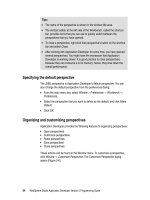
21.1 Common embolic and thrombotic causes of
pediatric brain ischemia.
Category Condition
Heart disease Congenital cardiac defects
Cyanotic congenital heart
disease
Atrial and ventricular septal
defects
Patent ductus arteriosus
Aortic and mitral stenosis
Mitral valve prolapse
Coarctation
Acquired heart disease
Rheumatic fever
Endocarditis
Myocarditis
Cardiomyopathies
Cardiac arrhythmia
Atrial myxoma
Hematological abnormalities Sickle cell anemia
Disorders causing a
hypercoagulable state:
—Antithrombin III deficiency
—Protein C/S deficiency
—Lupus anticoagulant
Leukemia
Polycytemia
Trombocytosis
Liver disorders
Vasculitis/vasculopathy Moya-moya disease
Fibromuscular dysplasia
Infectious and autoimmune
vasculitides
Primary cerebral angiitis
Venous thrombosis
Metabolic and genetic disorders Homocystinuria
Fabry’s disease
Mitochondrial disorders
(MELAS)
Methylmalonic aciduria
Neurofibromatosis
Migraine Migrainous stroke
Drug ingestion, toxins
causing vasospasm and
stroke
Cocaine or amphetamines use
Glue sniffing
Oral contraceptives
Systemic disorders Hypertension
Diabetes
Systemic hypotension
Hypernatremia
Genetic disorders Mitochondrial disorders
Homocystinuria
Fabry’s disease
Pseudoxanthoma elasticum
ogy of brain ischemia due to embolism and thrombosis
in pediatric patients.
Embolic Stroke
Cerebral embolism is characterized by a sudden neuro-
logical deficit that is maximal at onset and may show a
partial or total improvement due to lysis and reinstate-
ment of the perfusion. Emboli in children usually origi-
nate from the heart when congenital or acquired structural
abnormalities are present. Sources of cerebral emboli in
children include
Cardiac sources
• Congenital heart defects.
• Cyanotic congenital heart disease.
• Atrial and ventricular septal defect.
• Coarctation of the aorta.
• Transposition of great vessels.
• Acquired heart disease.
• Rheumatic heart disease.
• Bacterial and nonbacterial endocarditis.
• Cardiomyopathy.
• Atrial myxoma.
• Mitral valve prolapse.
• Arrhythmias: Atrial fibrillation occurs in children
with rheumatic heart disease, Ebstein’s anomaly,
atrial septal defect, and total anomalous pulmonary
venous return (Riela).
Arterial sources
• Vasculopathies: Moya-moya, fibromuscular dysplasia.
• Catheterization and other procedures.
• Arteritis and arterial aneurysms.
• Trauma.
Other sources
• Air/fat embolism.
• Paradoxical emboli.
Paradoxical Emboli and Differential Diagnosis
of an Acute Focal Event
Paradoxical embolization occurs when a cardiac defect
allows direct entrance of embolic formations into the sys-
temic circulation. The source of embolization derives
from thrombi that form in the lower extremities or pelvic
veins but also from pulmonary fistulas. Congenital heart
defects, such as atrial or ventricular septal defects, patent
foramen ovale with significant shunt, truncus arteriosus,
and so on, or large pulmonary arteriovenous fistulas that
can be found in children with hereditary hemorrhagic tel-
angiectasias, can result in the occurrence of paradoxical
embolism.
In the differential diagnosis of the vignette, an acute
vascular event is first considered but other causes of acute
focal weakness need to be presented.
Space-occupying lesions, such as neoplasms, usually
manifest with progressive hemiparesis but if a hemor-
rhage acutely occurs into the tumor, this will result in an
acute focal deficit in addition to headache and decreased
level of consciousness.
Complicated migraines can manifest with transitory
neurological deficits, particularly hemiplegia and less
commonly ophthalmoplegia, that can occur prior to or
Homocystinuria 199
after the headache and also in the absence of headache.
This is not always an easy diagnosis, particularly if the
characteristic migraine symptoms are not present. Other
etiologies that need to be excluded are antiphospholipid
antibodies and other disorders that can cause a hyper-
coagulable state.
Trauma and infections can also cause acute hemiplegia
but can easily be excluded from the vignette. Bacterial
and viral infections can be responsible for an acute focal
neurological deficit because of various mechanisms, in-
cluding vascular inflammation, cerebral infarction, sinus
occlusion, and parenchymal necrosis. Additional symp-
toms are usually present, such as fever, nausea, vomiting,
altered sensorium, and seizures.
Focal seizures, particularly if prolonged, can be fol-
lowed by hemiplegia and may suggest an underlying
vascular lesion, such as a cerebral malformation or an
infarction.
Metabolic disorders, particularly hypoglycemia, dia-
betes mellitus, or homocystinuria need to be mentioned
as causes of acute hemiplegia that enter into the differ-
ential diagnosis of this vignette.
Diagnosis
• Physical and neurological evaluation.
• Laboratory studies.
• Blood count PT and PTT.
• Special studies, in selected cases.
• Hemoglobin electrophoresis.
• Protein C/S.
• Antithrombin III.
• Antiphospholipid antibodies.
• Lupus anticoagulant.
• Lactate pyruvate (for mitochondrial
dysfunction).
• HIV, VDRL.
• Neuroimaging studies.
•CT.
• MRI.
• MRA and angiography in selected cases.
• Cardiac studies.
• EKG.
• Transesophageal echocardiogram in cases of
congenital cardiac defects or to demonstrate an
intracardiac thrombus or valvular vegetations.
Treatment
Roach and Riela recommend the short-term use of hep-
arin for patients at risk for recurrent, nonseptic cerebral
embolism and with minimal risk of secondary hemor-
rhage. The long-term use of anticoagulation with warfarin
is based on situations that carry a high risk of stroke, such
as in children with congenital and acquired heart disease,
venous sinus thrombosis, coagulopathies and hyperco-
agulable states, arterial dissection, and so on.
The use of antiplatelet agents in children is controver-
sial, particularly regarding the efficacy and effective dose
of aspirin, which has been used in low daily doses.
Bacterial endocarditis and septic embolism are treated
with intravenous antibiotics for at least six to eight weeks.
Homocystinuria
Vignette
A 10-year-old boy, mildly retarderd and with his-
tory of cataract, underwent an emergency appen-
dectomy. The postoperative period was compli-
cated by right hemiplegia and aphasia. There was
no history of heart disease, TIA, seizures, trauma,
or infections. He never experienced migraine and
his family history was unremarkable. He was tall
and slender. The pediatric resident noted that he
had pes cavus, hyposcoliosis, highly arched palate,
and multiple erythematous spots over his cheeks but
did not detect any organomegaly. Neurological ex-
amination showed expressive aphasia and dense
right hemiplegia, more severe in the face and upper
extremities with relative sparing of the lower
extremities.
Summary A 10-year-old boy experiencing an acute vas-
cular event after surgery. Involvement of several other
systems is indicated:
• Ocular system: Cataract.
• Skeletal system: Pes cavus, hyposcoliosis, highly
arched palate.
• Skin: Multiple erythematous spots over the cheeks.
• CNS: Mental retardation, acute hemiplegia, and
aphasia.
Localization and Differential Diagnosis
The expressive aphasia with right hemiplegia more severe
in the face and upper extremity, with relative sparing of
the lower extremity, localized to a lesion involving the
upper trunk of the left middle cerebral artery. The in-
volvement of multiple systems, including skeletal, eye,
skin, and central nervous system, points to a neurometa-
bolic disorder where stroke is a significant part of the
clinical manifestations.
Four neurometabolic genetic disorders—homocystin-
uria, Fabry’s disease, MELAS, and methylenetetrafolate
reductase deficiency—are responsible for strokes in chil-
dren and young adults due to vasculopathies and venous
or arterial occlusion.
Homocystinuria is the most common genetic disorder
that affects the brain vasculature and leads to premature
atherosclerosis and stroke (Caplan). The clinical symp-
200 21. Pediatric Cerebrovascular Disorders
tomatology involves multiple systems with skeletal de-
formities such as pes cavus and hyposcoliosis, derma-
tological features such as malar flush, ocular
abnormalities with lens dislocation, cataract, and so on,
and neurological abnormalities with mental retardation
and multiple cerebrovascular accidents. The clinical vi-
gnette clearly describes a case of homocystinuria.
MELAS (mitochondrial encephalomyopathy with lac-
tic acidosis and stroke-like episodes) is a mitochondrial
disorder characterized by multiple manifestations that in-
clude stroke-like episodes, migraine-type headache, re-
current vomiting, epileptic seizures, proximal muscle
weakness, short stature, and exercise intolerance. Lactic
acid levels are increased in blood and CSF and muscle
biopsy demonstrates ragged red fibers.
Fabry’s disease is a sex-linked lysosomal storage dis-
ease due to deficiency of alpha-galactosidase A. The
clinical manifestations include signs of peripheral neu-
ropathy manifesting with painful paresthesias, cutaneous
lesions presenting with a red-purple maculopapular rash,
and cerebrovascular complications, in particular hemiple-
gia and aphasia due to premature atherosclerosis.
Methylenetetrafolate reductase deficiency can manifest
with cerebrovascular complications due to thrombotic oc-
clusion, but also vomiting, seizures, mental deterioration,
and so on, in the absence of any ocular or skeletal
abnormalities.
Clinical Features
Homocystinuria is a disorder of methionine metabolism,
due to a defect of cystathionine B-synthase, which cata-
lyzes the conversion of homocystine and serine to cys-
tathionine. This abnormality results in homocystinuria
and increased plasma and CSF levels of homocystine and
methionine. The transmission is autosomal recessive.
Homocystinuria is responsible for a multitude of man-
ifestations due to involvement of ocular, skeletal, cuta-
neous, vascular, and CNS systems. Ocular manifestations
are represented by ectopia lentis, glaucoma, retinal de-
tachment, and cataracts. Skeletal abnormalities include
pes cavus, hyposcoliosis, high-arched palate, arachno-
dactyly, and so on. Children and adolescents are tall and
slender and have features that simulate Marfan’s syn-
drome. Skin anomalies manifest with livedo reticularis
and multiple erythematous spots over the maxillary area
and cheeks.
Mental retardation may occur and cognitive impair-
ment can also be attributed to multiple infarcts. Focal and
generalized seizures have been described, even in the ab-
sence of strokes.
Vascular complications that can occur particularly fol-
lowing surgery, even if minor, or intravenous injection,
are responsible for a multitude of manifestations that in-
clude myocardial infarction, deep venous thrombosis
with pulmonary embolism, renal artery and vein throm-
bosis, and cerebral thromboembolic events.
Diagnosis
The diagnosis of homocystinuria can be demonstrated by
the increased urinary excretion of homocystine, elevated
plasma levels of methionine and homocystine, and a posi-
tive urinary cyanide-nitroprusside reaction.
It is important to reach the diagnosis as promptly as
possible because early therapeutic intervention may pre-
vent some of the complications.
Treatment
Pyridoxine or betaine therapy and dietary manipulation
with restriction of methionine and cystine supplementa-
tion have shown efficacy in some patients.
Intracranial Hemorrhage
Vignette
An 8-year-old girl was playing basketball with her
teammates when she suddenly screamed, com-
plained of headache, and vomited. Her mother
could not keep her awake. There was no previous
history of trauma or seizure disorder. In the emer-
gency room she was drowsy and her neck was rigid.
Preretinal hemorrhages were present on the left
eye. During the next several hours she experienced
two generalized tonic-clonic seizures.
Summary A previously healthy 8-year-old girl experi-
encing sudden onset of headache, vomiting, decreased
level of consciousness, stiff neck, and seizures.
Localization
A sudden onset of headache, vomiting, and decreased
level of consciousness accompanied by signs of menin-
geal irritation and increased intracranial pressure in the
absence of focal neurological deficits is highly suggestive
of subarachnoid hemorrhage (SAH).
Infants and young children may have a less typical pre-
sentation with low-grade fever, hypersensitivity, irritabil-
ity, seizures, and vomiting.
Focal and generalized convulsions can occur and focal
neurological deficits are not noted unless there is exten-
sion into the brain parenchyma or if vasospasm causes
brain infarcts. Signs of increased intracranial pressure
manifest with headache, vomiting, and papilledema. Cra-
nial nerve dysfunction mainly affects the sixth and third
Acute Hemiplegia 201
T
ABLE
21.2 Etiology of pediatric subarachnoid and
intraparenchymal hemorrhage.
Category Condition
Trauma The most common cause of
intracranial hemorrhage in children.
In infants SAH should always bring
into consideration the possibility of
child abuse.
Prematurity Germinal matrix hemorrhage.
Structural vascular
malformations
Cerebral aneurysm. Symptomatic
intracranial aneurysms are
uncommon in the pediatric group.
Children tend to have more
aneurysms in the posterior
circulation and carotid bifurcation
and tend to have larger aneurysm.
Males are more affected than
females. Subarachnoid hemorrhage
is usually the initial presentation of
an intracranial aneurysm in both
children and adults.
Arteriovenous malformations.
Characterized by direct
communication of arteries with
veins. The symptoms of AVMs are
influenced by size, location, and
age at presentation. Vein of Galen
malformations manifest in the
neonatal period with congestive
heart failure and in infants with
macrocephaly, hydrocephalus and
so on. In older children or
adolescents, AVM typically
manifests with headache, seizures
and intraparenchymal or
subarachnoid hemorrhage.
Cavernous malformations.
Characterized by well-
circumscribed, dilated vessels,
sometimes multiple, and manifesting
with headache, recurrent seizures,
intracranial hemorrhage, etc.
Coagulopathies Hereditary Hemophilia A, B, and other
factor deficiency.
Thrombocytopenia.
Acquired Vitamin K deficiency. Liver
dysfunction with coagulation
defects.
Hemoglobinopathies
Vasculitis
Sickle cell anemia.
Sinovenous thrombosis
Hemorrhagic infarction
Hemorrhagic
encephalopathy
due to hypernatremia
Tumor, infections
nerve, the latter in particular can be an indication of a
posterior communicating artery aneurysm.
Subarachnoid hemorrhage in children is attributed pri-
marily to trauma.
Nontraumatic causes of SAH include sickle cell dis-
ease and coagulopathies, aneurysmal rupture, arterio-
venous malformations, and so on.
Table 21.2 presents the etiology of intracranial (sub-
arachnoid and intraparenchymal) hemorrhage in children.
Acute Hemiplegia
Vignette
A previously healthy, 20-month-old girl started ex-
periencing attacks of head shaking and eye rolling
several days after a febrile upper respiratory infec-
tion. She then developed acute left-sided weakness.
On examination, left hemiparesis, hyperreflexia and
a left Babinski’s sign were noted. Cranial nerves
were normal. She was drowsy and uncooperative
during the rest of the examination.
Summary A previously healthy, 20-month-old girl ex-
periencing episodes that could represent seizures (head
shaking and eye rolling) after a respiratory infection with
subsequent acute left hemiplegia.
Differential Diagnosis
The differential diagnosis of acute hemiplegia in children
includes several categories of disorders, and among them,
stroke is the most common cause of weakness.
Acute hemiplegia can be due to a vascular disorder,
can follow an epileptic seizure, or can be a migraine com-
ponent (hemiplegic migraine). Other possibilities include
metabolic abnormalities, infectious processes, trauma, or
a neoplastic lesion (Griesemer). Etiological factors pre-
disposing to an acute vascular event such as congenital
or acquired heart disease, sickle cell anemia, coagulopa-
thies, vasculitis, or vasculopathies can be recognized in
many but not all cases of strokes in children.
Cerebrovascular disorders have been divided based on
the pathophysiology into ischemic (embolic and throm-
botic) and hemorrhagic.
Cardiac abnormalities, congenital or acquired, are usu-
ally the source of emboli in children. They include dis-
orders such as septal defects, aortic and mitral valve in-
sufficiency, complex cardiac abnormalities, rheumatic
valvular disease, myocarditis, cardiomyopathy, atrial
myxoma, and so on.
Vasculitis of the intracranial vessels, which is usually
attributed to infections or autoimmune disorders, may
202 21. Pediatric Cerebrovascular Disorders
manifest with arterial thrombosis, intraparenchymal or
subarachnoid hemorrhage, or sinovenous occlusion. In-
fections may predispose to cerebrovascular occlusive dis-
ease, and often an upper respiratory infection may pre-
cede the onset of the stroke. Bacterial meningitis can be
complicated by cerebral vasculitis and strokes in children
due to acute inflammation of the vessel’s wall and
occlusion.
Other causes of intracranial arteritis include tubercu-
lous meningitis, HIV, varicella infection, and so on.
Among the autoimmune vasculitides, systemic lupus ery-
thematosus can manifest with cerebral infarction due to
arterial thrombosis, but also with hemorrhage and venous
occlusion.
Hematological disorders may be characterized by ar-
terial or venous occlusion or hemorrhage. Sickle cell dis-
ease in particular can predispose to stroke, especially is-
chemic infarction, often during the time of a crisis when
the child is febrile or dehydrated following an infection.
Venous occlusion and subarachnoid hemorrhage are also
complications of sickle cell disease. Other hematological
disorders, such as trombocytopenia, polycytemia, and
disorders of coagulation such as hemophilia A (X-linked
factor VIII deficiency) may be responsible for stroke and
acute hemiplegia.
Metabolic disease (homocystinuria, Fabry’s disease,
MELAS) can produce arterial and venous occlusions.
Among the vasculopathies, moya-moya syndrome can
present with acute hemiplegia. Clinical symptoms vary
from transitory ischemic attacks to strokes, seizures, and
cognitive decline. The Japanese word moyamoya mean-
ing “like a puff of smoke” best describes the angiographic
picture of abnormal vascular network at the base of the
brain.
Trauma can cause carotid occlusion in children, for
example, after a fall when the child is carrying some ob-
ject in the mouth such as a lollipop or a pencil, and can
be responsible for acute hemiparesis.
In the differential diagnosis of acute hemiplegia in chil-
dren, other categories aside from stroke (most common
form of weakness) need to be considered, such as epi-
lepsy, encephalitis, cerebral abscess, tumor, trauma, mi-
graine, metabolic disorders, etc.
Hemiplegia can follow a jacksonian seizure (Todd’s
paralysis), usually lasting a few hours, but can also be an
expression of prolonged focal seizures such as seen with
Rasmussen’s encephalitis, herpes encephalitis, or as a
manifestation of an underlyng vascular malformation
(Griesemer).
Brain neoplasm complicated by acute hemorrhage can
present with acute hemiplegia or focal seizures followed
by postictal hemiparesis.
Acute focal deficit can also be associated with meta-
bolic abnormalities such as hypoglycemia or diabetes
mellitus.
Transient neurological deficits, particularly hemiple-
gia, accompany complicated migraine in children. In al-
ternating hemiplegia, which has been described as a form
of complicated migraine, there are recurrent episodes of
unexplained hemiplegia often associated with head pain
prior to or following the attack and accompanied by other
neurological symptoms and developmental abnormalities.
Finally, multiple sclerosis can present with acute hemi-
plegia but the clinical diagnosis requires the presence of
neurological deficits disseminated in time and space.
Diagnosis
An accurate history and physical and neurological ex-
amination are very important in the formulation of the
diagnosis, particularly considering the possibilities of
trauma, convulsions, developmental status, cognitive im-
pairment, family history, and so on. The examination of
the cardiovascular system should cautiously consider
murmurs, abnormal heart sounds, abnormal rhythms, hy-
pertension, and bruits. The funduscopic examination may
reveal retinal pigmentation, hemorrhages, or exudates,
and also inspection of the skin may show abnormalities
such as rash, hyper-/hypopigmentation, and so on.
The diagnostic workup should include laboratory tests
such as complete blood count to rule out infection, sickle
cell anemia, polycythemia, leukemia, or thrombocyto-
penia. Hemoglobin electrophoresis is important if he-
moglobinopathies are considered in the differential di-
agnosis. Also, sedimentation rate, prothrombin time, and
partial prothrombin time are obtained. Serum chemistries
will rule out the possibility of hyperglycemia and
hypoglycemia.
Neuroimaging (CT/MRI of the brain) and cardiac stud-
ies are essential in the evaluation of a child with acute
hemiplegia. Lumbar puncture is important if there is no
cerebral mass effect and there is suspicion that the hemi-
plegia is due to a brain infection.
Angiography may be reserved for selected cases of ar-
terial dissection, moya-moya disease, cerebral vasculitis,
and so on.
Treatment
The treatment of acute hemiplegia, medical or surgical,
is based on the underlying etiology.
Subdural Hematoma
Vignette
A 6-month-old boy, previously in good health, was
found unresponsive in his crib by his babysitter. He
then experienced a generalized seizure and in the
Headache 203
T
ABLE
21.3 Causes of coma in children.
Category Condition
Traumatic injuries Hemorrhage
Epidural
Subdural
Subarachnoid
Malignant brain edema
Vascular disorders Intracranial
Nontraumatic
Hemorrhagic
Vasculitis
Venous thrombosis
Cerebral infarction
Infectious/parainfectious disorders Meningitis
Encephalitis
Encephalomyelitis
Cerebral abscess
Metabolic and systemic disorders Hyper/hypoglycemia
Hyper/hyponatremia
Hepatic coma
Uremic coma
Hypophosphatemia
Toxic disorders
Brain tumors
Hydrocephalus
emergency room was comatose. Pupils were poorly
reactive to light and bilateral retinal hemorrhages
were noted. He was afebrile and normotensive. A
chest x-ray indicated possible healing fractures of
the posterior rib cage.
Summary A 6-month-old boy suddenly became coma-
tose. Poorly reactive pupils and bilateral retinal hemor-
rhages were noted, as well as possible healing fractures
on chest x-ray.
Localization and Differential Diagnosis
In the differential diagnosis of a comatose child, several
causes are considered, including trauma, vascular disor-
ders, infections, tumors, toxic, metabolic, and systemic
disorders (Table 21.3). In this particular case, a traumatic
etiology is highly suspicious particularly because of heal-
ing fracture of the posterior rib cage.
Child abuse is an important consideration in the etiol-
ogy of intracranial vascular lesions. Cranial trauma due
to direct punch to the head with or without a skull frac-
ture, can be responsible for subdural, subarachnoid, or
intraparenchymal bleeding, swelling, and herniation.
Shaken baby syndrome may be responsible for a coma-
tose baby due to posttraumatic subarachnoid hemorrhage
or subdural hematoma even in the absence of signs of
external injury. The ophthalmoscopic examination may
demonstrate retinal hemorrhages, which are commonly
seen in child abuse after inflicted trauma, particularly
when there are no other signs of external injuries.
Subdural hematoma is common in battered babies and
can be bilateral, particularly in infants.
Clinical Features
Infantile subdural hematoma can be acute or chronic, and
when presenting acutely, manifests with altered level of
consciousness, generalized seizures, vomiting, and bulg-
ing fontanelle. Retinal or subhyoid hemorrhages are fre-
quently encountered. A skull fracture can also be dem-
onstrated in almost half the patients. Acute subdural
hematoma usually is due to tearing of cerebral veins
bridging to the sagittal sinus, with blood accumulating
beneath the dura against the brain parenchyma.
Diagnosis
The CT scan in acute subdural hematoma may show a
high-density, crescent-shaped extracerebral fluid collec-
tion or signs of cerebral mass effect and swollen brain.
MRI can give further details.
Treatment
The treatment is based on surgical intervention with evac-
uation of large hematoma with mass effect.
Headache
Basilar Migraine
Vignette
While playing basketball in school, a 14-year-old
boy complained of sudden visual loss and fainted.
When he regained consciousness, he had a throb-
bing headache and was vomiting. In the emergency
room, pupillary testing and an ophthalmoscopic ex-
amination were unremarkable.
Summary A 14-year-old boy with bilateral visual loss,
syncope, and headache.
Localization and Differential Diagnosis
The character of the visual loss reflects its posterior visual
pathway origin and localizes to the occipital cortical area.
All the possible causes of bilateral visual loss of cortical
origin should be considered. Even if more benign con-
ditions, such as basilar artery migraine, are suspected,
alternative diagnoses need also to be ruled out.
Vascular disorders involving the posterior circulation,
characterized by infarction of the posterior cerebral ar-
teries bilaterally due to embolization with occlusion of
the distal basilar artery, may present with cortical blind-
ness and headache, although this event is not common in
children. Subacute bacterial endocarditis and a prolapsing
204 21. Pediatric Cerebrovascular Disorders
mitral valve are the most common sources of such emboli
(Pellock).
Consideration needs to be given also to other disorders
such as vertebral artery dissection, cerebral vasculitis,
moya-moya disease, and vasospasm following subarach-
noid hemorrhage. Hematological disorders creating a hy-
percoagulable state and sickle cell disease may also cause
occipital lobe dysfunction. Hemorrhage, such as those
due to arteriovenous malformations, also needs to be
considered.
Tumors of the posterior fossa usually manifest with
progressive symptoms, mostly dominated by signs of in-
creased intracranial pressure, cranial nerve dysfunction,
ataxia, and so on. Traumatic injuries to the occipital lobe
can be responsible for cortical visual loss. The head in-
juries are usually mild, frequently involving blows to the
frontal or occipital region. Commonly, loss of vision is
complete or almost complete (Pellock). The association
of migraine or seizure disorder increases susceptibility to
posttraumatic transient cerebral blindness (Albert). Blind-
ness can also follow severe generalized convulsions in
infants or toddlers. It can be easily excluded in this
vignette.
Other causes of acquired cerebral visual impairment
during childhood that need to be mentioned, even if easily
excluded from this vignette, are CNS infections such as
meningitis and encephalitis (SSP, CJD, and so on) and
hypoxic-ischemic encephalopathies due to asphyxia, car-
diac arrest or hypotension during surgical procedure.
Visual loss of psychogenic origin in absence of organ-
icity can manifest in preadolescent and adolescent chil-
dren and needs to be carefully evaluated in the above
vignette.
Finally, hereditary metabolic disorders such as ME-
LAS may also present with occipital blindness in addition
to a multitude of symptoms.
Basilar artery migraine is an important consideration
in the differential diagnosis but because the history is
limited and there is no evidence in this child of other
features common to migraine, a more cautious and ag-
gressive approach should be mantained by obtaining MRI
of the brain to exclude structural lesions and even MRA
or angiography to rule out aneurysmal formations, vas-
culitis, and so on.
Basilar migraine is the most common type of compli-
cated migraine variant in children and manifests with aura
symptoms indicative of dysfunction of the brainstem or
both occipital lobes. The headache classification com-
mittee of the International Headache Society has designed
diagnostic criteria for basilar migraine that, in addition to
the criteria of migraine with aura, should include two or
more of the following: visual symptoms in the temporal
and nasal field of both eyes, dysarthria, vertigo, tinnitus,
hearing loss, diplopia, ataxia, bilateral paresthesias, par-
aparesis, and altered level of consciousness.
Clinical Features
The clinical presentation includes different symptoms, in
particular visual abnormalities characterized by blurred
vision, bilateral visual loss, tunnel vision, scintillating
scotoma, and positive or negative hallucinations. The vi-
sual disturbances during an attack indicate a posterior
visual pathway involvement with normal pupillary re-
sponses and funduscopic examinations. Ataxia and ver-
tigo with or without tinnitus also commonly occur as well
as dysartria.
Altered level of consciousness is also common and can
manifest with syncope, or drop attacks accompanied by
loss of consciousness and amnesia.
The aura generally lasts 10 to 60 minutes.
Diagnosis
Even if the history is suggestive, a cautious approach
should always be maintained in order to rule out alter-
native diagnoses.
MRI and MRA should be included in the diagnos-
tic studies as well as hematological tests such as cell
count, hemoglobin, anticardiolipid antibodies, VDRL,
and so on.
Treatment
The treatment is symptomatic and preventive for
reoccurrences.
Ophthalmoplegic Migraine
Vignette
A 3-year-old girl started experiencing severe right
retroorbital pain, irritability, vomiting, drowsiness,
and abdominal pain for two days. On the third day,
her right pupil dilated and she developed right pto-
sis and outward deviation of the eye. On examina-
tion, she was alert, comfortable, afebrile, and had
no physical or neurological abnormalities except
complete right ptosis, pupillary dilatation, and the
inability to move the right eye in any direction ex-
cept laterally. She was a full-term product of a nor-
mal pregnancy and vaginal delivery. Her neonatal
period was uneventful and she had developed nor-
mally from all points of view. She is the only child
of healthy parents.
Summary A 3-year-old girl developed a right, third
nerve palsy after two days of systemic symptoms: irrita-
bility, drowsiness, vomiting, abdominal pain, and right
retroorbital pain.
References 205
Localization and Differential Diagnosis
The differential diagnosis of a child presenting with acute
onset of third nerve palsy includes several possibilities.
Trauma is an important and the most common cause
of an acquired third nerve palsy in the pediatric popula-
tion (Liu). Other disorders include neoplastic processes,
infectious and inflammatory disorders, and ophthalmo-
plegic migraine. Severe head injuries accompanied by an
orbital or base of skull fracture or midbrain hemorrhage
may be responsible for cranial neuropathies (Liu). The
vignette does not mention or imply any previous trau-
matic event, so this cause can be easily ruled out.
Intracranial tumors must always be considered in a
child presenting with ophthalmoplegia. Brainstem
gliomas may be characterized by ophthalmoplegia, usu-
ally in combination with progressive ataxia and other cra-
nial nerve abnormalities and long tract signs. When
tumor-related third nerve palsies occur, lesions affecting
the orbit, orbital apex, and leptomeninges may also be
involved and other signs and symptoms can be present,
such as abducens paresis and proptosis with orbital
lesions.
Infectious and inflammatory processes are other im-
portant causes of third nerve palsies. Chronic sinusitis
with a mucocele of the sphenoid sinus may be associated
with recurrent headache and third nerve palsies (Hocka-
day). Patients usually have a history of chronic sinus in-
fection. Meningitis due to pneumococci and H. influen-
zae, as well as tuberculous meningitis, may present with
third nerve palsy, usually in association with headache
and systemic symptoms.
Tolosa-Hunt syndrome, characterized by nonspecific
granulomatous inflammation of the cavernous sinus and
superior orbital fissure, is rare in children and is charac-
terized by painful ophthalmoplegia with partial or total
involvement of extraocular muscles innervated by nerves
III, IV, or VI in any combination; various pupillary dys-
functions, and sensory abnormalities in the area of the
ophthalmic-trigeminal nerve. Tolosa-Hunt syndrome can
sometimes simulate ophthalmoplegic migraine but the
course is prolonged and headache and ophthalmoplegia
occur at the same time.
Isolated third nerve palsies due to posterior commu-
nicating aneurysms are very uncommon in the pediatric
population and usually occur in combination with hydro-
cephalus and signs of SAH.
Cranial neuropathies due to diabetes are exceptionally
rare in children.
Myasthenia gravis can be easily excluded because it is
usually characterized by bilateral signs that fluctuate and
do not involve the pupils.
Finally, we need to consider ophthalmoplegic migraine
as the appropriate diagnosis after excluding other, more
severe causes. Ophthalmoplegic migraine is a rare variant
of complicated migraine that usually causes an isolated
third nerve paresis. The onset of symptoms is usually in
the first decade of life. The diagnostic workup in this
child should include
• Careful history and neurological evaluation.
• MRI and MRA in order to exclude orbital or cavernous
sinus pathology or aneurysm.
• Lumbar puncture if the neuroimaging studies are neg-
ative and an infectious process is suspected.
• Cerebral angiogram in a patient 10 years old or older
to exclude aneurysm.
Clinical Features
Ophthalmoplegic migraine is characterized by one or re-
current episodes of ophthalmoplegia associated with se-
vere headache that usually precede the ocular paresis.
The third nerve is affected in the majority of the cases
with involvement of the pupil but the sixth nerve can also
be involved, and rarely the fourth nerve. The pain is com-
monly ipsilateral, localized in the orbital, retroorbital, and
temporal area and associated with nausea and vomiting.
With the onset of ophthalmoplegia, the headache often
subsides.
The episodes of ophthalmoplegic migraine, which usu-
ally involve the same eye, vary in frequency of attacks,
and the duration of the ophthalmoplegia is also variable
from a few hours up to several months.
The International Headache Society has defined diag-
nostic criteria for ophthalmoplegic migraine that include
at least two attacks characterized by headache associated
with paresis of one or more of the cranial nerves III, IV,
and VI in the absence of parasellar lesion excluded by
the appropriate investigations.
Diagnosis
The diagnostic workup in an infant or young child should
include magnetic resonance imaging (MRI) and magnetic
resonance angiography. If the patient is over 12 years of
age, angiography to rule out posterior communicating an-
eurysm is indicated.
Treatment
Full recovery is the rule, but after repeated severe attacks
residual deficits can be noted. Prevention of repeated ep-
isodes and residual abnormalities by the use of prophy-
lactic drugs is important.
References
Paradoxical Emboli
Caplan, L. Stroke: A Clinical Approach, ed. 2. Boston:
Butterworth-Heinemann, 1993.
206 21. Pediatric Cerebrovascular Disorders
Fenichel, G. Clinical Pediatric Neurology, ed. 3. Philadelphia:
W.B. Saunders, 1997.
Griesemer, D.A. Acute hemiplegia in childhood. Neurobase
MedLink, Arbor, 1993–2000.
Jones, H.R. Jr. et al. Cerebral emboli of paradoxical origin. Ann.
Neurol. 13:314–319, 1983.
Loscalzo, J. Paradoxical embolism: Clinical presentation, di-
agnostic strategies, and therapeutic options. Am. Heart J.
112:141–149, 1986.
Mendoza, P. and Conway, E.E. Jr. Cerebrovascular events in
pediatric patients. Pediatr. Ann. 27:665–674, 1998.
Nagaraja, D. et al. Cerebrovascular disease in children. ACTA
Neurol. Scand. 90:251–255, 1994.
Nicolaides, P. and Appleton, R.E. Stroke in children. Dev. Med.
Child Neurol. 38:172–180, 1996.
Rivkin, M.J. and Volpe, J.J. Strokes in children. Pediatr. Rev.
17:265, 1996.
Roach, E.S. and Riela, A.R. Pediatric Cerebrovascular Disor-
ders, ed. 2. New York: Futura, 1995.
Homocystinuria
Brett, E.M. Paediatric Neurology, ed. 2. New York: Churchill
Livingstone, 1991.
Lyon, G. et al. Neurology of Hereditary and Metabolic Diseases
of Children, ed. 2. New York: McGraw-Hill, 264–268, 1996.
Menkes, J.M. and Sarnat, H.B. Cererebrovascular Disorders in
Child Neurology, ed. 6. Philadelphia: Lippincott Williams &
Wilkins, 885–917, 2000.
Roach, E.S. and Riela, A.R. Pediatric Cerebrovascular Disor-
ders, ed. 2. New York: Futura, 1995.
Intracranial Hemorrage/Acute Hemiplegia
Berg, B.O. Principles of Child Neurology. New York: McGraw-
Hill, 1996.
Biller, J. et al. Strokes in children and young adults. Boston:
Butterworth-Heinemann, 1994.
Griesemer, D.A. Acute hemiplegia in childhood. Neurobase
MedLink Arbor, 1993–2000.
Mendoza, P.L. and Conway, E.E. Jr. Cerebrovascular events in
pediatric patients. Pediatr. Ann. 27:665–674, 1998.
Pellock, J.M. and Myer, E.C. Neurologic Emergencies in
Infancy and Childhood, ed. 2. Boston: Butterworth-
Heinemann, 1993.
Riela, A.R. and Roach, E.S. Etiology of stroke in children.
J. Child Neurol. 8:201–220, 1993.
Rivkin, M.J. and Volpe, J.J. Strokes in children. Pediatr. Rev.
17:265, 1996.
Roach, E.S. et al. Cerebrovascular disease in children and ad-
olescents. American Academy of Neurology, 52nd Annual
Meeting, San Diego, 2000.
Subdural Hematoma
Berg, B.O. Principles of Child Neurology. NewYork: McGraw-
Hill, 937–952, 1996.
Fenichel, G.M. Clinical Pediatric Neurology: A Sign and Symp-
tom Approach. Philadelphia: W.B. Saunders, 71–75, 1997.
Pellock, J.M. and Myer, E.C. Neurologic Emergencies in In-
fancy and Childhood. Boston: Butterworth-Heinemann, 91–
102, 1993.
Roach, E.S. and Riela, A.R. Pediatric Cerebrovascular Disor-
ders, ed. 2. New York: Futura, 291–312, 1995.
Basilar Migraine
Albert, D.M. et al. Principle and Practice of Ophthalmology.
Philadelphia: W.B. Saunders, 2634–2639, 1994.
Davidoff, R.A. Migraine: Manifestations, Pathogenesis and
Management. Philadelphia: F.A. Davis, 1995.
Hockaday, J.M. Migraine in Childhood and Other Nonepileptic
Paroxysmal Disorders. Boston: Butterworths, 1988.
Hockaday, J.M. Migraine in childhood. In: Berg, B.O. (Ed.).
Principles of Child Neurology. New York: McGraw-Hill,
693–706, 1996.
Molofski, W.J. Headaches in children. Pediatr. Ann. 27:614–
621, 1998.
Pellock, J.M. and Myer, E.C. Neurologic Emergencies in
Infancy and Childhood, ed. 2. Boston: Butterworth-
Heinemann, 268–269, 1993.
Rothner, A.D. The migraine syndrome in children and adoles-
cents. Pediatr. Neurol. 2:121–126, 1986.
Singer, H.S. Migraine headaches in children. Pediatr. Rev.
15:94–101, 1994.
Welch, K.M.A. Basilar Migraine. Neurobase MedLink, Arbor,
1993–2000.
Wright, K.W. Pediatric Ophthalmology and Strabismus. St.
Louis: Mosby, 801–805, 1995.
Ophthalmoplegic Migraine
Davidoff, R.A. Migraine: Manifestations, Pathogenesis, and
Management. Philadelphia: F.A. Davis, 1995.
Glaser, J. S. and Bachynski, B. Infranuclear disorders of eye
movement. In: Glaser, J.S. Neuroophthalmology, ed 2. Phila-
delphia: J.B. Lippincott, 361–419, 1990.
Hockaday, J.M. Migraine in childhood. Boston: Butterworths,
1988.
Lee, A.G. and Brazis, P. Ophthalmoplegic migraine. Neurobase
MedLink Arbor, 1993–2000.
Liu, G.T. Pediatric 3rd, 4th and 5th nerve palsy. American Acad-
emy of Neurology, 51st Annual Meeting, Toronto, 1999.
207
22
Pediatric Neurocutaneous Disorders
N
EUROFIBROMATOSIS
207
Neurofibromatosis
Vignette
A 15-year-old boy from Santo Domingo has com-
plained of bifrontal headache and intermittent vom-
iting for one month. His past medical history is sig-
nificant for generalized seizures since the age of 12
months. His developmental history is normal. On
examination, several hyperpigmented spots, skin-
fold axillary freckling, and subcutaneous nodules
are noted. He is alert and cooperative. Fundu-
scopic examination shows absent venous pulsa-
tions. Bilateral horizontal nystagmus, left dysme-
tria, and wide-based gait are also noted.
Summary A 15-year-old boy with headache and inter-
mittent vomiting for one month. Past medical history is
significant for generalized seizures since 12 months of
age. The neurological examination shows absent venous
pulsation on funduscopic examination, left dysmetria,
and gait ataxia. Also, neurocutaneous findings, hyperpig-
mented spots, axillary freckling, and subcutaneous nod-
ules are described.
Localization and Differential Diagnosis
The clinical findings indicate signs of increased intracra-
nial pressure as well as signs of left cerebellar dysfunc-
tion. There is also a long-standing history of generalized
convulsions, which point to a cortical irritative process.
An important finding in the vignette is the description of
the cutaneous lesions, which are represented by hyper-
pigmented macules, skinfold freckling, and subcutaneous
nodules. All these features point to a neurocutaneous
disorder.
Neurocutaneous syndromes include disorders charac-
terized by cutaneous and neurological manifestations.
The major neurocutaneous syndromes include
• Neurofibromatosis (Von Recklinghausen’s disease).
• Tuberous sclerosis.
• Sturge-Weber syndrome.
• Von Hippel-Lindau syndrome.
• Ataxia-telangiectasia.
In this vignette, the clinical findings described suggest
the diagnosis of neurofibromatosis (NF). The cutaneous
manifestations are characteristic and the signs of cere-
bellar dysfunction may indicate the possibility of an in-
tracranial tumor. Hyperpigmented macules (“cafe´ au lait
spots”) are an important cutaneous feature of neurofibro-
matosis type 1, which is the most common type, but are
nonspecific and can be observed in other neurocutaneous
syndromes and less frequently in neurofibromatosis
type 2. Skinfold freckling is usually seen in the axillary
and inguinal area. Subcutaneous neurofibromas as well
as plexiform neurofibromas are also common manifes-
tations of NF type 1.
Intracranial, spinal, and peripheral nerve tumors can
complicate NF type 1 but are more common in the type
2. Unilateral or bilateral optic nerve glioma is considered
the most commonly observed in NF type 1.
Clinical Features
There are two distinct types of neurofibromatosis: type 1
and type 2. Neurofibromatosis type 1 (NF1), or Von
Recklinghausen disease, is the most common form af-
fecting 1 in 4000 to 5000 individuals (Menkes and Maria)
and resulting from a spontaneous mutation in almost 50
percent of the cases. The cutaneous manifestations char-
acteristic of NF1 include cafe´ au lait spots, skinfold freck-
ling, and neurofibromas. Cafe´ au lait spots are character-
ized by hyperpigmented macules widely distributed over
the body, manifesting at birth and clearly obvious during
the first year of life. According to the diagnostic criteria,
at least six or more cafe´ au lait spots greater than 5 mm
in diameter need to be present in prepubertal children and
greater than 15 mm in postpubertal patients (Robertson).
208 22. Pediatric Neurocutaneous Disorders
Skinfold freckling consists of small pigmented lesions,
usually noted in the areas not exposed to the sun, such as
the axillary, inguinal area, inferior part of the chin, and
so on.
Neurofibromas, which can be dermal or subcutaneous,
are benign tumors that originate from peripheral nerves
and tend to increase after puberty. They vary in size and
number and can cause nerve compression with pain and
loss of function. Plexiform neurofibromas can affect the
trunk, face, and neck and cause significant deformity.
Lisch nodules are pigmented hamartomas of the iris
and are usually asymptomatic.
The neurological manifestations of NF1 include the
possible occurrence of tumors, particularly involving the
brain, spinal cord, and peripheral nerves. Among the cen-
tral nervous system tumors, optic nerve glioma is the
most commonly found in NF1 and may manifest with
progressive visual loss and optic atrophy.
Meningiomas, ependymomas and astrocytomas can
also be discovered in NF1. Skeletal abnormalities include
bone dysplasia of the sphenoid wing of the temporal bone
and pseudoarthrosis of the tibia.
Diagnosis
Neurofibromatosis is a hereditary disorder transmitted
with an autosomal dominant trait. The gene for NF1 is
linked on the long arm of chromosome 17 (17g11.2) that
of NF2 is on the long arm of chromosome 22 (22g11.2).
Several criteria have been established in order to fulfill
the diagnosis of NF1. They include
• Six or more “cafe´ au lait spots” greater than 5 mm in
diameter in prepubertal children and greater than 15
mm in postpubertal patients.
• Two or more neurofibromas of any type or one plexi-
form neurofibroma.
• Axillary or inguinal freckling.
• Two or more iris hamartomas (Lisch nodules).
• Optic glioma.
• Typical osseous lesions, such as sphenoid dysplasia or
tibial pseudoarthrosis.
• One or more first-degree relatives with NF1.
For NF2, any of the following:
• Bilateral vestibular schwannomas seen with imaging
techniques.
• Unilateral vestibular schwannoma in association with
any two of the following: meningioma, neurofibroma,
schwannoma, and juvenile posterior subcapsular len-
ticular opacity.
• Unilateral eighth nerve tumor or other spinal or brain
tumor in first-degree relative.
Neurofibromatosis type 2, which is less common than
type 1, is characterized by less consistent cutaneous man-
ifestations than type 1 and the typical occurrence of bi-
lateral vestibular schwannomas. Symptoms include hear-
ing loss, tinnitus, headache, and vertigo. Meningiomas of
the brain and spine can also occur.
References
Aicardi, J. Diseases of the nervous system in childhood.
McKeith Press. 1992. 203–11.
Berg, B.D. Child neurology: a clinical manual. Second ed.
Philadelphia: J.B. Lippincott Co. 1994. Ch. 9: 185–95.
Brett, E.M. Paediatric neurology. Second ed. New York: Chur-
chill Livingstone. 1991.
Conneally, M., Bird, T.D. et al. Neurocutaneous syndromes in
Neurogenetics Continuum Part A program of the American
Academy of Neurology Vol. 6, No. 6, Dec. 2000.
35–58.
Gutman, D.H. The diagnosis and management of neurofibro-
matosis 1. The neurologist. Nov. 1998; Vol. 4: 313–38.
Mackool, B.T. and Fitzpatrick, T.B. Diagnosis of neurofibro-
matosis by cutaneous examination. Semin. Neurol. 1992; Vol.
12: 358–63.
Menkes, J.H. and Maria, B.L. Neurocutaneous syndromes in
child neurology. Menkes, J.H. and Sarnat, H.B. Sixth ed.,
Philadelphia: Lippincott Williams & Wilkins 2000. Ch 11:
859–884.
Roach, E.S. Diagnosis and management of neurocutaneous syn-
dromes. Semin. Neurol. 1988; Vol. 8: 83–96.
Robertson P. Neurofibromatosis type 1; Neurofibromatosis type
2, Medlink Arbor-Publishing Corp. 1993–2001.
209
23
Pediatric Movement Disorders
H
UNTINGTON
’
S
D
ISEASE
209
S
YDENHAM
’
S
C
HOREA
210
D
YSTONIA
M
USCULORUM
D
EFORMANS
212
T
IC
D
ISORDERS
213
Huntington’s Disease
Vignette
An 8-year-old girl became irritable, apathetic, dis-
tractible, and lost interest in her schoolwork and
dance classes. She was noted to have sudden jerk-
ing movements in her arms and started experienc-
ing generalized tonic-clonic seizures. A year later
she was more withdrawn, not following questions
or commands, sometimes remaining in a catatonic
posture. On examination, there was rigidity with
loss of facial expression. Her prior developmental
history was unremarkable. She had no siblings. Her
father had involuntary movements and grimacing
and was demented.
Summary An 8-year-old girl with progressive cognitive
impairment associated with seizures and parkinsonian
features (rigidity, loss of facial expression). In the family
history, her father has dementia, facial grimacing, and
involuntary movements.
Localization and Differential Diagnosis
The vignette describes an extrapyramidal disorder that
occurs during childhood and is associated with progres-
sive dementia and seizures.
The family history with a father affected by dementia
and involuntary movements suggests a hereditary domi-
nant disorder. Among the hereditary, predominantly ex-
trapyramidal, syndromes occurring during late child-
hood and adolescence the following should be considered
first:
• Childhood and juvenile forms of Huntington’s
disease.
• Wilson’s disease.
• Hallervorden-Spatz disease.
Huntington’s disease is a progressive degenerative dis-
order with an autosomal dominant pattern of transmis-
sion. The clinical manifestations in children are domi-
nated by cognitive and behavior abnormalities, rigidity,
loss of facial expression, decreased voluntary move-
ments, and seizures. In the majority of childhood-onset
cases there is an affected father.
Wilson’s disease, which always needs to be ruled out
in a child presenting with signs of extrapyramidal system
dysfunction is an autosomal recessive disorder character-
ized by the accumulation of copper in the liver, basal
ganglia, and cornea. Younger children usually present
with signs and symptoms of significant liver dysfunction
rather than neurological involvement. Neurological man-
ifestations, with only minimal symptoms of liver disease,
are more likely when the onset of symptoms is in the
second decade (Fenichel). Speech abnormalities with
dysarthria as well as tremor dystonia and gait distur-
bances are often the presenting neurological symptoms.
Emotional lability and psychosis can also be the initial
feature, but seizures and marked dementia are not usually
a significant characteristic of the disease except in few
cases.
Hallervorden-Spatz disease is a familial disorder that
manifests with signs of involvement of the extrapyrami-
dal system such as rigidity, dysarthria, choreoathetosis,
and gait dysfunction, in association with signs of pyra-
midal involvement such as spasticity and hyperreflexia.
Behavioral abnormalities and cognitive impairment can
occur and visual abnormalities such as retinitis pigmen-
tosa and optic atrophy can also be present. Seizures are
not common. Typical pathological findings include hy-
perpigmentation of the pallidum and substantia nigra.
Other extrapyramidal disorders such as idiopathic tor-
sion dystonia, familial calcification of the basal ganglia,
juvenile paralysis agitans, chorea-acantocytosis, and so
on are easily differentiated by their clinical features.
210 23. Pediatric Movement Disorders
Considering the information presented in the vignette,
Huntington’s disease is the preferred diagnosis.
Clinical Features
Huntington’s disease (HD) in the pediatric population
usually presents in the first decade of life (between 5 and
12 years of age) with symptoms characterized by behav-
ioral and cognitive deterioration, rigidity, dystonia, and
seizures.
Seizures, which are usually not observed in adult pa-
tients with HD, can be a prominent initial manifestation
and may affect about 50 percent of children with HD
(Menkes). Epileptic seizures can be represented by tonic-
clonic convulsions, absence, and myoclonic seizures.
Tonic-clonic or myoclonic status can also occur.
Rigidity causing gait disturbances is common, and
dystonia, loss of facial expression and associated move-
ments, and decreased voluntary movements are signifi-
cant features in the majority of pediatric patients. Cho-
reoathetosis and hyperkinesia are not common in the
pediatric age group with HD.
Mental deterioration with progressive dementia is an
important characteristic feature. Behavior abnormalities
manifest with irritability, distractibility, emotional labil-
ity, negativism, and even catatonia. Most of childhood-
onset cases have inherited the gene from an affected fa-
ther. The HD gene has been localized to the short arm of
chromosome 4 and contains an abnormal repeat of the
trinucleotide CAG (cytidine-adenine-guanidine).
Diagnosis
The diagnosis is based on the clinical features and family
history. Neuroimaging studies demonstrate caudate atro-
phy and PET studies reveal significant reduction in cau-
date glucose metabolism. DNA analysis detects the ab-
normal gene.
Treatment
The treatment is symptomatic and is based on the use of
anti-parkinsonian medications to control rigidity and dys-
tonia. Behavioral abnormalities may respond to neurolep-
tics. The use of baclofen (GABA agonist) and diltiazem
(calcium-channel blocker that might block the action of
glutamate on calcium channels) is controversial.
Sydenham’s Chorea
Vignette
A 10-year-old Mexican immigrant was reported by
her teacher as being restless, inattentive, over-
emotional, and fidgety. Irregular jerking move-
ments of her distal upper extremities and face were
noted, and she seemed particularly troubled when
eating, drinking from a cup, or writing. Her family
and developmental histories were normal. Six
months earlier, while still in Mexico, she had ex-
perienced knee pain and swelling accompanied
with fever. Her family reported no other medical
history.
Summary A 10-year-old girl with onset of involuntary
movements and prior history of knee pain, swelling, and
fever.
Localization and Differential Diagnosis
The involuntary, irregular jerking movements that inter-
fere with activities such as writing or feeding in this pa-
tient, plus the fidgety, restless, and overemotional behav-
ior observed by her teacher most likely are indications of
a choreic disorder. Childhood chorea can be attributed to
various etiologies:
• Infectious disorders, such as Sydenham’s chorea, diph-
theria, viral encephalitis, and so on.
• Immunological disorders, such as systemic lupus ery-
thematosus, periarteritis nodosa, and sarcoidosis.
• Drug-induced causes, such as related to the use of neu-
roleptics, anticonvulsants, and so on.
• Toxic causes, such as due to manganese, carbon mon-
oxide, toluene, and alcohol.
• Metabolic and endocrine disorders, such as hypogly-
cemia, hyperglycemia, hypocalcemia, hyperthyroid-
ism, and Addison’s disease.
• Structural disorders, such as tumors and arteriovenous
malformations.
• Bilateral cerebral dysfunction, such as postanoxia.
• Genetic and hereditary degenerative disorders, such
as childood Huntington’s disease, Hallervorden-Spatz
disease, Lesch-Nyhan syndrome, and so on.
Sydenham’s chorea (St. Vitus’ dance) is a well-known
choreic sequelae of infection with group A streptococcus.
It affects children between 5 and 15 years of age, par-
ticularly females. A beta-hemolitic streptococcal infec-
tion of the pharynx may occur 1 to 7 months prior to the
onset of the neurological manifestations in most patients.
The movements are typically choreoathetoid and prefer-
entially involve the face and upper extremities, unilater-
ally or bilaterally. Sydenham’s chorea, polyarthritis, and
carditis are important features of rheumatic fever, the re-
sult of an antecedent group A streptococcal pharyngeal
infection. A prior history of pharyngitis is not always
given by the patient and families. The duration of the
chorea varies from three months to two years.
Other infectious processes that can be responsible for
Sydenham’s Chorea 211
the occurrence of chorea include bacterial, such as sub-
acute bacterial endocarditis, neurosyphilis, diphtheria, tu-
berculosis, Lyme disease, and viral infections, such as
viral encephalitis, mononucleosis, HIV, Epstein-Barr,
varicella, pertussis, and so on.
Immunological causes of chorea include, in particular,
systemic lupus erythematosus, Behc¸et’s disease,
Schonlein-Henoch purpura, antiphospholipid antibodies
syndrome, and so on. Systemic lupus erythematosus in
children can manifest with psychosis, seizures, cranial
neuropathy, and rarely with chorea as the only presenta-
tion. The presence of systemic symptoms such as fever,
rash, lymphadenopathy, hematuria, albuminuria, and so
on, and laboratory studies, particularly antibodies against
DNA, help confirm the diagnosis.
Drug-induced causes are now considered the most
common cause of chorea in children (Robertson et al.).
Among the drugs, neuroleptics, anticonvulsants, anti-
emetic, noradrenergic stimulants, and so on, can be in-
cluded. Tardive dyskinesia indicates a condition associ-
ated with the use of neuroleptics and characterized by
abnormal involuntary movements such as choreic move-
ments involving the face and limbs. Withdrawal emergent
syndrome (Robertson et al.) refers to the first appearance
of involuntary movements and chorea after interruption
of neuroleptic treatment.
Toxic agents that may induce chorea include carbon
monoxide, thallium, toluene (glue sniffing), and so on.
Metabolic and endocrine disturbances can also cause sec-
ondary chorea. Electrolyte disturbances such as hypo-
glycemia, hyperglycemia, hypocalcemia, hypomangane-
semia, and hepatic and renal failure can be responsible
for secondary chorea. The endocrine disorders primarily
include hyperthyroidism, but also hypoparathyroidism,
Addison’s disease, and so on. Some vitamin deficiencies
such as vitamin B
12
, beriberi, and pellagra can present
with chorea.
Chorea can also be secondary to diffuse cerebral dys-
function due to perinatal anoxia or decreased cerebral
perfusion due to postcardiopulmonary bypass. Structural
cerebral lesions like tumor, arteriovenous malformations
or cerebrovascular accidents can also present with chorea.
Trauma has also been involved in some cases.
Hereditary degenerative disorders manifesting with
chorea include the following:
• Juvenile Huntington’s disease, as previously described,
is an autosomal dominant disorder usually transmitted
by the affected father and characterized by progressive
cognitive impairment, rigidity, seizures, and choreo-
athetosis.
• Wilson’s disease is an autosomal recessive disorder of
copper metabolism characterized by hepatic failure and
neurological features particularly involving the extra-
pyramidal system with tremor, rigidity, dystonia, dys-
arthria, choreoathetosis, and so on.
• Hallervorden-Spatz disease is a rare autosomal reces-
sive disorder of iron metabolism, manifesting with
choreic movements, athetosis, dystonia, rigidity, cog-
nitive impairment, retinitis pigmentosa, seizures, and
so on.
• Pelizaeus-Merzbacher disease is an X-linked recessive
disorder of myelin formation characterized by invol-
untary movements with chorea or athetosis, cerebellar
ataxia, pendular nystagmus, developmental regression,
spasticity, optic atrophy, and so on.
• Fahr’s disease, or familial calcification of the basal
ganglia, manifests with choreoathetosis, mental im-
pairment, microcephaly, and seizures. There is pro-
gressive calcification of the basal ganglia.
• Neuroacanthocytosis is characterized by chorea in as-
sociation with seizures, orolingual dystonia, and acan-
thocytosis (acanthocytes are abnormal erythrocytes
that have thorny projections from the cell surface).
• Ataxia-telangiectasia is a hereditary autosomal reces-
sive disorder clinically characterized by progressive
ataxia, telangiectasias, and recurrent sinopulmonary in-
fections. Choreoathetosis can also be observed, par-
ticularly in infants.
• Benign familial hereditary chorea is an autosomal
dominant hereditary disorder manifesting with chorea,
dysarthria, and normal cognitive function.
• Genetic metabolic disorders such as GM
1
and GM
2
gangliosidosis, Leigh syndrome, lipofuscinoses, and so
on, can also include chorea in their symptomatology.
Hereditary paroxysmal choreas need also to be
mentioned:
• Paroxysmal dystonic choreoathetosis is an autosomal
dominant hereditary disorder that manifests with epi-
sodes of choreic movements and dystonia of various
duration from minutes to hours.
• Familial paroxysmal kinesiogenic choreoathetosis is a
hereditary disorder characterized by brief, recurrent ep-
isodes of unilateral choreoathetosis precipitated by a
sudden movement (Robertson).
Clinical Features
Sydenham’s chorea represents a late sequelae of group A
streptococcal pharyngitis. The neurological manifesta-
tions usually tend to present one to six months after the
streptococcal infection. Affected children range from 5 to
15 years of age and are preferentially girls. The disorder
manifests insidiously or acutely with involuntary move-
ments that involve the face and distal part of the upper
extremities. The involuntary movements disappear dur-
ing sleep or sedation. The child is first noted to be restless,
clumsy, and fidgety. The speech becames dysarthric, and
hypotonia may create abnormal postures. The hand grip
waxes and wanes when the child is asked to squeeze the
212 23. Pediatric Movement Disorders
examiner’s hand, a phenomenon called “milkmaid sign.”
Seizures rarely occur. Behavioral dysfunction, includes,
in particular, tics and obsessive-compulsive disorder.
Diagnosis
MRI of the brain, which is important in order to rule out
structural lesions, is usually normal but may show high
signal on T
2
-weighted images in the head of the caudate
and in the putamen.
Some laboratory tests should be considered including
• Blood count and differential.
• Blood chemistry.
• Thyroid function tests, erythrocyte sedimentation rate.
• Antinuclear antibodies titer.
• Anticardiolipid antibodies.
• Antistreptolisin O titer.
In selected cases, other laboratory studies include
• Blood smear for acantocytes.
• Ceruloplasmin, serum copper.
• VDRL.
• HIV.
• Heavy metal screen.
• Lysosomal enzymes.
Treatment
Streptococcal infection should be aggressively treated
with penicillin. Treatment of chorea is based on the use
of dopamine antagonists, benzodiazepines, or valproate.
Neuroleptics with more specific D
2
receptor antagonism
(such as haloperidol) are effective for the more intense
chorea, but carry a risk of tardive dyskinesia (O’Brien).
Dystonia Musculorum Deformans
Vignette
A 10-year-old boy started having difficulty walking
at the age of 6 because of intermittent abnormal
posture of his left foot with plantar flexion and in-
version as it approached the ground. The symptoms
slowly progressed and, at age 9, the boy was unable
to walk because both feet were constantly flexed.
Eventually, involuntary flexion appeared at the left
wrist as well as torticollis and facial grimacing. His
medical and developmental history were normal.
The patient was the product of a full-term, uncom-
plicated pregnancy. A paternal uncle in the family
history had difficulty with handwriting. Upon ex-
amination the boy had normal intelligence. Cranial
nerves, motor strength, reflexes, and sensation were
intact.
Summary A 10-year-old boy with involuntary move-
ments of his lower extremities consisting of abnormal
plantar flexion and inversion of his ankles that progressed
from age 6. In addition, left wrist flexion torticollis and
facial grimacing are described. Birth and developmental
history are normal. Mental status, cranial nerves, motor
strength, sensation, and reflexes are normal. In the family
history, one uncle has trouble with handwriting.
Localization
The disorder affecting this child may be localized to pa-
thology involving the extrapyramidal system. The vi-
gnette describes a case of dystonia, which by definition
is characterized by sustained muscle contraction of ago-
nist and antagonist muscles, frequently causing repetitive
abnormal movements and posture.
Diagnosis and Differential Diagnosis
The vignette indicates a normal perinatal and develop-
mental history and no past history of exposure to drugs
or toxins. The neurological examination shows a child
with normal cognitive function and normal strength, sen-
sation, and reflexes. This helps in narrowing the diag-
nostic possibilities.
A family history consistent with an uncle with “hand-
writing problems” points to a hereditary disorder. Torsion
dystonia can clearly explain all the symptoms expressed
in the vignette. It is a hereditary disorder characterized
by involuntary, sustained muscular contractions com-
monly involving the foot, with movements of plantar
flexion and inversion, which initially occur intermittently
and then became constant.
The most important consideration in the differential di-
agnosis is Wilson’s disease since it is a treatable condition
and needs to be excluded in all patients developing move-
ment disorders. In Wilson’s disease, signs of hepatic dys-
function may predominate in children. Neurological
symptoms include rigidity, tremor, bradykinesia, and dys-
arthria in addition to dystonia. Kaiser-Fleisher rings are
characteristic and the serum ceruloplasmin is generally
decreased.
Hereditary neurodegenerative disorders, such as Hunt-
ington’s disease, Hallervorden-Spatz syndrome, Fahr’s
disease, ceroid lipofuscinosis, ataxia-telangiectasia, neu-
roacanthocytosis, and so on, may manifest with dystonia
but usually they are also characterized by other neurolog-
ical and multifocal abnormalities, such as mental deteri-
oration, seizures, retinitis pigmentosa, and so on.
Symptomatic generalized dystonia may be secondary
to a neoplastic or vascular process, trauma, encephalitis,
or hypoxic or metabolic encephalopathy. Secondary dys-
tonia in children is often caused by perinatal asphyxia
(Menkes). Vascular cerebral malformations and neoplas-
Tic Disorders 213
tic conditions can present with localized or generalized
dystonia that may mimic the idyopathic type.
Dystonia can also be related to an acute brain infection
or trauma, or can be secondary to toxic agents such as
manganese or carbon monoxide, or drug ingestion such
as neuroleptics, phenytoin, phenobarbital, anthistamines,
and so on.
Psychogenic dystonia is also a consideration in a small
percentage of children but some clinical characteristics
such as bizarre movements, gait inconsistency, and de-
creased movement when the child is distracted, may help
the correct diagnosis.
In summary, dystonia can be etiologically distin-
guished into primary, or idiopathic, and secondary, or
symptomatic. The idiopathic group is characterized by
disorders with dystonic postures as the only abnormality
and with absence of other neurological symptomatology.
Symptomatic dystonias, which are associated with hered-
itary or acquired disorders, usually present with a multi-
tude of symptoms including dementia, seizures, spastic-
ity, hyperreflexia, ataxia, retinitis pigmentosa, and so on.
Clinical Features
Idiopathic torsion dystonia is a familial or sporadic dis-
order with various modes of inheritance: autosomal dom-
inant, autosomal recessive, or X-linked recessive. Gen-
eralized dystonia is the most common form observed in
children. The age of presentation varies between 6 and
12 years in children who have a normal developmental
history.
The first symptoms can present with intermittent in-
voluntary posturing of the foot with plantar flexion and
inversion while the child walks, but not during rest or
when he is running or walking backwards. With progres-
sion of the disease, the motor abnormalities became per-
sistent and may spread to involve contiguous areas, such
as the pelvic girdle muscles, shoulders, and spinal and
neck muscles, often interfering with daily activities. Al-
most all children for whom the dystonia begins in the
legs progress to have generalized dystonia within one to
five years (Robertson et al.). Dystonia of the tongue and
pharyngeal and laryngeal muscles may cause dysarthria
and dysphagia. Paroxysmal dyspnea has also been de-
scribed (Menkes). The dystonic movements disappear
during sleep and are exacerbated by stress, fatigue, and
excitement.
The neurological examination in idiopathic torsion
dystonia does not reveal any abnormality except for the
dystonic posture and movements. The intellectual func-
tion is normal.
Dopa-responsive dystonia, which affects children in
the first decade of life, needs to be differentiated from
idiopathic torsion dystonia because of its characteristic
diurnal fluctuations and excellent response to levodopa
treatment.
Myoclonic dystonia is an inherited condition charac-
terized by torsion dystonia in association with myoclonic
jerks.
Treatment
The treatment of torsion dystonia is based on the use of
anticholinergic agents such as trihexyphenidyl (Artane),
which is given in a dose that starts at 2 to 4 mg/day and
is gradually increased up to 60 to 80 mg/day until the
maximum benefit or intolerable side effects are encoun-
tered (Menkes). Baclofen has been beneficial in some pa-
tients. Intratheral baclofen has been used in selective
cases of severe intractable torsion dystonia. Levodopa ap-
pears to be effective in patients with late-onset dystonia.
Botulinum toxin can be utilized in the treatment of facial
dystonia, but not in the generalized form. In intractable
cases, surgery may represent an option, particularly uni-
lateral or bilateral pallidotomy.
Tic Disorders
Vignette
An 8-year-old boy was referred to an allergist after
the teacher noticed that he was sniffing, coughing,
and clearing his throat with unusual frequency. The
mother admitted that at home he seemed very ner-
vous, often blinking, grimacing, grunting, or shoul-
der shrugging, especially while watching televi-
sion. These symptoms probably started at age 6. On
examination he was a very bright boy, with
occasional squeezing of his eyelids and nasal
twitches. The neurological examination was unre-
markable. Past medical and developmental history
were normal.
Summary An 8-year-old boy with history of involuntary
movements (motor tics) and involuntary making of
sounds (phonic tics) since age 6. The neurological and
medical history are normal.
Localization, Differential Diagnosis, and
Diagnosis
Tics, characterized by involuntary, sudden, purposeless,
repetitive, stereotyped, motor movements or vocaliza-
tions, are the most common involuntary movement dis-
orders of childhood (Erenberg). Tic disorders vary in se-
verity from a transient tic disorder to Tourette’s syndrome
(TS). Transient tic disorder, which is common in children,
has a duration of less than one year. Chronic tic disorder,
characterized by motor or vocal tics but not both, has a
duration longer than a year.
214 23. Pediatric Movement Disorders
The boy described in the vignette has experienced both
motor and vocal tics for over a year, therefore he strongly
represents a case of Tourette’s syndrome. Diagnostic cri-
teria for Tourette’s syndrome, according to the DSM-IV-
TR, include
• Onset before 18 years of age.
• Presence of multiple motor tics and one or more vocal
tics.
• Recurrence of the tics many times a day, nearly every
day, or intermittently throughout a period of more than
one year.
• Etiology not related to the use of medications or other
medical conditions.
Tics can be motor or vocal, simple or complex. Simple
motor tics usually affect only one muscle and can be rep-
resented by eye blinking, eye movement, nose twitching,
shoulder shrugging, mouth opening, and so on. Complex
motor tics can include more complex movement, often in
sequence, such as jumping, twisting, spitting, touching,
smelling, rubbing, and copropraxia (obscene gestures).
Simple vocal tics are represented by various noises or
sounds, such as throat clearing, snorting, sniffing, cough-
ing, or barking. Complex vocal tics include words,
phrases, echolalia, and coprolalia (obscene words or
phrases). Patients describe an “involuntary urge” like tin-
gling or itching to perform the movement or make the
sound. The Tourette’s syndrome classification study
group has defined these feelings as sensory tics: uncom-
fortable sensations that can be focal, localized, or gen-
eralized, and are relieved by the movement of the affected
body part.
TS usually manifests in the first decade of life and has
a male predominance. Motor and vocal tics are precipi-
tated by stress, fatigue, and emotional excitement, and
can be temporarily suppressed, for example, when the
child is in school. Typically they increase when the child
is relaxing, for example, when watching television. Tou-
rette patients tend to have obsessive compulsive behav-
iors in over half the cases. Other disorders associated with
Tourette syndrome include attention deficit–hyperactivity
disorder, mood disorder, depression, antisocial behavior,
anxiety disorder, dyslexia, and so on. The long-term prog-
nosis of TS is favorable with spontaneous remission or
marked improvement of the symptoms in over half of the
cases.
Other movement disorders need to be distinguished
from tics and enter in the differential diagnosis of the
patient in the vignette. Hyperkinetic movement disorders
that need to be differentiated from tics include myo-
clonus, dystonia, chorea, akathisia, tardive dyskinesia,
stereotypes and psychogenic movement disorders.
Myoclonus is defined as a brief, sudden, shock-like
movement caused by an abrupt contraction of a muscle
or a group of muscles. It can be focal, multifocal, seg-
mental, or generalized, and can be physiological, e.g.,
associated with epilepsy or secondary to hypoxia or met-
abolic, or toxic disorders.
Dystonia manifests with prolonged muscle contrac-
tions causing repetitive movements or abnormal postures.
Dystonic tics, such as twisting, pulling, or squeezing,
usually are preceded by an urge and are responsible for
abnormal twisting or posturing that only last as long as
the tic.
Chorea is characterized by involuntary, irregular, rapid,
purposeless movements that cannot be suppressed but can
be incorporated by the patient in a semipurposeful move-
ment and is not preceded by an urge to make the
movement.
Akathisia is definied as motor restlessness that cannot
be suppressed and does not have an urge to make the
movement, and varies in severity from jumpiness and
fidgetiness to inability to sit or stand still.
Tardive dyskinesia, which typically occurs in patients
treated with neuroleptics, includes a variety of involun-
tary movements that can be choreoathetoid and dystonic,
and preferentially involve the oral-buccal and lingual
region.
Stereotypes are involuntary stereotyped movements,
such as arm flapping and hand waving, that can occur
during stress or excitement, and can decrease if the child
is distracted.
Tics can be secondary to acute and chronic insult caus-
ing cerebral dysfunction, such as trauma, cerebrovascular
accident, encephalitis, and so on, or can be secondary to
metabolic disorders such as hypoglycemia, toxic agents
such as carbon monoxide, or drug ingestion such as neu-
roleptics, lithium, levodopa, and so on.
Hereditary neurodegenerative disorders can also be
associated with tics, in particular neuroacanthocytosis,
Huntington’s disease, Hallervorden-Spatz disease, and
so on.
Treatment
The medical treatment of tic disorder is particularly im-
portant when tics affect the quality of life and create a
disabling psychosocial situation.
Alpha agonist agents, such as clonidine and guanadine,
are now the first line of treatment and may be particularly
useful in children with hyperactivity. Neuroleptic drugs,
such as pimozide, haloperidol, and fluphenazine, have
been widely used for TS. Pimozide is less sedative than
haloperidol but may cause prolonged QT interval. Halo-
peridol can have several adverse effects, such as acute
dystonic reactions, school phobia, depression, and par-
kinsonism. Atypical neuroleptics (risperidone, olanza-
pine, and ziprasidone) have fewer motor adverse effects
and are also used. Botulinum toxin has been considered
for patients with disabling intractable tics.
References 215
Alternative approaches include behavioral treatments
such as relaxation techniques, biofeedback, and hypnosis.
References
Huntington’s Disease
Brett, E.M. Paediatric Neurology, ed. 2. New York: Churchill
Livingstone, 223–262, 1991.
Fenichel, G.M. Clinical Pediatric Neurology, ed. 3. Philadel-
phia: W.B. Saunders, 293–309, 1997.
Lyon, G. et al. Neurology of Hereditary and Metabolic Diseases
of Children, ed. 2. New York: McGraw Hill, 199–219, 1996
Menkes, J. Heredodegenerative Diseases. In: Menkes, J.H. and
Sarnat, H.B. (Eds.). Child Neurology, ed. 6. Philadelphia:
Lippincott Williams & Wilkins, 171–239, 2000.
Robertson, M.M. et al. Movement and Allied Disorders in
Childhood. New York: John Wiley and Sons, 1995.
Sydenham’s Chorea
Allan, W.C. Acute hemichorea in 14-year-old boy. Semin. Neu-
rol. 3:164–169, 1996.
Fenichel, G.M. Movement disorders. In: Clinical Pediatric Neu-
rology: A Sign and Symptoms Approach, ed. 3. Philadelphia:
W.B. Saunders, 292–309, 1997.
Menkes, J.H. and Sarnat, H.B. Child Neurology, ed. 6.
Philadelphia: Lippincott Williams & Wilkins, 652–657,
2000.
O’Brien, C.F. Sydenham chorea. Neurobase MedLink, Arbor,
1993–2000
Robertson, M.M. and Eapen, V. Movement and Allied Disor-
ders in Childhood. New York: John Wiley and Sons, 1995.
Dystonia Musculorum Deformans
Eapen V, and Robertson, M.M. Movement and Allied Disorders
in Childhood. New York: John Wiley and Sons, 105–147,
1995.
Menkes, J.H. and Sarnat, H.B. Child Neurology, ed. 6. Phila-
delphia: Lippincott Williams & Wilkins, 177–181, 2000.
Tsui, J.K.C. Idiophatic torsion dystonia. Neurobase MedLink,
Arbor, 1993–2000.
Tic Disorders
Erenberg, G. The clinical neurology of Tourette syndrome. CNS
Spectrum 4:36–53, 1999.
Kurlan, R. Handbook of Tourette’s Syndrome and Related Tic
and Behavioral Disorders. New York: Marcel Dekker, 1993.
Marcus, D. and Kurlan, R. Tic and its disorders. In: Neurol.
Clin. 19:735–758, 2001.
Singer H.S. Tics, stereotypes and other movement disorders.
American Academy of Neurology, 53rd Annual Meeting,
Philadelphia, May 5–11, 2001.
217
24
Pediatric Neurometabolic Disorders
T
AY
-S
ACHS
D
ISEASE
217
K
RABBE
’
S
D
ISEASE
219
M
ETACHROMATIC
L
EUKODYSTROPHY
220
N
EURONAL
C
EROID
L
IPOFUSCINOSIS
221
A
DRENOLEUKODYSTROPHY
222
Early Infantile Neurometabolic
Disorders
Neurometabolic disorders occurring during early
infancy affect children in the first year of life. Some
clinical features can help identify these syndromes
and include
• Progressive encephalopathy with psychosensory-
motor regression with signs such as lack of in-
terest in the surroundings, poor head control, and
loss of milestones such as inability to roll over
or sit without support.
• Hypotonia, developmental delay.
• Neurological signs: Abnormal startle response,
tonic spasms, opistotonus, evidence of peripheral
neuropathy, chorea, athetosis, dystonia, and so on.
• Ocular findings: Cherry red spot, macular degen-
eration, optic atrophy, cataracts, and so on.
• Involvement of other organs: Hepatomegaly,
splenomegaly, kidney dysfunction, failure to
thrive due to unexplained nausea and vomiting,
dysmorphic features, and skin, air, and skeletal
abnormalities.
• Siblings or relatives with a similar or unex-
plained neurological syndrome.
GM
2
Gangliosidosis:
Tay-Sachs Disease
Vignette
An 8-month-old boy had a normal developmental
history till the age of 4 months, when he started
becoming increasingly restless, irritable, and over-
sensitive to sounds. During the evaluation he was
unable to sit, transfer objects from hand to hand,
did not babble, and had an exaggerated extension
response to unexpected sound. There was a red spot
in his macular area on funduscopic examination.
Increased DTR and bilateral Babinski’s signs were
present but no organomegaly. His paternal cousin
became blind and bedridden and died at 24 months
of uncontrolled seizures.
Summary An 8-month-old baby with developmental re-
gression starting at 4 months, hypersensitivity to sounds,
red spot in the macular area, and no organomegaly. The
history indicates a paternal cousin who become blind,
bedridden, and died of uncontrolled seizures.
Differential Diagnosis
First, it is important to determine in which category the
disorder described in the vignette belongs.
The vignette indicates several clinical features:
• Developmental delay and regression presenting in a
child who does not have major congenital abnor-
malities.
• Progressive neurological deterioration with focal find-
ings represented by hyperreflexia and bilateral Babin-
ski’s signs.
• Abnormal startle response.
• Ocular abnormalities consisting of red spot in the mac-
ular area (cherry red spot).
• No evidence of hepatosplenomegaly or other
organomegaly.
• A paternal cousin with blindness and uncontrolled sei-
zures who was bedridden and died at 24 months.
These characteristic clinical findings should raise the
suspicion of an hereditary neurometabolic disorder that
started during early infancy. In order to narrow the dif-
ferential diagnosis it is important to consider if the central
218 24. Pediatric Neurometabolic Disorders
or peripheral nervous system is involved, if there is any
evidence of organomegaly, and if preferentially the white
matter or gray matter is affected. The vignette describes
a case of progressive neurological deterioration with oc-
ular findings but without clinical evidence of extraneu-
rological involvement. Following are the disorders to be
considered:
• Tay-Sachs disease.
• Krabbe’s disease.
• Canavan-Van Bogaert-Bertrand disease.
Tay-Sachs disease (the best tentative diagnosis) is part
of the GM
2
gangliosidoses.
The GM
2
gangliosidoses are characterized by the ac-
cumulation of GM
2
ganglioside, due to lysosomal en-
zymes deficiency. The hydrolysis of gangliosides is de-
termined by
• Hexosaminidase A (which carries two subunits, alpha
and beta).
• Hexosaminidase B (which carries two beta subunits).
• The GM
2
activator protein.
Tay-Sachs disease is a hereditary autosomal recessive
disorder caused by hexosaminidase A alpha subunit de-
ficiency. The abnormal gene is linked to chromosome 15.
The clinical features are characterized by progressive
neurological deterioration that appears after the first few
months. Children became listless, irritable, and oversen-
sitive, and experience a characteristic startle response to
auditory and also visual and tactile stimuli. The startle
response is represented by a sudden extension of the arms
and legs, often accompanied by clonic jerks. Repetition
of the sound causes repetitive response without adapta-
tion as opposed to a normal startle reaction of infants that
rapidly shows adaptation. As the disease progresses the
child becomes unable to sit, roll, or vocalize. Hypotonia
and corticospinal signs are also present.
A cherry red spot of the macula can be observed in
more than 90 percent of cases (Lyon et al.). The finding
of a cherry red spot in the macula is characteristic but not
specific for Tay-Sachs disease and can also be identified
in other disorders (see below).
The neurological symptoms also include progressive
blindness and the occurrence of seizures that consist of
generalized, myoclonic, or gelastic convulsions.
After 2 to 3 years the children became demented, de-
cerebrate, and blind. Death occurs by the age of 5 to 6
years.
Diagnosis
The diagnosis is based on demonstration of deficiency of
hexosaminidase A with normal or elevated activity of
hexosaminidase B in white blood cells or serum. Prenatal
diagnosis is obtained by measuring hexosaminidase A
and B in amniotic fluid. There is no treatment available.
Krabbe’s and Canavan’s diseases are the other hered-
itary metabolic diseases without clinically evident extra-
neurological involvement (Lyon et al.). Krabbe’s disease
is characterized by onset before 6 months of age with
restless irritability, increased tone, tonic spasms, opistho-
tonic recurvation of the trunk and neck, and recurrent
fever without evidence of infection. Only rarely does au-
ditory stimulation induce a startle response, as in Tay-
Sachs disease, and signs of peripheral nerve involvement
are manifested early in the disease (Lyon et al.).
Canavan disease is characterized by hypotonia, tonic
spasms, psychomotor regression, progressive head en-
largement, and blindness. Tonic spasms are precipitated
by tactile and auditory stimuli, but the typical startle re-
sponse of Tay-Sachs patients that does not attenuate with
stimulus repetition, is not a feature.
Summary of GM
2
gangliosidoses
•GM
2
Gangliosidosis: Hexosaminidase A (subunits: al-
pha beta) and Hexosaminidase B (subunits: beta beta).
• Disorders due to deficiency in hexosaminidase al-
pha subunit, affecting hexosaminidase A (alpha,
beta).
• Tay-Sachs disease.
• Later onset variant of GM
2
gangliosidosis.
• Disorders due to deficiency of beta subunit, affect-
ing both hexosaminidase A and B.
• Sanhoff disease.
• Early infant.
• Juvenile.
•GM
2
activator deficiency.
• Disorders in which cherry red spots in the macula can
be observed.
• Storage diseases.
•GM
2
gangliosidosis.
•GM
1
gangliosidosis.
• Niemann-Pick disease.
• Metachromatic leukodystrophy.
• Farber’s lipogranulomatosis.
• Sialidoses.
• Cherry red spot–myoclonus syndrome.
• Vascular and traumatic disorders.
• Central retinal artery occlusion.
• Intramacular hemorrhage.
• Retinal trauma.
Leukodystrophies
The leukodystrophies are hereditary disorders of the
white matter presenting with a variety of manifestations
that reflect involvement of the long motor corticospinal
and corticobulbar tract, cerebellum, optic nerve, genicu-
localcarine structures, and peripheral nervous system (de-
myelinating neuropathy). Main disorders include
Krabbe’s Disease 219
• Krabbe’s disease.
• Metachromatic leukodystrophy.
• Juvenile X-linked adrenoleukodystrophy.
• Pelizaeus-Merzbacher disease.
• Sudanophilic leukodystrophies.
Krabbe’s Disease
Vignette
A 6-month-old baby boy started experiencing list-
lessness, irritability, bouts of inconsolable crying,
and regression of previously acquired motor skills
associated with episodes of extensor spasm precip-
itated by feeding and touching. His history was sig-
nificant for recurrent vomiting, feeding difficulties,
and unexplained fever. On examination the child
was extremely irritable. Tone was increased and
deep tendon reflexes could not be elicited. Optic
atrophy was noted but no organomegaly deformi-
ties or dysmorphic features.
Summary A 6-month-old baby with neurological re-
gression and history of vomiting, poor feeding, and un-
explained fever, and neurological signs suggestive of cen-
tral and peripheral nerve involvment, such as extensor
spasm, stimulus sensitive, hypertonia, and absent deep
tendon reflexes. Important findings include no organ-
omegaly deformities or dysmorphic features.
Localization and Differential Diagnosis
The signs point to a neurometabolic disorder of early in-
fancy. The combination of irritability, developmental re-
gression, extensor spasms, and peripheral neuropathy
may indicate the possibility of Krabbe’s disease (globoid
cell leukodystrophy).
Tay-Sachs disease, which also presents during early
infancy and is characterized by an exaggerated abnormal
startle reaction to auditory and other stimuli, can be ex-
cluded clinically by the signs of peripheral neuropathy,
and the absence of a cherry red spot in the macula.
The infantile form of Batten disease, which also shows
progressive psychomotor regression and visual dysfunc-
tion, does not have signs of peripheral nerve involvement.
Other disorders to be distinguished are the infantile
forms of Niemann-Pick and Gaucher’s diseases, but they
can be differentiated by the presence of organomegaly
(hepatomegaly and splenomegaly), cherry red spot, and
no evidence of peripheral neuropathy.
Pelizaeus-Merzbacher disease, another infantile leu-
kodystrophy, differs from Krabbe’s disease by the abnor-
mal eye movements and progressive significant cerebellar
and pyramidal signs.
Metachromatic leukodystrophy that clinically carries
some resemblance to Krabbe’s disease usually manifests
during the late infantile period.
Clinical Features
Globoid cell leukodystrophy, or Krabbe’s disease, is a
hereditary disorder transmitted with an autosomal recess-
sive pattern of inheritance and due to deficiency of the
lysosomal enzyme galactocerebrosidase. The clinical
manifestations are usually characteristic of the early in-
fantile period with vomiting and poor feeding. Other less
common variants are the late infantile, juvenile, and adult
forms.
In the early infantile form, the symptoms usually ap-
pear before 6 months of age with increased irritability,
apathy, bouts of inconsolable crying, failure to thrive due
to recurrent vomiting, increased tone, and tonic spasms
precipitated by external stimuli and feeding, in combi-
nation with signs of peripheral nerve dysfunction. Ocular
abnormalities include optic atrophy and blindness, and
rarely a cherry red spot. Seizures are not common. Tonic
spasms precede permanent opisthotonus characterized by
flexion of the arms, wrists, and fingers and marked ex-
tension of the legs. In the end, the infant becomes blind,
decerebrate, and in a chronic vegetative state.
The early infantile form of Krabbe’s disease has been
divided into three stages:
• Stage 1: Usually starts around 3 months of age, is char-
acterized by increased irritability and listlesness, epi-
sodes of severe crying, unexplained fever and vomit-
ing, psychomotor regression, and tonic spasm.
• Stage 2: A state of permanent opisthotonus develops,
characterized by flexion of the arms, wrists, and fingers
and marked extension of the legs. Seizures can be fre-
quent and optic atrophy and blindness occur.
• Stage 3: During this stage, the child is decerebrate and
hypotonic. Death usually occurs at a mean age of 1.2
years (Kolodny).
The late infantile form, which usually starts after the
second year of life is less common and is characterized
by progressive spastic quadriparesis, ataxia, marked vi-
sual dysfunction with optic atrophy and cortical blind-
ness, seizures, and signs of peripheral nerve involvement.
The juvenile variant, presenting in the second decade
of life, and the adult form, occurring in the third or fourth
decade of life, have features that include gait distur-
bances, progressive spastic weakness, pes cavus, and
signs of peripheral neuropathy with normal cognition in
50 percent of cases.
Diagnosis
The evaluation of a child suspected of having Krabbe’s
disease should include CSF sudies, which usually dem-
onstrate marked increase in the protein content. Nerve
220 24. Pediatric Neurometabolic Disorders
conduction studies may show a demyelinating neuropa-
thy with marked decreased conduction velocities. MRI of
the brain indicates diffuse demyelination of the white
matter.
The diagnosis is confirmed by the assessment of activ-
ity of galactosylceramide beta-galactosidase in leuko-
cytes or cultured fibroblasts.
Treatment
Treatment is limited and bone marrow transplantation has
been performed in selected cases.
Late Infantile Neurometabolic
Disorders
The late infantile progressive neurometabolic dis-
orders that usually appear after the first year of life
are characterized by loss of prior acquired motor
skills due to involvement of the corticospinal tract,
cerebellum, and extrapyramidal and peripheral ner-
vous system.
The presentation of the disorders that belong to
this group varies and includes;
• Progressive gait dysfunction due to involvement
of the central and peripheral nervous systems.
• Progressive cerebellar ataxia in combination
with involuntary movements, such as choreo-
athetosis or dystonia.
• Recurrent episodes of confusion or coma.
• Seizures, myoclonus, and blindness.
• Psychomotor regression, dysmorphic features,
and skeletal anomalies.
Metachromatic Leukodystrophy
Vignette
A 3-year-old girl had experienced gait difficulties
with unsteadiness and frequent falls since the age
of 20 months. One year later, she was only able to
stand with support and was unable to walk, often
refusing to try because of pain in her legs. Her
speech had become slurred, she had some problems
swallowing and was very irritable and sometimes
apathetic and unaware of her surrounding. On ex-
amination the girl responded only to some very sim-
ple commands, had a dysarthric speech, general-
ized spasticity, and bilateral Babinski’s signs. Ankle
jerks were absent. Bilateral optic atrophy was
noted.
Summary A 3-year-old girl with gait difficulties, dys-
arthria, dysphagia, and behavioral and cognitive prob-
lems. The neurological examination shows signs of in-
volvement of the central and peripheral nervous systems
(spasticity, bilateral Babinski’s signs, absent ankle jerk).
The age of onset is late infantile. Several clinical features
should suggest the possibility of a hereditary metabolic
disease:
• Progressive difficulty in walking due to involvement
of the central and/or peripheral motor system.
• Developmental regression with loss of prior acquired
motor skills.
• Ophthalmologic abnormalities (visual loss, bilateral
optic atrophy).
Differential Diagnosis
Among the hereditary metabolic disorders that cause
spastic weakness and/or peripheral neuropathy in com-
bination with mental regression and optic atrophy, meta-
chromatic leukodystrophy (MLD), particularly the late
infantile variant, which is the most common form, de-
serves serious consideration.
In the differential diagnosis of the vignette, late-onset
Krabbe’s disease needs to be considered because it can
create some diagnostic problems. In the late infantile
form of this disorder, the signs of CNS involvement pre-
dominate, with progressive spastic weakness, ataxia, op-
tic atrophy, cortical blindness, and cognitive dysfunction.
The peripheral nerve involvement is usually milder, par-
ticularly compared with the CNS manifestations and is
rarely the presenting symptom (Dumitru et al.).
Adrenoleukodystrophy can be differentiated because it
is an X-linked disorder that affects only males starting
during childhood and characterized by progressive neu-
rological deterioration, optic atrophy, cortical blindness,
dementia, spasticity and signs of adrenal insufficiency.
Neuroaxonal dystrophy is a rare disorder characterized
by developmental regression, progressive weakness, hy-
potonia, cortical spinal tract involvement, and hypore-
flexia. Therefore, it can create a diagnostic problem in
distinction from MLD. In neuroaxonal dystrophy, EMG
study shows findings consistent with denervation but mo-
tor and sensory conduction velocities are normal. CSF is
also normal. MRI of the brain demonstrates evidence of
cerebellar atrophy. The most valuable diagnostic test of
neuroaxonal dystrophy is the finding of typical neuroax-
onal spheroid aggregates in the distal part of peripheral
nerves or at the neuromuscular junction after nerve
biopsy.
Multiple sulfatase deficiency is an autosomal recessive
Neuronal Ceroid Lipofuscinosis 221
hereditary disorder that may simulate the early infantile
form of MLD but has some distinctive clinical features,
such as severe cognitive impairment since the early
stages, dysmorphic features, skeletal anomalies, and or-
ganomegaly (Lyon et al.).
Early-onset Strumpell-Lorrain familial spastic paraple-
gia and other rare forms of familial paraplegia can also
be distinguished by the fact that peripheral nerves are not
affected and CSF examination is normal (Lyon).
Clinical Features
Metachromatic leukodystrophy is a hereditary disorder
transmitted with an autosomal recessive pattern of inher-
itance characterized by the accumulation of metachro-
matic material in the central and peripheral nervous sys-
tems and visceral organs. It is caused by deficient activity
of the enzyme arylsulfatase A or, rarely, to a defect of a
nonenzymatic protein activator SAP 1 (in such case, aryl-
sulfatase levels are normal) (Lyon et al.). There are three
variants of this disorder: the late infantile form, which is
the most common, the juvenile form, and the adult form.
The late infantile form has its onset in the second year
of life, usually with walking difficulties. Hagberg, as re-
ported by Luijten, divided the disease into four stages
from stage 1 of progressive gait difficulties with unstead-
iness, pain, hypotonia, and mental and speech deteriora-
tion, to stage 4 characterized by a vegetative state. The
combination of progressive central nervous system dys-
function characterized by pyramidal involvement and
spasticity with signs of peripheral nerve dysfunction with
hyporeflexia is very important for the diagnosis of MLD
and is also a common presentation.
Cognitive and visual impairment are not noted in the
early stages but become evident as the disease progresses.
Seizures are not a prominent manifestation. Later on, pro-
gressive mental deterioration, spasticity and dysarthria
occur. Ocular abnormalities may demonstrate optic atro-
phy. In the late stage, the child is bedridden, blind, tetra-
plegic, and in a vegetative state.
The juvenile form manifests after the fourth year of life
with behavioral and emotional abnormalities associated
with progressive spastic paralysis and usually without
signs of peripheral nerve involvement.
The adult form presents in the second decade of life
with marked psychiatric symptoms and progressive cog-
nitive impairment.
Diagnosis
The diagnosis is based on demonstrating decreased or
absent arylsulfatase A (ASA) activity in serum, urine,
leukocytes, cultured skin fibroblasts, or amniotic fluid
cells. Rarely in symptomatic patients, arylsulfatase A ac-
tivity is normal and there is deficiency of the activator
protein SAP1. Additional diagnostic procedures include
CSF studies that show an elevated protein content (Ͼ100
mg/dl). MRI imaging demonstrates hyperintense signal
in the periventricular and central white matter on T
2
-
weighted images.
Nerve conduction velocities are decreased, particularly
in the late infantile variant. Sural nerve biopsy may dem-
onstrate metachromatic material in Schwann cells and
macrophages (Luijten).
The gene for ASA is linked to chromosome 22 (Lyon).
Treatment
The treatment is symptomatic. Selected cases may benefit
from bone marrow transplantation.
Neuronal Ceroid Lipofuscinosis
Vignette
A 5-year-old girl became clumsy, apathetic, and ir-
ritable, and had sleep difficulties beginning at age
18 months. She then experienced two generalized
tonic-clonic seizures that granted a neurological
evaluation with undetermined diagnosis. Several
months later she was reevaluated because of in-
creased incoordination and difficulty walking, se-
vere intractable seizures, irregular myoclonic jerks,
staring spells and visual difficulties. On examina-
tion she responded to voice and could follow very
simple commands. The ophthalmoscopic examina-
tion revealed optic atrophy and pigmentary
changes of the macular area. Deep tendon reflexes
were increased and bilateral Babinski’s sign was
noted. Gait was ataxic and very unsteady.
Summary A 5-year-old girl with progressive mental de-
terioration, seizures, myoclonus, and visual loss starting
at age 18 months. The neurological examination indicates
mental deterioration, optic atrophy, and increased reflexes
with Babinski’s sign and ataxia.
Localization and Differential Diagnosis
This is a disorder occurring in the late infantile period
and characterized by involvement of the central nervous
system and primarily affecting the gray matter, as sug-
gested by personality changes, seizures, and cognitive im-
pairment. Several characteristics should help categorize
this vignette into late infantile hereditary metabolic dis-
orders: progressive mental regression, seizures, myo-
clonus, ophthalmologic abnormalities, and progressive
ataxia.
222 24. Pediatric Neurometabolic Disorders
In the differential diagnosis, great consideration needs
to be given to neuronal ceroid lipofuscinosis, which has
been distinguished into the classic infantile, late infantile,
and juvenile variants.
The late infantile form (Jansky-Bielschowski disease)
is a hereditary disorder transmitted with an autosomal
recessive pattern of inheritance and onset of manifesta-
tions between the ages of 2 and 4 (Lyon et al.). Clinical
features include seizures of different types: generalized,
tonic-clonic, atonic, and myoclonic, and often refractory
to treatment. Stimulus-sensitive myoclonus also occurs.
Ataxia and progressive mental deterioration are important
clinical characteristics as well as progressive visual loss
that leads to blindness. Ocular abnormalities consist of
optic atrophy, macular degeneration, and hyperpigmen-
tation. The polymyoclonia is characterized by irregular
and asymmetrical myoclonic jerks, evoked by proprio-
ceptive stimuli, voluntary movement, or emotional ex-
citement (Lyon et al.).
Diagnostic tests include a careful ophthalmoscopic ex-
amination to identify the characteristic atrophy and pig-
mentary changes. The electroretinogram shows loss of
responses. The EEG may demonstrate occipital spikes in-
duced by low-frequency photic stimulation (Lyon et al.).
MRI may demonstrate diffuse brain and cerebellar atro-
phy. Skin or conjunctival biopsy may show intralysoso-
mal inclusions in mesenchymal cells.
Late infantile neuronal ceroid lipofuscinosis needs to
be distinguished from other disorders such as severe id-
iopathic epilepsy, Lennox-Gastaut syndrome, late infan-
tile GM
2
gangliosidosis, and so on (Lyon et al.).
Treatment
Treatment is not available.
Childhood-Onset
Neurometabolic Disorders
The childhood-onset neurometabolic disorders,
which cover the period between the fourth year and
adolescence, can include those disorders that pre-
sent early but allow survival until later years or
those typical of childhood onset. A variety of pre-
sentations and symptomatologies may occur, such
as progressive signs of central nervous system in-
volvement with spasticity and hyperreflexia asso-
ciated with peripheral neuropathy, progressive
ataxia with sensory loss, recurrent stroke-like epi-
sodes, extrapyramidal symptoms and ataxia, pro-
gressive dementia, and so on.
Adrenoleukodystrophy
Vignette
An 8-year-old boy started becoming moody, apa-
thetic, irritable, and less interested in school and
sports, often experiencing violent tantrums that re-
quired psychiatric evaluation with a final diagnosis
of attention deficit disorder with hyperactivity.
Later on, he seemed clumsy and unsteady and was
noted to have slurred speech and poor vision. His
previous history was unremarkable except for un-
explained episodes of vomiting and diarrhea since
the age of 4 and excessively tanned skin since than.
On examination he was inattentive, poorly coop-
erative, and clearly dysarthric with gross constric-
tion of the visual fields. DTR were hyperactive with
bilateral Babinski’s signs. Gait was ataxic. On in-
spection he had very dark areolae and gums. A ma-
ternal uncle died at the age of 10 of an unexplained
neurological disorder.
Summary An 8-year-old boy with progressive neurolog-
ical disorder involving visual, pyramidal, and cerebellar
systems associated with extraneurological symptoms,
which include hyperpigmentation (excessively tan skin
and very dark areolae and gums) and unexplained epi-
sodes of vomiting and diarrhea.
Localization and Differential Diagnosis
The case described in the vignette suggests a degenerative
disorder of childhood characterized by psychomotor re-
gression with loss of previously acquired functions. A
primary involvement of the white matter is indicated by
the symptoms of spasticity, cerebellar pathway involve-
ment, and visual dysfunction. Dementia and seizures tend
to occur later (Golden).
Some clues in the vignette should point to the most
likely diagnosis. These are the presence of nonneuro-
logical symptoms. Hyperpigmentation of areolae and
gums, excessively tan skin, diarrhea, and vomiting are all
indicative of adrenal insufficiency. The history of a ma-
ternal uncle who had died at 10 of a neurological disorder
suggests an X-linked transmission.
In summary, the case presented in the vignette localizes
to the white matter and indicates a progressive hereditary
X-linked degenerative disorder involving the nervous
system and adrenal glands in childhood.
Childhood cerebral adrenoleukodystrophy (ALD),
characterized by progressive demyelination of the central
nervous system associated with adrenal cortical insuffi-
ciency, is clearly an important first consideration in the
differential diagnosis in this child who presents with pro-