Ophthalmic Drug Delivery Systems - part 9 pot
Bạn đang xem bản rút gọn của tài liệu. Xem và tải ngay bản đầy đủ của tài liệu tại đây (594.31 KB, 64 trang )
junctival epithelium are also target tissues into which therapeutic genes can
be transferred with recombinant adenovirus. In an RGC model of degen-
eration, approximately 80% of RGCs could be induced to undergo apop-
tosis and degenerate following intraorbital transection of the optic nerve. A
single dose of adenovirus encoding brain-derived neurotrophic factor (Ad-
BDNF) coinjected with a free radical scavenger, N-tert-butyl-(2-sulfoph-
enyl)-nitrone (S-PBN), resulted in the survival of 63% axotomized RGCs,
indicating the clinical usefulness of the approach for treating RGCs follow-
ing optic nerve transection (115,116). Similar strategies were tested in the
management of corneal and conjunctival abnormalities. Adenovirus type 5
(Ad 5) vector is reported to successfully deliver the reporter lacZ gene to
these tissues in humans and rats. Maximum lacZ expression occurred 2–7
days after inoculation. Moreover, the nonspecific upregulation of the
inflammatory cytokines IL-6, IL-8, and ICAM-1 in the tissues, induced by
Ad5 infection, was suppressed with betamethasone, thereby allowing longer-
term transgene expression (117). Some groups have found that other com-
binations, such as coinjection of E1-deleted AV vectors carrying the lacZ
reporter gene with a modified adenovirus encoding a secreted immunomo-
dulatory molecule (CTLA4-Ig), could significantly reduce the immunologi-
cal consequences of gene transfer with adenoviruses and, thus, promote
prolonged transgene expression (118,119).
Corneal opacity, a condition associated with the expression of TGF-b,
is another serious cause of visual loss. The accessibility of the cornea has
encouraged many in the field to turn to gene therapy alternatives to reduce
this condition. An example of the potential usefulness of gene therapy
approach for treating corneal opacity is shown in a study that used an
adenoviral vector encoding a fusion gene containing the human type II
TGF-b receptor and the Fc fragment of human IgG (AdTbeta-ExR).
Transfection with the recombinant vector has proven successful in the
expression of a soluble TGF-b receptor in Balb/c mice (120). High levels
of the soluble receptor were found in serum and ocular fluids for at least 10
days after AdTbeta-ExR injection into the femoral muscle of the animals.
Furthermore, the overexpression of soluble TGF-b receptor inhibited TGF-
b signaling and may have resulted in the reduced corneal opacity observed in
mice subjected to silver nitrate-induced corneal injury. Angiogensis and
edema were also reduced in the injured corneas
Corneal opacity, a condition associated with the expression of TGF-,
is another serious cause of visual loss. the accessibility of the cornea has
encouraged many in the field to turn to gene therapy alternatives to reduce
this condition. An example of the potential usefulness of gene therapy
approach for treating corneal opacity is sown in a study that used an ade-
noviral vector encoding a fusion gene containing the human type II TGF-
574 Tombran-Tink
Copyright © 2003 Marcel Dekker, Inc.
receptorandtheFcfragmentofhumanIgG(AdTbeta-ExR).Transfection
withtherecombinantvectorhasprovensuccessfulintheexpressionofa
solubleTGF-receptorinBalb/cmice(120).Highlevelsofthesoluble
receptorwerefondinserumandocularfluidsforatleast10daysafter
AdTbeta-EXRinjectionintothefemoralmuscleoftheanimals.
Furthermore,theoverexpressionofsolubleTGF-receptorinhibited
TGF-signallingandmayhaveresultedinthereducedcornealopacity
observedimicesubjectedtosilvernitrate–inducedcornealinjury.
Angiogenesisandedemawerealsoreducedintheinjuredcorneaswiththe
overexpressionofTGF-b.Takentogether,theresultsfromtheseexperi-
mentssuggestthatadenoviral-mediateddeliveryoftherapeuticgenesisa
usefulapproachtoattenuatevisualloss.
c.Adeno-AssociatedViruses.Adeno-associatedviruses(AAVs)have
noknownpathogeniceffectsinhumans.Itisestimatedthat80%ofthe
populationdevelopantibodiestotheseviruses.AAVshaveawidehost
range,ahightransductionfrequency,andtheypreferentiallyintegrate
intothehumangenomeonchromosome19q13.4.Theydonotrequire
hostcellreplicationforintegration.TheAAVrepandcapgenecassettes
aredeletedbeforepackagingapassengergene.However,thetargetgene
capacityofAAVsislimitedtoapproximately4.8kb(Table1).AAVsin-
duce less host immune response than adenoviruses because a large percen-
tage of their genome is deleted to accommodate the transgene. Thus, few
viral proteins are expressed in vivo. The viruses are naturally replication
incompetent and usually require the gene function of a coinfected adeno-
virus of herpes simplex, as well as trans-complementation of the deleted
rep and cap genes to generate viral progeny. Production of high AAV ti-
ters is difficult to obtain and toxicity to the host is increased because of
the contaminating helper virus (121,122). While this is a potentially
powerful gene transfer vehicle, the transgene capacity is a limiting feature
in the widespread utility of AAVs in gene transfer approaches.
Until recently, transduction of normal, mature photoreceptor cells has
been inefficient and limited by toxicity and host immune response directed
against viral proteins. AAV transduction appears to obviate some of these
problems, but their use is constrained because of passenger gene size limita-
tion and low titer. Efficient transduction using recombinant AAVs, however,
has been achieved in all retinal layers, the pigment epithelium, and the optic
nerve. Stable transgene expression, lower cytopathology, and reduced
immunogenicity have been reported after transduction with the recombinant
virus in some retinal studies (123–129).
In one protocol, the AAV has shown considerable potential as an
effective system for delivering a functional active therapeutic gene in the
Experimental Approaches to Retinal Diseases 575
Copyright © 2003 Marcel Dekker, Inc.
treatment of Leber’s congenital amaurosis (LCA). LCA is a clinically severe
retinal degeneration causing near total blindness in children. It is associated
with a mutation in the RPE65 gene. In a breakthrough study, a recombinant
AAV vector encoding the RPE65 gene was used to test the effectiveness of
wide-type RPE65 in a spontaneously occurring RPE65 canine model. The
dogs suffer from an early onset of visual dysfunction similar to that seen in
humans affected with LCA. Intraocular innoculations with the recombinant
AAV-RPE65 construct resulted in effective transduction and expression of
the wild-type RPE65 gene product. Gene expression of the wild-type protein
correlated with a significant improvement in visual acuity in the transfected
animals (130). AAV transfer of another gene, CNTF, was shown to improve
photoreceptor function in a rhodopsin knockout mouse model for retinitis
pigmentosa. After transfection into the subretinal space, long-term expres-
sion of the biologically active, secreted CNTF resulted in prolonged survival
of photoreceptors in the rhodopsin knockout (131). In the retinal degenera-
tion slow (rds) mice, another mouse model for RP, the wild-type peripherin-
2 gene (Prph2), transferred by an AAV vector, promoted ultrastructure
stabilization of the photoreceptor layer in the retina. Prph2 is a photore-
ceptor-specific membrane glycoprotein found in the rims of the cell’s outer
segment discs. These discs contain photopigments essential for photon cap-
ture during visual transduction. Prph forms a complex with the rom-1 gene
product in the outersegments. The complex is essential to induce stable
generation of outer segments and formation of new stacks of discs.
During the study, it was noted that Prph2 transgene overexpression was
associated with the reestablishment of complex ultrastructure in the photo-
receptor layer. This subsequently led to an electrophysiological correction of
the outer nuclear layer of the Prph2 transfected retinas (132). Efficient
transduction and long-term gene expression of the wild-type bPDE were
also reported to preserve photoreceptor cells after AAV transduction in
the retina (133). The results, although preliminary, suggest that AAV is
another potentially useful vector for transferring therapeutic genes to the
human retina.
d. Herpes Simplex Virus. Herpes simplex viruses (HSVs) are large
DNA pathogens that infect approximately 60–90% of the world’s popula-
tion. The co-evolution of this virus with humans is due, in part, to its
ability to evade host immune surveillance. It establishes a latent infection
in its host, a condition that allows the virus to remain unnoticed. These
viruses are predominantly neurotrophic pathogens that can infect both
quiescent and proliferating cells, an important feature in gene therapy
protocols for central nervous system (CNS) disorders. HSVs have the po-
tential to establish a lifetime latent infection in cells of the nervous system.
576 Tombran-Tink
Copyright © 2003 Marcel Dekker, Inc.
AcuteinfectionorreactivationoflatentHSVelicitsastrongimmunere-
sponsefromthehost,oftenleadingtocornealblindnessorfatalsporadic
encephalitis.ChronicepisodesofreactivationoflatentHSVresultinstro-
malkeratitisandscarringcornealblindness.Theviralgenomeencodes81
knowngenes,38ofwhichareimportanttoinvitroviralreplication.Dur-
ingtheconstructionofarecombinantvector,severaloftheimmediate
earlygenesaredeletedtoaccommodateapassengergeneof>150kb
(134).Tropism,latentinfectiveactivity,largetransgenecapacity,andthe
abilitytoevadethehostimmunesystemaresomedesirablefeaturesinthe
useofthisvectorforgenetherapy(Table1).
Gene transfer experiments into the cornea, subconjunctiva and ante-
rior chamber of the mouse eye with HSV-1 indicate that the virus is an
effective gene delivery vehicle in the eye (135,136). The efficiency of HSV-
mediated transfer of the lacZ gene was also tested in monkey eyes, human
trabecular meshwork, and human ciliary muscle cells. Gene transfer was
reported to be successful after determination of the b-galactosidase activity
in the infected tissues. However, significant inflammation, mild vitritis, and
retinitis were observed in the eye after infection. Transgene delivery and
expression in RPE cells, optic nerve, retinal ganglion cells, and the iris
epithelium were also reported with HSV (137). The possibilities for HSV
as a gene delivery vehicle in retinal degenerative diseases are enormous
because the virus has a large gene transfer capacity and can infect a wide
range of retinal cells. However, its potential will only be fully realized with
modifications that will decrease the vector’s immunogenicity and reduce
packaging instability of the target gene.
The above-described four viruses are currently the most advanced gene
delivery system in clinical protocols, but others, including the lentivirus and
human immunodeficiency viruses (HIVs), are being approached as possibi-
lities for gene transfer therapy (138–143). They are endowed with features
that could be advantageous to the gene therapy approach. Engineering
second-generation viruses with predictable biological properties and
reduced immunogenicity or developing chimeric vectors that combine the
advantageous properties of several delivery systems will enhance the use of
gene therapy and provide flexibility in the treatment of many diseases.
4. Non Viral Vectors
One of the greatest concerns that researchers are faced with in gene therapy
is the safety of viral vectors as gene delivery vehicles. Consequently, con-
siderable effort has been devoted to evaluating and designing alternative
strategies for gene delivery. Nonviral approaches that are currently in devel-
Experimental Approaches to Retinal Diseases 577
Copyright © 2003 Marcel Dekker, Inc.
opment take into consideration the size of the therapeutic gene to be deliv-
ered, targeting specificity, immunogenicity, and toxicity.
a. Naked DNA. Perhaps the simplest nonviral gene delivery system
in use today is the transfer of naked DNA directly into cells. The overall
efficiency of this method, however, is very poor when compared to viral
gene transfer. Without mechanical or chemical help, naked DNA will not
enter cells rapidly, and once inside, the nucleic acid is exposed and suscep-
tible to enzymatic degradation. In addition, plasmid DNA carrying thera-
peutic gene does not usually integrate into the host genome, and gene
expression is transient in those cells that are successfully transfected. In
spite of these limitations, surprisingly high levels of gene expression have
been obtained in a few accessible tissues, such as skin and muscle, using
plasmid DNA. In such cases, treatment is carried out by directly injecting
the plasmid DNA into the tissue because the DNA is vulnerable to degra-
dation in body fluids. So far, the method is safe and nontoxic, but it lacks
the ability to transduce a large number of cells and requires surgical pro-
cedures to access internal tissues.
An improved strategy for delivery nucleic acid directly into cells is by
high velocity bombardment of the cells with DNA attached to gold particles
using a ‘‘gene gun’’ approach. Microparticle bombardment has shown some
impressive results in focal delivery of naked DNA to corneal cells with little
damage or irritation to the tissue (144). It is a method that is being devel-
oped for more widespread use and may be a solution to some of the pro-
blems encountered with viral vectors. Most gene transfer studies in the eye
are carried out using viral vector, but plasmid delivery of a few therapeutic
genes, such as tissue plasminogen activator and IFN-, has been tested and
shows potential benefits in treating corneal-related pathologies (145–157). A
few studies have also reported successful gene expression in the retina using
plasmid DNA. In one case it was demonstrated that condensed plasmids
containing the human fibroblast growth factor genes were able to transduce
a small population of choroidal and RPE cells after subretinal injections
into RCS rat eyes. FGF gene expression in those tissues consequently
resulted in a delay in photoreceptor degeneration (148). While the current
methods of delivering naked DNA are still very inefficient, the eye is in a
prime location to benefit from improvements in mechanical delivery strate-
gies that increase the therapeutic index of this approach.
b. Liposomes. Liposomes are probably the most widely known non-
viral vectors used to transfer DNA into cells. The strategy involves encap-
sulation of plasmid DNA in lipid complexes that are capable of fusing
with the cell membrane and delivering the therapeutic genes intracellu-
larly. Initially, this approach has encountered difficulties because classical
578 Tombran-Tink
Copyright © 2003 Marcel Dekker, Inc.
liposomes are negatively charged lipids that do not interact spontaneously
with DNA. Charge limitations and the need to separate DNA-liposome
complexes after delivery have led to the development of positively charged
cationic lipids. These interact with DNA more readily and have proven to
be valuable tools that can compact and deliver DNA across the cell mem-
brane with greater efficacy (149–170). Cationic liposomes are typically for-
mulated using a positively charged lipid and a co-lipid that will stabilize
the DNA complex. A commonly used formulation is a mixture of a cyto-
fectin with a neutral lipid component such as DOPE. This combination
can be formulated into unilammellar vesicles by several methods, includ-
ing reverse phase evaporation and microfluidization. Stable complexes are
formed when DNA is combined with the vesicles. The DNA is subse-
quently condensed in the vesicles, forming nanometric particles that are
referred to as lipoplexes. These complexes protect the DNA, interact with
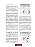
cell surface proteoglycans, and enter the cell by endocytosis (170) (Fig. 7).
Experimental Approaches to Retinal Diseases 579
Figure 7 Liposome-mediated transfer of nucleic acid. The nucleic acid is con-
densed in the liposome to form lipoplexes. These enter the cell by endocytosis.
The majority of lipoplexes are trapped in late endosome. A small percentage can
either be released into the cytoplasm (mRNA), where they are functional, or traffic
non-specifically to the nucleus, where they may form episomes.
Copyright © 2003 Marcel Dekker, Inc.
Currently, no more than 30 genes transfer–competent cationic liposomes
have been developed and are commercially available. Perhaps the most
widely used formulations are DOTAP and DOTMA, the latter of which
is sold as Lipofectin, an in vitro transfecting agent.
A disadvantage of current methods of liposome-mediated gene trans-
fer is due to the large percentage of the DNA-bound complexes trapped in
late endosomes, where they undergo enzymatic degradation and are no
longer therapeutically useful. Only a small percentage of the bound nucleic
acid escapes systemic inactivation and endosome entrapment. Those that
manage to escape face yet another hurdle of getting to the nucleus and
maintaining their functional integrity. As with viral delivery, in vitro effec-
tiveness of liposome-mediated gene transfer is often misleading and is a poor
guide for clinical efficacy. Conventional liposome formulations lack cell
specificity and can take hours for uptake into the cell. They are highly
susceptible to inactivation by a number of serum proteins that bind and
cause membrane destabilization, a major obstacle for systemic administra-
tion of liposomes. A current research focus in the pharmaceutical industry is
the development of sterically stable liposome formulations that are resistant
to serum disruption and that will not aggregate prior to delivery. One mod-
ification currently in development uses conventional liposome lipid mem-
branes to covalently attach polymers such as polyethylene glycol (PEG-
lipid) to create stealth liposomes (152,155,156,162,169). Properly formulated
polymer-grafted liposomes are shown to be sterically stabilized compounds
that have long residence times in circulation, increased biodistribution, and
reduction in uptake by cells of the reticuloendothelial system. Other clini-
cally advantageous features of pegylated liposome pharmacokinetics include
dose independence and increased efficacy as a slow release system for ther-
apeutically active drugs. This is a fascinating technology that has the poten-
tial of being a tailor-made delivery system that will improve the therapeutic
index of a number of drugs.
Another modification strategy under active investigation is the manu-
facture of ligand-targeted liposomal drugs using combinatorial approaches.
Such molecular conjugates could potentially be more versatile than the
conventional systems (159–164). In a recent study, transfection was
observed to be increased in hepatoma cells after the administration of mod-
ified lipoplexes containing triantennary galactosyl residues that specifically
target hepatoma cells (172). Targeted delivery of doxorubicin to human
umbilical vein endothelial cells and subsequent decrease in the survival of
the cells were also achieved with immunoliposomes that were conjugated to
a monoclonal antibody against E-selectin, a surface marker of HUVECs
(173). While targeting will increase transfection efficiency to specific tissues,
it does not address problems of DNA release encountered in the endosomes.
580 Tombran-Tink
Copyright © 2003 Marcel Dekker, Inc.
Some researchers have shown that the association of amphiphilic peptides,
such as GALA, a pH-sensitive peptide, with cationic liposomes can induce
fusion and permeabilization at acidic pH values and improve release of the
DNA from endosomes. The peptides induce osmotic swelling and subse-
quent rupture of the endosomal membranes so that the DNA can escape
easily (174). These new modifications, however, are not without problems.
Competition between ligand-mediated processes and nonspecific interac-
tions with the cell membrane can hinder the efficacy of gene delivery and
must be resolved before ligand-modified liposomes are of clinical relevance.
In addition to engineering modifications that result in specific cell
targeting and more efficient DNA release, mechanisms that will increase
nucleic acid condensation and promote nuclear targeting are promising
areas of research that will improve liposome-mediated gene delivery.
Modifications using DOTAP liposome-protamine sulfate-DNA (LPD) for-
mulations are shown to produce denser particles when bound to DNA and
result in consistently higher gene expression levels (175,176). Complexes
formed with polycations are also observed to be much smaller than those
formed with liposomes alone and have increased resistance to nuclease
degradation. Smaller-size complexes may allow for higher levels of gene
expression because of increased cellular internalization. An interesting var-
iation to this hybrid concept is the use of UV-irradiated Japanese Sendai
virus (HVJ)-cationic liposome to facilitate nuclear targeting. The binding of
high mobility group 1 protein (HMG-1) increase the potency of the complex
and enhances nuclear targeting and stability of the DNA after delivery into
the nuclear envelope. The success of HVJ-liposome complexes in cancer
applications is thought to result from the ability of the complex to bypass
the endocytosis process, thereby minimizing the difficulties encountered
when the DNA is released from the endosomes (171). The development of
‘‘synthetic chemical viruses’’ that are capable of (a) extended blood circula-
tion, (b) increased DNA microparticle condensation, (c) improved cellular
uptake, (d) flexible tropism, (e) escaping enzymatic degradation, and (f)
nuclear targeting is an attractive challenge in the area of biopharmaceutics.
If realized, such compounds have enormous potential in gene therapy pro-
tocols and may surpass the clinical usefulness of viral vectors.
Liposome-based techniques have been optimized to successfully trans-
fer functional genes into human primary RPE cells. In one study, differences
in the efficiency of transfection were observed between the types of lipo-
somes used in the assay. Nontoxic transfer was achieved after each liposome
treatment, but the Tfx-50 formulation showed the most significant results
when compared to transfection of the RPE cells with other liposome varia-
tions, including lipofectin, lipofectamine, Cellfectin, and DMRIE-C (177).
A fascinating variation of gene transfer by liposomes was achieved by a
Experimental Approaches to Retinal Diseases 581
Copyright © 2003 Marcel Dekker, Inc.
group of researchers who used liposome eye drops to transfer rat retinal
ganglion cells. Transfection was reported to be efficient and nontoxic to
ocular tissues. This approach represents an interesting development in non-
surgical gene delivery for retinal diseases (178). The use of another liposome
method, hemagglutinating virus of Japan liposomes, was tested for efficacy
in delivering tissue inhibitor of metalloproteinase-3 gene into rat RPE cells.
Not only was the transfection successful, but expression of the introduced
gene inhibited the development of experimental choroidal neovasculariza-
tion induced by laser photocoagulation after transfection of the tissue (179).
These are only a few examples showing the feasibility of using, nonviral,
nontoxic synthetic DNA-complexing derivatives to transfer therapeutic
genes to the retina.
The development of innovative nonviral delivery system is still in its
infancy, but many advantages are associated with their use in gene transfer
applications: (a) they can package and deliver a transgene of any size; (b)
packaging cell lines are not required to generate high titers; (c) they are non-
pathogenic and cannot replicate; (d) immunogenicity, toxicity, and inflam-
mation are minimized with their use; and (e) they can become completely
synthetic. While these are safe gene delivery systems, the disadvantages
currently lie in their overall inefficiency of transfection and their inability
to achieve cell-specific and nuclear targeting. Modifications that improve
these features will allow synthetic polymer-based gene vectors to be the
candidates of choice for pharmacological intervention in many diseases.
5. Gene Knock down Therapy
a. Antisense Drugs. Antisense technology is a novel gene delivery
method that is increasingly applied to knock down the expression of a
specific target gene for therapeutic purposes or to study the function of
that gene. The fundamental principle of the antisense approach is to si-
lence a gene using a short synthetic DNA or RNA sequence that is homo-
logous to that contained within the target gene. Antisense
oligodeoxynucleotides (ODNs) are synthesized in the opposite direction of
the known complementary DNA sequence and are designed to hybridize
specifically with their target sequences to interrupt the production of the
corresponding protein. Almost all human diseases are associated with a
dysfunctional protein. While most conventional drugs are designed to in-
hibit the disease-causing activity of a dysfunctional protein, antisense mo-
lecules are designed to inhibit the production of the protein. In principle,
gene silencing may be accomplished at the genetic level by inhibiting bio-
logical events, such as transcription, translation, or gene splicing (180–
183). During the inhibition of transcription events, ODNs bind to double-
582 Tombran-Tink
Copyright © 2003 Marcel Dekker, Inc.
stranded DNA to induce the formation of a short triple-helical structure.
This structure is mediated by Hoogsteen hydrogen bonds and sterically
hinders the transcription of a specific mRNA. In addition, translation of
RNA species can be interrupted by binding of ODNs to tRNA or pre-
RNA to prevent their transport from the nucleus or by directly interacting
with target mRNA molecule after transcription. In cases where inhibition
occurs after the transcript is matured, antisense binding to RNA is in-
tended to block ribosomal assembly or ribosomal sliding along the
mRNA during translation of the protein. ODNs can also be of therapeu-
tic value if they are designed to target the intron-exon junctions of prema-
ture RNA. In this regard, they prevent splicing events that are essential
for maturation of the RNA transcript. Three regions of the RNA that are
considered the best targets when designing ODNs are the 5
0
Cap region,
the AUG translational initiation codon, and the 3
0
untranslated region.
The concept of disabling the function of a mRNA by hybridization of
antisense reagents is a simple one, but, like other gene-based therapies, the
technology has encountered difficulties in the past. The technical problems
experienced in the early pioneering stages of antisense technology are only
now being elucidated and are the focus of active study. From these analyses,
several features are apparent in the design of effective antisense molecules:
determining the length of sequence with the greatest activity and specificity;
cellular uptake; specific targeting of the ODN; antisense stability; and toxi-
city. Other factors that have influenced the effectiveness of antisense mole-
cules are frequency of protein turnover, the intracellular environment of the
cell, and the extent of longevity of ODNs after administration.
Gene knockdown practices are still under development and will
require significant modifications before being clinically acceptable as a ther-
apeutic modality. Introducing variations in antisense chemistry by subtle
changes in the phosphate or sugar moieties of the nucleic acid backbone
is one method that shows success in minimizing nuclease degradation of the
molecules. Replacing a nonbridging oxygen with a sulfur atom in the phos-
phodiester bond between nucleotides on the phosphate backbone generates
a phosphorothioate linkage, which is reported to be one of the most success-
ful modification of antisense oligonucleotides to date (184,195).
Phosphorothioate compounds have shown efficacy in delivery and are less
vulnerable to intracellular nuclease degradation. A disadvantage of their
use, however, is that the constructs are chiral and form a racemix mixture
of ODN species that exhibit both desirable and undesirable properties in
vivo (186). Some ODNs are reported to be toxic, while others show non-
specific affinity for proteins (1987). The technical progress in chemical mod-
ification of antisense has recently shifted from the first-generation
phosphodiester oligonucleotides, which are still nuclease sensitive, to the
Experimental Approaches to Retinal Diseases 583
Copyright © 2003 Marcel Dekker, Inc.
more nuclease-resistant chimeric compounds that contain methoxyethyl
modifications at the end of the ODNs. The development of oligonucleotide
conjugates with cell-penetrating and nuclear-targeting peptides and colloidal
antisense carriers that protect against degradation is emerging rapidly and
will significantly improve cellular uptake, stability, subcellular trafficking,
and increased in vivo activity (188–192). Over 200 patents disclose antisense
sequences with therapeutic utility in the treatment of human diseases. It is a
powerful tool with exceptional clinical value and is being exploited to iden-
tify gene function and validate new drug targets.
Formivirsen (ISIS 2922) is the first antisense oligonucleotide drug
approved for the treatment of cytomegalovirus (CMV)–induced retinitis.
The 21-phosphorothioate oligonucleotide inhibits viral replication in the
human eye by binding to complementary sequences of early mRNA CMV
viral transcripts. In preliminary clinical trials, the progression of CMV reti-
nitis in AIDS patients is significantly delayed after intravitreal administra-
tion of formivirsen. Drug-clearance studies show that formivirsen exhibits
first-order kinetics with a half-life of 62 hours in rabbits and 78 hours in
monkey. A mild and transient inflammatory response and increase in intrao-
cular pressure are observed after treatment with formivirsen. These appear
to be resolved spontaneously or reversed with topical steroid treatment
(193–196).
Diseases characterized by retinal neovascularization are among the
principal candidates for antisense treatment. The use of antisense oligonu-
cleotide against vascular endothelial growth factor (VEGF) has shown pro-
mising results for the treatment of proliferative retinopathy. After
intraocular administration in a murine model of retinal neovascularization,
phosphorothioate antisense molecules reduced VEGF protein synthesis and
the growth of new blood vessels in a dosage-dependent manner. The study
shows the therapeutic potential of ODNs in ischemia-induced proliferative
retinopathies (197). Proliferative vitreoretinopathy (PVR) is an ocular dis-
order often associated with proliferating RPE cells. Antisense knockdown of
c-myc, a protein active in the mitogenic pathway, inhibits the proliferation
of human retinal pigment epithelial cells, suggesting that c-myc ODNs may
be an exciting perspective in the treatment of PVR (198).
Retinal ganglion cell death is associated with increased expression of
the Bax protein after transection of the optic nerve. A phosphorothioate
Bax antisense oligonucleotide was reported to show therapeutic utility in
preserving ganglion cell following axotomy. Bax expression was reduced
and the number of surviving neurons increased after treatment with Bax
ODNs. This represents a novel approach for neurodegeneration due to
optic nerve injury (199). The use of ODNs to silence the expression of
another retinal gene GLAST, a glial glutamate transporter, showed sig-
584 Tombran-Tink
Copyright © 2003 Marcel Dekker, Inc.
nificant changes in normal retinal transmission and indicates the impor-
tance of GLAST in maintaining retinal function (200). Similarly, an anti-
sense compound generated against the trkB receptor mRNA for brain-
derived neurotrophic factor (BDNF) alters the neurochemical phenotype
of retinal neurons (201). BDNF and its receptor are important to survival
and differentiation of the retina and are potentially useful targets in retinal
degenerative diseases. Antisense targeting of fibronectin transcripts was
also shown to reduce the expression of fibronectin in retinal vascular
cells (202). The use of antisense oligonucleotides in these studies reflects
the significance of the technology in understanding the function and reg-
ulation of a specific protein and the potential therapeutic benefits for
antisense-based ocular therapies.
b. Ribozymes. Ribozymes are naturally occurring catalytic RNA
and a new class of genetic tools used to inhibit gene expression. Designer
ribozymes are chemically designed to recognize and bind specific RNA
through complementary base-pair hybridization. Their value in human
therapeutics is dependent on their ability to distinguish between mutant
and wild-type RNA species and to act as molecular scissors to digest or
edit the target RNA in a way that will prevent translation of the corre-
sponding protein (203–205). There are developed as an alternate approach
to antisense drugs. Analysis of the physical, biochemical, and biological
properties of naturally occurring ribozymes has allowed researchers to
classify them according to their various catalytic functions:
1. Hammerhead ribozymes: These are approximately 30 nucleotides
long and the smallest ribozymes identified. They are found in
many viral DNA and are capable of site-specific cleavage of a
phosphodiester bond. Hammerhead ribozymes have been exten-
sively studied, and many have been synthesized against RNA
targets. In recent years they have emerged as a potentially effec-
tive therapeutic measure in models of retinitis pigmentosa. In
areas of the brain, hammerhead ribozymes have been directed
against the amyloid peptide precursor (B-APP), which is asso-
ciated with the pathogenesis of Alzheimer’s disease. Others, such
as angiozyme, have been synthesized against angiogenic pro-
cesses involved in the progression of tumor metastasis.
2. Group 1 and Group 11 intron ribozymes: These species can self-
splice, digest, and ligate phosphodiester bonds. They are found in
lower eukaryotes and some bacteria. Group 1 intron ribozymes
mediate trans-splicing of RNA targets and is considered a useful
genetic tool in repairing mutations in defective genes.
Experimental Approaches to Retinal Diseases 585
Copyright © 2003 Marcel Dekker, Inc.
3. Ribonuclease P: Cleaves phosphodiester bonds of tRNA precur-
sor molecules.
The catalytic activity of these molecules make them particularly interesting
in the treatment of dominantly inherited diseases. In autosomal dominant
retinitis pigmentosa (ADRP), a substitution of histidine for proline occurs
at codon 23 in the rhodopsin gene. This mutation is referred to as P23H and
is responsible for the synthesis of a mutant gene product that results in the
death of photoreceptor cells (206). Because the field is relatively new, only a
few studies have been carried out in the retina to test the therapeutic effect of
ribozymes in ocular diseases. One research team has now shown that in vivo
expression and activity of hairpin and hammerhead ribozymes can be
achieved in a transgenic rat model of ADRP. Efficient transduction and
stable expression of the ribozymes were accomplished using an adeno-asso-
ciated virus that contained a rod opsin promoter. The results suggested that
the expressed ribozymes discriminated between wild-type and mutant rho-
dopsin RNA and specifically destroyed the P23H mutant specie. As a result,
translation of the P23H protein was inhibited and progression of photore-
ceptor degeneration in ADRP model was significantly slowed down (206–
212). Combining the advantages of current gene delivery strategies with
catalytic ribozymes has broad therapeutic implications for dominantly
expressed retinal diseases where the disease is already in progression.
E. Neurotrophic Factors
The neurotrophic approach to treating retinal diseases is of therapeutic
relevance in ophthalmology because trophic factors target apoptotic
mechanisms that are independent of the genetic mutation(s) for the disease.
Treatment with soluble neurotrophic factors has been shown to prevent the
death of retinal neurons in complex or difficult-to-treat ocular diseases
where the etiologies are not completely defined or where mutations in sev-
eral genes are associated with the progression of the disease. The method of
delivering highly concentrated amounts of trophic factors to the eye is
straightforward and relatively simple to perform and bypasses the need
for complex viral or non viral delivery systems. Subretinal or intravitreal
injections are common routes of delivery to the affected area. Preparative
amounts of neurotrophic proteins can be easily purified from recombinant
expression systems, and combinations of several therapeutic proteins can be
administered simultaneously to the area of pathology. Another clinically
appealing feature of this approach is that the therapeutic efficacy of soluble
trophic factors is not hindered by the immunologic and toxic limitations
that are usually associated with vector-mediated delivery of DNA.
586 Tombran-Tink
Copyright © 2003 Marcel Dekker, Inc.
Designinganeffectivetreatmentprotocol,however,isbasedonadequate
knowledgeofthepharmacokineticsofthetrophicfactorinabiological
systemandestablishingitsabilitytofunctioninaphysiologicalenviron-
ment.Alimitationassociatedwiththeuseoftrophicfactorsinretinaldis-
easesistheneedformultipletreatmentstosignificantlyeffectreversalofthe
pathology.Unlesstheseagentsareadministeredtopicallytotheeyeor
packagedinaslow-releasesystem,themethod,whilesafe,isnotconvenient
forlong-termmanagementofretinaldiseases.
Oneendogenousneurotrophicfactor,whichwehaveisolatedand
characterizedinourlaboratory,isa50kDaprotein,pigmentepithelum-
derivedfactor(PEDF)(103,213),sonamedbecauseitwasinitiallyisolated
fromtheretinalpigmentepithelium.Functionally,thereisastrikingasso-
ciationbetweenPEDF,orthelackthereof,andbiologicalprocessesinvol-
vingsurvivalanddeathofretinalcellsaswellasangiogenicmechanismsin
theeye(35,215–219,229).ThePEDFgeneiswellcharacterizedandisclas-
sifiedasaserineproteaseinhibitorbecauseofitsstructuralandsequence
homologywithmembersofthisgroupofgenes(104,227).Inaddition,
PEDFmapstohumanchromosome17p13.3andistightlylinkedtoan
autosomaldominantretinitispigmentosalocusinthatregionofthechro-
mosome.Severalpolymorphismshavebeenidentifiedinthegene,butnone
hasshownadirectcorrelationbetweenPEDFandspecificretinalpatholo-
gies(220–224).However,invivoandinvitrostudieswiththesolubleprotein
consistentlydemonstratetheneuroprotectiveandantiangiogenicactivities
ofPEDF,suggestingapromisingfutureforthisproteinasatherapeuticthat
cancircumventtheeffectsofspecificmutationsorchemicalstimulatorsthat
causethedeathofvisualcells.
WefirstidentifiedthePEDFproteinintheconditionedmediumof
primaryculturesoffetalhumanRPEcellandintheinterphotoreceptor
matrix(IPM)locatedbetweentheRPEandneuralretina
(103,213,225,226).Theproteinisexpressedinhighconcentrationinfetal
andyoungadultRPEcellsbutappearstobeseverelydownregulatedin
senescingRPEcultures,afindingthatsuggeststhatitmayplayarolein
age-relatedretinaldysfunctions.Inoneofthefirststudies,weshowedthat
PEDFinhibitsthegrowthofahumanretinoblastomacellline(Y79)by
inducingdifferentiationofthetumorcellsintoaphenotypethatisreminis-
centofmaturedneurons.Innontreatedcultures,theY79cellsgrowas
clustersinsuspensionanddonotspontaneouslyattachordifferentiate.
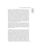
TreatmentofthesecellswithasmalldoseofPEDFiseffectiveinpromoting
extensiveneuriteoutgrowthsfromthetumorcells,upregulatingneurofila-
mentproteinsandneuron-specificenolase,andpromotingconnections
betweenthegrowingneuritesofnewlydifferentiatedcells(Fig.8).
Approximately 90% of the cultures attach and differentiate on poly-d-
Experimental Approaches to Retinal Diseases 587
Copyright © 2003 Marcel Dekker, Inc.
angiostatinandendostatin,itsefficacywasslightlymorepotentthanthose
inhibitors.Insupportoftheirstudy,weshowedhigherconcentrationsof
PEDFinthevitreousofpatientswithavascularproliferativevitrealretino-
pathyanddiabeticretinopathywhencomparedtopatientswithretinal
pathologiesassociatedwithincreasedangiogenicactivity(228).Basedon
theclinicaldata,aswellasvivostudiesusinganimalmodels,itappearsthat
theconcentrationofPEDFintheeyeisimportanttothevascularstateof
oculartissues.Theseresultshavestirredmuchinterestintheophthalmicfield
andhaveencouragedseveralgroupstoexploitthetherapeuticpotentialof
PEDFinoculardiseases,suchasage-relatedmaculardegeneration,where
bothcelldeathandincreasedangiogenesiscontributetoseverevisualloss.
Inseveralmodesofinducedretinaldegeneration,convincingevidence
thatphotoreceptorcellssurviveinthepresenceofPEDFhasbeenprovided.
Inaninvitromodelofretinaldamage,alargepercentageofretinalneurons
undergoapoptosisanddieafterexposuretohydrogenperoxide(H
2
O
2
)
(215–217).Hydrogenperoxideisareactiveoxygenspecies(ROS)foundin
elevatedconcentrationinlight-damagedretinas.ItisbelievedthatROS
contributetodegenerativeandagingprocessesintheeye.Totestthepro-
tectiveeffectsofPEDFinH
2
O
2
-damagedeyes,ratretinalcultureswere
treatedwithPEDFbeforetheywereexposedtoH
2
O
2
.Inthepresenceof
PEDF,apoptoticmechanismsthatledtocelldeathwereinhibited,and
approximately60%ofthecellsthatwouldhaveotherwisedegenerated
survived.Furthermore,ahighpercentageofthetreatedcellswererhodopsin
positiveand,therefore,highlylikelytoberodphotoreceptors.Invivo,the
retinacanalsobedamagedbyexposuretoconstantlight,inpartbecauseof
thegenerationofreactiveoxygenspeciesinahigh-lipid-contentregionof
theretina.Photoreceptordegenerationisvisibleasearlyasthethirddayof
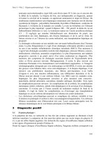
lightexposureintherat.InastudyaimedattestingtheeffectofPEDFin
light-damagedrateyes,wefoundthatasingleintravitrealinjectionof
PEDF,priortochroniclightexposure,waspotentenoughtoinhibitthe
lightdamageeffectsonphotoreceptor.Thiswasclearlyseeninhistological
preparationsofthetreatedretinaandelectrophysicalmeasurementsofthe
nucleiintheouternuclearlayer(ONL)(Fig.9).
In a similar study, photoreceptor survival with PEDF treatment was
examined in two mutant mice types, homozygous retinal degeneration (rd/
rd) and retinal degeneration slow (rds/rds), in which photoreceptor loss is a
hallmark of the mutations. Intravitreal injections of PEDF resulted in a
transient but significant delay in the death of photoreceptors in both
mutants (229). The efficacy of PEDF was also assessed in an embryonic
Xenopus model of retinal degeneration (218). In this model, mechanical
removal of the RPE cells from the Xenopus retina results in a distortion
of photoreceptor ultrastructure and disruption of outersegment formation,
Experimental Approaches to Retinal Diseases 589
Copyright © 2003 Marcel Dekker, Inc.
retinas was blocked by a neutralizing polyclonal antibody to the 50 kDa
native protein, suggesting that the rescuing effect was specific.
From these findings it appears that the course of photoreceptor degen-
eration can be altered by neuroprotective agents, like PEDF, which can
prevent pathomorphological and apoptotic effects of neurodegenerative
promoters. Other trophic factors, such as bFGF, CNTF, and BDNF have
shown similar results in promoting photoreceptor survival in naturally
occurring inherited retinal degeneration models with genetic defects similar
to those in human inherited retinal degeneration (230). Survival factors are,
therefore, particularly attractive therapeutic tools that may prove to be
increasingly important in treating retinal degenerations if long-term, sus-
tained delivery to the affected area is to be maintained.
REFERENCES
1. Adler, R., Curcio, C., Hicks, D., Price, D., and Wong, F. (1999). Cell death in
age-related macular degeneration. Mol. Vis., 5:31.
2. Votruba, M., and Gregor, Z. (2001). Neovascular age-related macular degen-
eration: present and future treatment options. Eye, 15(Pt 3):424–429.
3. Sickenberg, M. (2001). Early detection, diagnosis and management of chor-
oidal neovascularization in age-related macular degeneration: the role of
ophthalmologists. Ophthalmologica, 215(4):247–253.
4. Rubin, G. S. (2001). Vision rehabilitation for patients with age-related macu-
lar degeneration. Eye, 15(Pt 3):430–435.
5. Harding, S. (2001). Photodynamic therapy in the treatment of subfoveal chor-
oidal neovascularization. Eye, 15(Pt 3):407–412.
6. Schmidt-Erfurth U., and Hasan, T. (2000). Mechanisms of action of photo-
dynamic therapy with verteporfin for the treatment of age-related macular
degeneration. Surv. Ophthalmol., 45(3):195–214.
7. Verma, L., Das, T., Binder, S., Heriot, W. J., Kirchhof, B., Venkatesh, P.,
Krebs, I., Stolba, U., Jahn, C., Feichtinger, H., Kellner, L., Krugluger, H.,
Pawelka, I., Frohner, U., Kruger, A., Li, W., and Tewari, H. K. (2000). New
approaches in the management of choroidal neovascular membrane in age-
related macular degeneration. Indian J. Ophthalmol., 48(4):263–278.
8. Ho, Ac. (1999). Laser treatment in eyes with drusen. Curr. Opin. Ophthalmol.,
10(3):204–208.
9. Hageman, G. S., and Mullins, R. F. (1999). Molecular composition of drusen
as related to substructural phenotype. Mol. Vis., 5:28.
10. Blodi, C. F., and Stone, E. M. (1990). Best’s vitelliform dystrophy. Ophthal.
Paediatr. Genet., 11(1):49–59.
11. Bither, P. P., and Berns, L. A. (1988). Stargardt’s disease: a review of the
literature. J. Am. Optom. Assoc., 59(2):106–111.
Experimental Approaches to Retinal Diseases 591
Copyright © 2003 Marcel Dekker, Inc.
12. Phelan, J. K., and Bok, D. (2000). A brief review of retinitis pigmentosa and
the identified retinitis pigmentosa genes. Mol. Vis., 6:116–124.
13. Baumgartner, W. A. (2000). Etiology, pathogenesis, and experimental treat-
ment of retinitis pigmentosa. Med. Hypotheses, 54(5):814–824.
14. van Soest, S., Westerveld, A., de Jong, P. T., Bleeker-Wagemakers, E. M., and
Bergen, A. A. (1999). Retinitis pigmentosa: defined from a molecular point of
view. Surv. Ophthalmol., 43(4):321–334.
15. Shastry, B. S. (2000). Hereditary degenerative retinopathies: optimism for
somatic gene therapy. IUBMB Life, 49(6):479–484.
16. Eudy, J. D., and Sumegi, J. (1999). Molecular genetics of Usher syndrome.
Cell Mol. Life Sci., 56(3–4):258–267.
17. Perrault, I., Rozet, J. M., Gerber, S., Ghazi, I., Leowski, C., Ducroq, D.,
Souied, E., Dufier, J. L., Munnich, A., and Kaplan, J. (1999). Leber conge-
nital amaurosis. Mol. Genet. Metab., 68(2):200–208.
18. Harris, E. W. (2001). Leber’s congenital amaurosis and RPE65. Int.
Ophthalmol. Clin., 41(1):73–82.
19. Algvere, P. V., Berglin, L., Gouras, P., and Shen, Y. (1994). Transplantation
of fetal retinal pigment epithelium in age-related macular degeneration with
subfoveal neovascularization. Graefes Arch. Clin. Exp. Ophthalmol., 232:707–
716.
20. Mohand-Said, S., Hicks, D., Dreyfus, H., and Sashel, J. A. (2000). Selective
transplantation of rods delays cone loss in a retinitis pigmentosa model. Arch.
Ophthalmol., 118:807–811.
21. Sheedlo, H. J., Li, L., and Turner, J. E. (1991). Photoreceptor cell rescue at
early and late RPE-cell transplantation periods during retinal disease in RCS
dystrophic rats. J. Neural Transplant Plast., 2:55–63.
22. Rezai, K. A., Kohen, L., Wiedemann, P., and Heimann, K. (1997). Iris pig-
ment epithelium transplantation. Graefes Arch. Clin. Exp. Ophthalmol.,
235:558–562.
23. Bhatt, N. S., Newsome, D. A., and Fenech, T. (1994). Experimental trans-
plantation of human retinal pigment epithelial cells on collagen substrates.
Am. J. Ophthalmol., 117:214–221.
24. Jiang, L. Q., Jorquera, M., and Streilein, J. W. (1993). Subretinal space and
vitreous cavity as immunological privileged sites for retinal allografts. Invest.
Ophthalmol. Vis. Sci., 34:3347–3354.
25. Mohand-Said, S., Hicks, D., Dreyfus, H., and Sashel, J. A. (2000). Selective
transplantation of rods delays cone loss in a retinitis pigmentosa model. Arch.
Ophthalmol., 118:807–811.
26. Little, C. W., Castillo, B., DiLoreto, D. A., Cox, C., Wyatt, J., del Cerro, C.,
and del Cerro, M. (1996). Transplantation of human fetal retinal pigment
epithelium rescues photoreceptor cells from degeneration in the Royal
College of Surgeons on rat retina. Invest. Ophthalmol. Vis. Sci., 37(1):204–211.
27. Sauve, Y., Klassen, H., Whiteley, S. J., and Lund, R. D. (1998). Visual field
loss in RCS rats and the effect of RPE cell transplantation. Exp. Neurol.,
152(2):243–250.
592 Tombran-Tink
Copyright © 2003 Marcel Dekker, Inc.
28. Sharma, R. K., Bergstrom, A., Zucker, C. L., Adolph, A. R., and Ehringer, B.
(2000). Survival of long-term retinal cell transplants. Acta. Ophthalmol.
Scand., 78:396–402.
29. Aramant, R. B., Seiler, M. J., and Ball, S. L. (1999). Successful cotransplanta-
tion of intact sheets of fetal retinal with retinal pigment epithelium. Invest.
Ophthalmol. Vis. Sci., 40:1557–1564.
30. Woch, G., Aramant, R. B., Seiler, M. J., Sagdullaev, B. T., and McCall, M. A.
(2001). Retinal transplants restore visually evoked responses in rats with
photoreceptor degeneration. Invest. Ophthalmol. Vis. Sci., 42(7):1669–1676.
31. Schraermeyer, U., Kayatz, P., Thumann, G., Luther, T. T., Szurman, P.,
Kociok, N., and Bartz-Schmidt, K. U. (2000). Transplantation of iris pigment
epithelium into the choroid slows down the degeneration of photoreceptors in
the RCS rat. Graefes Arch. Clin. Exp. Ophthalmol., 238(12):979–984.
32. Schraermeyer, U., Kociok, N., and Heimann, K. (1999). Rescue effect of IPE
transplants in RCS rats: short-term results. Invest. Ophthalmol. Vis. Sci.,
40(7):1545–1556.
33. Thumann, G., Bartz-Schmidt, K. U., El Bakri, H., Schraermeyer, U., Spee,
C., Cui, J. Z., Hinton, D. R., Ryan, S. J., and Heimann, K. (1999).
Transplantation of autologous iris pigment epithelium to the subretinal
space in rabbits. Transplantation, 68(2):195–201.
34. Carwile, M. E., Culbert, R. B., Sturdivant, R. L., and Kraft, T. W. (1998).
Rod outer segment maintenance is enhanced in the presence of bFGF, CNTF
and GDNF. Exp. Eye Res., 66(6):791–805.
35. Abe, T., Tomita, H., Kano, T., Yoshida, M., Ohashi, T., Nakamura, Y.,
Nishikawa, S., and Tamai, M. (2000). Autologous Iris pigment epithelial
cell transplantation in monkey subretinal region. Curr. Eye Res., 20:268–275.
36. Humayun, M. S., de Juan, E., del Cerro, M., Dagnelie, G., Radner, W.,
Sadda, S. R., and del Cerro, C. (2000). Human neural retinal transplantation.
Invest. Ophthalmol. Vis. Sci., 41:3100–3106.
37. Algvere, P. V., Gouras, P., and Dafgard Kopp, E. (1999). Long-term outcome
of RPE allografts in non-immunosuppressed patients with AMD. Eur. J.
Ophthalmol., 9(3):217–230.
38. Thumann, G., Aisenbrey, S., Schraermeyer, U., Laufaut, B., Esser, P., Walter,
P., and Bartz-Schmidt, K. U. (2000). Transplantation of autologous iris pig-
ment epithelium after removal of choroidal neovascular membranes. Arch.
Ophthalmol., 118(10):1350–1355.
39. Bhatt, N. S., Newsome, Da., and French, T. (1994). Experimental transplan-
tation of human retinal pigment epithelial cells on collagen substrates. Am. J.
Ophthalmol., 117:214–221.
40. Fang, S. R., Kaplan, H. J., Del Piore, L. V., Liu, Y., Wang, X., Hornbect, R.,
Landgraf, M., Mason, G., and Silverman, M. S. (1993). Development of a
surgical procedure and instrument for transplantation of extended gelatin
sheets to the subretinal space. Invest. Ophthalmol. Vis. Sci., 34:1093.
Experimental Approaches to Retinal Diseases 593
Copyright © 2003 Marcel Dekker, Inc.
41. Thomas, J. A., Itskovitz-Eldor, J., Shapiro, S. S., Waknitz, M. A., Swiergiel, J.
J., Marshall, V. S., and Jones, J. M. (1998). Embryonic stem cell lines derived
from human blastocysts. Science, 282:1145–1147.
42. Shamblott, M. J., Axeman, J., Wang, S., Bugg, E. M., Littlefield, J. W.,
Donovan, P. J., Blumenthal, P. D., Huggins, G. R., and Gearhart, J. D.
(1998). Derivation of pluripotent system cells from cultured human primordial
germ cells. Proc. Natl. Acad. Sci. USA, 95:13726–13731.
43. Bjornson, C. R., Rietze, R. L., Reynolds, B. A., Magli, M. C., Vescovi, A. L.
(1999). Turning brain into blood: a hematopoietic fate adopted by adult
neural stem cells in vivo. Science, 283(5401):534–537.
44. Joshi, S. S., Tarantolo, S. R., Kuszynski, C. A., and Kessinger, A. (2000).
Antitumor therapeutic potential of activated human umbilical cord blood cells
against leukemia and breast cancer. Clin. Cancer Res., 6(11):4351–4358.
45. Gluckman, E. (2000). Current status of umbilical cord blood hematopoietic
stem cell transplantation. Exp. Hematol., 28(11):1197–1205.
46. Howery, R. P., Martin, P. L., Driscoll, T., Szabolcs, P., Kelly, T., Shpall, E. J.,
Bearman, S. I., Slat-Vasquez, V., Rubinstein, P., Stevens, C. E., and
Kurtzberg, J. (2000). Graft-versus-leukemia-induced complete remission fol-
lowing unrelated umbilical cord blood transplantation for acute leukemia.
Bone Marrow Transplant, 26(11):1251–1254.
47. Orlic, D., Kajstura, J., Chimenti, S., Jakoniuk, I., Anderson, S. M., Li, B.,
Pikel, J., McKay, R., Nadal-Ginard, B., Bodine, D. M., Leri, A., and
Anversa, P. (2001). Bone marrow cells regenerate infarcted myocardium.
Nature, 410(6829):701–705.
48. Brown, J. M., Weissman, I. L., and Shizuru, J. A. (2001). Immunity to infec-
tions following hematopoietic cell transplantation. Curr. Opin. Immunol.,
13(4):451–457.
49. Terskikh, A. V., Easterday, M. C., Li, L., Hood, L., Kornblum, H. I.,
Geschwind, D. H., and Weissman, I. L. (2001). From hematopoiesis to neu-
ropoiesis: evidence of overlapping genetic programs. Proc. Natl. Acad. Sci.
USA, 98(14):7934–7939.
50. Gokhan, S., and Mehler, M. F. (2001). Basic and clinical neuroscience appli-
cations of embryonic stem cells. Anat. Rec., 265(3):142–156.
51. Livesey, F. J., and Cepko, C. L. (2001). Vertebrate neural cell-fate determina-
tion: lessons from the retina. Nat. Rev. Neurosci., 2(2):109–118.
52. Tropepe, V., Coles, B. L., Chiasson, B. J., Horsford, D. J., Elia, A. J.,
McInnes, R. R., and van der Kooy, D. (2000). Retinal stem cells in the
adult mammalian eye. Science, 287(5460):2032–2036.
53. Ahmed, I., Tang, L., and Pham, H. (2000). Identification of neural progeni-
tors in the adult mammalian eye. Biochem. Biophys. Res. Commun.,
270(2):517–521.
54. Henderson, T. R., Coster, D. J., and Williams, K. A. (2001). The long term
outcome of limbal allografts: the search for surviving cells. Br. J. Ophthalmol.,
85(5):604–609.
594 Tombran-Tink
Copyright © 2003 Marcel Dekker, Inc.
55. Perron, M., and Harris, W. A. (2000). Retinal stem cells in vertebrates.
Bioessays, 22(8):685–688.
56. Tamalu, F., Chiba, C., Ishida, A. T., and Saito, T. (2000). Functional differ-
entiation of ganglion cells from multipotent progenitor cells in sliced retinal of
adult goldfish. J. Comp. Neurol., 419(3):297–305.
57. Layer, P. G., Rothermel, A., and Willbold, E. (2001). From stem cells towards
neural layers: a lesson from re-aggregated embryonic retinal cells.
Neuroreport, 12(7):A39–46.
58. Otteson, D. C., D. Costa, A. R., Hitchcock, P. F. (2001). Putative stem cells
and the lineage of rod photoreceptors in the mature retina of the goldfish. Dev.
Biol., 232(1):62–76.
59. Kurimoto, Y., Shibuki, H., Kaneko, Y., Ichikawa, M., Kurokawa, T.,
Takahashi, M., and Yoshimura, N. (2001). Transplantation if adult rat hip-
pocampus-derived neural stem cells into retina injured by transient ischemia.
Neurosci Lett., 306(1–2):57–60.
60. Takahashi, M., Palmer, T. D., Takahashi, J., and Gage, F. H. (1998).
Widespread integration and survival of adult-derived neural progenitor cells
in the developing optic retina. Molec. Cell Neurosci., 12(6):340–348.
61. Henderson, T. R., Coster, D. J., and Williams, K. A. (2001). The long term
outcome of limbal allografts: the search for surviving cells. Br. J. Ophthalmol.,
85(5):604–609.
62. Anderson, D. F., Ellies, P., Pires, R. T., and Tseng, S. C. (2001). Amniotic
membrane transplantation for partial limbal stem cell deficiency. Br. J.
Ophthalmol., 85(5):567–575.
63. Eiges, R., Schuldiner, M., Drukker, M., Yanuka, O., Itskovitz-Eldor, J., and
Benvenisty, N. (2001). Establishment of human embryonic stem cell-trans-
fected clones carrying a marker for undifferentiated cells. Curr. Biol.,
11(7):514–518.
64. Chow, A. Y. (1993). Electrical stimulation of the rabbit retinal with subretinal
electrodes and high density microphotodiode array implants. Invest.
Ophthalmol. Vis. Sci., 34:835.
65. Rizo, J. F. 3rd, Wyatt, J., Humayun, M., de Juan, E., Liu, W., Chow, A.,
Eckmiller, R., Zrenner, E., Yagi, T., and Abrams, G. (2001). Retinal prosthe-
sis: an encouraging first decade with major challenges ahead. Opthalmology,
108(1):13–14.
66. Rizzo, J. F., and Wyatt, J. (1997). Prospects for a visual prosthesis.
Neuroscientists, 3:251–262.
67. Eckmiller, R. (1997). Learning retina transplants with epiretinal contacts.
Ophthalmol. Res., 9:281–289.
68. Peyman, G., Chow, A. Y., Liang, C., Chow, V. Y., Perlman, J. I., and
Peachey, N. S. (1998). Subretinal semiconductor microphotodiode array.
Ophthalmic Surg. Lasers, 29(3):234–241.
69. Chow, A. Y., and Chow V. Y. (1997). Subretinal electrical stimulation of the
rabbit retina. Neurosci. Lett. 255:13–16.
Experimental Approaches to Retinal Diseases 595
Copyright © 2003 Marcel Dekker, Inc.
70. Zrenner, E., Stett, A., Weiss, S., Aramant, R. B., Guenther, E., Kohler, K.,
Miliczek, K. D., Seiler, M. J. and Haemmerle, H. (1999). Can subretinal
microphotodiodes successfully replace degenerated photoreceptors? Vision
Res., 39(15):2555–2567.
71. Peachey, N. S., and Chow, A. Y. (1999). Subretinal implantation of semicon-
ductor-based photodiodes: progress and challenges. J. Rehabil. Res. Dev.,
36(4):371–376.
72. Humayun, M. S., de Juan, G., Dagnelie, R. G., Greenberg, R. H., Propst, D.
H., and Phillips, (1996). Visual perception elicited by electrical stimulation of
the retina in blind humans. Arch. Ophthalmol., 114:40–46.
73. Zrenner, E., Miliczek, D. K., Gabel, V. P., Graf, H. G., Guenther, E.,
Haemmerle, H., Hoefflinger, B., Kohler, K., Nish, W., Schubert, M., Stett,
A., and Weiss, (1997). The development of subretinal microphotodiodes for
replacement of degenerated photoreceptors. Ophthalmic Res., 29:269–280.
74. Chow, A. Y., Pardue, M. T., Chow, V. Y., Peyman, G. A., Liang, C.,
Perlman, J. I., and Peachey, N. S. (2001). Implantation of silicon chip micro-
photodiode arrays into the cat subretinal space. IEEE Trans. Neural Syst.
Rehabil. Eng., 9(1):86–95.
75. Hesse, L., Schanze, T., Wilms, M., and Eger, M. (2000). Implantation of
retina stimulation electrodes and recording of electrical stimulation responses
in the visual cortex of the cat. Graefes Arch. Clin. Exp. Ophthalmol.,
238(10):840–845.
76. Walter, P., Szurman, P., Vobig, M., Berk, H., Ludtke-Handjery, H. C.,
Richter, H., Mittermayer, C., Heimann, K., and Sellhaus, B. (1999).
Successful long-term implantation of electrically inactive epiretinal microelec-
trode arrays in rabbits. Retina, 19(6):546–542.
77. Nadig, M. N. (1999). Development of a silicone retinal implant: cortical
evoked potentials following focal stimulation of the rabbit retina with light
and electricity. Clin. Neurophysiol., 110(9):1545–1553.
78. Grumet, A. E., Wyatt, J. L. Jr, and Rizzo, J. F. 3rd. (2000). Multi-electrode
stimulation and recording in the isolated retina. J. Neurosci. Methods,
101(1):31–42.
79. Hershey, A. D., and Chase, M. (1952). Independent functions of viral proteins
and nucleic acid in growth of bacteriophage. J. Gen. Physiol., 36:39–56.
80. Watson, J. D., and Crick, F. H. C. (1953). Molecular structure of nucleic
acids: a structure for deoxyribose nucleic acid. Nature, 171:737–738.
81. Crick, F. H. C., Barnett, L., Brenner, S., and Watts-Tobin, R. J. (1961).
General nature of the genetic code for proteins. Nature, 192:1227–1232.
82. Danos, O., and Mulligan, R. C. (1988). Safe and efficient generation of recom-
binant retroviruses with amphotropic and ecotropic host ranges. PNAS,
85:6460–6464.
83. Markowitz, D., Goff, S., and Bank, A. (1988). A safe packaging line for gene
transfer: separating viral genes on two different plasmids. J. Virol. 62:1120–
1124.
596 Tombran-Tink
Copyright © 2003 Marcel Dekker, Inc.
84. Cone, R. D., and Mulligan, R. C. (1992). High-efficiency gene transfer into
mammalian cells: generation of helper-free recombinant retrovirus with broad
mammalian host range. Biotechnology, 24:420–424.
85. Coffin, J. M. (1996). Retroviridae: the viruses and their replication. In: Fields
Virology (B. N. Fields, D. M. Knippe, and P. Howley, eds.). Lippincott-
Raven, Philadelphia, pp. 1767–1840.
86. Battini, J. L., Heard, J. M., and Danos, O. (1992). Receptor choice determi-
nants in the envelope glycoproteins of amphotropic, xenotropic, and polytro-
pic murine leukemia viruses. J. Virol, 66:1468–1475.
87. Sakamoto, T., Spee, C., Scuric, Z., Gordon, E. M., Hinton, D. R., Anderson,
W. F., and Ryan, S. J. (1998). Ability of retroviral transduction to modify the
angiogenic characteristics of RPE cells. Graefes Arch. Clin. Exp. Opthalmol.,
236(3):220–229.
88. Kido, M., Rich, K. A., Yang, G., Barron, E., Kohn, D. B., al-Ubaidi, M. R.,
Blanks, J. C., and Lang, G. (1996). Use of retroviral vector with an internal
opsin promoter to direct gene expression to retinal photoreceptor cells. Curr.
Eye Res., 15(8):833–844.
89. Couderec, B. C., de Neuville, S., Douin-Echinard, V., Serres, B., Manenti, S.,
Darbon, J. M., and Malecaze, F. (1999). Retrovirus-mediated transfer of a
suicide gene into lens epithelial cells in vitro and in an experimental model of
posterior capsule opacification. Curr. Eye Res., 19(6):472–482.
90. Schubert, C. A., Kimura, H., Spee, C., Hinton, D. R., Gordon, E. M.,
Anderson, W. F., and Ryan, S. J. (1997). Retrovirus-mediated transfer of
the suicide gene into retinal pigment epithelial cells in vitro. Curr. Eye Res.,
16(7):656–665.
91. Kimura, H., Sakamoto, T., Cardillo, J. A., Spee, C., Hinton, D. R, Gordon,
E. M., Anderson, W. F., and Ryan, S. J. (1996). Retrovirus-mediated suicide
gene transduction in the vitreous cavity of the eye: feasibility in prevention of
proliferative vitreoretinopathy. Hum. Gene Ther., 1:7(7):799–808.
92. Sakamoto, T., Kimura, H., Scuric, Z., Spee, C., Gordon, E. M., Hinton, D.
R., Anderson, W. F., and Ryan, S. J. (1995). Inhibition of experimental pro-
liferative vitreoretinopathy by retroviral vector-mediated transfer of suicide
gene. Can proliferative vitreoretinopathy be a target of gene therapy?
Ophthalmology, 102(10):1417–1424.
93. Seitz, B., Baktanian, E., Gordon, E. M., Anderson, W. F., LaBreed, L., and
McDonnell, P. J. (1998). Retroviral vector-mediated gene transfer into kera-
tocytes: in vitro effects of polybrene and protamine sulfate. Graefes Arch. Clin.
Exp. Ophthalmol., 236(8):602–612.
94. Bradshaw, J. J., Obritsch, W. F., Cho, B. J., Gregerson, D. S., and Holland, E.
J. (1999). Ex vivo transduction of corneal epithelial progenitor cells using a
retroviral vector. Invest. Ophthalmol. Vis. Sci., 40(1):230–235.
95. Murata, T., Cui, J., Taba, K. E., Oh, J. Y., Spee, C., Hinton, D. R., and Ryan,
S. J. (2000). The possibility of gene therapy for the treatment of choroidal
neovascularization. Ophthalmology, 107(7):1364–1373.
Experimental Approaches to Retinal Diseases 597
Copyright © 2003 Marcel Dekker, Inc.
96. Murata, T., Hoffman, S., Ishibashi, T., Spee, C., Gordon, E. M., Anderson,
W. F., Hinton, D. R., and Ryan, S. J. (1998). Retrovirus-mediated gene
transfer targeted to retinal photocoagulation sites. Diabetologia, 41(5):500–
506.
97. vanRaaij, M. J., Mitraki, A., Lavigne, G., and Cusack, S. (1999). A triple ?-
spiral in the adenovirus fibre shaft reveals a new structural motif for a fibrous
protein. Nature 401:935–938.
98. Shenk, T. (1996). Adenoviridae. The viruses and their replication. In: Fields
Virology (B. N. Fields, D. M. Knippe, and P. Howley, eds.). Lippincot-Raven,
Philadelphia, pp. 2111–2148.
99. Stratford-Perricaudet, L. D., Makeh, I., Perricaudet, M., and Briand, P.
(1992). Wide-spread long-term gene transfer to mouse skeletal muscles and
heart. J. Clin. Invest., 90:626–630.
100. Bett, A. J., Haddara, W., Prevec, L., and Graham, F. L. (1994). An efficient
and flexible system for construction of adenovirus vectors with insertions of
deletions in early regions 1 and 3. PNAS, 91:8802–8806.
101. Abraham, N. G., Da Silva, J. L., Dunn, M. W., Kigasawa, K., and Shibahara,
S. (1998). Retinal pigment epithelial cell-based gene therapy against hemoglo-
bin toxicity. Int. J. Mol. Med., 1(4):657–663.
102. Verma, L., Das, T., Binder, S., Heriot, W. J., Kirchhof, B., Venkatesh, P.,
Krebs, I., Stobla, U., Jahn, C., Feichtinger, H., Kellner, L., Krugluger, H.,
Pawelka, I., Frohner, U., Druger, A., Li, W., and Tewari, H. K. (2000). New
approaches in the management of choroidal neovascular membrane in age-
related macular degeneration. Indian J. Ophthalmol., 48 :263–278.
103. Tombran-Tink, J., Chader, G. G., and Johnson, L. V. (1991). PEDF: a pig-
ment epithelium-derived factor with potent neuronal differentiative activity.
Exp. Eye Res., 53(3):411–414.
104. Steele, F. R., Chader, G. J., Johnson, L. V., and Tombran-Tink, J. (1993).
Pigment epithelium-derived factor: neurotrophic activity and identification as
a member of the serine protease inhibitor gene family. Proc. Natl. Acad. Sci.,
15;90(4):1526–1530.
105. Dawson, D. W., Volpert, O. V., Gillis, P., Crawford, S. E., Xu, H., Benedict,
W., and Bouck, N. P. (1999). Pigment epithelium-derived factor: a potent
inhibitor of angiogenesis. Science, 9;285(5425):245–248.
106. Mori, K., Duh, E., Gehlbah, P., Ando, A., Takahashi, K., Pearlman, J., Mori,
K., Yang, H. S., Zack, D. J., Ettyreddy, D., Brough, D. E., Wei, L. L., and
Campochiaro, P. A. (2001). Pigment epithelium-derived factor inhibits retinal
and choroidal neovascularization. J. Cell Physiol., 188(2):253–263.
107. Honda, M., Sakamoto, T., Ishibashi, T., Inomata, H., and Ueno, H. (2000).
Experimental subretinal neovascularization is inhibited by adenovirus-
mediated soluble VEGF/flt-1 receptor gene transfection: a role of VEGF
and possible treatment for SRN in age-related macular degeneration. Gene
Ther., 7(11):978–985.
598 Tombran-Tink
Copyright © 2003 Marcel Dekker, Inc.
108. Cayouette, M., and Gravel, C. (1997). Adenovirus-mediated gene transfer of
ciliary neurotrophic factor can prevent photoreceptor degeneration in the
retinal degeneration (rd) mouse. Hum. Gene Ther., 8(4):423–430.
109. Cayouette, M., Behn, D., Sendtner, M., Lachapelle, P., and Gravel, C. (1998).
Intraocular gene transfer of ciliary neurotrophic factor prevents death and
increases responsiveness of rod photoreceptors in the retinal degeneration
slow mouse. J. Neurosci., 18(22):9282–9293.
110. Bennett, J., Pakola, S., Zeng, Y., and Maguire, A. (1996). Humoral response
after administration of E1-deleted adenoviruses: immune privilege of the sub-
retinal space. Hum. Gene Ther., 7(14):1763–1769.
111. Bennett, J., Tanabe, T., Sun, D., Zeng, Y., Kjeldbye, H., Gouras, P., and
Maguire, A. M. (1996). Photoreceptor cell rescue in retinal degeneration
(rd) mice by in vivo gene therapy. Nat. Med. 2(6):649–654.
112. Akimoto, M., Miyatake, S., Kogishi, J., Hangai, M., Okazaki, K., Takahashi,
J. C., Saiki, M., Iwaki, M., and Honda, Y. (1999). Adenovirally expressed
basis fibroblast growth factor rescues photoreceptor cells in RCS rats. Invest.
Ophthalmol. Vis. Sci., 40(2):273–279.
113. Bennett, J., Zehng, Y., Bajwa, R., Klatt, L., Li, Y., and Maguire, A. M.
(1998). Adenovirus-mediated delivery of rhodopsin-promoted bc1-2 results
in a delay in photoreceptor cell death in the rd/rd mouse.
114. Kumar-Singh, R., and Farber, D. B. (1998). Encapsidated adenovirus mini-
chromosome-mediated delivery of genes to the retina: application to the res-
cue of photoreceptor degeneration. Hum. Mol. Genet., 7(12):1893–1900.
115. Isenmann, S., Klocker, N., Gravel, C., and Bahr, M. (1998). Short commu-
nication: protection of axotomized retinal ganglion cells by adenovirally deliv-
ered BDNF in vivo. Eur. J. Neurosci., 10(8):2751–2756.
116. Weise, J., Isenmann, S., Klocker, N., Kugler, S., Hirsch, S., Gravel, C., and
Bahr, M. (2000). Adenovirus-mediated expression of ciliary neurotrophic fac-
tor (CNTF) rescues axotomized rat retinal ganglion cells but does not support
axonal regeneration in vivo. Neurobiol. Dis., 7(3):212–223.
117. Tsubota, K., Inoue, H., Ando, K., Ono, M., Yoshino, K., and Saito, I. (1998).
Adenovirus-mediated gene transfer to the ocular surface epithelium. Exp. Eye
Res., 67(5):531–538.
118. Ali, R. R., Reichel, M. B., Byrnes, A. P., Stephens, C. J., Thrasher, A. J.,
Baker, D., Hunt, D. M., and Bhattacharya, S. S. (1998). Co-injection of
adenovirus expressing CTLA4-Ig prolongs adenovirally mediated lacZ repor-
ter gene expression in the mouse retina. Gene Ther., 5(11):1561–1565.
119. Reichel, M. B., Ali, R. R., Thrasher, A. J., Hunt, D. M., Bhattacharya, S. S.,
and Baker, D. (1998). Immune response limit adenovirally mediated gene
expression in the adult mouse eye. Gene Ther., 5(8):1038–1046.
120. Sakamoto, T., Ueno, H., Sonoda, K., Hisatomi, T., Shimizu, K., Ohashi, H.,
Inomata, H. (2000). Blockage of TGF-beta by in vivo gene transfer of a
soluble TGF-beta type II receptor in the muscle inhibits corneal opacification,
edema and angiogenesis. Gene Ther., 7 (22):1915–1924.
Experimental Approaches to Retinal Diseases 599
Copyright © 2003 Marcel Dekker, Inc.
121. Berns, K. L. (1996). Parvoviridae: the viruses and their replication. In: Fields
Virology (B. N. Fields, D. M. Knippe, and P. Howley, eds.). Lippincot-Raven,
Philadelphia, pp. 2173–2196.
122. McKeon, C., and Samulski, R. J. (1996). NIDDK Workshop on AAV
Vectors: Gene Transfer into Quiescent Cells. Hum. Gene Ther., 7:1615–1619.
123. Ali, R. R., Reichel, M. B., Thrasher, A. J., Levinsky, R. J., Kinnon, C.,
Kanuga, N., Hunt, D. M., and Bhattacharya, S. S. (1996). Gene transfer
into the mouse retina mediated by an adeno-associated viral vector. Hum.
Mol. Genet., 5(5):591–594.
124. Flannery, J. G., Zolotukhin, S., Vaquero, M. I., LaVail, M. M., Muzyczka,
N., and Hauswirth, W. W. (1997). Efficient photoreceptor-targeted gene
expression in vivo by recombinant adeno-associated virus. Proc. Natl. Acad.
Sci. USA, 94(13):6916–6921.
125. Grant, C. A., Ponnazhagan, S., Wang, X. S., Srivastava, A., and Li, T. (1997).
Evaluation of recombinant adeno-associated virus as a gene transfer vector
for the retina. Curr. Eye Res., 16(9):949–956.
126. Lai, Y. K., Rakoczy, P., Constable, I., and Rolling, F. (1998). Adeno-asso-
ciated virus-mediated gene transfer into human retinal pigment epithelium
cells. Aust. NZ J. Ophthalmol., Suppl 1:S77–79.
127. Guy, J., Qi, X., Muzyczka, N., and Hauswirth, W. W. (1999). Reporter
expression persists 1 year after adeno-associated virus-mediated gene transfer
to the optic nerve. Arch. Ophthalmol., 117(7):929–937.
128. Dreyer, E. B., Vorwerk, C. K., Zurakowski, D., Simon, P. D., and Bennett, J.
(1999). Infection with adeno-associated virus may protect against excitotoxi-
city. Neuroreport, 10(14):2887–2890.
129. Bennett, J., Maguire, A. M., Cideciyan, A. V., Schnell, M., Glover, E., Anand,
V., Aleman, T. S., Chirmule, N., Gupta, A. R., Huang, Y., Gao, G. P.,
Nyberg, W. C., Tazelaar, J., Hughes, J., Wilson, J. M., and Jacobson, S. G.
(1999). Stable transgene expression in rod photoreceptors after recombinant
adeno-associated virus-mediated gene transfer to monkey retina. Proc. Natl.
Acad. Sci. USA, 96(17):9920–9925.
130. Acland, G. M., Aguirre, G. D., Ray, J., Zhang, Q., Aleman, T. S., Cideciyan,
A. V., Pearce-Kelling, S. E., Anand, V., Zeng, Y., Maguire, A. M., Jacobson,
S. G., Hauswirth, W. W., and Bennett, J. (2001). Gene therapy restores vision
in a canine model of childhood blindness. Nat. Genet., 28(1):92–95.
131. Liang, F. Q., Dejneka, N. S., Cohen, D. R., Krasnoperova, N. V., Lem, J.,
Maguire, A. M., Dudus, L., Fisher, K. J., and Bennett, J. (2001). AAV-
mediated delivery of ciliary neurotrophic factor prolongs photoreceptor sur-
vival in the rhodopsin knockout mouse. Mol. Ther., 3(2):241–248.
132. Ali, R. R., Sarra, G. M., Stephens, C., Alwis, M. D., Bainbridge, J. W.,
Munro, P. M., Fauser, S., Reichel, M. B., Kinnon, C., Hunt, D. M.,
Bhattacharya, S. S., and Thrasher, A. J. (2000). Restoration of photoreceptor
ultrastructure and function in retinal degeneration slow mice by gene therapy.
Nat. Genet., 25(3):245–246.
600 Tombran-Tink
Copyright © 2003 Marcel Dekker, Inc.