Antibody Phage Display Methods and Protocols - part 4 doc
Bạn đang xem bản rút gọn của tài liệu. Xem và tải ngay bản đầy đủ của tài liệu tại đây (462.39 KB, 39 trang )
10. Repeat steps 7 and 8 for phh3-V
L
-V
H
-lib (see Fig. 5) and store in aliquots at
–80°C as the original V
L
–V
H
library stock.
11. Plate an aliquot of the V
L
–V
H
library (1 × 10
9
bacteria for a library of 1 ×
10
7
members) at high density on LB–CARB–GLU plates. Scrape the bacterial
colonies and grow in LB–CARB–GLU containing 10 µg/mL tetracycline
(LB-CARB-GLU-TET) to an OD
600
of 0.5. Superinfect with VCSM13 helper
phage at a 20Ϻ1 ratio of phageϺbacteria and prepare phage as indexed elsewhere
in this volume. Also determine the cfu/pfu ratio by plating serial dilutions
of the phage on LB–CARB plates for phagemid colonies and on B plates
(per L: 10 g Bacto-tryptone, 8 g NaCl, 15 g agar) for plaques (a cfu/pfu ratio
≥ 1 is desirable).
12. Carry out positive/negative selection on the phage following appropriate methods.
See index for details.
13. After positive/negative selection, infect XL1-Blue supercompetent bacteria with
the selected phage. Plate the infected cells on LB-CARB-GLU plates in serial
dilutions to determine the size of the selected library and at high density to
recover the library. Scrape the bacterial colonies and superinfect a portion of
the culture with VCSM13 helper phage to produce phage for immunoassay
(e.g., enzyme-linked immunosorbant assay against the poly-Ag target). Store the
remainder of the selected library culture in aliquots in LB–CARB–GLU–15%
glycerol at –80°C.
14. Prepare dsDNA from the selected library and digest with SacI and XhoI.
Gel-purify the 5-kb backbone (see Note 4). Also isolate the 1.8-kb SacI/XhoI
fragment from vector no. 578 plPEHPl(+) (see Fig. 6), the bidirectional mam-
malian lPPl cassette, which carries the mammalian promoter and leader sequences
and the mouse Ig µ enhancer. Ligation of these fragments will generate phh3-
V
L
-m-V
H
-lib.
15. Transform phh3-V
L
-m-V
H
-lib into supercompetent HB101 cells and plate on
LB–CARB plates in serial dilutions to determine the size of the selected library
and to ascertain that ≥90% of library members have the correct-size insert (as
determined by diagnostic restriction enzyme digestion of selected clones) and at
high density to recover the library.
16. Prepare DNA from the recovered phh3-V
L
-m-V
H
-lib and digest with EcoRI
and HindIII. Gel-purify the 2.3-kb fragment containing the V
L
-V
H
pairs and
the mammalian lPPl cassette, and ligate with the 15.2-kb EcoRI/HindIII back-
bone from the mammalian vector no. 577 pMDV-IgG2b. This will generate
pM-DV-IgG2b-lib (see Fig. 6).
17. Repeat step 15 for pMDV-IgG2b-lib (see Note 6).
18. Prepare DNA from the recovered pMDV-IgG2b-lib and transfect into Sp2/0
mammalian cells (see Note 7).
19. Plate transfected cells in 96-well microtiter plates (0.1 mL/well) in IMDM/10%
FBS and 50 µg/mL gentamicin, in serial dilutions, to determine the size of
the transfected library (see Note 8), and at high density, to obtain multiple
clones/well (see Note 9). After overnight incubation, add 0.1 mL/well medium
108 Sharon et al.
Fig. 6. Transfer of V-region gene pairs between bidirectional phage-display and
mammalian expression vectors (partial maps and not to scale). Prokaryotic elements
are as in Fig. 5. Mammalian regulatory elements are oval shaped. amp
r
, ampicillin
resistance; ori, prokaryotic origin of DNA replication; P, promoter; E, enhancer;
l, leader sequence; ss, splice site; h, human (all other mammalian regulatory elements
are murine).
Polyclonal Antibody Library Construction 109
(IMDM, 20% FBS, 50 µg/mL gentamicin) containing 1/30X HMX. Two days
later, aspirate one-half the medium from each well and replace with 0.1 mL/well
medium containing 1/5X HMX and 10% (v/v) HES. Feed by replacement with
medium containing 1X HMX when the cell supernatants in the plates turn
orange-yellow about 1 wk later.
20. When clones appear in the dense plates, transfer entire library of transfectomas to
afl ask. Use one-half the cells for cryopreservation in several freezing vials. Grow
the other half of the cells as desired, and purify the Ab library for immunoassay
and further biological characterization (see Note 10). This is the PCAL.
4. Notes
1. Primers for cDNA synthesis and subsequent PCR steps must be designed for
every species.
2. The low-stringency fi rst PCR (37°C) ensures amplifi cation of a large repertoire
of V-region genes using a limited primer set; nesting of reverse primers in the
fi rst PCR, compared to the cDNA reaction, and in the second PCR, compared
to the fi rst PCR, minimizes amplifi cation of non-V-region sequences. Examples
of primer sequences for the mouse are shown in Fig. 3. The principles of design
can be adapted with ease to other species of interest.
3. The optimal number of cycles is the minimum number that will yield the
maximum amount of V-region gene PCR product. To determine this, sample
small volumes from a test PCR after 10, 15, 20, 25, and so on, cycles for
gel analysis, and use the lowest cycle number yielding a strong-staining band
(15 cycles in this lab).
4. For backbone preparation, the vector is linearized by cutting with the fi rst (less-
effi cient) enzyme, gel-purifi ed and the recovered DNA fragment is then cut with
the second enzyme and gel-purifi ed. This procedure minimizes the amount of
uncut vector in the backbone sample.
5. A library size ≥1 × 10
6
members is desirable for phh3-V
H
-lib. A library size
≥1 × 10
8
members is desirable for phh3-V
L
-V
H
-lib, although at the time of
writing, our largest library has comprised 2 × 10
7
clones.
6. A library size ≥10× the size of the poly-Ag-selected library is desirable, to ensure
good representation of every member of the selected library.
7. Transfection into Sp2/0 cells can be done by electroporation (7) of 2 × 10
7
Sp2/0
cells in 0.8 mL PBS/cuvet with 10 µg DNA, linearized by prior digestion with
SalI, followed by gel purifi cation. Electroporation conditions are 960 µF and
240 V. Alternatively, transfection can be achieved by spheroplast fusion (8).
Prepare spheroplasts from about two OD
550
of chloramphenicol-treated bacterial
culture, and add 13 mL DMEM/sucrose/MgCl
2
to a DMEM-washed monolayer
of Sp2/0 cells in a 10-cm tissue culture dish. Centrifuge 5 min at 1200g in
appropriate plate carriers, and aspirate the medium. Add 4 mL 50% PEG, and
70 s later, dilute the PEG, and gently wash with DMEM. Resuspend in complete
medium and incubate for 4 h at 37°C, then harvest the cells by scraping. To
110 Sharon et al.
avoid expression of more than one pair of HC and LC per transfected cell,
electroporation should be done at a limiting DNA concentration that favors
integration and expression of a single plasmid molecule. Spheroplast fusion
should be done at limiting spheroplast number that favors fusion of a single
spheroplast; this may contain up to 1000 copies of the same plasmid per mam-
malian cell.
8. A transfected library size ≥10× the size of the poly-Ag-selected library is
desirable to ensure good representation of every member of the selected library.
9. The library of transfected cells is initially plated in 96-well microtiter plates, to
allow development of clones in an immobile crossfeeding environment.
10. The library can be regenerated by growth from cryopreserved aliquots of the
transfection mixture or by retransfection of pMDV-IgG2b-lib.
Acknowledgments
We thank Liyan Chen for discussion and Steven Pageau for computer
graphics and manuscript preparation. This work was supported by grant no.
AI23909 from the National Institutes of Health to J. Sharon. Seshi Sompuram
and Chiou-Ying Yang have contributed equally to establishment of this method.
Chiou-Ying Yang was formerly known as Chiou-Ying Y. Kao.
References
1. Sharon, J. (1998) Basic Immunology. Williams & Wilkins, Baltimore, MD.
2. Sarantopoulos, S., Kao, C. Y., Den, W., and Sharon, J. (1994) A method for linking
VL and VH region genes that allows bulk transfer between vectors for use in
generating polyclonal IgG libraries. J. Immunol. 152, 5344–5351.
3. Den, W., Sompuram, S. R., Sarantopoulos, S., and Sharon, J. (1999) A bidirectional
phage display vector for the selection and mass transfer of polyclonal antibody
libraries. J. Immunol. Methods 222, 45–57.
4. Baecher-Allan, C. M., Santora, K., Sarantopoulos, S., Den, W., Sompuram,
S. R., Cevallos, A. M., et al. (1999) Generation of a polyclonal Fab phage display
library to the protozoan parasite Cryptosporidium parvum. Combinatorial Chem.
High Throughput Screening 2, 299–305.
5. Santora, K. E., Sarantopoulos, S., Den, W., Petersen-Mahrt, S., Sompuram,
S. R., and Sharon, J. (2000) Generation of a polyclonal fab phage display library
to the human breast carcinoma cell line BT-20. Combinatorial Chem. High
Throughput Screening 3, 51–57.
6. Sharon, J., Sarantopoulos, S., Den, W., Kao, C Y., Baecher-Allan, C. M., Santora,
K. E., et al. (2000) Recombinant polyclonal antibody libraries. Combinatorial
Chem. High Throughput Screening 3, 185–196.
7. Sharon, J., Gefter, M. L., Wysocki, L. J., and Margolies, M. N. (1989) Recurrent
somatic mutations in mouse antibodies to p-azophenylarsonate increase affi nity
for hapten. J. Immunol. 142, 596–601.
Polyclonal Antibody Library Construction 111
8. Sharon, J., Gefter, M. L., Manser, T., and Ptashne, M. (1986) Site-directed
mutagenesis of an invariant amino acid residue at the variable-diversity segments
junction of an antibody. Proc. Natl. Acad. Sci. USA 83, 2628–2631.
9. Kabat, E. A., Wu, T. T., Perry, H. M., Gottesman, K. S., and Foeller, C. (1991)
Sequences of Proteins of Immunological Interest. U.S. Department of Health and
Human Services, Bethesda, MD.
10. Barbas, C. F. I., Kang, A. S., Lerner, R. A., and Benkovic, S. J. (1991) Assembly
of combinatorial antibody libraries on phage surfaces: the gene III site. Proc. Natl.
Acad. Sci. USA 88, 7978–7982.
112 Sharon et al.
113
From:
Methods in Molecular Biology, vol. 178: Antibody Phage Display: Methods and Protocols
Edited by: P. M. O’Brien and R. Aitken © Humana Press Inc., Totowa, NJ
7
Antigen-Driven Stimulation
of B-Lymphocytes In Vitro
Zhiwei Hu
1. Introduction
When attempting to establish libraries of immunoglobulins (Igs) from
human subjects during the course of infection or other illness, a number of
basic problems present themselves. First, the only source of lymphocytes that
can be easily sampled is the peripheral blood in which the representation of
antibodies (Abs) against the chosen target is likely to be low. Since direct
immunization to increase representation is unethical, alternative means must
be devised to drive the proliferation of the clones of interest in vitro. In our
own studies of the colorectal cancer (CRC)-associated antigen (Ag) CA-Hb3,
a 50-kDa protein that is recognized by monoclonal antibody (MAb), Hb3 (1),
procedures were developed to drive the proliferation of specifi c B cells from the
blood of patients, through exposure to Ag in vitro. This has enabled generation,
through phage display, of recombinant human Abs against CA-Hb3.
2. Materials
1. Affinity-purified Ag in phosphate-buffered saline (PBS) or crude Ag (see
Notes 1 and 2).
2. Recombinant human interleukin-2 (rhIL-2).
3. Pokeweed mitogen (PWM).
4. Lymphocyte separation solution.
5. Dulbecco’s modifi ed Eagle’s medium (DMEM) culture medium.
6. Fetal bovine serum (FBS) heat-inactivated at 56°C for 30 min.
7. Hank’s balanced salt solution (HBSS).
8. Heparin diluted in PBS or heparinized tubes.
Ag Stimulation of B-Lymphocytes In Vitro 113
9. Glutaraldehyde (1%).
10. Butanol-1.
11. 50 mM Carbonate buffer, pH 9.6.
12. 1% Bovine serum albumin (BSA) in PBS.
13. Antihuman IgG and IgM horseradish peroxidase conjugates.
14. O-phenylenediamine (OPD).
15. Hydrogen peroxide (30%).
16. 2 M Sulfuric acid.
17. Ampicillin and streptomycin.
18. Trizol reagent.
19. Standard reagents for polymerase chain reaction (PCR) (Taq polymerase, buffer,
deoxyribonucleoside triphosphates [dNTPs], primers, and so on).
3. Methods
3.1. Screening for Seropositive Donors
1. In order to drive a secondary immune reaction during in vitro stimulation,
enzyme-linked immunosorbent assay (ELISA) assay should be used to select
patients with Abs against the given Ag and/or the Ag itself, if possible. If samples
are negative for Ag and/or Ab, it may still be worthwhile to go ahead with in
vitro stimulation (see Subheading 3.2.).
2. To test for Abs in serum (plasma, if heparin has been used) against CA-Hb3, the
Ag of interest here, culture the cancer cells overnight at 10
4
cells/well in 100
µL medium in 96-well plates at 37°C and 5% CO
2
, then fi x cells with 0.24%
glutaraldehyde at room temperature for 10 min. Alternatively, coat microtiter
wells with 100 µL of 10 µg/mL crude butanol extraction (CBE) Ag at 37°C for
2 h then 4°C overnight. The Ag is extracted from cells with 2.5% 1-butanol (2)
and diluted in 0.05 M bicarbonate buffer, pH 9.6 (coating buffer).
3. Wash the plates 3 × 3 min with PBS.
4. Block wells with 200 µL of 1% BSA in PBS at room temperature for 30 min.
5. Incubate each well with 100 µL of serially diluted plasma at 37°C for 2 h.
6. Wash 3 × 3 min with PBS.
7. Incubate each well with 100 µL of 1Ϻ2000 diluted anti-human IgG + IgM HRP
conjugate in 1% BSA at 37°C for 1 h.
8. Wash 3 × 3 min with PBS.
9. Incubate each well with 100 µL OPD (1 mg OPD powder in 2 mL PBS containing
1 µL 30% H
2
O
2
) as HRP substrate at room temperature for 15 min.
10. Add 50 µL 2 M sulfuric acid to each well to stop reaction and read absorbance
at 490 nm in a ELISA reader.
3.2. Screening for Ag in Patient Sera
1. To test for Ag in blood samples, sandwich or indirect ELISA procedures can be
used if MAbs or polyclonal Abs are available.
114 Hu
2. To conduct a sandwich ELISA, dilute a mAb against the Ag of interest to 10 µg/mL
in carbonate buffer and add to a 96-well plate for 37°C for 2 h, then 4°C
overnight.
3. Follow the procedure above (see Subheading 3.1.), except use an HRP labeled
MAb against the Ag of interest in place of the anti-IgG + IgM HRP conjugate.
4. To conduct an indirect ELISA, coat serially diluted plasma to a 96-well plate at
37°C for 2 h, then 4°C overnight.
5. Wash and incubate wells with 10 µg/mL of a MAb or polyclonal Ab against the
Ag of interest at 37°C for 1 h.
6. After washing, incubate wells with HRP-labeled second Ab conjugate at 37°C
for 1 h.
7. After washing, incubate wells with OPD, then read A
490
nm, as described (see
Subheading 3.1., step 10).
8. Control blood sample should come from the peripheral blood of a healthy
volunteer and should be diluted identically.
9. Phage libraries are best constructed from patients who are positive for both Ab
and Ag (see Note 3).
3.3. Recovery and Culture of Lymphocytes
1. Sterile plastic tubes and fl asks are used throughout. All solutions and reagents
are fi ltered through 0.22-µmfi lter.
2. Take 10-mL blood samples from either a cancer patient or a patient with another
disease of interest. Blood should be collected into a tube containing heparin (up
to 50 U/mL blood) or a heparinized tube.
3. Dilute the blood sample with 10 mL HBSS.
4. Add 6 mL diluted blood sample to the top of 6 mL lymphocyte separation
solution in a wide transparent centrifuge tube with a cap.
5. Centrifuge at 4°C or room temperature for 15 min at 250g.
6. Carefully pipet out the white layer containing peripheral blood lymphocytes
(PBL) into a fresh 50-mL centrifuge tube.
7. Resuspend PBL with 20 mL HBSS and centrifuge at room temperature at 100g
for 3 min.
8. Gently resuspend PBL pellet again in 20 mL HBSS.
9. Count PBL numbers and viable cells using 0.4% trypan blue exclusion assay
(see Note 5), then centrifuge at 100g for 3 min.
10. Gently resuspend PBL with appropriate volume of DMEM supplemented with
50 U/mL ampicillin and 50 µg/mL streptomycin and 15% heat-inactivated FBS
to adjust cell density to 10
6
cells/mL in a fl ask.
11. For in vitro stimulation, add affi nity-purifi ed Ag to a fi nal concentration of
10 µM (10 µM is equal to 0.5 µg/mL CA-Hb3) or CBE Ag (see Notes 1 and
2; 2). Then add rhIL-2 (see Note 6; 3) to a fi nal concentration of 20 U/mL and
PWM to 10 µg/mL into the PBL culture.
Ag Stimulation of B-Lymphocytes In Vitro 115
12. Incubate the PBL at 37°C and 5% CO
2
for 5 d. Do not change the DMEM–15%
FBS supplemented with Ag, rhIL-2, and PWM during these 5 d.
13. At d 5, remove and keep old medium and add 10 mL fresh DMEM–15% heat-
inactivated FBS, Ag, rhIL-2, PWM, and antibiotics in the same concentrations as
above (see Subheading 3.3., steps 10 and 11) and culture the PBL for 2 d more or
until cell colonies and lymphoblast cells form (see Fig. 1 and Notes 3 and 7).
14. Collect the PBL, using a cell scraper for extraction of total RNA and/or further
purifi cation of mRNA. Total RNA samples can be used to assay Ig transcript
levels (see Subheading 3.4.) or for making phage Ab libraries (see Note 4).
3.4. Assay of Ig Transcript Levels
by Reverse Transcriptase (RT)-PCR
1. Collect in vitro stimulated PBL from tissue culture fl asks by scraping with a cell
scraper and spin briefl y to remove culture medium.
2. Resuspend the PBL in 10 mL PBS and count cell numbers using trypan blue
exclusion assay (see Note 5).
3. Extract total RNA of the PBL with Trizol reagent or other total RNA extraction
reagent according to the manufacturer’s instructions. In vitro stimulation
procedure should increase total RNA content of the PBL and the abundance of
Ig mRNA. For example, 10 µg total RNA was extracted from 10 mL peripheral
blood from a colon cancer patient without in vitro stimulation, but 25 µg total
RNA was extracted from 10 mL peripheral blood from the same patient (number 1
in Table 1) after in vitro stimulation.
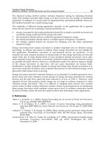
Fig. 1. Typical cellular morphology of PBL from colon cancer patient no. 1 from Table 1
at d 7 after in vitro stimulation with a colorectal cancer-associated CA-Hb3 Ag.
116 Hu
4. To synthesize complementary DNA (cDNA) from total RNAs from the stimulated
and unstimulated PBLs, add 1 µg total RNA to 0.2 µg oligo(dT), 10 U RNase
inhibitor, 5 mM dNTPs, 1X RT buffer and 5 U avian myeloblastosis virus RT in a
reaction volume of 20 µL. Incubate the reaction tubes at 42°C for 60 min.
5. To amplify V
H
–C
H
1 (λ) and V
L
–C
L
(κ), a touchdown PCR procedure was used
(4). The 5′ primer for amplifi cation of V
H
–C
H
1 is 5′-GAGGTGCAGCTGKT
GSAGTCTGS-3′, 3′ primer is 5′-GTCCACCTTGGTGTTGCTGGGCTT-3′. For
amplifi cation of V
L
–C
L
, 5′ primer is 5′-GAWRTTGTGMTGACKCAGTCTCC-3′
and 3′ primer is 5′-AGACTCTCCCCTGTTGAAGCTCTT-3′, where R is A or
G, W is A or T, S is C or G, K is T or G. β-actin can be used as an internal
control (5′-primer is 5′-CTTCTACAATGAGCTGCGTG-3′, and 3′ primer
5′-TCATGAGGTAGTCAGTCAGG-3′). Set up 50-µL PCR reactions con-
taining 2 µL cDNA from stimulated or unstimulated PBL, 1X PCR buffer,
200 µM of dNTPs, 20 pmol of each 5′-primer or 3′-primer, and 2.5 U Taq DNA
polymerase.
6. Amplify with a modifi ed touchdown procedure consisting of three cycles each
of denaturation at 94°C for 30 s, annealing at 55°C for 1 min, and elongation
at 74°C for 1.5 min. Repeat for annealing temperatures reduced in steps of
1°C, from 55° to 46°C. Follow the touchdown cycles with 10 cycles using an
annealing temperature of 45°C and a 10-min extension at 74°C.
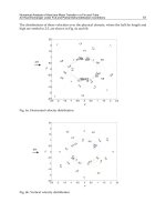
7. Analyze one-tenth of the PCR reaction by electrophoresis on 1% agarose gels.
In our experience, V
H
–C
H
1 and V
L
–C
L
amplifi cation yields from stimulated PBL
were 0.3× greater than from the unstimulated PBL (Fig. 2).
4. Notes
1. The use of an affi nity-purifi ed Ag is important since it determines the specifi city
of the phage Abs. To make an affi nity column, if the MAb is available, it could
Table 1
Numbers of Total Peripheral Blood Lymphocytes Counted by Trypan
Blue Exclusion Assay in 10 mL Peripheral Blood from Four Colorectal
Cancer Patients, Before and after In Vitro Stimulation Driven by a
Colorectal Cancer-Associated CA-Hb3 Ag
Total cell no. Total cell no.
CRC patient before stimulation after stimulation
1 1.00 × 10
7
0.98 × 10
7
2 1.52 × 10
7
0.85 × 10
7
3 0.75 × 10
7
0.68 × 10
7
4 1.20 × 10
7
0.48 × 10
7
Ag Stimulation of B-Lymphocytes In Vitro 117
be conjugated with cyanogen bromide-activated Sepharose 4B according to
manufacturer’s instructions.
2. If there is no existing MAb against the Ag of interest, crude Ag or recombinant
sources of protein or synthetic peptides can be used. Because 2.5% 1-butanol
in PBS extracts tumor-specifi c transplantation Ag from cancer cell membranes,
CBE Ag extracted in this way from tumor cell lines can be used at a fi nal
concentration of 10 µg/mL for in vitro stimulation (2).
3. Successful in vitro stimulation can be judged from the following: morphology
changes to cells in culture refl ecting a secondary immune response, specifi cally,
the size of PBL and colony formation; the appearance of specifi c Ab against the
Ag of interest in culture supernatant over the 7 d of culture (this can be assessed
by ELISA) (see Subheading 3.1.); the amounts of total RNA from PBL before
and after in vitro stimulation; the yields of PCR products from Ig RT-PCR (see
Subheading 3.3., step 7).
4. For screening of phage Ab libraries, progressive decreases in the concentration of
binding Ag are suggested, i.e., use 1 µg/mL affi nity-purifi ed Ag or recombinant
protein or synthetic peptide for the fi rst panning, 0.1 µg/mL for the second
panning, then 0.01 µg/mL for the third panning step. If pure Ag is not available,
but a MAb can be obtained, a sandwich procedure can be used for screening. To
Fig. 2. Assay of Ig transcript levels by RT-PCR. The amounts of Ig from the
stimulated PBL (V
H
-C
H
1 in lane 1 and V
L
–C
L
in lane 3) were 0.3× more than those
from the unstimulated PBL (V
H
–C
H
1 in lane 2 and V
L
–C
L
in lane 4) estimated by
band brightness. β-actin is the internal control. The marker (M) was 100 bp DNA
ladder (Life Technologies).
118 Hu
carry this out, 1–5 µg/mL MAb is coated onto a Petri dish. After washing 3 ×
3 min with PBS and blocking with 1% BSA, 50 µg/mL crude Ag (e.g., CBE Ag)
is added to the dish for 1 h at 37°C. After washing 3 × 3 min with PBS, the dish
is ready for the fi rst panning; for the second or third screening, concentrations
of the crude Ag can be reduced to 5 or 0.5 µg/mL, respectively. If the MAb is
not available, crude Ag (50, 5, and 0.5 µg/mL for the fi rst, second, and third
screening, respectively) could still be used for screening of phage Ab libraries.
The step-by-step decreases in Ag concentration may increase the chances of
recovering phage clones of high affi nity.
5. In my experiments, PBL numbers from 10 mL peripheral blood from a typical
colon cancer patient were 1 × 10
7
before in vitro stimulation and the numbers
were 0.98 × 10
7
7 d later after in vitro stimulation. The cell numbers were
counted with trypan blue exclusion assay and a hemocytometer, viable cells
comprising more than 95% before and after in vitro stimulation. After in vitro
stimulation, the total numbers of PBLs from 10 mL peripheral blood per patient
from four colorectal cancer patients fell to 40–98% of their original numbers
(Table 1).
6. It should be noted that IL-2 alone will induce apoptosis of T-lymphocytes (3).
Therefore, IL-2 and pokeweed mitogen should be added after or simultaneously
with Ag.
7. After in vitro stimulation, PBLs become rounder and bigger and the classical
morphology of a secondary immune response appears. In detail, lymphocytes
at d 0 are small and round, lymphoblast-like cells appear at d 3, some colonies
form and lymphoblast cells can be observed at d 5, and at d 7, colonies are more
numerous, bigger, and lymphoblast-like cells can still be seen (Fig. 1).
References
1. Sun, Q. B., Ho, J. I. L., and Kim, Y. S. (1986) Human colonic cancer associated
antigens detected by three monoclonal antibodies. Chin. Med. J. 99, 63–74.
2. Coggin, J. H., Gillis, L. D., and Payne, W. J., Jr. (1984) Differential extraction of
tumor-transplantation antigen and embryonic antigen from simian virus 40- and
adenovirus 7-induced sarcoma cells of hamsters with 1-butanol and 3 M potassium
chloride. J. Natl. Cancer Inst. 72, 853–862
3. Lenardo, M. J. (1991) Interleukin-2 programs mouse αβ T lymphocytes for
apoptosis. Nature 353, 858–861.
4. Cai, X. and Garen, A. (1995) Anti-melanoma antibodies from melanoma patients
immunized with genetically modifi ed autologous tumor cells: Selection of specifi c
antibodies from single-chain Fv fusion phage libraries. Proc. Natl. Acad. Sci.
USA 92, 6537–6541.
Ag Stimulation of B-Lymphocytes In Vitro 119
121
From:
Methods in Molecular Biology, vol. 178: Antibody Phage Display: Methods and Protocols
Edited by: P. M. O’Brien and R. Aitken © Humana Press Inc., Totowa, NJ
8
The Recovery of Immunoglobulin Sequences
from Single Human B Cells by Clonal Expansion
Ruud M. T. de Wildt and René M. A. Hoet
1. Introduction
The development of phage-display technology and the construction of huge
libraries of antibody (Ab) fragments have provided an unlimited source of
binders to virtually any antigen (Ag) (1). However, it is unlikely that the heavy
(V
H
) and light (V
L
) chains of the Abs obtained from these libraries resemble
original in vivo pairings. In certain autoimmune diseases and immunological
processes, such as B-cell tolerance, these V
H
and V
L
combinations can be of
crucial importance. To be able to determine the original V
H
and V
L
combina-
tions of Abs, we have set up a single B-cell culture system. This comprises the
sorting of individual lymphocytes into culture wells using fl ow cytometry, a
culture system to expand these cells (2) and polymerase chain reaction (PCR)
amplifi cation of their variable-region genes, thereby immortalizing the V
H
and
V
L
regions from individual human B cells. The method relies on the clonal
expansion of single B cells in which cell–cell interactions (CD40–CD40L), as
well as soluble factors, have been shown to be essential. One advantage beyond
conventional hybridoma technology is that this method circumvents laborious
plating and screening; the advantage compared to phage-display technology
is that original V
H
and V
L
pairings can be isolated. This system has been
used to analyze V
H
and V
L
pairings of human immunoglobulin G
+
(IgG+)
B cells of unknown specifi city (3) and, combined with a selection on the Ag
U1A, a frequent autoantigenic protein target in patients with systemic lupus
erythematosus, to analyze pairings in Ag-specifi c B cells (4). The effi ciency of
the system makes it possible to analyze large numbers of B cells and should
therefore allow rare B-cell activities to be studied.
Recovery of Immunoglobulin Sequences 121
Other technologies that retain original V
H
/V
L
pairings involve PCR assembly
of V
H
and V
L
within a single cell (5), which has been achieved with hybrid-
omas but has yet to be routinely applied to populations of B cells because of
technical problems. Others have isolated single Ag-specifi c lymphocytes using
micromanipulation of lymphocytes bound to Ag-coated erythrocytes (6) or
Ag-coated beads (7). The V
H
and V
L
genes from these single cells are amplifi ed
using reverse transcriptase (RT)-PCR, and cloned as functional Ab fragments.
However, these techniques involve laborious manipulation of every cell of
interest and hence suffer low throughput.
2. Materials
2.1. Preparation of Lymphocytes
1. Heparinized blood from a patient group of interest.
2. Phosphate-buffered saline (PBS) with and without 0.3% Na citrate.
3. Ficoll-Paque (Pharmacia Biotech).
4. Dulbecco’s modified Eagle’s medium (DMEM)–HAM’s F12 (1Ϻ1) (Gibco
product code 21331).
5. Supplemented calf serum (CS) (Hyclone product code A 2151L).
6. Dimethyl sulfoxide.
7. Fetal calf serum (Gibco).
8. Fluorescein isothiocyanate (FITC)-conjugated anti-human IgG (Kallestadt,
Amiter, TA).
9. Phycoerythryin-conjugated anti-CD19 (Dako).
10. Coulter Epics Elite fl ow cytometer equipped with an automatic deposit unit
(Coulter, Hialeah, FL).
11. Target Ag of interest (e.g., U1A).
12. 6-Well culture plates (Greiner).
13. 0.1 M NaHCO
3
, pH 9.6.
14. Tissue culture incubator with associated gas supply.
15. Trypsin (Gibco).
16. Ethylenediamine tetraacetic acid (EDTA).
17. FITC-conjugated anti-CD19 and anti-CD20 monoclonal antibodies (MAbs)
(Dako).
2.2. Culture of B Cells
1. 96-Well round-bottomed plates (Costar).
2. Phytohemagglutinin (Murex).
3. β-Phorbol-12-myristate-13-acetate (PMA) (Sigma).
4. Freshly cultured EL4-B5 thymoma cells obtainable from R. Zubler (see
Note 4).
122 de Wildt and Hoet
2.3. Enzyme-Linked Immunosorbant Assay (ELISA)-Testing
of Culture Supernatant
1. 96-Well plates (Nunc, Maxisorp).
2. 0.1 M NaHCO
3
, pH 9.2 or pH 9.6, depending on application (see Subheading
3.3., step 1).
3. Anti-human IgG, IgM, and total Ig (Dako).
4. 2% Skimmed milk powder in PBS (PBSM).
5. Tween-20 in PBS (PBST).
6. Horseradish-peroxidase conjugated anti-human IgG, IgM, and total Ig (Dako).
7. PBSM containing 2% CS.
8. Substrate solution: 100 mM sodium acetate (NaAc), pH 6.0, containing 100 µg/mL
3′3′5′5′-tetramethylbenzidine and 0.5 µL/mL 30% hydrogen peroxide solution.
Add the hydrogen peroxide solution immediately before use of the substrate
solution.
9. 1 M Sulphuric acid.
2.4. Cloning of V
H
/V
L
Regions from B-Cell Clones
1. RNAzol (Cinna/Biotecx Laboratories).
2. Chloroform.
3. 20 mg/mL Glycogen (Boehringer Mannheim) dissolved in Millipore fi ltered H
2
O.
4. Ethanol–NaAc mix: combine 96 mL absolute ethanol with 4 mL 3 M NaAc,
pH 5.0.
5. 70% Ethanol.
6. RNasin (Promega).
7. 10 pmol/µL 15-mer Oligo(dT) primer (Boehringer Mannheim).
8. RT buffer: 250 mM Tris-HCl, pH 8.3, 375 mM KCl, 15 mM MgCl
2
.
9. 0.1 M Dithiothreitol.
10. SuperScript II RT (100 U/µL; Gibco).
11. Deoxyribonucleoside triphosphate (dNTP) mix (10 mM each nucleotide).
12. Taq DNA polymerase and 10X reaction buffer.
13. QIAquick PCR purifi cation kit (Qiagen, CA).
14. Phage-display or expression vector (e.g., pHENIX).
15. Mouse MAb P5D4 (Boehringer Mannheim).
16. Electrocompetent Escherichia coli TG1 and electroporation apparatus.
17. TYE agar plates: 15 g Bacto-agar, 8 g Na chloride, 10 g tryptone, 5 g yeast
extract in 1 L.
18. Ampicillin.
19. 20% Glucose.
20. 2TY: 16 g tryptone, 10 g yeast extract, and 5 g Na chloride in 1 L.
Recovery of Immunoglobulin Sequences 123
21. Isopropyl thiogalactopyranoside (IPTG).
22. Extraction buffer: 200 mM Na borate, pH 8.0, 160 mM NaCl, 1 mM EDTA.
23. Rabbit anti-mouse Ig horseradish peroxidase conjugate (Dako).
2.5. Sequencing of V
H
/V
L
Regions
1. ABI PRISM Big Dye Terminator Cycle Sequencing Kit (Perkin Elmer, Foster
City, CA).
2. Automated sequencer (Applied Biosystems 373A, Perkin-Elmer).
3. Methods
3.1. Preparation of Lymphocytes
1. Dilute heparinized blood with an equal amount of PBS–0.3% Na citrate. Care-
fully layer 20–30 mL diluted blood onto 15 mL Ficoll-Paque. Centrifuge at 500g
for 30 min at room temperature.
2. Remove the layer containing the peripheral blood mononuclear cells (PBMC),
transfer to another tube and add at least 3 vol DMEM–HAM’s F12 containing
10% CS.
3. Centrifuge the cells for 10 min at 200g, resuspend in DMEM–HAM’s F12–10%
CS, centrifuge, and resuspend in the same medium.
4. At this stage, PBMC are either used directly or frozen in culture medium
containing 10% dimethylsufl oxide and 50% fetal calf serum.
5. When no Ag selection is preferred, PBMC can be used directly to sort single
IgG- or IgM-positive B cells (see Note 8). Label the cells with FITC-conjugated
anti-human IgG and phycoerythryin-conjugated anti-CD19 for 10 min at room
temperature at a concentration of 1 µg/10
6
cells. Centrifuge the cells for 5 min
at 200g and resuspend in 0.5 mL PBS. Viable, single IgG
+
, CD19
+
lymphocytes
are then sorted into 96-well plates using a Coulter Epics Elite fl ow cytometer
equipped with an automatic cell deposit unit. Continue from Subheading 3.2.
6. When Ag selection is preferred, fi rst remove monocytes from the PBMC by
incubating the cells for 1–2 h at 1–2 × 10
6
cells/mL in DMEM–HAM’s F12–10%
CS at 37°C, >98% humidity, and 5% CO
2
. Recover nonadherent cells for further
selection (see Note 1).
7. Coat 6-well culture plates with of the target Ag (e.g., recombinant U1A) at a
concentration of 5 µg/mL in 0.1 M NaHCO
3
, pH 9.6, overnight at 4°C.
8. Wash the coated plates 3× with PBS and add 1–5 × 10
6
monocyte-depleted PBMC
in 4 mL DMEM–HAM’s F12–10% CS. Incubate for 1–2 h at 37°C (see Note 2).
9. Remove unbound cells by washing 6× with PBS. Collect those cells
that have adhered to the target Ag using 300 µL PBS containing 0.05% trypsin,
1.1 mM EDTA. Terminate trypsin treatment after 5 min at 37°C by adding 5 mL
DMEM–HAM’s F12–10% CS.
124 de Wildt and Hoet
10. Harvest the cells and label with a mixture of anti-CD19 and anti-CD20 MAbs
conjugated to FITC for 10 min at room temperature at a concentration of 1 µg/10
6
cells. Centrifuge the cells for 5 min at 200g and resuspend in 0.5 mL PBS.
11. Sort viable, single CD19
+
/CD20
+
cells into 96-well plates using the flow
cytometer.
3.2. Culture of B Cells
1. First, human T-cell–macrophage supernatant (TSN) is prepared from freshly
isolated PBMC (buffycoat) using Ficoll-Paque density centrifugation as described
(see Subheading 3.1., step 1).
2. Wash the cells 3× with DMEM–HAM’s F12–10% CS and culture in the presence
of 5 µg/mL phytohemagglutinin and 10 ng/mL PMA, at a concentration of
1.5 × 10
6
cells/mL.
3. After 48 h, centrifuge the cell suspension for 10 min at 1000g. Harvest the cell
supernatant (TSN) and store in aliquots at –70°C (see Note 3).
4. Single, sorted B cells (see Subheading 3.1., step 5 or Subheading 3.1.,
step 11) are deposited in 96-well plates containing 200 µL/well DMEM–HAM’s
F12–10–15% TSN–10% CS and 20,000 irradiated (2500 rad) EL4-B5 thymoma
cells/well (see Notes 3–5).
5. Remove 100 µL from each well at d 3 and 6 and replace with DMEM–HAM’s
F12–10% TSN–10% CS.
6. Test culture supernatants from the B-cell cultures for (Ag-specifi c) Ab production
(see Subheading 3.3.) at d 10 or 11 (see Notes 6–9).
3.3. ELISA-Testing of Culture Supernatant
1. To detect the production and Ag-specifi c Ig, coat 96-well plates with 100 µL/well
of an Ag solution at 1 µg/mL Ag (e.g., recombinant U1A) in 0.1 M NaHCO
3
,
pH 9.6. Incubate overnight at 4°C. To detect the production of IgG, IgM, or total
Ig (see Note 3), coat plates with the same volume of 1 µg/mL anti-human IgG,
IgM, or total Ig in 0.1 M NaHCO
3
, pH 9.2.
2. Block the plates with 200 µL/well PBSM for 2 h at room temperature, then
wash 3× with PBS.
3. Mix 40 µL culture supernatant with an equal volume of PBSM, add to the coated
plates, and incubate for 1 h at room temperature.
4. Wash the plates 3× with PBST and 3× with PBS.
5. Detect the binding of IgG, IgM, or total Ig by adding 100 µL/well of the cor-
responding horseradish peroxidase conjugated anti-human Ab, diluted 1Ϻ5000
in PBSM containing 2% CS. Dilute the conjugates 1Ϻ1000 for detection of
Ag-specifi c Ab production.
6. Wash the plates 3× with PBST and 3× with PBS.
Recovery of Immunoglobulin Sequences 125
7. Add 100 µL/well substrate solution. Stop the reactions when the color has
developed by adding 50 µL/well 1 M sulphuric acid. Measure the OD
650
–OD
450
(see Notes 3 and 6)
3.4. Cloning and Expression of V
H
/V
L
Regions from B-Cell Clones
(
see
Note 10)
1. Using a Pasteur pipet, remove the medium carefully from wells containing IgG
+
or Ag-specifi c Ab-producing cells. Resuspend all cells in 200 µL RNAzol and
transfer to 1.5-mL microcentrifuge tubes. Add 20 µL chloroform, mix the tube
contents, and incubate 5 min at 4°C. Centrifuge the samples in a microcentrifuge
for 15 min and collect the aqueous phase.
2. Add 2 µL glycogen solution and precipitate the RNA by adding 2 vol ethanol–
NaAc. Mix and incubate for 45 min at 4°C. Spin the tubes for 15 min, 15,000g
at 4°C. Carefully remove the ethanol–NaAc mix without disturbing the RNA
pellet. Add 0.5 mL 70% ethanol and spin again for 5 min at 4°C. Air-dry the
RNA and dissolve in 100 µL Millipore-fi ltered H
2
O containing 20 U RNasin.
Precipitate the RNA again using ethanol/NaAc and store at –70°C until further
use (see Note 10).
3. Recover, wash, and air-dry the RNA as above (see Subheading 3.4., step 2).
Dissolve in 20 µL Millipore-fi ltered H
2
O containing 20 U RNasin. Use half of
the RNA for fi rst-strand cDNA synthesis and store the remainder at –70°C.
4. Add 2 µL of 10 pmol/µL oligo(dT) primer and briefl y centrifuge. Heat the
mixture to 70°C for 5–10 min, then chill on ice to anneal the primer. Add 4 µL
RT buffer, 2 µL 0.1 M dithiothreitol, 1 µL (100 U) SuperScript II RT and
2 µL dNTP mix. Mix and incubate for 1 h at 42°C. Inactivate the RT reaction
by heating for 2 min at 80°C.
5. Use 5-µL aliquots of these cDNAs in separate PCRs to amplify V
H
, V
κ
, and
V
λ
genes using family-specific 5′ primers and 3′ constant-region primers
(Table 1; see Note 11).
6. To carry out PCRs, add 20 pmol of each primer in 1X Taq reaction buffer containing
1.5 mM MgCl
2
, 250 µM dNTPs, and 2.5 U Taq polymerase. Carry out 25 cycles
of 94°C, 1 min; 55°C, 1 min; and 72°C, 1.5 min (see Note 12).
7. Purify the PCR products using a QIAquick PCR purifi cation kit, following the
manufacturer’s protocol.
8. At this stage, PCR products can be used for direct sequencing (see Subheading
3.5.), or for cloning as scFv.
9. In a 3′-nested second PCR, use 1 µL of the fi rst PCR product under the same
conditions as described above (see Subheading 3.4., steps 5 and 6) with primers
containing appropriate restriction sites for cloning. As 5′ primers, the same
primers as in Table 1 can be used, extended with Sfi I/NcoI restriction sites for
V
H
primers (8) and ApaL1 restriction sites for V
κ
and V
λ
primers (9). As 3′
primers for the heavy chains (HC), J
H
forward primers with a SalI site (10)
are used, and for the light chains (LC), J
κ
or J
λ
primers containing a NotI site
(8) are used.
126 de Wildt and Hoet
10. Clone HCs and LCs sequentially into a phagemid vector, such as pHENIX (11),
in which a peptide epitope of the vesicular stomatitis virus glycoprotein is fused
at the C-terminus as a tag for detection using mouse MAb, P5D4.
11. Electroporate the ligated vector into electrocompetent TG1 and plate onto TYE
plates containing 100 µg/mL ampicillin and 1% glucose.
12. To determine whether isolated clones are reactive with the Ag of interest in
ELISA, pick single colonies into 2TY containing 100 µg/mL ampicillin and 1%
glucose and grow overnight at 37°C.
Recovery of Immunoglobulin Sequences 127
Table 1
Primers for Amplifying Rearranged Ab V Genes
V
H
1Back: CAG (AG)T(CGT) CAG CTG GTG CAG TCT GG
V
H
2Back: CAG (AG)TC ACC TTG AAG GAG TCT GG
V
H
3Back: GAG GTG CAG CTG GTG GAG TCT GG
V
H
4Back: CAG GTG CAG CTG CAG GAG T(CG)(CG) GG
V
H
5Back: GAG GTG CAG CTG GTG CAG TCT GG
V
H
6Back: CAG GTA CAG CTG CAG CAG TCA GG
V
κ
1Back: G(AC)C ATC C(AG)G ATG ACC CAG TCT CC
V
κ
2Back: GAT GTT GTG ATG ACT CAG TCT CC
V
κ
3Back: GAA ATT GTG (AT)TG AC(AG) CAG TCT CC
V
κ
4Back: GAC ATC GTG ATG ACC CAG TCT CC
V
κ
5Back: GAA ACG ACA CTC ACG CAG TCT CC
V
κ
6Back: GAA ATT GTG CTG ACT CAG TCT CC
V
λ
1Back: CAG TCT GTG (CT)TG AC(GT) CAG CC
V
λ
2Back: CAG TCT GCC CTG ACT CAG CCT GC
V
λ
3aBack: TCC TAT GAG CTG AC(AT) CAG CC
V
λ
3bBack: TCT TCT GAG CTG ACT CAG GAC CC
V
λ
4Back: CAG C(CT)T GTG CTG ACT CAA TC
V
λ
5Back: CAG (CG)CT GTG CTG ACT CAG CC
V
λ
6Back: AAT TTT ATG CTG ACT CAG CCC CA
V
λ
7/8Back: CAG (AG)CT GTG GTG AC(CT) CAG GAG
V
λ
9/10Back: CAG (CG)C(TA) G(GT)G CTG ACT CAG CCA
IgG1-4C
H
1For GTC CAC CTT GGT GTT GCT GGG CTT
C
κ
For AGA CTC TCC CCT GTT GAA GCT CTT
C
λ
For TGA AGA TTC TGT AGG GGC CAC TGT CTT
Sequencing primers
C
H
1.lib.seq primer GGT GCT CTT GGA GGA GGG TGC
C
κ
lib.seq CAA CTG CTC ATC AGA TGG CG
C
L
.seq AGT GTG GCC TTG TTG GCT TG
fdseq1 GAA TTT TCT GTA TGA GG
forlinkseq GCC ACC TCC GCC TGA ACC
13. Inoculate 2TY containing 100 µg/mL ampicillin and 0.1% glucose with 0.01 vol
from the overnight culture. Grow with shaking at 37°C until the OD
600
is approx
0.9. Add IPTG to a fi nal concentration of 1 mM and shake the cultures at 30°C for
3 h (for periplasmic fractions) or overnight (expression in supernatant).
14. For the isolation of periplasmic fractions, centrifuge the bacteria at 4000g at
4°C for 10 min. Resuspend the pellet in 20 mL/L culture cold-extraction buffer.
Centrifuge the fractions at 8000g at 4°C for 10 min and fi lter-sterilize.
15. Test soluble scFv in periplasmic fractions, or in the culture supernatant, for
binding to the Ag (recombinant auto-Ag U1A) in ELISA, which is performed
as described (see Subheading 3.3.), except that scFv are detected with mouse
MAb P5D4 at a dilution of 1Ϻ1000 and rabbit anti-mouse Ig HRP conjugate
(1Ϻ1000 in PBSM) (see Note 13).
3.5. Sequencing of V
H
/V
L
Regions (
see
Note 14)
1. PCR products can be directly sequenced from amplifi ed rearranged human
variable–constant region genes using C
H
1.lib.seq primer for the HCs. C
κ
lib.seq
for the κ LCs and C
L
.seq for the λ LCs (Table 1). These primers anneal
~50 nucleotides from the 5′ end of the constant-region genes.
2. For sequencing cloned scFv fragments, fdseq1 and forlinkseq are used. We use
Big Dye reagents and analyze on an Applied Biosystems 373A machine.
3. Nucleotide sequences are aligned to their germline counterparts using the
V-BASE Sequence Directory (12) ( />index.html).
4. Notes
1. Removal of monocytes by plastic adherence, the enrichment for Ag-specifi c
B cells, and subsequent culturing are performed essentially as has been described
in ref. 13.
2. As described, Ag selection is performed on Ag-coated plates. Immobilization of
Ag on superparamagnetic minibeads (Mylteni Biotech, Germany) has also
been effective. A major advantage of these magnetically sorted cells is that
they can be used directly for fl ow cytometry analyses in contrast to Dynabeads
(Dynal, Norway), from which the cells must be detached before use on the
fl ow cytometer.
3. TSN may contain a small amount of human Ig, which may interfere with ELISA
testing for Ig production. This can be depleted from the TSN using Protein G
Sepharose, although we have found that positive signals in ELISA can be clearly
distinguished from background. The optimum amount of TSN for effi cient
outgrowth of B cells can be established by titration (13), but we found that 10%
TSN routinely gave good results.
4. EL4-B5 thymoma cells are routinely cultured in DMEM–HAM’s F12 (1Ϻ1)–10%
CS between 1 × 10
4
and 1 × 10
6
cells/mL. EL4-B5 cells can be obtained with
permission from Dr. R. Zubler (Hopital Cantonal Universitaire de Geneve,
128 de Wildt and Hoet
Centre de Transfusion Sanguine, Division d’Hematologie, CH 1211 Geneva 14,
Switzerland) or from R. D. W.
5. Hyclone-supplemented CS batches gave best results with B-cell outgrowth and
no stimulation of the irradiated EL4-B5 cells was observed.
6. Typical percentages of Ig-positive cultures determined by ELISA after 10–11 d
culture varies between 50 and 70%. The frequency of U1A-specifi c B-cell clones
varies between 1 and 2.5% as a percentage of Ig-positive wells. As a control,
cells from a healthy donor were used and subjected to the same procedure. No
U1A-specifi c Ab production could be detected in these cultures; the percentage
of Ig-producing wells was similar to those found with the systemic lupus
erythematosus patient B cells. Distributions of IgG, IgM, and IgG–IgM double-
positive isotypes in Ig-producing single B-cell cultures were 3Ϻ3Ϻ1.
7. Assuming that the frequency of Ag-specifi c B cells in the periphery varies
between 10
–4
and 10
–5
(14), a frequency of 1–2.5% of Ag-specific B cells
indicates an enrichment factor of 100–1000. Other groups have also succeeded
in the isolation of Ag-specifi c B cells from peripheral blood using an expansion
B-cell culture system using virally infected donors (15) or donors vaccinated with
bacterial Ags (7). The frequency of specifi c cells to those Ags in the periphery
is most likely much higher compared to the auto-Ag-specifi c B cells analyzed
in our studies.
8. We have sorted single IgG
+
B cells of unknown specifi city and used this system
to analyze V
H
and V
L
pairings (3,16) and to compare V
H
and V
L
pairings between
healthy and autoimmune disorders (17).
9. After culture in the EL4-B5 system, the B cells obtained a plasmablast-like
phenotype expressing CD38
HIGH
and syndecan-1
MODERATE
, a plasma cell marker
stained with MAb B-B4 (18). One B cell generates about 400 cytoplasmic Ig
positive cells after 8–10 d in culture (2), but, because of the large number of
EL4-B5 cells present (~20,000 cells/well), these B-cell clones are not easily
distinguishable under the microscope.
10. The expansion step results in an increase of mRNA levels derived from one clone,
which avoids the risk of contamination in downstream procedures and makes it
more convenient to analyze single peripheral B cells, which are mostly resting
cells with low mRNA levels. One major consideration in studying peripheral
B cells often is the lack of other available patient materials.
11. PCR products amplifi ed with a mixture of V
H
family-specifi c primers and a
constant-region primer should give rise to a product of ~750 (for V
H
) or 700
nucleotides (for V
L
). With the LCs, V
κ
and V
λ
should never be found together
in the same clone, indicating clonality. As a control for the PCR, cDNA isolated
from a well in which no B cell was used. Such control reactions should never
result in a PCR product.
12. With the current set of primers, almost all functional V genes should be amplifi ed.
Indeed, using these primers, we detected the majority of expressed V genes:
86% V
H
, 80% V
λ
, and 58% of the functional V
κ
segments (3). Recently, other
primer sets has been published, which theoretically should be able to amplify
Recovery of Immunoglobulin Sequences 129
all functional V genes (12,19), although mixes of these primers have never been
used to amplify V regions from single B-cell clones.
13. We were able to detect fi ve U1A-specifi c B-cell clones. Two of these (B5 and C9)
were cloned into a phagemid vector for scFv expression. Soluble scFvs present in
bacterial supernatant or periplasmic fractions were tested for binding in ELISA
on a number of auto-Ags. Indeed, this showed that these clones specifi cally
recognized the U1A protein (4).
14. For a more detailed description of the analysis of human Ab sequences, see
ref. 20.
References
1. Winter, G., Griffi ths, A. D., Hawkins, R. E., and Hoogenboom, H. R. (1994)
Making antibodies by phage display technology. Annu. Rev. Immunol. 12, 433–455.
2. Zubler, R. H., Erard, F., Lees, R. K., Van Laer, M., Mingari, C., Moretta, L., and
MacDonald, H. R. (1985) Mutant EL-4 thymoma cells polyclonally activate murine
and human B cells via direct cell interaction. J. Immunol. 134, 3662–3668.
3. de Wildt, R. M. T., Hoet, R. M. A., van Venrooij, W. J., Tomlinson, I. M., and
Winter, G. (1999) Analysis of heavy and light chain pairings indicates that receptor
editing shapes the human antibody repertoire. J. Mol. Biol. 285, 895–901.
4. de Wildt, R. M. T., Steenbakkers, P. G., Pennings, A. H. M., van den Hoogen,
F. H. J., van Venrooij, W. J., and Hoet, R. M. A. (1997) A new method for the
analysis and production of monoclonal antibody fragments originating from single
human B cells. J. Immunol. Methods 207, 61–67.
5. Embleton, M. J., Gorochov, G., Jones, P. T., and Winter, G. (1992) In-cell PCR
from mRNA: amplifying and linking the rearranged immunoglobulin heavy and
light chain V-genes within single cells. Nucleic Acids Res. 20, 3831–3837.
6. Babcook, J. S., Leslie, K. B., Olsen, O. A., Salmon, R. A., and Schrader, J. W.
(1996) A novel strategy for generating monoclonal antibodies from single, isolated
lymphocytes producing antibodies of defi ned specifi cities. Proc. Natl. Acad. Sci.
USA 93, 7843–7848.
7. Lagerkvist, A. C. S., Furebring, C., and Borrebaeck, C. A. K. (1995) Single,
antigen-specifi c B cells used to generate Fab fragments using CD40-mediated
amplifi cation or direct PCR cloning. Biotechniques 18, 862–869.
8. Marks, J. D., Hoogenboom, H. R., Bonnert, T. P., McCafferty, J., Griffi ths, A. D.,
and Winter, G. (1991) By-passing immunization. Human antibodies from V-gene
libraries displayed on phage. J. Mol. Biol. 222, 581–597.
9. Jespers, L. S., Roberts, A., Mahler, S. M., Winter, G., and Hoogenboom, H. R.
(1994) Guiding the selection of human antibodies from phage display repertoires
to a single epitope of an antigen. Biotechnology 12, 899–903.
10. Figini, M., Marks, J. D., Winter, G., and Griffi ths, A. D. (1994) In vitro assembly
of repertoires of antibody chains on the surface of phage by renaturation. J. Mol.
Biol. 239, 68–78.
130 de Wildt and Hoet
11. Finnern, R., Pedrollo, E., Fisch, I., Wieslander, J., Marks, J. D., Lockwood, C. M.,
and Ouwehand, W. H. (1997) Human autoimmune anti-proteinase 3 scFv from a
phage display library. Clin. Exp. Immunol. 107, 269–281.
12. Tomlinson, I. M., Williams, S. C., Ignatovich, O., Corbett, S. J., and Winter,
G. (1999) V Base Sequence Directory, MRC Centre for Protein Engineering,
Cambridge, UK.
13. Steenbakkers, P. G., Hubers, H. A., and Rijnders, A. W. (1994) Effi cient generation
of monoclonal antibodies from preselected antigen-specifi c B cells. Effi cient
immortalization of preselected B cells. Mol. Biol. Rep. 19, 125–134.
14. Zubler, R. H., Perrin, L. H., Doucet, A., Zhang, X., Huang, Y. P., and Miescher,
P. A. (1992) Frequencies of HIV-reactive B cells in seropositive and seronegative
individuals. Clin. Exp. Immunol. 87, 31–36.
15. Steenbakkers, P. G., van Wezenbeek, P. M., and Olijve, W. (1993) Immortalization
of antigen selected B cells. J. Immunol. Methods 163, 33–40.
16. de Wildt, R. M., van Venrooij, W. J., Winter, G., Hoet, R. M., and Tomlinson, I. M.
(1999) Somatic insertions and deletions shape the human antibody repertoire.
J. Mol. Biol. 294, 701–710.
17 de Wildt, R. M., Tomlinson, I. M., van Venrooij, W. J., Winter, G., and Hoet, R. M.
(2000) Comparable heavy and light chain pairings in normal and systemic lupus
erythematosus IgG(+) B cells. Eur. J. Immunol. 30, 254–261.
18. Wijdenes, J., Vooijs, W. C., Clement, C., Post, J., Morard, F., Vita, N., et al. (1996)
A plasmocyte selective monoclonal antibody (B-B4) recognizes syndecan-1. Br.
J. Haematol. 94, 318–323.
19. Sblattero, D. and Bradbury, A. (1998) A defi nitive set of oligonucleotide primers
for amplifying human V regions. Immunotechnology 3, 271–278.
20. Walter, G. and Tomlinson, I. (1996) Analysis of human antibody sequences, in
Antibody Engineering (McCafferty, J., Hoogenboom, H., and Chiswell, D., eds.),
Oxford University Press, Oxford, pp. 119–145.
Recovery of Immunoglobulin Sequences 131
133
From:
Methods in Molecular Biology, vol. 178: Antibody Phage Display: Methods and Protocols
Edited by: P. M. O’Brien and R. Aitken © Humana Press Inc., Totowa, NJ
9
Panning of Antibody Phage-Display Libraries
Standard Protocols
David W. J. Coomber
1. Introduction
Recombinant antibody (Ab) libraries have been constructed from a wide
range of B-lymphocyte sources using a number of different approaches. Sizes
of the libraries that have been produced vary considerably, from small libraries
of 10
6
up to large libraries >10
10
. Often an Ab with the desired specifi city exists
at low frequencies in the recombinant Ab repertoire. It is therefore necessary to
have an effective technique for the enrichment and identifi cation of a desired
Ab from a heterogenous repertoire.
The process for the selection of specifi c Abs is referred to as “panning,”
and, in principle, involves the selection of Abs on the basis of their affi nity.
The isolation of a desired Ab generally involves repeated rounds of panning,
with each successive round resulting in the enrichment of the desired Ab. Each
round of Ab selection can be divided into panning, removal of nonspecifi c
phage, and the elution and amplifi cation of phage Abs for the next round
(Fig. 1). In this way, it has been shown that antigen (Ag)-specifi c Ab that occur
at low frequencies in a library can be enriched by over a million-fold (1).
The methods for the selection of Ab from phage-display Ab libraries are
many and varied, of which some appear later in this chapter. One of the more fre-
quently used methods is panning against purifi ed Ag coated on a well of a micro-
titer plate or in an immunotube. Using this approach, the methods presented
below have been successfully used to isolate Ag-specifi c Abs (see Fig. 2).
There are several points that should be noted about the protocols that are
presented below. First, the libraries used for panning are constructed in the
Panning Ab Phage-Display Libraries 133
MCO phage-display vector system (2), which is derived from pComb3 (3), and
was specifi cally designed for the production, selection, and screening of Fab
phage. These protocols are therefore also suitable for Fab libraries produced in
other pComb3-based vectors. In addition, the MCO vector contains an amber
codon between the heavy chain (HC) gene-cloning site and gene III, which
enables the expression of soluble Fab in nonsuppressor strains of Escherichia
coli. This feature has also been included in some other derivatives of pComb3.
Second, protocols for the panning of scFv phage libraries, although similar, vary
slightly from these protocols because of the use of different expression vectors:
These protocols have been extensively detailed elsewhere (4). However, the
basic principles of the panning process are the same. Therefore, these protocols
can be modifi ed according to the expression vector and Ab system of choice.
Fig. 1. Schematic diagram of the panning process. (A) Library of phage Abs with
a range of Ab specifi cities is applied to an Ag bound to a solid phase. (B) Surface is
washed to remove nonbinding phage Abs, which are then eluted from the surface. (C)
Eluted phage are used to infect E. coli for the production of fresh phage Abs, which
will be used in the next round of panning. Repeated rounds of panning lead to the
enrichment of those phage Abs that are specifi c to the Ag.
134 Coomber