Antibody Phage Display Methods and Protocols - part 5 docx
Bạn đang xem bản rút gọn của tài liệu. Xem và tải ngay bản đầy đủ của tài liệu tại đây (503.29 KB, 39 trang )
6. Anti-tag monoclonal Ab (e.g., 9E10 for myc-tagged Abs). Dilute in 2% PBSM
according to the supplier’s recommendations.
7. Rabbit anti-mouse peroxidase (RAMPO). Dilute in 2% PBSM at a concentration
recommended by the supplier.
8. 10X Tetramethylbenzidine buffer (TMB). Dissolve 37.4 g Na acetate–3H
2
O in
230 mL of H
2
O. Adjust the pH with saturated citric acid (92.5 g citric acid–
50 mL H
2
O) and adjust the volume to 250 mL.
9. TMB stock. Dissolve 10 mg TMB in 1 mL DMSO.
10. TMB staining solution. Mix 1 mL 10X TMB buffer with 9 mL H
2
O/microtiter
plate. Add 100 µL TMB and 1 µL 30% hydrogen peroxidase. Make this solution
fresh and keep it in the dark.
11. 96-Well, fl at-bottomed ELISA microtiter plates (2 plates to screen 96 colonies).
12. For IE: microtiter plates with low coating effi ciency (2/96 colonies).
13. Microtiter plate reader (for optical density 450 nm [OD
450
] measurements).
3. Methods
3.1. Biotinylation of Ag
This method describes chemical biotinylation, which is the most common
way to obtain a biotinylated Ag. For other alternatives, see Notes 1–3.
3.1.1. Chemical Biotinylation of Ag
1. Dissolve the peptide/protein of interest at a concentration of 1–10 mg/mL in
50 mM NaHCO
3
, pH 8.5. If the peptide/protein is in another solvent, dialyze for
at least 4 h against 1 L 50 mM NaHCO
3
, changing the buffer 2–3×.
2. Calculate the amount of NHS-SS-Biotin required using a molar ratio of
biotin:protein between 5 and 20Ϻ1 (see Note 5).
3. Dissolve the required amount of NHS-SS-Biotin in dH
2
O (see Note 6) and
immediately add to the protein sample, or, alternatively, when using larger
amounts of protein, add NHS-SS-Biotin directly to the protein solution.
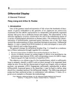
Fig. 2. ELISA using biotinylated antigen and soluble antibody fragments.
150 Chames, Hoogenboom, and Henderikx
4. Incubate for 30 min at room temperature or for 2 h on ice if the protein is
temperature-sensitive.
5. Add 1 M Tris-HCl, pH 7.5, to a fi nal concentration of 50 mM and incubate for
1 h on ice to block any free NHS-SS-Biotin.
6. To remove the free NHS-SS-Biotin, dialyze for at least 4 h (to overnight) at
4°C against PBS, changing the buffer. Alternatively, follow steps 7–9 below.
For small peptides (<20 amino acids), alternative separation protocols (e.g.,
affi nity chromatography, high-performance liquid chromatography) should be
followed.
7. Alternative to step 6: spin the solution at 1000–5000g in an ultrafi ltration device
(e.g., Centricon 10 or 30) to concentrate the sample in 100 µL.
8. Dilute the sample in PBS to dilute out free NHS-SS-Biotin left after concentration.
9. Repeat steps 6 and 7 twice more.
10. Add Na azide to a fi nal concentration of 0.1%.
11. Store in small aliquots at –20°C or at 4°C. Storage conditions should be tested
for individual proteins.
3.1.2. Determination of Biotinylation Effi ciency
It is important to determine the percentage of protein that has actually been
biotinylated. If the Ag has to be used for selection in solution, the nonbiotinyl-
ated part of the preparation will be detrimental to selection, blocking specifi c
phages, and impairing their binding to the biotinylated fraction. Hence, this
nonbiotinylated fraction must represent less than 10–15%. This protocol
is also used to determine the amount of biotinylated peptide captured by a
certain amount of magnetic beads. Extrapolation of the results can be used for
determining the concentration of Ag and amount of beads to be used during
phage library selection.
1. Resuspend the streptavidin Dynabeads with gentle shaking.
2. Make fi ve dilutions of the biotinylated protein/peptide between 5 and 50 nM
in 200 µL PBS.
3. Transfer 50 µL beads into a tube that fi ts into the magnetic separator and add an
excess of PBS; shake gently to mix.
4. Put the tube into the magnetic separation device for 2 min and pipet off the PBS.
5. Add 0.5 mL PBST and incubate for 60 min.
6. Remove the PBST as in step 4 and resuspend the beads in 50 µL of PBST.
7. Aliquot 10 µL of the beads into 5 tubes and add 100 µL diluted peptide/protein
to each tube. Seal the tubes and incubate for 30 min at room temperature in an
end-over-end rotator. The remaining 100 µL of each dilution (fraction 0) will be
used to evaluate the percentage of biotinylation.
8. Place the tubes into the magnet for 2 min, remove, and store 100 µL of the
supernatants (fraction 1).
Ab Selection Against Biotinylated Ags 151
9. Resuspend the Dynabeads in 1 mL PBST, place the tubes into the magnet, and
discard the supernatant. Repeat 4×.
10. If the protein measurements are to be performed by SDS-PAGE, resuspend
the beads in 110 µL 1X reducing SDS-PAGE sample buffer and incubate for
10 min (fraction 2a; eluted protein). Alternatively, the protein concentration can
be measured by UV 280 nm. In this case, resuspend the beads in PBS containing
10 mM DTT (fraction 2b).
11. For SDS-PAGE measurements, add 10 µL 10X reducing loading buffer to
fractions 0 and 1, and load samples (e.g., 10 µL and 50 µL) of fractions 0, 1, and
2a to a gel of suitable acrylamide percentage for the protein of interest. Perform
SDS-PAGE, stain gel with Coomassie blue, and destain.
12. Alternatively, dilute fractions 0, 1, and 2b in an amount of PBS suitable for the
quartz cuvet. Measure UV
280
absorption.
13. The percentage of protein found in fraction 2 is the percentage of biotinylation.
The proteins in fraction 1 are not biotinylated.
14. If the biotinylation was effi cient, check the maximum amount of biotinylated
protein able to bind 10 µL streptavidin dynabeads (the highest concentration
for which there is almost no protein in fraction 1). Extrapolate this amount to
phage-selection conditions (e.g., a maximum of 30 nM can be bound at >85%
to 10 µL beads: therefore, for 500 mM Ag used during the selections, 166 µL of
magnetic beads should be used).
3.2. Selection of Abs by Means of Phage Display
1. Mix equal volumes of the phage library and 4% PBSM in a total volume of
0.5 mL. During the fi rst selection, the number of phage particles should be
at least 100× higher than the library size (e.g., 10
12
cfu for a library of 10
10
clones). Diversity drops to 10
6
after the fi rst round and is thus not limiting in
the next rounds.
2. Incubate on a rotator at room temperature for 60 min.
3. While preincubating the phage, wash 100–200 µL streptavidin Dynabeads/Ag
sample in a tube with 1 mL PBST using the magnetic separation device as
described in Subheading 3.1.2. The minimal amount of beads for selection can
be calculated as described in Subheading 3.1.2.
4. Resuspend the beads in 1 mL 2% PBSM.
5. Equilibrate the beads at room temperature for 1–2 h using a rotator.
6. Add the biotinylated Ag (100–500 nM) diluted in 0.5 mL PBS (+ 5% DMSO if
the Ag solubility is an issue, e.g. for certain peptides) directly into the equilibrated
phage mix. Incubate on a rotator at room temperature for 30 min–1 h.
7. Using the magnet, draw the equilibrated beads to one side of the tube and remove
the PBSM.
8. Resuspend the Dynabeads in the phage–Ag mix and incubate on a rotator at
room temperature for 15 min (see Note 7).
152 Chames, Hoogenboom, and Henderikx
9. Place the tubes in the magnetic separator and wait until all the beads are bound
to the magnetic site (1 min).
10. Tip the rack upside down and back again with the caps closed, which will wash
down the beads from the cap. Leave the tubes in the rack for 2 min, then aspirate
the tubes carefully, leaving the beads on the side of the tube.
11. Using the magnet, wash the beads carefully 6× with 1 mL PBSMT.
12. Transfer beads to a new Eppendorf tube and wash the beads 6× with 1 mL
PBSMT.
13. Transfer the beads to a new Eppendorf tube and wash the beads 2× with
1 mL PBS.
14. Transfer the beads to a new tube and elute the phage from the beads by adding
200 µL 10 mM DTT and rotate the tube for 5 min at room temperature (see
Note 8). Place the tubes in the magnetic separator and transfer the supernatant
containing the phages to a new tube.
15. Infect a fresh exponentially growing culture of Escherichia coli TG1 with the
eluted phage and amplify according to standard protocols (see Chapter 9) to
perform further rounds of selection (see Notes 9 and 10). Store any remaining
phage eluate at 4°C.
16. Express soluble Ab fragments from the selected phage clones using standard
protocols for the particular expression system.
3.3. Inhibition ELISA
The purpose of this ELISA is to identify binders among phages retrieved
after each selection round. The setup of this ELISA is similar to the setup
used for selection. It uses the same biotinylated Ag and an indirect coating
via streptavidin, to ensure maintenance of the native structure of the Ag and
precoating of the plastic panning surface with biotinylated BSA is used to
circumvent the low adsorption properties of streptavidin. This ELISA uses an
anti-tag (myc) Ab to detect soluble Ab bound to biotinylated Ag. The use of
other Ab expression systems will necessitate the use of a different detection Ab.
An optional competition step (IE) allows one to ensure that the Ag is also
recognized in solution by the binders. These extra steps are in parentheses at
the end of some of the following steps.
1. Add 100 µL biotinylated BSA to each well of the microtiter plate. For screening
colonies in 96-well plates, coat two plates (negative control and positive plates).
Incubate for 1 h at 37°C or overnight at 4°C.
2. Discard the coating solution and wash the plates 3× in PBST for 5 min by
submerging the plate into the wash buffer and removing the air bubbles by
rubbing the plate. Following the fi nal wash, remove any remaining wash solution
from the wells by tapping on paper towels.
Ab Selection Against Biotinylated Ags 153
3. Add 100 µL/well of streptavidin to both plates. Incubate for 1 h at room
temperature while shaking gently.
4. Wash the plates as described in step 2.
5. Add 100 µL biotinylated Ag diluted in PBS (1–10 µg/mL) to each well of the
positive plate and add 100 µL of 2% PBSM to the wells of the negative control
plate. Incubate for 1 h at room temperature. (For IE only: add the biotinylated
Ag to both plates.)
6. Wash the plates 3× with PBST (+ DMSO) (see Note 4) as described in step 2.
7. Block the plates with 200 µL/well 2% PBSM–DMSO and incubate for at least
30 min at room temperature.
8. Discard the blocking solution and add 50 µL/well 4% PBSM–DMSO to all the
wells of both plates. (For IE only: this step must be done in two other noncoated
plates with low coating effi ciency. It will be used to incubate the Abs and the
nonlabeled Ag).
9. Add 50 µL/well culture supernatant containing soluble Ab fragment and mix
by pipeting. (For IE: add also 10 µL/well PBSM to one of the plates from step
8 [positive] and add 10 µL/well nonbiotinylated Ag to the other plate from
step 8 [negative]. Mix by pipeting and incubate for 30 min. Discard the blocking
agent of plates from step 7. Add 100 µL positive mix to one plate and 100 µL
negative mix to the other.)
10. Incubate for 1.5 h at room temperature with gentle shaking.
11. Wash 3× with PBST as described in step 2.
12. Add 100 µL/well diluted detection Ab (e.g., 9E10) to all of the wells and incubate
for 1 h at room temperature with gentle shaking.
13. Wash as in step 2.
14. Add 100 µL/well RAMPO solution to all of the wells and incubate for 1 h at
room temperature with gentle shaking.
15. Wash as in step 2.
16. Develop the ELISA by adding 100 µL/well TMB substrate solution. Incubate
for 10–30 min in the dark until suffi cient color has developed. Stop the reaction
by adding 50 µL/well 2 M H
2
SO
4
.
17. Measure the optical density at 450 nm. If the optical density of a clone on the
positive plate is higher than 2× the optical density of the same clone on the
negative plate, it can be considered positive and should be tested further.
4. Notes
1. There are many commercially available reagents that can be used for biotinylation
using a variety of chemistries. For most biotinylations, we prefer to use the
chemical reagent NHS-SS-Biotin (sulfo-succinimidyl-2-[biotinamido]ethyl-1,3-
dithiopropionate, mol wt 606.70). This molecule is a unique biotin analog
with an extended spacer arm of approx 24.3 Å in length, capable of reacting
with primary amine groups (lysines and NH
2
termini). The long chain reduces
154 Chames, Hoogenboom, and Henderikx
steric hindrances associated with binding of biotinylated molecules to avidin or
streptavidin and should not interfere with the structure of the protein/peptide
involved.
2. It is also possible to effi ciently biotinylate proteins using an enzymatic reaction.
E. coli possesses a cytoplasmic enzyme, BirA, which is capable of specifi cally
recognizing a sequence of 13 amino acids, and adding a biotin on a unique lysine
present on this sequence (14). If this sequence is fused as a tag to the N- or
C-terminal part of a protein, the resulting fusion will also be biotinylated.
The chief advantage of this system is that the protein remains fully intact.
Conversely, chemical biotinylation randomly modifi es any accessible lysine.
Overbiotinylation often leads to inactivation of the protein of interest, especially
if a lysine is present in the active site of the protein. The use of a low ratio
of biotinϺprotein may reduce this problem, but this may lead to poor yield of
biotinylation. The enzymatic biotinylation avoids this drawback, leading to a
100% active protein, but also to a high yield of biotinylation (typically 85–95%).
The “tagged” enzymatic method of biotinylating Ag has another important
advantage: it allows an ideal orientation of the protein during the selection or
the ELISA analysis. In both instances, the tag will be bound to streptavidin and
will thus be directed toward the solid surface (beads or plastic); the rest of the
molecule is perfectly oriented, available for interaction with the phage-Ab. This
allows a uniform presentation of the Ag, whereas chemical biotinylation will lead
to a number of Ags having the epitope of interest directed toward streptavidin
and thus not available for phage-Ab binding.
3. It is also possible to perform enzymatic biotinylation in vivo if the Ag is produced
in the cytoplasm of E. coli. In this case, the only requirement is to overexpress
birA and add free biotin to the culture medium. The biotinylation is also effi cient
on intracellularly expressed proteins that form inclusion bodies. However, if
the Ag has to be produced in the periplasm of E. coli, the biotinylation yield is
poor (0.1–1%) (Chames et al., unpublished). In this case, and when the Ag is
produced in another expression system, the biotinylation of the tag can still be
performed in vitro on the purifi ed protein using purifi ed commercially available
BirA. The main drawbacks of the enzymatic methods are that they cannot be
applied on nonrecombinant proteins, and that the link between biotin and the
Ag cannot be broken using DTT. In addition, failure to obtain good yields of
biotinylation may occur because of degradation of the biotinylation tag caused
by the presence of proteases co-purifi ed with the protein of interest. Therefore,
protease inhibitors must be included.
4. Check whether the Ag is water-soluble in the buffers used. If the Ag (peptide) is
too hydrophobic, one must fi nd alternative buffer conditions in which it remains
in solution and use these conditions for the selection. We have, for example,
successfully used 5% DMSO in all solutions.
5. Although the amount of NHS-SS-Biotin required depends on the number of
lysines present within the protein, a ratio of 5Ϻ1 proteinϺbiotin usually works
Ab Selection Against Biotinylated Ags 155
well. When enough protein is available, it is advised to test different ratios
of proteinϺbiotin. Overbiotinylation often results in nonfunctional protein
(aggregation, and so on), therefore, the best molar ratio of biotinϺprotein must
be determined empirically. Ideally, 1–2 biotinylated residues should be present
per molecule. To determine the number of biotin molecules per protein/peptide,
the HABA method can be used (see www.piercenet.com) (NHS-SS-Biotin, mol
wt 606.70; NHS-LC-Biotin, mol wt 556.58).
6. Avoid buffers containing amines (such as Tris-HCl or glycine) since these
compete with peptide/protein during the biotinylation reaction. In addition,
reducing agents should not be included in the conjugation step to prevent cleavage
of the disulfi de bond within NHS-SS-Biotin.
7. If a signifi cant proportion of the peptide/protein is not labeled, one can incubate
the Ag fi rst with the streptavidin beads, taking into account the molarity of the
biotinylated peptide/protein and wash away the nonbiotinylated peptide. The
beads are then used directly for the selection.
8. The presence of the S-S linker in NHS-S-S-Biotin enables the use of a reducing
agent (DTT, DTE, β-mercaptoethanol) to separate the Ag and all phage-Abs
bound to it from the beads. This feature allows a more specifi c elution, which
is useful when unwanted streptavidin binders are preferentially selected from a
phage-Ab repertoire. For other biotinylation chemistries, elute the bound phage
with 1 mL 100 mM triethylamine, then transfer the solution to an Eppendorf tube
containing 0.1 mL 1 M Tris-HCl, pH 7.4, and mix by inversion. It is necessary
to neutralize the phage eluate immediately after elution.
9. For the selection of high-affi nity Abs, it is advisable to perform further rounds
of selection with a decreasing Ag concentration. For example, use 100 nM
biotinylated Ag for the fi rst round, 20 nM for the second round, 5 nM for the
third round, and 1 nM for the fourth round.
10. The use of 10 mM DTT as elution buffer should avoid the preferential selection
of streptavidin phage binders. However, if this still occurs (which may be the
case when using nonimmunized or synthetic Ab libraries), deplete the library
by incubating for 1 h (from round 2 on, and later) with 100 µL streptavidin-
Dynabeads before adding the biotinylated Ag to the depleted library.
References
1. Winter, G., Griffi ths, A. D., Hawkins, R. E., and Hoogenboom, H. R. (1994)
Making antibodies by phage display technology. Ann. Rev. Immunol. 12, 433–455.
2. Davies, J., Dawkes, A. C., Haymes, A. G., Roberts, C. J., Sunderland, R. F.,
Wilkins, M. J., et al. (1994) Scanning tunnelling microscopy comparison of
passive antibody adsorption and biotinylated antibody linkage to streptavidin on
microtiter wells. J. Immunol. Methods 167, 263–269.
3. Butler, J., Ni, L., Nessler, R., Joshi, K. S., Suter, M., Rosenberg, B., et al.
(1992) The physical and functional behaviour of capture antibodies adsorbed on
polystyrene. J. Immunol. Methods 150, 77–90.
156 Chames, Hoogenboom, and Henderikx
4. Oshima, M. and Atassi, M. Z. (1989) Comparison of peptide-coating conditions
in solid phase plate assays for detection of anti-peptide antibodies. Immunol.
Invest. 18, 841–851.
5. Pyun, J. C., Cheong, M. Y., Park, S. H., Kim, H. Y., and Park, J. S. (1997)
Modifi cation of short peptides using epsilon-aminocaproic acid for improved
coating effi ciency in indirect enzyme-linked immunosorbent assays (ELISA).
J. Immunol. Methods 208, 141–149.
6. Loomans, E. E., Gribnau, T. C., Bloemers, H. P., and Schielen, W. J. (1998)
Adsorption studies of tritium-labeled peptides on polystyrene surfaces. J. Immunol.
Methods 221, 131–139.
7. Tam, J. P. and Zavala, F. (1989) Multiple antigen peptide: a novel approach to
increase detection sensitivity of synthetic peptides in solid-phase immunoassays.
J. Immunol. Methods 124, 53–61.
8. Ivanov, V. S., Suvorova, Z. K., Tchikin, L. D., Kozhich, A. T., and Ivanov, V. T.
(1992) Effective method for synthetic peptide immobilization that increases
the sensitivity and specifi city of ELISA procedures. J. Immunol. Methods 153,
229–233.
9. Henderikx, P., Kandilogiannaki, M., Petrarca, C., von Mensdorff-Pouily, S., Hilg-
ers, J. H., Krambovitis, E., Arends, J. W., and Hoogenboom, H. R. (1998) Human
single-chain Fv antibodies to MUC1 core peptide selected from phage display
libraries recognize unique epitopes and predominantly bind adenocarcinoma.
Cancer Res. 58, 4324–4332.
10. de Haard, H. J., van Neer, N., Reurs, A., Hufton, S. E., Roovers, R. C., Henderikx,
P., et al. (1999) A large nonimmunized human Fab fragments phage library that
permits rapid isolation and kinetic analysis of high affi nity antibodies. J. Biol.
Chem. 274, 18,218–18,230.
11. Hawkins, R. E., Russell, S. J., and Winter, G. (1992) Selection of phage antibodies
by binding affi nity. Mimicking affi nity maturation. J. Mol. Biol. 226, 889–896.
12. Schier R. and Marks, J. D. (1996) Effi cient in vitro affi nity maturation of phage
antibodies using BIAcore guided selections. Hum. Antibodies Hybridomas 7,
97–105.
13. Schier, R., Bye, J., Apell, G., McCall, A., Adams, G. P., Malmqvist, M., Weiner,
L. M., and Marks, J. D. (1996) Isolation of high-affi nity monomeric human anti-
c-erbB-2 single chain Fv using affi nity-driven selection. J. Mol. Biol. 255, 28–43.
14. Schatz, P. J. (1993) Use of peptide libraries to map the substrate specifi city of a
peptide-modifying enzyme: a 13 residue consensus peptide specifi es biotinylation
in Escherichia coli. Biotechnology 11, 1138–1143.
Ab Selection Against Biotinylated Ags 157
159
From:
Methods in Molecular Biology, vol. 178: Antibody Phage Display: Methods and Protocols
Edited by: P. M. O’Brien and R. Aitken © Humana Press Inc., Totowa, NJ
11
Isolation of Anti-Hapten Specifi c Antibody
Fragments from Combinatorial Libraries
Keith A. Charlton and Andrew J. Porter
1. Introduction
The generation of high-affi nity antibodies (Abs) against hapten targets
(molecular weight below 1000 Dalton) presents particular problems not
encountered with larger antigens (Ags). By their nature, haptens are invisible to
the host immune system unless presented as an epitope conjugated to a suitable
immunogenic carrier protein, such as bovine thyroglobulin. The principal
interest in anti-hapten Abs is as detection molecules for use in diagnostic
assays. These typically use dipstick (qualitative) or, more commonly, enzyme-
linked immunosorbant assay (ELISA) formats, for the quantifi cation and/or
detection of targets such as environmental pollutants or for monitoring the
presence of drugs in clinical samples. There are also applications related to
biological functions, e.g., Abs directed against signal molecules enhance the study
of cell signaling pathways and have potential as candidate therapeutic agents.
When designing methodologies to select or generate Abs against a hapten,
it is necessary to consider how the Ag will be presented at the binding site
on the Ab. Specifi cally, it is important to estimate which atoms or groups
will be signifi cant in Ab-Ag interactions and therefore how to conjugate the
Ag to the carrier protein (1–3). Halogens and other strongly electronegative
atoms, charged groups, and groups capable of forming H-bonds are all good
candidates for enhancing Ab binding and so should not be used for conjugation
where alternative sites exist. Many hapten Ags belong to groups of structurally
related compounds and Abs may be required that are either specifi c for one
particular compound or are able to bind to all members of the family. In the
former case, those regions that distinguish the compound of interest should
Anti-Hapten Specifi c Ab Fragments 159
be exposed when conjugated, and, in the latter, it is the conserved structural
elements that are more important.
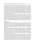
Most applications of anti-hapten Abs involve their use in competitive-
inhibition ELISA using either of two formats. With direct-competition assays,
native and enzyme-labeled Ag in solution compete for the Ab-binding site
(Fig. 1). The Ab is captured by an immobilized secondary Ab directed against a
suitable affi nity tag, for example, the c-myc and FLAG tags. Residual enzyme
activity is then measured across a range of native Ag concentrations. With
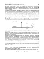
indirect competition assays, native Ag in solution competes with immobilized
Ag conjugate and with residual immobilized anti-hapten Ab detected using
a labeled secondary Ab (Fig. 2). In both cases, increasing the concentration
of native hapten results in a signal reduction, allowing a calibration curve to
be constructed (Fig. 3).
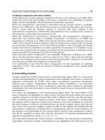
In order to be effective, Abs are required that bind to conjugate with suffi cient
affi nity to generate a usable signal in ELISA, but which also bind preferentially
to free hapten. A high affi nity for the conjugate is generally undesirable because
such Abs do not dissociate readily and reduce the sensitivity of the assay.
Care must also be taken to avoid selection of interface binders (3). These Abs
recognize the hapten Ag in the context of the conjugate and bind to some extent
to the linker used in conjugation and perhaps to the carrier protein itself in the
vicinity of the point of conjugation. As a result, they show higher affi nity for
conjugate than for free hapten and so are unsuitable (Fig. 3).
160 Charlton and Porter
Fig. 1. Schematic representation of a direct competition ELISA. (1) Anti-affi nity tag
polyclonal antibody; (2) scFv with affi nity tag; (3) hapten; (4) alkaline-phosphatase [E]
labeled hapten; (5) and (6) unbound free and labeled hapten removed by washing.
Anti-Hapten Specifi c Ab Fragments 161
Fig. 2. Schematic representation of an indirect competition ELISA. (1) Immobilized
hapten conjugate; (2) scFv; (3) horse radish peroxidase [E]-labeled anti-affi nity tag
polyclonal antibody; (4) free and (5) scFv-bound soluble hapten removed by washing.
Fig. 3. Indirect competition ELISA data of two antibodies selected from the same
immune library against the hapten antigen atrazine. (•) Selected against atrazine-BSA
and eluted with triethylamine (see Subheading 3.2.); (o) selected against atrazine-
BSA and eluted with free atrazine (round 3 onwards). Broken vertical lines indicate
IC
50
values.
This chapter describes protocols for the isolation of anti-hapten Abs from Ab
phage-display libraries. The method utilizes two different hapten conjugates
for alternative rounds of selection, therefore avoiding the selection of phage to
the carrier protein and also uses different phage elution methods for each step
of the selection, which aid in the isolation of high-affi nity anti-hapten Abs.
The technique may be performed using any of the available types of phage
Ab libraries, e.g., those constructed from naïve repertoires (whether native,
semisynthetic, or fully synthetic) or a custom-made library produced from
an animal immunized against the hapten conjugate in the same way as for
generating hybridomas. Such immune libraries offer the advantage of being
biased in favor of Abs recognizing the hapten of interest, although they require
time to construct and separate libraries are usually required for each target
of interest. However, it is possible to immunize an animal with several Ags
simultaneously and to isolate phage Abs specifi c for each target (4). A single
suitable naïve library can also be used to select Abs to any number of targets
with an equal chance of success. However, in order to yield Abs with affi nities
comparable to those from an immune library, large (>10
10
) naïve libraries
are normally required. For anti-hapten diagnostic Abs, the typical limits of
sensitivity achievable (IC
50
) are <1 nM (immune library) vs >100 nM (naïve
library).
2. Materials
1. Phage Ab library, freshly amplifi ed and titered (see Note 1).
2. Hapten conjugate 1 and hapten conjugate 2, purifi ed (see Note 2).
3. Free Ag (hapten).
4. Phosphate-buffered saline (PBS).
5. PBS containing 2% (w/v) and 4% skim milk powder (PBSM) (make fresh as
required).
6. PBS containing 0.1% (v/v) Tween-20 (PBST).
7. Elution buffers. 100 mM TEA: 70 µL triethylamine (7.18 M) in 5 mL H
2
O (dilute
on day of use). For KM13 helper phage: trypsin stock solution at 10 mg/mL.
Dilute 50 µL stock solution in 450 µL PBS for use.
8. 1 M Tris-HCl, pH 7.4.
9. Escherichia coli TG1 (Stratagene).
10. Helper phage VCSM13 (Stratagene) or KM13 (MRC Laboratory, Cambridge,
UK) (see Note 3).
11. 2TY medium. 2TY containing 15% (v/v) glycerol. Antibiotic stock solutions:
100 mg/mL ampicillin in H
2
O; 50 mg/mL kanamycin in H
2
O; both fi lter-sterilized
(0.2 µm).
12. TYE agar plates containing 100 µg/mL ampicillin and 1% glucose (TYE–AMP–
GLU), in standard and large-size diameter Petri dishes.
13. PEG–NaCl: 20% (w/v) polyethylene glycol 6000, 2.5 M NaCl.
162 Charlton and Porter
14. 4 mL Nunc Maxisorb immunotubes (Gibco-BRL); Immulon-4 96-well fl at-
bottomed ELISA plates (Dynex Technologies); 96-well fl at-bottomed tissue
culture plates.
15. Peroxidase-conjugated anti-M13 Ab (Pharmacia).
16. Tetramethyl benzidine (TMB) microwell peroxidase substrate (Dynex Technolo-
gies); tetramethyl benzidine (TMB) tablets (Sigma).
17. 1 M H
2
SO
4
.
3. Methods
3.1. Selection on Immunotubes
3.1.1. Round 1
1. Coat an immunotube with 4 mL 10–100 µg/mL hapten conjugate 1 in PBS
overnight at 4°C (see Note 4).
2. Discard the contents of the tube, and wash 3× with PBS (pour in and immediately
pour out).
3. Block the tube with 4 mL 2% PBSM at room temperature for 1–2 h.
4. Wash as in step 2.
5. Add approx 10
12
cfu phage library (see Note 5) in 4 mL 2% PBSM and incubate
at room temperature by tumbling on an over-under turntable for 30 min, followed
by 90 min without tumbling (see Note 6).
6. Discard the phage solution (see Note 7) and wash the tube 10× with PBST,
followed by 10× with PBS as in step 2. Shake out any remaining wash buffer.
(For subsequent rounds, wash at least 20× with each of PBST and PBS).
7. Elute the bound phage (see Subheading 3.2.).
3.1.2. Further Rounds of Selection
1. For the second round, repeat Subheading 3.1.1. with the following modifi cation:
at step 1, coat the immunotube with hapten conjugate 2 at 10 µg/mL.
2. For subsequent rounds, revert to coating with hapten conjugate 1 at 1 µg/mL
(see Note 8).
3.2. Elution of Bound Phage
The panning strategy employed and the elution steps, in particular, are
critical to the isolation of Abs with high affi nity for hapten Ags. The approaches
used vary with the different stages of selection and are covered under separate
subheadings.
3.2.1. Elution with Triethylamine (Rounds 1 and 2)
1. From a fresh overnight culture of E. coli TG1 cells in 2TY broth (no antibiotics
or glucose), make a 1Ϻ100 dilution in fresh media and grow, shaking at 37°C, to
optical density 600 nm (OD
600
) 0.4–0.5 (1–2 h) (see Note 9).
Anti-Hapten Specifi c Ab Fragments 163
2. Add 1 mL 100 mM TEA to the immunotube and incubate with tumbling for
10 min (see Note 10).
3. Immediately pour the contents of the immunotube into 500 µL of 1 M Tris-HCl
(pH 7.4) to neutralize the pH.
4. Add one-half (0.75 mL) of the eluted phage to 5.25 mL log-phase TG1 cells
(from step 1). Add a further 4 mL log-phase TG1 cells to the immunotube.
Incubate both for 30 min without shaking in a 37°C water bath.
5. Pool the cells, and prepare 4–5 serial 10-fold dilutions (100 µL in 900 µL 2TY).
Plate 100 µL of each dilution on TYE–AMP–GLU plates and incubate overnight
at 30°C to titer the number of infective phage eluted (see Note 11).
6. Centrifuge the remaining cells at 3000g for 10 min at 4°C, resuspend in 1 mL
fresh media, then spread over a large-diameter TYE–AMP–GLU plate, and
incubate at 30°C overnight.
7. Rescue the phage for use in round 2 as detailed in Subheading 3.3.
3.2.2. Elution with Trypsin (Rounds 1 and 2 if Using KM13 Helper Phage)
1. To the washed immunotube (see Subheading 3.1.1., step 6) add 500 µL trypsin–
PBS, and rotate on an over-under turntable for 10 min at room temperature
(see Note 12).
2. Add 250 µL eluted phage to 9.75 mL log-phase TG1 cells (store the remaining
250 µL at 4°C). Incubate for 30 min at 37°C in a water bath.
3. Use 100 µL infected cells to prepare 4–5 serial 10-fold dilutions. Spread these
on TYE–AMP–GLU plates and incubate overnight at 30°C, to titer the eluted
phage.
4. Centrifuge the remaining cells at 3000g for 10 min at 4°C, then resuspend in
1 mL fresh media, spread over a large-diameter TYE–AMP–GLU plate, and incu-
bate at 30°C overnight.
5. Rescue the phage as detailed in Subheading 3.3.
3.2.3. Elution with Free Ag (Round 3 Onwards)
1. Add 4 mL 10 µM solution (see Note 13) of free Ag (hapten) in PBS to the
immunotube and incubate on an over–under turntable for 1 h (see Note 14).
2. Pour out the contents of the immunotube (DO NOT DISCARD) (see Note 15).
Add one-half of the eluted phage to 8 mL log-phase TG1 cells (the remaining
2 mL should be stored at 4°C) and incubate without shaking in a 37°C water
bath for 30 min.
3. Prepare serial 10-fold dilutions (100 µL in 900 µL 2TY). Plate 100 µL of each
dilution on TYE–AMP–GLU plates and incubate overnight at 30°C to titer the
number of infective phage eluted.
4. Centrifuge the remaining cells at 3000g for 10 min at 4°C, resuspend in 1 mL of
media, then spread over a large-diameter TYE–AMP–GLU plate, and incubate
at 30°C overnight.
164 Charlton and Porter
5. Rescue phage as detailed in Subheading 3.3.
6. For subsequent rounds, reduce the concentration of free Ag used to elute the
phage by 100–1000-fold for each successive round (see Notes 16 and 17).
3.3. Rescue of Enriched Phage Abs
1. Add 2–3 mL 2TY–15% glycerol to the agar plate and scrape off the cells with
a glass spreader. Inoculate 50–100 µL cell suspension into 100 mL 2TY–100
µg/mL ampicillin/1% glucose (2TY–AMP–GLU) and check that OD
600
nm is
≤0.1. Incubate at 37°C with shaking until the OD
600
reaches 0.4–0.5. Store the
remaining glycerol stock in aliquots at –70°C.
2. To 10 mL culture, add a 20-fold excess of helper phage (see Note 18) and
incubate without shaking in a 37°C water bath for 30 min.
3. Spin the infected cells at 3000g for 10 min and resuspend the cell pellet in 50 mL
2TY–100 µg/mL ampicillin/50 µg/mL kanamycin (2TY–AMP–KAN). Incubate
at 30°C with shaking overnight.
4. Spin the cells at 10,000g for 10 min (or 3000g for 30 min).
5. Add one-fi fth vol (10 mL) PEG–NaCl to the supernatant, briefl y mix by vortex-
ing, and leave on ice for at least 1 h.
6. Spin at 10,000g for 10 min and pour off the supernatant. Respin briefl y and
remove any remaining supernatant by pipeting or aspiration.
7. Resuspend the pellet in 2 mL PBS and spin at maximum speed for 10 min,
to remove any remaining bacterial debris. Use 1 mL phage suspension for the
next round of selection. Add glycerol (15%) to the remaining aliquot and store
at –70°C.
3.4. Screening Phage Abs by ELISA
3.4.1. Polyclonal Phage ELISA
1. Coat duplicate wells of a 96-well ELISA plate with 100 µL hapten conjugate 1
and with each hapten carrier protein alone at 1 µg/mL in the same buffer as used
for panning. Incubate the plates overnight at 4°C (see Note 19).
2. Wash the plate 3× with PBS by fi lling the wells using a multichannel pipet or
squeezy bottle, inverting the plate, and shaking. Residual wash buffer can be
removed by patting the plate onto paper towels.
3. Block the wells with 200 µL/well 2% PBSM at 37°C for 1–2 h, then wash 3×
with PBS as in step 3.
4. Dilute 10 µL PEG-precipitated phage from the end of each round of selection
and from the initial library rescue in 100 µL 2% PBSM and incubate for 1 h at
room temperature. Include wells that contains PBSM only.
5. Discard the phage solution (see Note 7) and wash the plate 3× with PBST.
6. Add 100 µL/well 1Ϻ5000 dilution of horseradish peroxidase (HRP)–anti-M13
Ab in 2% PBSM and incubate for 1 h at room temperature.
7. Wash the wells 3× with PBST, then 3× with PBS.
Anti-Hapten Specifi c Ab Fragments 165
8. Add 100 µL/well TMB solution (see Note 20) and incubate at room temperature
until a blue color develops (2–20 min or until color appears in the control [no
phage] wells).
9. Stop the reaction by adding 50 µL 1 M H
2
SO
4
(the blue color will turn yellow).
Using a plate reader, measure the OD at 450 nm and 650 nm. Subtract OD
650
from OD
450
, to determine the reading for each well.
3.4.2. Monoclonal Phage ELISA
1. Inoculate individual colonies from the plates generated by the titration of eluted
phage (see Subheading 3.2.) into 100 µL 2TY–AMP–GLU in 96-well tissue cul-
ture plates and incubate with shaking (250 rpm) at 37°C overnight (see Note 21).
2. Using a multichannel (96-well) pipeting device, inoculate a second replicate
96-well plate containing 175 µL/well 2TY–AMP–GLU with 25 µL overnight
culture, then touch the pipet tips to the surface of a large-diameter TYE–AMP–
GLU agar plate (see Note 22). Incubate the 96-well plate at 37°C with shaking
(250 rpm) for 2 h, then proceed to step 3. Incubate the agar plate at 30°C
overnight. Add glycerol to the fi rst 96-well plate (overnight culture) to a fi nal
concentration of 15% and store at –70°C.
3. Add 25 µL 2TY–AMP–GLU containing 10
9
helper phage to each well and
incubate for 30 min at 37°C without shaking followed by 30 min with shaking
(250 rpm).
4. Spin at 1800g for 15 min, then aspirate off the supernatant and discard.
5. Resuspend the pellet in 200 µL 2TY–AMP–KAN and incubate with shaking
(250 rpm) overnight at 30°C.
6. Coat three 96-well ELISA plates overnight at 4°C with 100 µL/well Ag as
follows: plates 1 and 2, hapten conjugate 1; plate 3, carrier protein 1. Wash and
block the plates as in Subheading 3.4.1., steps 2 and 3.
7. Add 50 µL/well 4% PBSM to plates 1 and 3 and 50 µL 4% PBSM containing
1–10 µM free hapten to plate 2. Spin the plates from step 5 at 1800g for 15 min.
Add 50 µL/well of the phage supernatant to each plate and incubate for 1 h at
room temperature.
8. Continue the ELISA as detailed in Subheading 3.4.1., steps 5–9.
9. Select those clones for further analysis that bind to plate 1, do not bind to plate 3,
and do not bind or give reduced signals to plate 2 (see Note 23).
3.5. Competitive Inhibition ELISA
Competition ELISA is best performed with soluble Ab fragments (see Notes
24 and 25).
3.5.1. Indirect Competition ELISA
1. Coat a 96-well ELISA plate with 100 µL/well of hapten conjugate 1 at
1 µg/mL.
2. Wash the plate 3× with PBS.
166 Charlton and Porter
3. Block the wells with 200 µL/well 2% PBSM at 37°C for 1–2 h (see Note 26)
and wash 3× with PBS.
4. Prepare serial (2- or 4-fold) dilutions of free Ag (hapten) in PBS in microcentri-
fuge tubes, including a tube with PBS only. Add an equal volume of the Ab
fragment to a fi nal subsaturating concentration (see Note 27) and incubate at
4°C for 1 h.
5. Apply 100 µL of the Ab–Ag solution to replicate wells of the blocked plate and
incubate at room temperature for 1 h. Wash the plate 3× with PBST.
6. Continue the ELISA as before (Subheading 3.4.1., steps 6–9) using a suitable
labeled secondary reagent diluted in PBST (see Note 28).
7. Plot the signal generated for each concentration of free Ag as a percentage of
that obtained without free Ag against free Ag concentration and determine the
concentration that reduces the signal by 50% (IC
50
) (see Fig. 3).
3.5.2. Direct Competition ELISA
1. Coat a 96-well ELISA plate with 100 µL/well anti-affi nity tag Ab (Protein A or
Protein L can be used as a alternative).
2. Wash the wells 3× with PBS, block with PBSM, then wash 3× with PBS, as
mentioned previously.
3. Prepare serial (2- or 4-fold) dilutions of free Ag in PBS. Include a tube without
free Ag. Add each dilution to tubes containing a constant concentration of
enzyme labeled (usually alkaline phosphatase) Ag (see Note 29).
4. Add an equal volume of soluble Ab fragment to the predetermined fi nal subsatu-
rating concentration and incubate at 4°C for 1 h.
5. Add 100 µL of the Ab–Ag solution to replicate wells of the blocked plate and
incubate at room temperature for 1 h.
6. Develop the ELISA with pNPP substrate according to the manufacturer’s
instructions and measure the optical density at 405 nm and 650 nm. Subtract the
OD
650
from OD
405
to determine the reading for each well.
7. Plot a curve as in Subheading 3.5.1., step 7 (see Fig. 3).
4. Notes
1. For the isolation of hapten-specifi c Abs, a library based on a phagemid system
is preferable to one using an entire functional phage genome. Phagemid vectors
allow expression of a single Ab fragment per virus particle and so avoid problems
associated with avidity effects, which are encountered with multivalent display.
The protocols in this chapter are based on a phagemid expression system that
encodes ampicillin resistance.
2. Prepare two hapten conjugates for panning (hapten conjugate 1, hapten conjugate
2) using two different carrier proteins, which, where applicable, differ from that
used for immunization, e.g., bovine serum albumin, keyhole limpet hemocyanin
(KLH), and bovine thyroglobulin. These should be purifi ed if possible, for
example, by high-performance liquid chromatography to avoid the selection of
Ab against protein contaminants common to both preparations.
Anti-Hapten Specifi c Ab Fragments 167
3. There are several strains of helper phage available, e.g., VCSM13 (Stratagene),
M13KO7 (Pharmacia), and KM13 (MRC, Cambridge, UK). KM13 differs in that
it includes a trypsin-cleavage site within the minor coat protein (gIII). Therefore,
bound phage Abs can be eluted by incubation with 500 µL trypsin solution (1
mg/mL in PBS) for 10 min at room temperature. Only those phage that include
a displayed Ab fragment fused to the noncleavable product of gIII, will be
infective, so reducing the background of nonspecifi c binders carried through to
subsequent rounds. All of the helper phage above encode a selectable kanamycin
resistance gene.
4. It is important to recover as many different clones as possible that recognize the
target Ag from the library in the fi rst round of selection so a high concentration
of coating Ag is used. The incubation temperature and buffering solution may
need to be altered for different carrier proteins. The conditions given are suitable
for BSA and KLH conjugates.
5. The number of phage applied to the immunotube is particularly important during
the first round of selection. Aim to include ~10
3
–10
4
copies of each clone
represented (10
3
–10
4
× library size); however, consideration should be given to
the size of the library and the number of hapten molecules conjugated to the
carrier protein. An excessive number of phage from a library of limited diversity
and a low coating Ag density may lead to exclusion of all but those clones with
a high affi nity for the hapten conjugate.
6. In order to reduce the number of phage selected against the carrier proteins, each
protein used can be added to the immunotube during the phage-binding step
at fi nal concentrations of 1 mg/mL. If using an immune library, the immunizing
carrier protein can also be included.
7. Dispose of solutions containing unwanted phage directly into a viracidal solution,
such as Virkon to prevent accidental infection of TG1 cells during later stages.
8. Alternation of the carrier protein during the initial rounds of selection is necessary
to remove phage that bind to the carrier. It is not necessary to continue alternating
beyond round 3.
9. Effi cient infection of E. coli cells by phage is dependant on cells being in log
phase (OD
600
0.4–0.5). Cells can be kept on ice for up to 30 min before infection,
if necessary, but procedures should be timed to avoid this if possible.
10. TEA is destructive to phage and incubation should not exceed 10 min.
11. The number of phage recovered will vary with the stage of the selection process
and the library used. When using a library of good size, i.e., >10
8
clones, and
particularly when using an immune library, expect to get at least 10
4
phage back
after the fi rst round of panning. Naïve libraries and those of smaller size will
yield less. The fi rst round of selection is the most important and errors made
here will be amplifi ed during later rounds. If less than 1000 phage are recovered,
repeat the infection and rescue (see Subheading 3.2.1., steps 4–7) using the
remaining 0.75 mL eluted phage. If a similar recovery is seen, check that the Ag
is coating effi ciently under the conditions used and alter conditions if necessary.
Store titration plates containing colonies for later monoclonal analysis.
168 Charlton and Porter
12. By reducing the carry-through of nonspecifi c phage, the use of trypsin as a
means of eluting bound phage increases the rate of enrichment of Ag-specifi c
clones (5).
13. For the fi rst round of free Ag elution, a high concentration of free Ag is used in
order to recover as many different phage Abs as possible from the immobilized
population, which are able to recognize soluble Ag. The concentration used
may be restricted by the solubility of the Ag in aqueous solution. If the Ag is
particularly insoluble in H
2
O, then methanol up to 10% (v/v) can be used without
any signifi cant effect on Ab binding.
14. The incubation time with free Ag can be varied and consideration should be
given to the effects of this. The Ab–Ag interaction is a dynamic process with
ligand and analyte continually dissociating and reassociating. In the absence of
free Ag, a large number of phage Abs will be found in the liquid phase at any
time. Excessive incubation times will increase the number of clones displaying
Abs with high affi nities for the hapten conjugate, which are carried through to
the next round. Shorter times may help to select clones with a rapid dissociation
rate from the hapten conjugate, but incubations of less than 30 min are not
recommended.
15. If using the KM13 helper phage, then nondisplaying background phage can be
reduced at this stage by adding 50 µL trypsin stock to the eluted phage and
incubating at room temperature for 10 min prior to infection.
16. Reducing the concentration of free Ag used to elute bound phage with successive
rounds can help to select those Abs with the highest affi nities for the native Ag.
Care should be taken not to use too low a concentration.
17. The number of phage recovered from each round by elution with free Ag may
only increase slowly (if at all) when the concentration of free Ag is progressively
reduced. If numbers fall signifi cantly, rescue the remaining stored eluted phage
or repeat the round of selection.
18. An OD
600
of 1.0 = approx 8 × 10
8
E. coli/mL.
19. All Ag-coating and blocking steps can be carried out at 37°C for 1–2 h or
overnight at 4°C.
20. If not available, TMB tablets are available from Sigma. 30% H
2
O
2
and citric
phosphate buffer will be required. Dissolve 2.55 g citric acid and 3.545 g
NaH
2
PO
4
in 400 mL of H
2
O. Adjust the pH to 5.0 with 5 M NaOH, add H
2
O to
500 mL, and autoclave. This substrate is generally slower to develop color and
gives lower OD
450
readings, but is otherwise suitable.
21. Place the plate into a suitable container and surround with damp paper towels
to prevent evaporation.
22. It is convenient to inoculate an agar plate with phage clones for further analysis
to prevent repeated thawing of the glycerol stock.
23. The signal generated from plate 1 results from a combination of the binding
kinetics of the Ab and the expression level of the phage-Ab clone. High signals do
not necessarily indicate the best diagnostic clone. Similarly, a low % reduction of
signal on plate 2, relative to plate 1, may result from the presence of a saturating
Anti-Hapten Specifi c Ab Fragments 169
concentration of phage regarding the coating Ag. Select several clones displaying
a range of apparent free Ag binding for more detailed analysis.
24. Most commonly used phagemid vectors, e.g., pHEN and pCANTAB, allow for
the expression of soluble Ab fragments by including a lac promoter and an
amber (TAG) stop codon between the Ab genes and the gIII minor coat protein
gene. Infection of phage Abs into a nonsuppressor strain of E. coli, such
as HB2151, permits the induction of Ab expression with 1 mM isopropyl-
β-thiogalactopyranoside (IPTG). When using a synthetic or semisynthetic library,
ensure that the coding variability does not allow for the inclusion of TAG codons
within the variable region genes. When this is the case, TG1 cells can also be
induced with IPTG. However, the amber codon between the scFv and gIII
will also be suppressed, leading to expression of both scFv and scFv–pIII
fusions. Ideally scFv genes should be cloned into a dedicated soluble expression
vector, e.g., pIMS147 (6). Amber codons, where present, can be altered to GAG
(glutamate) by site-directed mutagenesis.
25. Soluble Ab fragments should be purifi ed for use in ELISAs, for example, by
Protein-A or Protein-L affi nity column or by immobilized metal affi nity chro-
matography if the construct includes a histidine tag. Crude culture supernatants
or periplasmic extracts can be used, but it is possible that contaminating proteins
may infl uence the results.
26. For assays using soluble Ab fragments, 3% BSA in PBS can be used as an
alternative blocking agent when high-background binding of enzyme-labeled
secondary Ab is experienced.
27. Prepare a binding profi le of a range of serial dilutions of soluble Ab fragment
to 1 µg/mL hapten-conjugate coating concentration. Select a concentration of
Ab fragment that lies on the linear portion of the sigmoidal curve and gives a
suitable ELISA signal, i.e., >0.5 absorbance.
28. Most phagemid/soluble expression vectors include a tag 3′ of the Ab fragment
for purifi cation/detection purposes. Suitable detection reagents include an HRP-
labeled Ab to the affi nity tag or either Protein A–HRP or Protein L–HRP, if
this is not available. Weak signals can be amplifi ed by using an unlabeled anti-
affi nity tag secondary Ab in a sandwich ELISA format and detecting with an
enzyme-labeled anti-species Ab.
29. Empirically determine the optimum concentrations of Ab fragment and enzyme-
labeled Ag. Ab fragment should be subsaturating regarding the immobilized
capture Ab (see Subheading 3.5.2., step 1) and labeled Ag (just) subsaturating
regarding the captured Ab fragment.
Acknowledgments
Data included in this manuscript is drawn from research funded by the
Biotechnology and Biological Sciences Research Council, UK.
170 Charlton and Porter
References
1. Goodrow, M. H., Harrison, R. O., and Hammock, B. D. (1990) Hapten synthesis,
antibody development, and competitive inhibition enzyme immunoassay for
s-triazine herbicides. J. Agric. Food Chem. 38, 990–996.
2. Karu, A. E., Goodrow, M. H., Schmidt, D. J., Hammock, B. D., and Bigelow,
M. W. (1994) Synthesis of haptens and derivation of monoclonal antibodies
for immunoassay of the phenylurea herbicide diuron. J. Agric. Food Chem. 42,
301–309.
3. Tuomola, M., Harpio, R., Mikola, H., Knuuttila, P., Lindström, M., Mukkala,
V M., Matikainen, M T., and Lövgren, T. (2000) Production and characterization
of monoclonal antibodies against a very small hapten, 3-methylindole. J. Immunol.
Meth. 240, 111–124.
4. Li, Y., Cockburn, W., Kilpatrick, J. B., and Whitelam, G. C. (2000) High affi nity
ScFvs from a single rabbit immunized with multiple haptens. Biochem. Biophys.
Res. Commun. 268, 398–404.
5. Kristensen, P. and Winter, G. (1998) Proteolytic selection for protein folding using
fi lamentous bacteriophages. Folding Design 3, 321–328.
6. Hayhurst, A. and Harris, W. J. (1999) Escherichia coli Skp chaperone co-expression
improves solubility and phage display of single chain antibody fragments. Protein
Exp. Purif. 15, 336–343.
Anti-Hapten Specifi c Ab Fragments 171
173
From:
Methods in Molecular Biology, vol. 178: Antibody Phage Display: Methods and Protocols
Edited by: P. M. O’Brien and R. Aitken © Humana Press Inc., Totowa, NJ
12
Blocking Immunodominant Epitopes
by Competitive Deselection
Roberto Burioni
1. Introduction
The development of combinatorial antibody (Ab) libraries displayed on
the surface of phage has led to the production of a wide range of human
monoclonal antibodies (MAbs) against a plethora of viral antigens (Ag) (1–5).
However, sometimes the isolation of a given Ab can be particularly diffi cult
because of the predominance of some epitopes, and, in the case of impure
Ags, because the protein or compound of interest is present in a low amount.
Many techniques have been developed for overcoming this problem, including
epitope-masking (6) or sandwich-capture (7) procedures. These approaches
require a MAb against the Ab or epitope of interest, which is not always avail-
able. In this chapter, a new procedure of preadsorption panning is described,
which facilitates the molecular cloning of MAb fragments against nonim-
munodominant Ag determinants using phage-display immunoselection. The
procedure, called competitive deselection, is easy, fast, inexpensive, and can be
coupled with other described panning techniques; however, its chief advantage
is in the isolation of Abs against less-represented Ag determinants.
The procedure described in this chapter also increases the effi ciency of
cloning Abs of rare specifi cities. In our hands we have successfully isolated
human recombinant Fabs from phage display libraries able to distinguish
herpes simplex virus type 1 from type 2 in immunofl uorescence assays (8).
We have also been able to increase the effi ciency of selection of anti-idiotype
mouse monoclonal Fabs bearing the internal image of the template Ag (Burioni,
R., in press).
Blocking Immunodominant Epitopes 173
The protocol is a modifi cation of a standard panning protocol, except that
the phage library is fi rst preabsorbed on the Ag of interest to remove phage
that react with the immunodominant epitope. The unbound phage are then
incubated a second time with Ag and eluted and amplifi ed according to normal
protocols. One caveat that must be kept in mind when using this approach is
that, as shown clearly by our results (8), the subtraction of unwanted clones
is only partial and should be considered as a negative enrichment rather than
a complete subtraction. Subtracted clones are always present at the end of the
panning procedure, but their frequency is lower than that obtained with an
unmodifi ed panning procedure, which appears to leave room for rare clones to
be selected and analyzed. Therefore, this limitation does not appear to affect
the success of the technique.
2. Materials
1. Ab phage library, freshly amplifi ed according to standard protocols, resuspended
in phosphate-buffered saline (PBS)–1% (w/v) bovine serum albumin (BSA), and
titered (colony-forming units [cfu]/mL) (see Notes 1 and 2).
2. PBS or 0.1 M carbonate buffer, pH 8.6: for 1 L, dissolve 8.4 g NaHCO
3
in H
2
O,
adjust the pH to 8.6, then fi lter, and store at 4°C.
3. Ag of interest, diluted in PBS or 0.1 M carbonate buffer (see Note 3).
4. PBS–1% BSA; PBS–0.5% (v/v) Tween-20.
5. Elution buffer: for 200 mL, add 1.6 mL 12 M HCl to H
2
O and adjust to pH 2.2
with solid glycine. Autoclave and store at room temperature.
6. 2 M Tris-HCl base.
7. Escherichia coli strain XL1 Blue; VCSM13 helper phage (Stratagene).
8. Superbroth medium (SB), variably containing antibiotics at the following
fi nal concentrations: tetracycline (10 µg/mL); carbenicillin (20 µg/mL in low-
carbenicillin SB; 50 µg/mL in high-carbenicillin SB); kanamycin (70 µg/mL).
9. Luria-Bertani agar plates containing 100 µg/mL carbenicillin.
10. Polyethylene glycol (PEG)–NaCl: 20% (w/v) PEG-8000, 2.5 M NaCl. Autoclave
and store at room temperature.
11. Enzyme-linked immunosorbant assay (ELISA) plates (half area, high-affinity
binding: Costar cat. no. 3690); Oak Ridge centrifuge tubes (Sigma, St. Louis, MO).
3. Methods
1. Coat ELISA plate wells with the appropriate amount of Ag in each well (see
Note 4). Coat both the adsorption and panning plate at the same time. For the
adsorption plate, coat at least 10 wells for each phage selection. For the fi rst
round of panning, coat four wells; for the following rounds, coat two wells/phage
selection.
2. Incubate the sealed plate at 4°C overnight.
174 Burioni
3. Wash both plates 5× with dH
2
O (100 µL/well), then blot dry on paper towel.
Block both plates with 150 µL/well PBS–1% BSA for 2 h at 37°C. Do not let
the plates dry out (see Note 5).
4. Inoculate a single fresh colony of Escherichia coli XL1 Blue into 15 mL
SB–tetracycline in a 50-mL tube. Grow at 37°C in a rotatory shaker. Start the
culture in time to have a exponential growth culture (OD
600
= 0.6) for infection
at step 9 (see Note 6).
5. Remove the block solution from the wells of the adsorption plate with a pipet and
add 20 µL (>10
10
cfu) of freshly amplifi ed library phage to each of the 10 wells
(see Note 7). Seal the plate and incubate at 37°C for 2 h (see Note 8).
6. Carefully remove 15 µL phage from each well of the adsorption plate and
combine in a 0.5-mL microcentrifuge tube and keep on ice. Remove the blocking
solution from the panning plate and immediately add 25–35 µL combined
adsorbed phage to each well for the fi rst round of panning (using four wells) or
50–70 µL phage for subsequent panning rounds (two panning wells). Seal the
plate and incubate for an further 2 h at 37°C.
7. Wash the wells 10× with PBS–0.5% Tween-20 by adding 100 µL/well
incubating for 5 min, then discarding (see Note 9). Use barrier tips to avoid
contamination.
8. Elute the phages bearing specifi c Abs by adding 50 µL elution buffer/well and
incubating at room temperature for 3 min. Remove the elution buffer into a
microcentrifuge tube and neutralize immediately by adding 3 µL 2 M Tris-HCl
per 50 µL elution buffer.
9. Add the eluted phage to 2 mL exponential growth-phase E. coli XL1 Blue and
incubate for 15 min at 37°C.
10. Add 10 mL prewarmed SB–low carbenicillin + tetracycline to the 2 mL infected
cells. Plate 10-, 1-, and 0.1-µL aliquots of the infected cell suspension on Luria-
Bertani–CARB plates and incubate overnight at 37°C. Calculate the approximate
number of eluted phages from the number of colonies. Incubate the remaining
cell suspension for 1 h at 37°C in a shaker.
11. Add 100 mL prewarmed SB (high carbenicillin + tetracycline) and incubate for
1 h at 37°C in a shaker.
12. Add 10
12
pfu helper phage VCSM13. Incubate at 37°C in a shaker for a further 2 h.
13. Add kanamycin to a fi nal concentration of 70 µg/mL and incubate the culture
overnight at 30°C in a shaker.
14. Centrifuge the cultures at 2000g for 10 min at room temperature.
15. Precipitate the phage from the resulting supernatant by adding 7 mL PEG–NaCl
solution to 30 mL supernatant in Oak Ridge tubes. Incubate on ice for 30 min.
16. Centrifuge at 15,000g for 20 min at 4°C and discard the supernatant. Let the
tubes dry upside down on paper towel for 2–4 min.
17. Carefully resuspend the phage pellet in 1 mL PBS–1% BSA per tube. Be careful
to also resuspend the pellet that usually forms on the wall of the tube. Transfer
Blocking Immunodominant Epitopes 175
the suspension to a microcentrifuge tube and mix the tube by inverting several
times (do not vortex).
18. Centrifuge at 10,000g for 15 min at 4°C, then transfer the supernantant into a
clean microcentrifuge tube.
19. Use this phage suspension to perform further rounds of panning (see Note 10),
or once several rounds have been completed, for the infection of E. coli for the
subsequent production of soluble Fab (9).
4. Notes
1. This protocol uses a Fab library constructed in pComb3 or its derivatives. The
use of alternative expression systems may require a modifi cation to the antibiotic
selection used in the amplifi cation of eluted phage.
2. The phage library, or subsequently selected phage, need to be freshly amplifi ed
for each panning cycle. Although phage molecules themselves are stable and can
be stored for years at –70°C without losing infectivity, displayed Ab molecules
on the surface are not stable. Panning of a stored phage preparation can yield
unpredictable results.
3. Optimal conditions for binding, including temperature of binding and coating
buffer, need to be determined experimentally for each individual Ag. Most
proteins bind well in PBS or in 0.1 M carbonate buffer. Do not reuse the plates.
Use a fresh plate for each round of panning and a (fresh) different one for
adsorption each time. The best results are obtained using plates freshly coated
with Ag.
4. As a rule, dilute the Ag to a concentration 5× greater than that used in ELISA for
detection of Abs. If this ELISA concentration is not known, use 500 ng/well Ag
for panning and 100 ng/well for ELISA. This concentration is usually suitable
for the isolation of Ab-bearing phages. The volume in which the Ag is added
can range from 25 to 50 µL.
5. Proper blocking of the wells is crucial. The procedure must be performed
simultaneously for both the adsorption and panning plate. Do not let the wells
dry out at any stage.
6. Infection of bacteria is a critical step. It is important that the OD of the E. coli
culture is approximately that indicated (i.e., exponential growth) in order to
obtain maximal infection. Do not dilute the bacterial culture to obtain the correct
OD, but schedule the time of inoculation of the culture appropriately.
7. The amount of starting phage is critical. A low phage titer (<10
11
cfu/mL) usually
results in an unsuccessful subtraction experiment. The fi rst round of panning is
crucial for a successful selection.
8. Subtraction is not as effi cient as selection. For this reason, it is necessary to use
a higher number of wells (10) for absorption than used for panning (four in the
fi rst round, two in subsequent rounds). In the case of an over-representation of
dominant clones, the number of adsorption wells can be doubled, while trying
to keep to a minimum the total volume of phage. An alternative or additional
procedure is to increase the adsorption time. If this is shown to be required,
176 Burioni