Antibody Phage Display Methods and Protocols - part 7 pdf
Bạn đang xem bản rút gọn của tài liệu. Xem và tải ngay bản đầy đủ của tài liệu tại đây (480.6 KB, 39 trang )
splenocytes (absorber cells), in order to remove phage of undesired specifi cities.
The thymic tissue was fi xed using total body perfusion fi xation (6), then minced
into small fragments and nonadherent thymocytes were removed by vigorous
shaking. The selection of the preabsorbed library on the thymic fragments was
performed overnight at 4°C in the presence of a fresh batch of fi xed absorber
cells. After extensive washing, the bound phages were eluted and amplifi ed
before being used for the next selection round (Fig. 1). Following three and
four rounds of selection, we analyzed scFv Abs from individual phage clones
for reactivity against thymus and various lymphoid and nonlymphoid organs
using immunohistochemistry. Using this subtractive selection protocol, we
were able to isolate scFv Abs that bind to murine thymic stromal cells (selector
tissue); Abs reactive with lymphoid cells (absorber cells) were not detected.
Furthermore, some of the isolated clones crossreacted with human thymic
stromal cells, indicating that Abs recognizing evolutionary conserved epitopes
were recovered (Fig. 2).
The subtractive selection of phage Ab libraries on tissue fragments should
be adaptable for use against tissues other than the thymus with the aim of
generating Abs against tissue-specifi c antigens. The choice of selector tissue
and absorber cells/tissue, as well as incubation conditions, will depend on the
individual research question and desired application. In general, this approach
could be applied in the studies of all disease processes that involve qualitative
changes in the histology of the affected tissue. One possible application is in
tumor biology, in which tumor-cell-specifi c markers might easily be lost during
the preparation of single-cell suspensions because of the isolation procedure.
Furthermore, abnormalities related to a tumor may not only be located on the
tumor cells, but also in the extracellular matrix. Therefore, using this approach
with tumor tissue as the selector and normal healthy tissue of the same type as
the absorber tissue, it may be possible to isolate Ab clones that identify cellular
and histological abnormalities of a tumor.
2. Materials
1. Mice as a source of selector tissue and absorber cells (see Note 1).
2. For total body perfusion fi xation: phosphate-buffered saline (PBS)–70 mg/mL
Nembutal; PBS–0.1% procaine–HCl.
3. PBS–0.05% glutaraldehyde: freshly prepared monomeric-distilled glutaraldehyde
(e.g., Polysciences) in PBS, adjusted to pH 7.4.
4. PBS; PBS–1% fetal calf serum (FCS), fi lter-sterilized; PBS–4% skim milk
powder (block solution); PBS–0.05% Tween-20.
5. Nylon sieve with 100 µm pores.
6. Phage-Ab library, freshly amplifi ed and titered.
7. Elution buffer: 76 mM citric acid, pH 2.5.
236 Radoˇsevi´c and van Ewijk
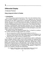
Fig. 1. Schematic diagram of the selection protocol. The numbers correspond to the
steps described in Subheading 3.
Isolation of Single-Chain Antibodies 237
8. 1 M Tris-HCl, pH 7.4.
9. XL1 Blue Escherichia coli.
10. 2TY medium: containing 12 µg/mL tetracycline, 100 µg/mL ampicillin, and
5% (w/v) glucose (TAG medium); large 2TY agar plates containing 12 µg/mL
tetracycline, 100 µg/mL ampicillin, and 5% (w/v) glucose (TAG plates).
11. 5-mL polystyrene round-bottomed centrifuge tubes; 50-mL conical-bottomed
centrifuge tubes.
Fig. 2. Immunohistological identifi cation of epithelial cells in the human thymus
using TB4-20 scFv Ab (objective: ×40).
238 Radoˇsevi´c and van Ewijk
3. Methods
The method described here uses thymic tissue as selector tissue, splenocytes
as absorber cells, and a scFv phage Ab library. These protocols should be
adapted accordingly for each individual system. The individual steps below
(steps 1–19) are schematically presented in Fig. 1.
1. Fix the thymic tissue by total body perfusion fi xation (see Notes 2–4; 6).
2. Isolate the thymus, mince with scissors or a razor blade, and transfer into a
50 mL tube fi lled with PBS.
3. Remove the nonadherent cells (thymocytes) by vigorously vortexing the thymic
fragment suspension for 15 min.
4. Let the fragments sediment by standing the tube at room temperature for
5–10 min, then pipet off the PBS containing the nonadherent cells, and transfer
to a clean tube. Centrifuge the nonadherent cells at 200g for 5 min and resuspend
them either in 5 mL PBS–1% FCS to store (see Note 5) or in block solution (at
concentration of 10
8
/mL) for selection (these are the thymocyte absorber cells).
Resuspend the thymic fragments either in 5 mL PBS–1% FCS to store (see Note 5)
or in 1 mL block solution for selection.
5. Prepare the splenocyte absorber cells: mince a (nonfi xed) spleen through a nylon
sieve (100-µm pores) into 50 mL PBS. Centrifuge the cells at 200g for 5 min and
resuspend them in 10 mL PBS–0.05% glutaraldehyde. Incubate for 15 min at
room temperature. Wash the cells once with 50 mL PBS, then resuspend either in
5 mL PBS–1% FCS to store (see Note 5) or in block solution (at a concentration
of 10
8
/mL) for selection (see Note 6).
6. Preabsorb, and preblock the library: mix 0.5 mL freshly amplifi ed phage library
(approx 10
13
phages/mL) with 1 mL thymocyte absorber cells and 1 mL of
splenocyte absorber cells in a 5 mL tube. Incubate the tube on an end-over-end
rotator for 1 h at room temperature. Centrifuge the tube at 200g for 5 min and
collect the supernatant. This represents the preabsorbed/preblocked library.
7. Preblock the fi xed-tissue fragments (from step 4): incubate the fragments in
block solution for 1 h at room temperature.
8. Add the preabsorbed/preblocked library (2.5 mL) and a fresh batch of fi xed absorber
cells (a mix of 10
8
thymocyte and 10
8
splenocyte absorber cells in 0.5 mL block
solution) to the tissue fragments. This represents the selection mixture (see Note 7).
9. Incubate the suspension overnight at 4° on an end-over-end rotator with slow
rotation.
10. Let the fragments sediment, then pipet off the supernatant and discard.
11. Wash the fragments thoroughly using a total volume of 1–2 L PBS–0.05%
Tween-20 in order to remove unbound phages (see Note 8).
12. To elute the bound phages, resuspend the fragments after the fi nal wash in
450 µL 76 mM citric acid (pH 2.5) and incubate for 5 min at room temperature.
Add 900 µL 1 M Tris-HCl, pH 7.4, to neutralize the pH and mix gently.
Isolation of Single-Chain Antibodies 239
13. Allow the fragments to sediment and pipet off the supernatant (containing the
eluted phages) into a fresh tube (see Note 9).
14. Add 3 mL 2TY medium and 3 mL fresh log-phase culture of E. coli XL1 Blue
(optical density 590 nm = 0.5) to the eluted phages and infect for 30 min at 37°C.
15. Centrifuge the bacterial culture at 2000g for 15 min and resuspend the bacterial
pellet in 0.5 mL 2TY. Spread the bacteria on a TAG plate and incubate overnight
at 37°C.
16. Add 3 mL 2TY medium to the plate and loosen the colonies with a sterile
spreader. Collect the bacterial suspension into a clean tube.
17. Inoculate 100 µL bacteria into 50 mL TAG medium and amplify and precipitate
the phage, according to standard protocols. Make a 15% (v/v) glycerol stock from
the remaining bacterial suspension and freeze in aliquots at –70°C.
18. Repeat the selection for the desired number of rounds (usually 3–4).
19. Using standard protocols, isolate soluble scFv Ab from randomly selected
individual clones and check the specifi city of binding to thymus and lymphoid and
nonlymphoid tissue (or other appropriate tissue) using immunohistochemistry
and/or fl uorescence-activated cell sorting (FACS) analysis (see Notes 10 and 11).
4. Notes
1. In order to avoid isolation of phages directed to major histocompatibility complex
(MHC) antigens, mouse strains of different MHC haplotypes should be used as
a source of cells/tissue for individual selection rounds (i.e., change the strain
each round).
2. Total body perfusion fi xation is performed as follows (6): anesthetize a mouse
by intraperitoneal injection of 200 µL PBS–70 mg/mL Nembutal. Incise the
thorax to expose the heart. Insert a cannula in the tip of the left ventricle. Incise
the right atrium and start the total body perfusion with a prewashing solution
of PBS–0.1% procaine-HCl for 2 min (procaine is used for the dilatation of
blood vessels, it may be omitted). Keep the fl ow rate at 0.5 mL/s at a pressure of
40 mm Hg. After prewashing, switch the perfusion to PBS–0.05% glutaraldehyde
for 10 min.
3. Instead of fi xation by total body perfusion, the tissue can also be fi xed by
immersion fi xation as follows: using scissors, mince the thymic tissue on a
nylon sieve above a glass beaker. Rinse thoroughly to remove the nonadherent
cells (thymocytes) by pipeting 50 mL PBS onto the tissue fragments. Transfer
the fragments to a tube, a fix with 10 mL PBS–0.05% glutaraldehyde for
15 min at room temperature. Let the fragments sediment, pipet off the fi xative, and
resuspend in 50 mL PBS. Let the fragments sediment, pipet off the supernatant,
and resuspend either in 5 mL PBS–1% FCS to store or in 1 mL block solution, for
selection (selector tissue). Collect the nonadherent cells that were rinsed out of
the tissue (thymocyte absorber cells) and fi x them as described for the splenocyte
absorber cells in Subheading 3., step 5. Proceed with step 5 in Subheading 3.
240 Radoˇsevi´c and van Ewijk
4. The mild fi xation used might be advantageous for the selection protocol for
several reasons. The epitopes remain well-preserved during overnight incubation
(no internalization or proteolytic cleavage) and the tissue fragments can be
shaken vigorously in order to effi ciently remove nonadherent cells (thymocytes),
thus exposing the thymic stromal cells for selection.
5. Fixed tissue fragments and absorber cells can be stored in PBS–1% FCS at
4°C for 1–4 wk.
6. It is also possible to use appropriate tissue fragments, instead of a single-cell
suspension as an absorber population. The absorber tissue fragments should be
prepared as described previously for the selector tissue fragments.
7. If using tissue fragments, instead of a single-cell suspension as the absorber, only
the preabsorbed/preblocked library is added to the selector tissue fragments.
8. Transfer the fragments to a 50 mL tube, and wash at least 20×. Each washing
step is performed as follows: add 50 mL PBS–0.05% Tween-20, vortex, incubate
for 5–10 min at room temperature, then remove and discard the supernatant
using a capillary pipet.
9. An alternative is to allow the fragments to sediment during the elution, then to
pipet off the supernatant (containing the eluted phages) into a tube containing
1 M Tris-HCl, pH 7.4, in order to prevent the possible rebinding of phages to
the tissue upon neutralization.
10. In general, for preliminary screenings of scFv Abs we prepare periplasmic (TES)
extracts from the output (selected) clones in strain XL1 Blue. Although this is a
suppressor E. coli strain, the suppression is not complete, resulting in the produc-
tion of a mixture of scFv and fusion-scFv (scFv coupled to the pIII protein). In
addition, we recently used mini-scFv preparations for immunohistochemistry
and FACS screenings. Mini-scFv preparations are supernatants of individual
clones (either in suppressor or nonsuppressor E. coli strains) grown in 96-well
plates and induced with isopropyl thiogalactopyranoside. The volume obtained
from one well is suffi cient for a single immunostaining. The signals obtained
using these preparations are usually weaker than from the periplasmic prepara-
tions, but they do enable high-throughput preliminary screenings. A limiting
factor in the number of clones that can be screened in one experiment is the
number of sections or FACS samples that can be handled at one time. For further
screenings, we transform a nonsuppressor strain of E. coli (e.g., SF110) with the
scFv DNA and prepare periplasmic extracts for binding analysis. A fl ow diagram
of our current screening strategy is shown in Fig. 3.
11. To date, we have isolated a limited repertoire of thymus-reactive clones following
three and four rounds of selection. The reasons for this are as yet unclear, but
may partly result from the vigorous washing step following incubation with the
phage library, in which only the clones with the highest affi nity would remain
bound to epitopes on the stromal cells. It is also possible that clones with other
specifi cities were recovered in the fi rst and second selection rounds, but that they
Isolation of Single-Chain Antibodies 241
were lost (overselected) during further selection rounds because of the growth
advantage of dominant clones.
References
1. van Ewijk, W. (1991) T-cell differentiation is infl uenced by thymic microenviron-
ments. Annu. Rev. Immunol. 9, 591–615.
2. van Ewijk, W., Shores, E. W., and Singer, A. (1994) Crosstalk in the mouse thymus.
Immunol. Today 15, 214–217.
Fig. 3. Screening strategy for postpanning analysis of isolated scFv Abs using
immunohistochemistry and FACS (see Note 10).
242 Radoˇsevi´c and van Ewijk
3. van Ewijk, W., Wang, B., Hollander, G., Kawamoto, H., Spanopoulou, E., Itoi,
M., et al. (1999) Thymic microenvironments, 3-D versus 2-D? Semin. Immunol.
11, 57–64.
4. van Ewijk, W., de Kruif, J., Germeraad, W. T. V., Berendes, P., Röpke, C.,
Platenburg, P. P., and Logtenberg, T. (1997) Subtractive isolation of phage-
displayed single-chain antibodies to thymic stromal cells using intact thymic
fragments. Proc. Natl. Acad. Sci. USA 94, 3903–3908.
5. de Kruif, J., Boel, E., and Logtenberg, T. (1995) Selection and application of
human single chain Fv antibody fragments from a semi-synthetic phage antibody
display library with designed CDR3 regions. J. Mol. Biol. 248, 97–105.
6. van Ewijk, W., Brons, N. H. C., and Rozing, J. (1975) Scanning electron micros-
copy of homing and recirculating lymphocyte populations. Cell Immunol. 19,
245–261.
Isolation of Single-Chain Antibodies 243
245
From:
Methods in Molecular Biology, vol. 178: Antibody Phage Display: Methods and Protocols
Edited by: P. M. O’Brien and R. Aitken © Humana Press Inc., Totowa, NJ
21
Selection of Antibodies Based on Antibody
Kinetic Binding Properties
Ann-Christin Malmborg, Nina Nilsson, and Mats Ohlin
1. Introduction
Molecular evolution approaches to developing molecules with characteristics
particularly suited for specifi c applications have become important tools in
biomedicine and biotechnology. Not only is it possible to identify molecules
with specifi cities that cannot easily be obtained by other means, but it is also
possible to fi ne-tune in an effi cient manner the properties for, in principle,
any specifi ed application. Attention has particularly been put into identifying
molecules with specifi c reaction-rate and affi nity properties. Depending on
the intended application, the binding of a molecule to its target is desired to
be long-lived or short-lived. In biosensors, it will generally be appropriate for
the association between the ligand and its receptor to be rapid. However, the
dissociation of the complex should also be fast to ensure a rapid response
of the sensor to a changing environment, particularly in on-line systems. In
contrast, stable, nondissociating interactions are favored when, for example,
an antibody (Ab) is used for tumor imaging or tumor therapy. In conventional
immunoassays, high affi nity (and specifi city) is often sought to ensure a high
sensitivity of the assay. However, under conditions in which a high throughput
rather than a highly sensitive format is necessary, it may be more important to
have a rapid association rate and a rapid establishment of equilibrium of the
assay system than simply to have an assay based on high affi nity alone.
Mostly independent of the requirements of the system to be developed, tools
are now available to identify molecules with kinetic and affi nity properties that
are appropriate for the specifi c application being developed. It is now possible
to devise systems based on display of libraries that select for molecular
Selection by Antibody Kinetic-Binding Properties 245
variants with such specifi c properties. These systems may be developed using
a variety of display technologies, but the following discussion focuses on
the identifi cation of receptors displayed on the surface of fi lamentous phage.
Although the examples are limited to display of Ab fragments, many of the
principles could be applicable to any receptor–ligand pair.
Most conventional selection systems based on interaction of phage-displayed
molecules with soluble ligands, followed by a step through which the complexes
are caught onto a solid matrix, tend to select for a slow dissociation rate of the
complex. These systems usually depend on using low concentrations of the
ligand in a monomeric, soluble format. Binders that, because of their reaction
rate and affi nity properties, are able to bind the ligand under the conditions
employed, will subsequently be retrieved. Theoretical considerations, describ-
ing how such selections should be carried out, have been put forward (1). In
all of these systems, specifi c attention must be paid to problems associated
with avidity effects that will result from multivalent display of binders on
the surface of the protein-displaying particle (see Note 1). Furthermore, it is
not easy to fi ne-tune the selection to achieve specifi c reaction-rate properties.
However, the kinetic parameters for antigen (Ag)–Ab interactions, rather than
the affi nity alone, have been shown to correlate with biological or technological
performance, as outlined above, which points at the importance of being able
to effi ciently select for and evaluate kinetic parameters of conventional and
recombinant Abs. Approaches to specifi cally identify and retrieve clones, based
on their reaction rate kinetics, have also been established (2–4). This chapter
describes procedures for isolating Abs from phage libraries by employing
the Biacore technology to select for displayed molecular variants, which is
primarily based on a reduced dissociation rate, and the specifi c amplifi cation
of phages (SAP) approach (see Note 2) to identify molecules dependent on
either their association rate constant (k
ass
) or dissociation rate constant k
diss
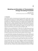
(see Fig. 1).
2. Materials
1. BIACORE biosensor (Biacore, Uppsala, Sweden) equipped with an elution
device, i.e., BIACORE
®
2000 and BIACORE
®
3000. Older models may be
upgraded for this purpose.
2. Phage-Ab library constructed in an appropriate phagemid vector, which encodes
the C-terminal domain of the bacteriophage, gene III protein (gIIIp) (6).
3. Ag of interest, purifi ed. For SAP experiments, fusion proteins consisting of
the N1 and N2 domain of gIIIp fused to the Ag of interest should be prepared
according to Nilsson et al. (7) and Krebber et al. (8).
4. Relevant Escherichia coli strain of male origin (e.g., Top10F′). This strain is
used as indicator bacteria and to harbor and propagate phagemids and phage.
246 Malmborg, Nilsson, and Ohlin
Fig. 1. Summary of procedures followed in Biacore-based and SAP-based proce-
dures to enrich for clones displaying diverse affi nity and reaction rate characteristics.
Numbers in parenthesis refer to steps in Subheading 3.
5. For Biacore, conventional helper phage (e.g., VCSM13). A gIII-deleted helper
phage, e.g., R408d3 (5), is required for SAP.
6. Liquid media (e.g., 2TY), antibiotics, and agar plates for selection, according to
the requirements of the specifi c phage Ab library expression system.
3. Methods
3.1. Selections Using the BIACORE Biosensor
1. Amplify the phage library using helper phage, VCSM13, according to standard
protocols and determine the titer (cfu/mL).
2. Immobilize the Ag according to appropriate coupling routines to the sensor chip,
preferably a Pioneer Chip C1 (see Note 3). The amount of Ag immobilized to the
chip should be optimized, according to specifi c requirements (see Note 4).
3. Inject the phage library at 1 µL/min (see Notes 5 and 6) undiluted or diluted in
the running buffer provided by the manufacturer or in any other buffer known
to be compatible with Ab recognition of the Ag of interest. The injected volume
Selection by Antibody Kinetic-Binding Properties 247
will determine the association time, i.e., injection of 10 µL at the specifi ed fl ow
rate will give an association time of 10 min.
4. Collect 10 µL fractions of the eluate at the desired time-points (see Note 7).
The longer the dissociation time, the more likely it is to fi nd an Ab fragment of
slower k
diss
(see Notes 8–10).
5. Infect a freshly grown log-phase culture of E. coli (optical density 600 nm =
0.4–0.6) with dilutions of the eluate by adding 10 µL of each phage dilution to
100 µL bacteria. Incubate at room temperature for 30 min and plate on agar plates
with the appropriate antibiotics for selection. Incubate at 37°C overnight.
6. Screen the individual colonies by monoclonal phage enzyme-linked immuno-
sorbent assay to determine Ag specifi city. Repeat the selection process if necessary.
7. Evaluation of the ranking of k
diss
of positive clones can be performed directly on
the monoclonal phage stocks using Biacore (see Note 11). For determination of
absolute k
diss
and k
ass
and therefore affi nity constants for the selected Abs, it is
advisable to express the Abs as soluble fragments.
3.2. SAP Selections
This protocol is designed to select specifi c phage binders of ranging affi nity
from a library of noninfectious Ab-displaying, phagemid-containing phage
particles, i.e., SAP phage particles.
1. Amplify the phage Ab library using standard protocols using gIII-deleted helper
phage at a multiplicity of infection (MOI) of 10–100. Grow the SAP phage
particles for 6–16 h at 37°C (see Note 12), then precipitate the phage particles
using polyethylene glycol and resuspend the pellet in phosphate-buffered saline.
2. Incubate the phage (normally 10
7
–10
10
phage/selection) with the N1/N2-domain
fused Ag, using a series of increasing Ag concentrations (see Notes 13 and 14)
in a total volume of 100–150 µL of PBS. Depending on the desired affi nity, use
fusion protein concentrations ranging from 10
–6
to 10
–11
M (see Notes 15–17).
Incubate at room temperature for 3 h with moderate shaking (in order to avoid
precipitation of the phage and to increase the mobility of the interacting pairs).
3. Add 100–500 µL freshly grown log-phase E. coli and infect for 30 min at 37°C
(no shaking).
4. Remove the unbound-input phage particles by centrifugation for 10 min at
2000g. It is important to remove unbound-input phage since these phage might
give rise to nonspecifi c interactions, which will compromise the specifi city of
the selection and the amplifi cation.
5. Resuspend the bacterial pellet in 100–500 µL growth medium and plate onto agar
plates supplemented with selective antibiotics and grow overnight at 30°C.
6. Using a small amount of 2TY, scrape the bacterial cells from the plates and
amplify according to standard protocols using gIII-deleted helper phage at a MOI
of 10–100 to generate secondary stocks of SAP phage particles.
7. The selection is repeated until satisfactory results (e.g., as evaluated by standard
immunoassay procedures) are obtained. It is advisable to analyze the material
248 Malmborg, Nilsson, and Ohlin
after each round of selection using standard polymerase chain reaction procedures
with Ab gene-specifi c primers because a large accumulation of clones lacking an
Ab gene insert suggests that the selection process does not operate properly.
4. Notes
1. Avidity effects have been shown to be a particular problem when displaying
single-chain Ab fragments (scFvs) because many of them tend to dimerize under
conditions in which for example, the linker causes hindrance to formation of
the V
H
–V
L
interaction within the same scFv molecule. Similarly, high levels of
display may also, in the absence of dimerization, cause some phage particles
to carry multiple copies of the displayed protein. Unless appropriate selection
conditions are used, avidity effects, rather than reaction rate properties of the
displayed protein, will come to dominate the selection process. However, the use
of monovalent Ag and stringent conditions under which phage carrying specifi c
binders are caught (9) have mostly eliminated the problems associated with
avidity-based, rather than affi nity-based, selection conditions, allowing retrieval
of high-affi nity clones recognizing essentially any ligand.
2. The SAP procedure is performed in solution and is therefore based on affi nity,
rather than avidity, which is often the case in standard selection procedures
involving selection against immobilized Ag. Consequently, despite multivalent
display of the Ab fragment (all gIIIp C-terminal domains display the Ab frag-
ment) on SAP phage particles, high-affi nity binders are preferentially selected.
In addition, it is possible to select lower-affi nity binders and binders displaying
specifi c reaction rate properties under certain circumstances (see Note 16).
3. The properties of the sensor chip used for the analysis can infl uence the size
of the signal. A conventional CM sensor chip consists of a three-dimensional
dextran matrix, which allows the Ag to be immobilized not only on the surface
of the dextran layer, but also within the matrix. However, because of the size of
the phage, only the Ag on the surface of the dextran is accessible to the bulky
phage, thus giving a lower-than-expected signal. For this reason, Biacore has
developed two new types of sensor chips, especially suitable for analysis of
phage-displayed molecules. These are the Sensor Chip C1, with a fl at carboxy-
methylated surface, and the Sensorchip F1, with a short carboxy-methylated
dextran matrix. Both have proven to be more effi cient when working with phage-
displayed molecules, probably as a combination of altered charge and reduction
of steric effects. More effi cient, in this context, means that lower titers of phage
are needed to observe the binding and binding of phages displaying low-affi nity
Abs can be analyzed.
4. Optimization of the density of immobilized Ag is important to obtain true kinetic
properties. An increased Ag density gives rise to an apparent slower dissociation
rate, because a surface with a high surface density of Ag increases the probability
for a dissociated Ab to rebind to the surface before it reaches the bulk buffer fl ow.
Consequently, this applies not only to di/multivalent Abs, but also to monovalent
binders, which may be infl uenced by the Ag density.
Selection by Antibody Kinetic-Binding Properties 249
5. The signals obtained from phage libraries in Biacore are low, considering the
size of the phage itself, which may result from steric hindrance occurring when
the large phage particles are to fi nd their immobilized target antigens. A titer of
~1 × 10
9
cfu/mL is usually necessary for observing any signal. However,
selections may be performed even if no signal is visible.
6. There may be a problem with the rebinding of dissociated phages (as discussed in
Note 4), which reduces the effi ciency by which phage-displaying Ab fragments
of low k
diss
are enriched. One way to overcome this problem is to increase
the fl ow rate. A higher fl ow rate gives rise to a faster dissociation, probably
because of more effi cient removal of dissociated phages. This is probably an
effect of a reduced thickness of the stationary liquid layer above the surface, and
consequently, the residence time of molecules in this layer, i.e. mass transport
limitations are minimized at high fl ow rates. However, bulky molecules such as
phage may be diffusion-limited at high fl ow rates in the small channels of the
IFC. For this reason, the fl ow should be kept as low as possible.
7. Another approach to minimizing the effect of rebinding of dissociating phages
and Ab fragments, resulting in an ineffi cient enrichment of phage displaying slowly
dissociating Ab fragments, would be to add a competing soluble Ag in the fl ow
buffer during the dissociation phase. This would increase the apparent k
diss
.
8. After a long period, the remaining fraction of bound phage may display multiple
copies of the Ab fragment. Collect the eluate before such phages come to
dominate the eluted fraction. A suitable time-point can only be determined by
experience, and it will differ between different experimental systems. Some
guidance might be obtained by assessing the theoretical rate by which binders
displaying different dissociation rates ought to dissociate. The theoretical
dissociation of complexes follows the relationship
m(t) = m(0) × e
(–k
diss
× t)
in which m(0) is the amount of complexes at time-point 0, m(t) is the amount
of complexes at time-point t, t is the time of dissociation (s), and k
diss
is the
dissociation rate constant (s
–1
).
9. In order to retrieve the binders with the highest affi nity, fractions can be collected
during a regeneration step. However, a regeneration step is a general washing
step, and the number of nonbinders and Abs of lower affi nity is often higher
than expected. Furthermore, regeneration is usually performed at either reduced
or elevated pH, meaning that an immediate neutralization step is essential for
the survival of the phage.
10. The BIAcore can be used to evaluate conditions for elutions in conventional
selection systems, e.g., panning or magnetic beads. These so-called BIA-guided
selections were evaluated by Schier and Marks (10), who determined optimal
conditions for elution of a phage-displayed Ab library, to ensure selection based
on increased affi nity, and not on irrelevant parameters, such as decreased toxicity
or increased expression levels. This was evaluated based on the percentage
250 Malmborg, Nilsson, and Ohlin
eluted phage derived from a polyclonal library bound to an Ag immobilized to
the sensor chip surface using different eluants. Furthermore, they determined
the concentration of competing Ag for each round of the panning by testing in
Biacore in a similar manner.
11. Direct determination of the k
ass
from sensorgrams using phage-displayed mole-
cules is not advisable since the signal is limited by mass transport, and thus
determination of the k
diss
may also be diffi cult. However, a relative ranking of
molecules could be obtained by comparing their dissociation curves.
12. When using the SAP selection system to select specifi c phage binders, whether
peptides, Ab fragments (e.g., scFv, Fab), or any other protein, it is of utmost
importance that the phage particles do not display wild-type gIIIp. The SAP
phage particles need to be checked thoroughly for their display content, which
can be performed by an anti-gIIIp Western blot analysis. The presence of wild-
type gIIIp will destroy the selectivity of the selection, thereby making it diffi cult
to select low abundant binders. R408-generated gIII-deleted helper-phage stocks
have proven to be more stable than VCSM13- and M13KO7-derived helper-
phage stocks. The former phage shows considerably lower frequency of reverting
to wild-type genotype than other deleted helper phages (5).
13. To be able to accomplish effi cient and highly specifi c SAP experiments, it is
crucial to determine the exact and preferably functional, active concentration
of the respective parts of the selection, i.e., phage particles and fusion proteins.
This can be achieved through conventional protein concentration assays (such
as bicinchoninic acid protein assay kit) (7). Ab-displaying phage particles to be
used in SAP selections can be stored at 4°C for several weeks if polyethylene-
glycol-precipitated and appropriate protease inhibitors are added, but freshly
produced phage stocks perform better.
14. Even though the generated SAP phage particles are free of wild-type gIIIp, they
can infect bacterial cells by a pilus-independent mechanism. The receptor, if
one exists, for this kind of infection is currently not known. Furthermore, if a
library of Ab fragments is displayed on the surface of the phage, there is a high
probability for antibacterial Abs to be present in the large pool of Abs. Phage
displaying such antibacterial Abs will hamper the specifi city and thereby the
effi ciency of the system. It is therefore necessary to evaluate the phage particles to
be used in selection for nonspecifi c binding to bacterial cells or to irrelevant Ag.
15. Important parameters when selecting for specifi c binders using the SAP proce-
dure, is the time of interaction and the concentration of fusion protein (4).
Through modulation of these two parameters, it is possible to select specifi c
binders with different affi nity properties. Shorter incubation times will favor
the selection of high-affinity binders; longer incubation times, exceeding
3–4 h at room temperature and with moderate shaking will decrease the amount
of specifi c binders because of decreased stability of the fusion protein–phage
complex (4). Furthermore, to select high-affi nity binders, it is advisable to keep
the fusion protein concentration low (the molarity of the fusion protein should
Selection by Antibody Kinetic-Binding Properties 251
be below the desired affi nity constant), since high amounts of fusion protein will
lead to increased levels of nonspecifi c background infections.
16. The k
ass
between the interacting pairs most infl uences the SAP event (4). SAP
experiments with shorter incubation times and low concentration of fusion
protein will favor the selection of binders with fast k
ass
, and particularly those
binders showing a fast k
diss
. To obtain binders with slower k
diss
values, competing
free Ag (i.e., without the N1 and N2 domains) can be added during the selection,
to capture the fast dissociating binders.
17. The SAP procedure favors the selection of high-affi nity binders, and the number
of selected clones of Ab-displaying phage increases with the affi nity of the
interacting Ag–Ab complex (7). To select low-affi nity binders, it is necessary to
increase the concentration of the fusion protein, thereby increasing the number
of nonspecifi c binders. To circumvent this problem, it is possible to perform a
subtractive preselection step, and, in doing so, deleting the high-affi nity binders.
The preselection is achieved in the presence of a low concentration of fusion
protein selecting high-affi nity Abs. The nonbinders remain in the supernatant,
and are used for a second selection experiment with high amounts of fusion
protein, favoring the retrieval of low-affi nity Abs.
References
1. Levitan, B. (1998) Stochastic modeling and optimization of phage display.
J. Mol. Biol. 277, 893–916.
2. Hawkins, R. E., Russell, S. J., and Winter, G. (1992) Selection of phage antibodies
by binding affi nity. Mimicking affi nity maturation. J. Mol. Biol. 226, 889–896.
3. Malmborg, A C., Dueñas, M., Ohlin, M., Söderlind, E., and Borrebaeck, C. A. K.
(1996) Selection of binders from phage displayed antibody libraries using
BIACORE™ biosensor. J. Immunol. Methods 198, 51–57.
4. Duenas, M., Malmborg, A C., Casalvilla, R., Ohlin, M., and Borrebaeck, C. A. K.
(1996) Selection of phage displayed antibodies based on kinetic constants. Mol.
Immunol. 33, 279–285.
5. Rakonjac, J., Jovanovic, G., and Model, P. (1997) Filamentous phage infection-
mediated gene expression: construction and propagation of the gIII deletion
mutant helper phage R408d3. Gene 198, 99–103.
6. Johansen, L. K., Albrechtsen, B., Andersen, H. W., and Engberg, J. (1995) pFab60:
a new, effi cient vector for expression of antibody Fab fragments displayed on
phage. Protein Eng. 8, 1063–1067.
7. Nilsson, N., Karlsson, F., Rakonjac, J., and Borrebaeck, C. A. K. (2000) Dissecting
selective infection of E. coli based on specifi c protein-ligand interactions, in
press.
8. Krebber, C., Spada, S., Desplancq, D., Krebber, A., Ge, L., and Pluckthun, A.
(1997) Selectively-infective phage (SIP): a mechanistic dissection of a novel in
vivo selection for protein-ligand interactions. J. Mol. Biol. 268, 607–618.
252 Malmborg, Nilsson, and Ohlin
9. Schier, R., Bye, J., Apell, G., McCall, A., Adams, G. P., Malmqvist, M., Weiner,
L. M., and Marks, J. D. (1996) Isolation of high-affi nity monomeric human
anti-c-erbB-2 single chain Fv using affi nity-driven selection. J. Mol. Biol. 255,
28–43.
10. Schier, R. and Marks, J. D. (1996) Effi cient in vitro affi nity maturation of phage
antibodies using BIACore guided selections. Hum. Antibodies Hybridomas 7,
97–105.
Selection by Antibody Kinetic-Binding Properties 253
255
From:
Methods in Molecular Biology, vol. 178: Antibody Phage Display: Methods and Protocols
Edited by: P. M. O’Brien and R. Aitken © Humana Press Inc., Totowa, NJ
22
Selection of Functional Antibodies
on the Basis of Valency
Manuela Zaccolo
1. Introduction
Antibodies (Abs) displaying an agonist or antagonist activity are powerful
tools for mimicking or blocking physiological functions in the cell. A number of
applications of Abs in diagnosis and therapy require multivalent reagents, either
because biological activity depends on the polymeric nature of the antigen
(Ag), or because biological activity depends on an effect on the formation of
homodimeric species. Often dimerization is a prerequisite for activation of
a number of surface receptors by their natural ligands and divalent Abs are
typically required for mimicking or blocking the activity of such ligands.
Ab fragments can be generated by using phage-display technology, but these
are normally monomeric fragments (Fvs, scFvs, and Fabs) (1). Strategies for
engineering multivalent fragments have been described (2–4), but they are
laborious and inappropriate for mass screening. The methodology presented
here allows for the selection from phage-display libraries of Ab fragments
capable of modulating cell surface receptor functions when in a divalent format
(5). This approach combines the advantage of easy selection offered by phage
display of monovalent Ab fragments with an approach to isolating Abs whose
function depends on divalency. A two-step selection protocol is used: the fi rst
step consists of the selection of monovalent recombinant Ab fragments from
phage-display libraries using standard protocols. Selection at this stage is based
on the specifi city of binding to the Ag of interest and the only requirement for
the next step is that the recombinant Ab fragment is tagged with an epitope
recognized by a specifi c anti-tag Ab (e.g., a Myc tag). The selected Ab fragment
Selection on the Basis of Valency 255
is then expressed in Escherichia coli and purifi ed before testing its ability to
interfere with a specifi c cellular function.
The second step consists of the identifi cation of those Ab fragments that
show biological activity when in a dimeric format. To this end, the Ab fragments
are dimerized using the anti-tag Ab as a dimerization domain: two identical
Ab fragments bind via their tag to each of the two binding sites of a divalent
(immunoglobulin G) anti-tag Ab, thus generating a divalent binding site
for the Ag of interest. Cells can subsequently be challenged with the anti-
tag–Ab-fragment complexes and inhibition or enhancement of specifi c cellular
functions can be evaluated.
This approach is versatile and allows for conditional selection of monomeric
or dimeric Abs and is readily suited to mass-screening for activity. Abs that
prove to be active as dimers can be further engineered for multivalency (e.g., as
complete immunoglobulin G expressed in mammalian cells).
This chapter contains the detailed protocol for the selection of Ab fragments
(Fab) capable of interfering with the cell-proliferation signal induced by bind-
ing of a growth factor (hepatocyte growth factor/scatter factor [HGF/SF]) to
its transmembrane receptor (Met). In this specifi c case, the selection procedure
relies on a DNA–thymidine incorporation assay to evaluate cell proliferation
as an indication of function. For other applications, the assay of choice for
the isolation of functionally active Ab fragments will necessarily depend on
the specifi c system and on the particular function the Ab is expected to mimic
or inhibit.
2. Materials
This method is based on dimerization using Myc-tagged recombinant Ab
fragments.
1. Recombinant Ag-specifi c Ab (in this case, anti-HGF/SF), expressed as an affi nity-
tagged fusion protein (e.g., Myc) and purifi ed using affi nity chromatography
(see Note 1).
2. Mouse keratinocyte cell line expressing the HGF/SF receptor on cell surface.
3. Serum-free medium (SFM) basal medium (Gibco LRT, 041-17005 M); purifi ed
epidermal growth factor (Gibco LRT cat. no. 13029-012); bovine pituitary extract
(Gibco LRT, cat. no. 13028-014).
4. 96-Well fl at-bottomed tissue culture plates.
5. Purified anti-Myc tag monoclonal Ab (e.g., 9E10, which is commercially
available).
6.
3
H-methylthymidine (Amersham, TRA 120, 1 mCi/mL and 5 Ci/mmol): 25X
stock solution at 10 µCi/mL in SFM.
7. Purifi ed recombinant HGF/SF.
8. 0.2 M NaOH.
256 Zaccolo
9. Phosphate-buffered saline (PBS).
10. Ecolume liquid scintillation solution, 5 mL scintillation vials, and liquid scintil-
lation β analyzer.
3. Methods
1. Plate the mouse keratinocytes in a 96-well plate at 5 × 10
3
cells/well in 200 µL
keratinocyte SFM basal medium supplemented with 5 ng/mL epidermal growth
factor and 50 µg/mL bovine pituitary extract. Incubate at 37°C in a 5% CO
2
humidifed atmosphere until confl uent (approx 2–3 d).
2. Once confl uent, wash the cells once by adding 200 µL warm sterile PBS/well,
then aspirating off.
3. Add 200 µL SFM basal medium (no additives) to each well, and incubate for
20–24 h, to growth-arrest the cells.
4. When the cells are ready for the experiment, preincubate 10
–7
M of Ab fragments
(fi nal concentration) (see Note 2) with 0.5 × 10
–7
M of anti-tag Ab (e.g., 9E10)
in a total volume of 100 µL PBS for 1 h at 37°C (see Note 3). As a control, set
up the same experiment omitting the anti-tag Ab.
5. Aspirate the media from the cells and replace it with 200 µL (total volume) of
SFM basal medium containing the 100 µL preincubated Ab mix and 30 pmol/mL
HGF/SF (see Note 4).
6. Add 20 µL
3
H-methylthymidine in SFM basal medium to give a fi nal concentra-
tion of 2 µCi/well. Incubate the plate for 24 h at 37°C.
7. Wash the cells twice with 200 µL ice-cold PBS. Keep the cells on ice throughout
the washing procedure.
8. Remove the plate from the ice and add 200 µL of 0.2 M NaOH to each well and
incubate for 30 min at 37°C.
9. Transfer the 200 µL medium from each well to a 5 mL scintillation vial. Wash
the wells with an additional 200 µL 0.2 M NaOH and also add to the scintillation
vial.
10. Add 5 mL scintillation fl uid/vial, mix thoroughly, and count on a liquid scintil-
lation analyzer for 1 min.
11. Compare the counts from the wells with dimerized Ab fragments to the counts
from control wells (no anti-tag Ab). Abs with agonist or antagonist activity
will generate an increase or reduction in counts, respectively, compared to the
control wells.
4. Notes
1. If the available Ab clone does not express a tag, this can be easily rectifi ed
by subcloning into an appropriate expression vector. In the specifi c example
described in Subheading 3., the Abs against HGF/SF were fi rst selected as Fabs
displayed on phage, then were subcloned into pUC119His
6
MycXba vector (6),
which includes two different tags: a His
6
tag for purifi cation of Ab fragments
and a Myc tag for dimerization. The Abs were then purifi ed via the His tag using
Selection on the Basis of Valency 257
immobilized metal affi nity chromatography on Ni-agarose resin. However, it
is not necessary to use two different tags for purifi cation and dimerization and
there are other phage display and/or recombinant protein expression plasmids
that would also be appropriate.
2. This corresponds to 5 µg/mL if purifi ed Fabs are used. For different Ab frag-
ments, the amount must be calculated according to the molecular weight of
the fragment.
3. A twofold molar excess of Ab fragment to anti-tag Ab ensures that most binding
sites are in a divalent conformation.
4. A growth-response curve was determined empirically by evaluating the cell
growth rate (as measured by
3
H-thymidine incorporation) using increasing
amounts of HGF/SF. The resulting curve is a sigmoid and 30 pmol/mL is the
amount of HGF/SF that gives half-maximal stimulation of DNA synthesis in
mouse keratinocyte cells. This is the optimal concentration of growth factor
to use, because small changes in ligand concentration result in maximal effect
on growth rate.
References
1. Winter, G., Griffi ths, A. D., Hawkins, R. E., and Hoogenboom, H. R. (1994)
Making antibodies by phage display technology. Ann. Rev. Immunol. 123,
443–455.
2. Pack, P. and Pluckthun A. (1992) Miniantibodies: use of amphipathic helices to
produce functional, fl exibly linked dimeric Fv fragments with high avidity in
Escherichia coli. Biochemistry 31, 1579–1584.
3. Ito, W. and Kurosawa, Y. (1993) Development of an artifi cial antibody system
with multiple valency using an Fv fragment fused to a fragment of Protein A.
J. Biol. Chem. 268, 20668–20675.
4. Kipriyanov, S. M., Little, M., Kropshofer, H., Bretling, F., Gotter, S., and Dubel, S.
(1996) Affi nity enhancement of a recombinant antibody: formation of complexes
with multiple valency by a single-chain Fv fragment-core streptavidin fusion.
Protein Eng. 9, 203–211.
5. Zaccolo, M., Griffi ths, A. D., Prospero, T. D., Winter, G., and Gherardi, E. (1997)
Dimerization of Fab fragments enables ready screening of phage antibodies
that affect hepatocyte growth factor/scatter factor activity on target cells. Eur.
J. Immunol. 27, 618–623.
6. Griffi ths, A., Williams, S. C., Hartley, O., Tomlinson, I. M., Waterhouse, P.,
Crosby, W. L., et al. (1994) Isolation of high affi nity human antibodies directly
from large synthetic repertoires. EMBO J. 13, 3245–3260.
258 Zaccolo
259
From:
Methods in Molecular Biology, vol. 178: Antibody Phage Display: Methods and Protocols
Edited by: P. M. O’Brien and R. Aitken © Humana Press Inc., Totowa, NJ
23
Two-Step Strategy for Alteration of Immunoglobulin
Specifi city by In Vitro Mutagenesis
Yoshitaka Iba, Chie Miyazaki, and Yoshikazu Kurosawa
1. Introduction
A two-step strategy for changing the specifi city of antibodies (Abs) is
presented, which we have used to change the specificity of an Ab from
11-deoxycortisol (11-DOC) to cortisol (CS). Two kinds of in vitro mutagenesis
are utilized in this protocol: fi rst, mutations are introduced at restricted posi-
tions in the complementarity-determining regions (CDRs) by site-directed
mutagenesis; second, mutations are introduced into the entire V-coding regions
by random mutagenesis.
Prior to manipulation, the genes encoding the Fab form of the original Ab
were isolated and inserted into a phage-display expression vector. Based on
computer modeling of the antigen (Ag)–Ab complex, several residues thought
to be directly involved in forming the Ag-binding pocket were selected as
targets for mutation. A library of Abs was constructed in which mutations were
introduced by polymerase chain reaction (PCR) with degenerate oligonucle-
otide primers. Using this procedure, several clones can usually be isolated,
which have gained a new Ag specifi city. In many cases, however, the isolated
Abs retain the ability to bind to the original Ag. Therefore, a second library
was constructed, in which mutations were introduced at random by error-prone
PCR. Clones were then selected for altered Ag specifi city.
These strategies generated mutants with different characteristics. In the case
of site-directed mutagenesis, the constructed library carried a large number
of different sequences, but the mutants appeared to have some limitations,
in terms of fi ne-tuning and/or fi tting to the Ag. On the other hand, random
mutagenesis may generate too many clones to be entirely represented in the
In Vitro Mutagenesis of Ig Specifi city 259
constructed library, and many of the Abs that have acquired random mutations
may be unable to fold properly (1). Nevertheless, this approach generated a
better resource for the isolation of anti-CS Abs when coupled with a competitive
selection strategy. In order to change the specifi city of Abs, we recommend the
two-step strategy described here.
2. Materials
1. A hybridoma line secreting an monoclonal Ab specifi c for a relevant Ag or
recombinant Ab clone. In the example described, the monoclonal Ab, SCET,
which is specifi c for 11-DOC, was used as a starting material (2).
2. A phage-display vector for expression of Fab–cp3 fusions, mutagenesis, and
selection. Our methods describe the use of vector, pAALFab (3), which permits
the simultaneous introduction of highly diverged sequences into six CDRs of
an Ab by PCR with degenerate oligonucleotide primers (3). Helper phage (e.g.,
M13KO7) will be required for the production of phage stocks from Escherichia
coli-carrying phagemids.
3. Ags for screening. In the case described here, cortisol conjugated with ovalbumin
(CS-OVA) was used as an Ag to screen for alteration in Ab specifi city following
mutagenesis. Free 11-DOC, the cognate Ag for Fab, SCET, was used as a
competitor in panning.
4. Tubes, buffers, and immunochemicals for screening. Immunotubes and enzyme-
linked immunosorbant assay (ELISA) plates for Ag immobilization. Phosphate-
buffered saline (PBS), PBS supplemented with 2% skimmed milk (PBSM), 0.1%
Tween-20 (PBST), or both additives (PBSMT). 100 mM triethylamine; 1 M
Tris-HCl, pH 6.8; anti-M13 Ab, Ab–enzyme conjugate, and substrate solution
for phage ELISA.
5. An appropriate E. coli host (e.g., DH12S) and microbial growth media (2TY
liquid and solid media), antibiotics, glucose supplements.
6. Reagents for conventional and error-prone PCR. 10X buffer for conventional
PCR: 100 mM Tris-HCl (pH 8.3), 500 mM KCl, 1 mg/mL gelatine and
25 mM MgCl
2
. 10X buffer for error-prone PCR: 100 mM Tris-HCl (pH 9.0),
500 mM KCl,75 mM MgCl
2
, 5 mM MnCl
2
, 1% Triton X-100. Separate nucleotide
solutions, to enable preparation of deoxyribonucleside triphosphate stocks of
different concentration and composition. Taq DNA polymerase. Oligonucleotide
primers.
3. Methods
3.1. Construction of Plasmid DNA Encoding Fab Form
of Ab Fused with Truncated cp3 (
see
Note 1)
1. Amplify DNA fragments encoding the V
H
DJ
H
and V
L
J
L
genes of the starting
Ab by PCR from either cloned DNA or mRNA using back and forward primers
carrying restriction sites, which change minimally the original amino acid
sequence.
260 Iba, Miyazaki, and Kurosawa
2. Digest the amplifi ed DNA fragments and the chosen phage-display vector with
appropriate restriction enzymes. Several unique restriction sites are used in this
protocol. If these sites also exist in the V-coding regions, they should be eliminated
by site-directed mutagenesis (4). Note that the SCET V
H
template used here
for mutagenesis to specifi city to CS carried an NdeI site in CDR2 (Fig. 1).
3. Clone the PCR products into the phage-display vector. Methods for these steps
can be found in Chapter 2.
3.2. Structural Modeling of Ag-Binding Pocket
The three-dimensional structure formed by the main chains of V domains
can be predicted from their amino acid sequences (5). It will be diffi cult,
however, to predict the three-dimensional (3D) structure of the Ag–Ab complex
without prior knowledge from X-ray crystallographic analysis. Since the 3D
structure of a complex between progesterone and a progesterone-specifi c
monoclonal Ab had been reported, we were able to construct structural
models of the Ag-binding pocket (6,7). From these models, target residues for
mutagenesis were identifi ed (see Note 2).
3.3. Introduction of Mutations at Restricted Positions
by PCR with Degenerate Oligonucleotide Primers
1. In the case described here, it was predicted that the CDRs of the V
L
domain
would form the binding pocket for the A ring of the steroid (6–8). In an attempt
to alter the specifi city of the Ab, mutations were introduced into the V
H
gene
only. In many other cases, it would prove necessary to apply the protocol that
follows to both the V
H
and the V
L
sequences.
2. After identifying the regions to be targeted and the kinds of amino acids to be
introduced, introduce mutations by PCR with degenerate primers as shown in
Fig. 1 (5,6,9,10; see Notes 2 and 3). Primer sequences used to diversify the CDRs
of the SCET Fab are shown in Fig. 1B. Perform the PCR in 100 µL 1X standard
PCR buffer: 1 µM of each of the primers; 10 ng/mL of plasmid DNA, 0.2 mM
each of dATP, dCTP, dGTP, and dTTP; and 2.5 U Taq DNA polymerase.
3. Cycle the mixture 25× through 94°C for 1 min, 55°C for 2 min, and 72°C for
1.5 min.
4. In the example shown in Fig. 1, the SCET template was initially amplifi ed with
HI-B (in addition to diversifying CDR1, this oligonucleotide carries a SnaI site)
and HI-F (encodes a PstI site) and separately with HII-B (diversifi es CDR2,
eliminating the NdeI site present in the template) and HII-F (diversifi es CDR3
and carries a BstPI site).
5. Mix the products of the primary reactions and reamplify with fl anking primers,
to create products diversifi ed in all three CDRs by overlap extension. In the
example, reamplifi cation with HIII-B and F-I retained SnaI and BstPI sites at
the termini of the amplicons.
In Vitro Mutagenesis of Ig Specifi city 261
Fig. 1 (A) Method used for introduction of mutations into three CDRs of the V
H
gene. Plasmid DNA encoding the original monoclonal Ab was used as the template.
Fortuitously, an NdeI site was present in the SCET V
H
sequence (indicated by a circle).
PCR reactions were performed with primers HI-B plus HI-F and HII-B plus HII-F.
Wavy portions of respective primers indicate the presence of degenerate codons (see
1B). A PstI site was introduced on the HI-F primer (indicated by a circle). The products
of the fi rst PCRs were combined and reamplifi ed with primers HIII-B and F-I. The
resulting amplicons were digested with NdeI and PstI to eliminate those fragments in
which diversifi cation of CDR2 had not occurred, but, as a general principle, digestion
with two restriction enzymes is not obligatory. Primers B-I and F-I were used in error-
prone PCR. Ps, PstI; Sn, SnaI; Nd, NdeI; Bs, BstPI. Other unique restriction sites could
be used. (B) Sequences of degenerate primers aligned with the SCET sequence.
262 Iba, Miyazaki, and Kurosawa