Introduction to Medical Immunology - part 7 pps
Bạn đang xem bản rút gọn của tài liệu. Xem và tải ngay bản đầy đủ của tài liệu tại đây (861.49 KB, 71 trang )
Page 451
23.4 Which one of the following pharmacological agents should be immediately administered to a patient with
suspected anaphylaxis?
A. Antihistaminics
B. Cholinergic drugs
C. Epinephrine
D. Methylxanthines
E. Sodium cromoglycate
23.5 Which of the following is coupled to the solid phase in a radioallergosorbent test (RAST) assay?
A. A given allergen
B. Anti-IgE
C. Antibodies to a given allergen
D. IgE
E. The patient's serum
23.6 Which of the following newly synthesized mast-cell mediators is responsible for the attraction of eosinophils to the
peribronchial tissues in the late stages of an asthma attack?
A. Eosinophil chemotactic factor-A
B. Leukotriene B-4 (LTB-4)
C. Major basic protein
D. Platelet aggregation factor
E. Prostaglandin E2
23.7 Of the following reagents, which one will be able to induce the release of histamine from the mast cells of a
ragweed-sensitized individual?
A. A univalent fragment of ragweed
B. F(ab')2 from an anti-IgE antibody
C. Fab from an anti-IgE antibody
D. IgE antiragweed
E. IgG antiragweed
23.8 A major control mechanism of type I hypersensitivity reactions mediated by eosinophils is the release of:
A. Cationic proteins
B. Histaminase
C. Leukotrienes C4, D4, and E4
D. Major basic protein
E. Platelet activating factor
23.9 Which of the following mediators is NOT involved (directly or indirectly) in negative feedback reactions in
immediate hypersensitivity?
A. Eosinophil chemotactic factor-A
B. Histaminase
C. Histamine
D. Phospholipase D
E. Prostaglandin D2
23.10 What is the meaning of the incidental finding of an elevated serum IgE level of 500 IU/mL in a clinically
asymptomatic individual?
A. The subject is atopic
B. HLA-B7 is likely to be represented on the individual's phenotype
C. Hyposensitization is not likely to be effective
Page 452
D. The individual is a high IgE producer
E. The individual is likely to develop allergies
Answers
23.1 (B) The Fc
ε
-RI is the Fc receptor with highest affinity for its ligand (IgE). It is only expressed on basophils and
mast cells, which do not mediate ADCC reactions, and is structurally unrelated to the immunoglobulin superfamily.
23.2 (A) Aerosolized glucocorticoids are preferred to systemic glucocorticoids because they are effective in most cases
with less risk of development of side effects. Antihistaminics and methylxanthines may have anti-inflammatory
properties, but are not sufficiently effective to be useful as primary treatment for chronic asthma.
23.3 (B) Hyposensitization appeared to induce and to increase the activity of antigen-specific suppressor cells that will
lead to a decrease in serum IgE levels.
23.4 (C) Epinephrine is the drug of choice for immediate treatment of anaphylaxis.
23.5 (A) The allergen is coupled to the solid phase; if IgE antibodies are present in the patient's serum, they will become
bound to the antigen in the solid phase, and their presence can be revealed with a radiolabeled anti-IgE antibody.
23.6 (B) The two other chemotactic factors for eosinophils, platelet activating factor (PAF) and eosinophil chemotactic
factor-A (ECF-A), are preformed and released in the early phase.
23.7 (B) The release of histamine requires the cross-linking of membrane IgE which can be induced either by complete
anti-IgE antibodies, bivalent F(ab')
2
fragments of anti-IgE antibodies, or multivalent antigen of the right specificity.
23.8 (B)
23.9 (E) Histamine, by reacting with type III histamine receptors in basophils and mast cells, will inhibit further
histamine release; phospholipase D degrades PAF; histaminase degrades histamine; LTB-4 is chemotactic for
eosinophils, which release phospholipase D, histaminase, and other protective factors.
23.10 (D) Many nonallergic individuals may have IgE values above the upper limit of normalcy. It is not possible to
conclude that an individual with high IgE levels is more likely to become allergic.
Bibliography
Bochner, B.S., Undem, B.J., and Lichtenstein, L.M. Immunological aspects of asthma.
Annu. Rev. Immunol., 12:
295,
1994.
Bracquet, P., Touqui, L., Shen, T.Y., and Vargaftig, B.B. Perspectives in platelet-activating factor research.
Pharmacol.
Rev., 39:
97, 1987.
Burrows, B., and Lebowitz, M.D. The
β
-agonist dilemma.
N. Engl. J. Med., 326:
560, 1992.
Geha, R.S. Regulation of IgE synthesis in humans.
J. Allergy Clin. Immunol., 90:
143, 1992.
Goetzl, E.J., Payan, D.G., and Goldman, D.W. Immunopathogenetic role of leukotrienes in human diseases.
J. Clin.
Immunol., 4:
79, 1984.
International Consensus Report on Diagnosis and Management of Asthma. U.S. Dept. of Health and Human Services
Publication 92–3091, 1992.
Postma D., Bleekeer E.R., Amelung P.J., Holroyd, K.J., Jianfeng, X., Panhuysen, C.I.M., Meyers, D., and Levitt, R.C.
Genetic susceptibility to asthma-bronchial hyperresponsiveness coinherited with a major gene for atopy.
N. Engl. J.
Med., 333:
894, 1995.
Weller P.F. The immunobiology of eosinophils.
N. Engl. J. Med., 324:
1110, 1991.
Page 453
24
Immunohematology
Gabriel Virella and Mary Ann Spivey
I. Introduction: Blood Groups
A. The ABO System.
The first human red-cell antigen system to be characterized was the ABO blood group system.
Specificity is determined by the terminal sugar in an oligosaccharide structure. The terminal sugars of the
oligosaccharides defining groups A and B are immunogenic. In group O the precursor H oligosaccharide is unaltered.
The red cells express either A, B, both A and B, or neither, and antibodies are found in serum to antigens not expressed
by the red cells, as shown in Table 24.1.
1. The ABO group of a given individual is determined by testing both cells and serum. The subject's red cells are
mixed with serum containing known antibody, and the subject's serum is tested against cells possessing known
antigen. For example, the cells of a group A individual are agglutinated by anti-A serum but not by anti-B serum,
and his serum agglutinates type B cells but not type A cells. The typing of cells as group O is done by exclusion (a
cell not reacting with anti-A or anti-B is considered to be of blood group O).
2. The anti-A and anti-B isoagglutinins are synthesized as a consequence of cross-immunization with
enterobacteriaceae that have outer membrane oligosaccharides strikingly similar to those that define the A and B
antigens (see Chap. 13). For example, a newborn with group A blood will not have anti-B in his or her serum,
since there has been no opportunity to undergo cross-immunization. When the intestine is eventually colonized by
the normal microbial flora, the infant will start to develop anti-B, but will not produce anti-A because of tolerance
to his or her own blood group antigens (see Table 24.1).
3. The inheritance of the ABO groups follows simple Mendelian rules; with three common allelic genes: A, B, and
O (A can be subdivided into A
1
and A
2
), of which any individual will carry two, one inherited from the mother, and
one from the father.
B. The Rh System
1.
Historical overview.
In 1939,
Philip Levine
discovered that the sera of most women who gave birth to infants
with hemolytic disease contained an antibody that reacted with the red cells of the infant and with the red cells of
85% of Caucasians. In 1940,
Landsteiner and Wiener
injected blood from the
Page 454
Table 24.1
The ABO System
Red-cell antigen
Serum isoagglutinins
Blood group
A Anti-B
A
B Anti-A
B
A and B None
AB
None Anti-A and -B
O
monkey
Macacus rhesus
into rabbits and guinea pigs and discovered that the resulting antibody agglutinated
Rhesus red cells and appeared to have the same specificity as the neonatal antibody. The donors whose cells were
agglutinated by the antibody to Rhesus red cells were termed Rh positive; those whose cells were not agglutinated
were termed Rh negative. It is now known that the antibody obtained by Landsteiner and Wiener reacts with an
antigen (LW) that is different but closely related to the one that is recognized in human hemolytic disease, but the
Rh nomenclature was retained.
2.
Theories, nomenclatures, and antigens of the Rh system.
The Rh system is now known to have many
antigens in addition to the one originally described, and several nomenclature systems are in use.
a. According to the
Fisher-Race
theory, the Rh gene complex is formed by combinations of three pairs of
allelic genes: Cc, Dd, Ee. The possible combinations are: Dce, DCe, DcE, DCE, dce, dCe, dcE, and dCE. The
three closely linked gene loci are inherited as a gene complex. Thus a DCe/DcE individual can only pass DCe
or DcE to his offspring and no other combination. The original antigen discovered is called
D
and people who
possess it are called
Rh positive.
The antigen
d
has never been discovered, and the symbol “d” is used to
denote the absence of
D.
All individuals lacking the D antigen are termed
Rh negative.
The most frequent
genotype of D-negative individuals is dce/dce. The lack of one
Table 24.2
Comparison of the Fisher-Race and Wiener Notations for the Rh
System
Fisher-Race notation
Wiener notation
Gene complex
Antigens
Genes
Agglutinogens
Factors
Dce
D,c,e
R
0
Rh
o
Rh
o
,hr',hr''
DCe
D,C,e
R
1
Rh
1
Rh
o
,rh',hr''
DcE
D,c,E
R
2
Rh
2
Rho,hr',rh"
DCE
D,C,E,
R
z
Rh
z
Rh
o
,rh',rh"
dce
d,c,e
r
rh
hr',hr"
dCe
d,C,e,
r'
rh'
rh',hr"
dcE
d,c,E
r"
rh"
hr',rh"
dCE
d,C,E
r
y
rh
y
rh',rh"
Page 455
of the postulated alleles seems to imply that the genetic basis of the Fisher-Race theory and nomenclature are
not correct, but the use of this nomenclature has been retained, since it is easier to understand than any other.
b. The second most common nomenclature is that proposed by Wiener, who theorized multiple alleles at a
single complex locus, each locus determining its particular agglutinogen comprising multiple factors that
were designated by bold-face type. The equivalents of the most common Rh factors in the Fisher-Race and
Wiener nomenclature are shown in Table 24.2.
c. Recent studies analyzing DNA from donors of different Rh phenotypes have found two structural genes
within the Rh locus of Rh (D) positive individuals and only one present in Rh-negative persons. Therefore,
one gene appears to encode the D protein and the other governs the presence of C, c, E, and e.
C. Other Blood Groups.
Several other blood group systems with clinical relevance have been characterized. Other than
those caused by clerical error, most transfusion reactions are due to sensitization against alloantigens of the Rh, Kell,
Duffy, and Kidd systems, of which the Kell system is the most polymorphic. In contrast, most cases of autoimmune
hemolytic anemia involve autoantibodies directed to public antigens (antigens common to most, if not all, humans), such
as the I antigen or core Rh antigens.
D. Laboratory Determination of Blood Types
1.
Reagents.
Most reagents consist of monoclonal antibodies, usually of mouse origin, used individually or
blended, and directed against the different blood group antigens that are used for blood group typing. A major
advantage of the use of monoclonals is their specificity, minimizing the possibility of false-positive reactions due
to additional contaminating antibodies found in human serum reagents. An important disadvantage derives from
the fact that monoclonal antibodies react with a single epitope and the blood group antigens have multiple
epitopes. Thus, individuals missing the epitope recognized by the antibody may be typed as negative. This problem
is significantly reduced by using a blend of monoclonal antibodies, each one of them recognizing a different
epitope of a given antigen.
2.
Tests
a.
Direct hemagglutination
is the simplest, preferred test. It is easy to perform with typing reagents
containing IgM antibodies that directly agglutinate cells expressing the corresponding antigen. Reagents
containing IgG antibodies can also be used in a direct hemagglutination test. In one approach, protein is
added in relatively high concentration to the reagent for the purpose of dissipating the repulsive forces that
keep the red cells apart. As a consequence, the red cells can be directly agglutinated by IgG antibodies. A
second approach involves modification of the IgG antibodies by mild reduction of their interchain disulfide
bonds to produce “unfolded” molecules, capable of direct agglutination of red cells.
b.
Indirect antiglobulin test.
In general, reagents containing IgG anti-
Page 456
bodies are used in an indirect antiglobulin test (see below) as a way to induce the agglutination of red cells
coated with the corresponding antibodies.
E. Direct and Indirect Antiglobulin (Coombs) Tests.
In 1945,
Coombs, Mourant,
and
Race
described the use of
antihuman globulin serum to detect red cell-bound nonagglutinating antibodies. There are two basic types of
antiglobulin or Coombs tests.
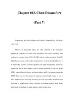
1. The
direct antiglobulin test
is performed to detect in vivo sensitization of red cells or, in other words,
sensitization that has occurred in the patient (Fig. 24.1). The test is performed by adding antihuman IgG (and/or
antihuman complement, to react with complement components bound to the red cells as a consequence of the
antigen-antibody reaction) to the patient's washed red cells. If IgG antibody is bound to the red cells, agglutination
(positive result) is observed after addition of the antiglobulin reagent and centrifugation. The
direct
antiglobulin
test is an aid in diagnosis and investigation of: hemolytic disease of the newborn; autoimmune hemolytic anemia;
drug-induced hemolytic anemia; and hemolytic transfusion reactions.
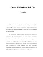
2. The
indirect antiglobulin
test detects in vitro sensitization, which is sensitization that has been allowed to occur
in the test tube under optimal conditions (Fig. 24.2). Therefore, the test is used to investigate the presence of
nonagglutinating red-cell antibodies in a patient's serum. The test is performed in two steps (hence the designation
indirect):
a serum suspected of containing red-cell antibodies is incubated with normal red blood cells; and after
washing unbound antibodies, antihuman IgG (and/or anticomplement) antibodies are added to the red cells as in
the direct test.
The
indirect
antiglobulin test is useful in: detecting and characterizing red-cell antibodies using test cells of known
antigenic composition (antibody screening); crossmatching; and phenotyping blood cells for antigens not demonstrable
by other techniques
Figure 24.1
Diagrammatic representation of a direct Coombs test
using anti-IgG antibodies.
Page 457
Figure 24.2
Diagrammatic representation of an indirect Coombs' test.
II. Blood Transfusion Immunology
A. Blood Testing
1.
Compatibility testing.
Before a blood transfusion, a series of procedures needs to be done to establish the
proper selection of blood for the patient. Basically, those procedures try to establish ABO and Rh compatibility
between donor and recipient and to rule out the existence of antibodies in the recipient's serum which could react
with transfused red cells.
a. To establish the ABO and Rh compatibility between donor and recipient, both the recipient and the blood
to be transfused are
typed.
b. To rule out the existence of antibodies (other than anti-A or anti-B), a general
antibody screening test
is
performed with
group O red cells of known composition,
which are first incubated with the patient's serum
to check for agglutination; if the direct agglutination test is negative, an indirect antiglobulin (Coombs) test is
performed.
2.
The cross-match.
The most direct way to detect antibodies in the recipient's serum that could cause hemolysis
of the transfused red cells is to test the patient's serum with the donor's cells
(major cross-match).
Page 458
a. The complete cross-match also involves the same tests as the antibody screening test described above.
b. An
abbreviated
version of the
cross-match
is often performed in patients with a negative antibody
screening test. This consists of immediately centrifuging a mixture of the patient's serum and donor cells to
detect agglutination; this primarily checks for ABO incompatibility.
3. The
minor cross-match,
which consists of testing a patient's cells with donor serum, is of little significance and
rarely performed, since any donor antibodies would be greatly diluted in the recipient's plasma and rarely cause
clinical problems.
4.
Implications of positive antibody screening for transfusion.
Donor blood found to contain antibodies can be
safely transfused as packed red cells, containing very little plasma. This is a routine blood bank procedure, and no
whole blood units containing clinically significant red-cell antibodies are issued. Such blood is issued as packed
red cells and the plasma is discarded. If a patient has a positive antibody screening test due to a clinically
significant antibody, the antibody is identified using a panel of cells of known antigenic composition and antigen
negative blood is selected for transfusion.
B. Blood Transfusion Reactions.
Transfusion reactions may occur due to a wide variety of causes (Table 24.3). Among
them, the most severe are those associated with hemolysis, which may be life-threatening. A list of the causes of fatal
transfusion reactions reported to the FDA from 1985–1987 is reproduced in Table 24.4.
1. The most frequent cause is an ABO mismatch due to clerical error, resulting in the transfusion of the wrong
blood.
2. Transfusion of blood incompatible for other blood groups to a patient previously sensitized during pregnancy or
as a consequence of earlier transfusions can also cause a hemolytic reaction.
3. Patients with autoimmune hemolytic anemia often have antibodies reacting with “public” antigens expressed by
red cells from virtually all donors as well as their own, and are likely to develop hemolysis whenever a transfusion
is administered to them.
C. Hemolytic Reactions
1.
Pathogenesis.
Hemolytic reactions can be classified as intravascular or extravascular.
a.
Intravascular hemolytic reactions
are triggered by the binding of preformed IgM antibodies to the red
cells.
i. IgM antibodies are very effective in causing the activation of the complement system. Massive
complement activation by red-cell
Table 24.3
Classification of Transfusion Reactions
A. Nonimmune
B. Immune
1. Red-cell incompatibility
2. Incompatibilities associated with platelets and leukocytes
3. Incompatibilities due to antiallotypic antibodies (anti-Gm or Am antibodies)
Page 459
Table 24.4
Summary of Fatal Transfusion Reactions
a
Causes
No.
Hemolytic reactions
ABO incompatible transfusions
29
Collection errors
7
Blood bank clerical errors
8
Blood bank technical errors
1
Nursing unit errors
11
Undetermined
2
Non
-ABO incompatible transfusions
b
6
No detectable antibody
3
Glycerol
1
Nonhemolytic reactions 26
Bacterial contamination
11
c
Acute respiratory distress
9
Anaphylaxis
6
a
Reported to the Food and Drug Administration from
1985 to 1987.
b
Including anti-Jk
b
, -c, Fy
a
, and -K.
c
In nine cases, the source of contamination was a
platelet preparation.
Source: Beig, K., Calhoun, A., and Petz, L.D. ISBT &
AABB Joint Congress, Los Angeles, CA, 1990,
Abstract S282.
antibodies causes intravascular red-cell lysis, with release of hemoglobin into the circulation. Most of
the free hemoglobin forms complexes with haptoglobin.
ii. Due to the massive release of soluble complement fragments (e.g., C3a and C5a) with anaphylotoxic
properties, the patient may suffer generalized vasodilatation, hypotension, and shock.
iii. Because of the interrelationships between the complement and clotting systems, disseminated
intravascular coagulation may occur during a severe transfusion reaction.
iv. As a consequence of the nephrotoxicity of free hemoglobin, the patient may develop acute renal
failure, usually due to acute tubular necrosis. This only happens when the amount of release hemoglobin
exceeds the binding capacity of haptoglobin.
b.
Extravascular hemolytic reactions
are caused by the opsonization of red cells with IgG antibodies.
i. IgG red-cell antibodies can activate complement but do not cause spontaneous red-cell lysis.
ii. Red cells opsonized with IgG (often with associated C3b) are efficiently taken up and destroyed by
phagocytic cells, particularly splenic and hepatic macrophages.
iii. These reactions are usually less severe than intravascular transfusion reactions. In addition,
transfusion reactions may be delayed
Page 460
when an anamnestic response in a patient with undetectable antibody is the precipitating factor.
iv. Typically, a positive direct antiglobulin (Coombs) test will be noted after transfusion in association
with a rapidly diminishing red-cell concentration.
2.
Clinical presentation
a. The most common initial symptom in a hemolytic transfusion reaction is fever, frequently associated with
chills.
b. Dark urine (due to hemosiderinuria or, rarely, to hemoglobinuria) may be the first symptom noticed by the
patient in cases of rapid intravascular hemolysis.
c. During surgery, the only symptom may be bleeding and/or hypotension.
d. With progression of the reaction, the patient may experience chest pains, dyspnea, hypotension, and shock.
e. Renal damage is indicated by back pain, oliguria, and in most severe cases, anuria.
3.
Laboratory investigation
a. Immediately after a hemolytic transfusion reaction is suspected, the following procedures must be done:
i. A clerical check to detect any errors that may have resulted in the administration of a unit of blood to
the wrong patient.
ii. Confirmation of intravascular hemolysis by visual or photometric comparison of pre- and
postreaction plasma specimens for free hemoglobin (the prereaction specimen should be light yellow,
and the postreaction sample should have a pink/red discoloration).
iii. Direct antiglobulin (Coombs) test on pre- and postreaction blood samples.
b. If any of the above procedures gives a positive result supporting a diagnosis of intravascular hemolysis,
additional serological investigations are indicated, including the following:
i. Repeat ABO and Rh typing on patient and donor samples.
ii. Repeat antibody screening and cross-matching.
iii. If an anti-red-cell antibody is detected, determine its specificity using a red-
cell panel in which group
O red cells of varied antigenic composition are incubated with the patient's serum to determine which
RBC antigens are recognized by the patient's antibody.
c. Additionally, one or several of the following confirmatory tests may be performed:
i. Measurement of serum haptoglobin which decreases due to the uptake of hemoglobin-haptoglobin
complexes by the reticulo-endothelial system.
ii. Measurement of unconjugated bilirubin on blood drawn 5 to 7 hours after transfusion (the
concentration should rise as the released hemoglobin is processed).
iii. Determination of free hemoglobin and/or hemosiderin in the urine (neither is normally detected in
the urine).
D. Nonhemolytic Immune Transfusion Reactions
1.
Antileukocyte antibodies
a. When a patient has antibodies directed to leukocyte antigens, a transfu-
Page 461
sion of any blood product containing cells expressing those antigens can elicit a febrile transfusion reaction.
Leukocyte-reduced blood products should be used for transfusions in patients with recurrent febrile reactions.
b. Special problems are presented by patients requiring platelet concentrates that have developed anti-HLA
antibodies or antibodies directed to platelet-specific antigens (HPA antigens). In such cases, it will be necessary to
give HLA- or HPA-matched platelets, since platelets will be rapidly destroyed if given to a sensitized individual
with circulating antibodies to the antigens expressed by the donor's platelets.
c. Transfusion of blood products containing antibodies to leukocyte antigens expressed by the patient receiving the
transfusion can induce intravascular leukocyte aggregation. These aggregates are usually trapped in the pulmonary
microcirculation, causing acute respiratory distress, and, in some cases, noncardiogenic pulmonary edema. A
similar situation may emerge when granulocyte concentrates are given to a patient with antileukocyte antibodies
reactive with the transfused granulocytes.
2.
Anti-IgA antibodies.
The transfusion of any IgA-containing blood product into a patient with high titers of
preformed anti-IgA antibodies can cause an anaphylactic transfusion reaction.
a. Anti-IgA antibodies are mostly detected in immunodeficient individuals, particularly those with IgA
deficiency.
b. It is very important to test for anti-IgA antibodies in any patient with known IgA deficiency who is going
to require a transfusion, even if the patient has never been previously transfused. If an anti-IgA antibody is
detected in a titer judged to represent a risk for the patient, it is important to administer packed red cells with
all traces of plasma removed by extensive washing. If plasma products are needed, they should be obtained
from IgA-deficient donors.
III. Hemolytic Disease of the Newborn (Erythroblastosis Fetalis)
Case 1
A 25-year-old gravida 1, para 0, woman who had not received prenatal care appeared at the
emergency room just prior to delivering a 3.5-kg baby girl. The mother was found to be group O,
Rh negative, and her antibody screen was negative. Twenty hours later, the nurse observed that the
neonate was jaundiced. A hemogram with differential showed WBC of 6200/
µ
L, RBC of 4.1 × 10
6
µ
L, hemoglobin of 15 g/dL. The differential showed 5% reticulocytes.
This case raises several questions:
• What is the most probable cause of the neonatal jaundice, and what treatment, if any, is usually
indicated in such cases?
Page 462
• What laboratory tests should be ordered to investigate the cause of this newborn's jaundice?
• Can this situation be prevented? How?
A. Pathogenesis
1. Immunological destruction of fetal and/or newborn erythrocytes is likely to occur when
IgG antibodies
are
present in the maternal circulation directed against the antigen(s) present on the fetal red blood cells (only IgG
antibodies can cross the placenta and reach the fetal circulation).
2. The two types of incompatibility most usually involved in hemolytic disease of the newborn are anti-D and anti-
A or anti-B antibodies. Anti-A or anti-B antibodies are usually IgM, but, in some circumstances, IgG antibodies
may develop (usually in group O mothers). This can be secondary to immune stimulation (some vaccines contain
blood group substances or cross-reactive polysaccharides), or may occur without apparent cause for unknown
reasons.
3.
Mechanisms of sensitization
a. Although the exchange of red cells between mother and fetus is prevented by the placental barrier during
pregnancy, about two
-thirds of all women, after delivery (or miscarriage), have fetal red cells in their
circulation.
b. If the mother is Rh-negative and the infant is Rh-positive, the mother may produce antibodies to the D
antigen. The immune response is usually initiated at term, when large amounts of fetal red cells reach
maternal circulation. In subsequent pregnancies, even the small number of red cells crossing the placenta
during pregnancy are significant to elicit a strong secondary response, with production of IgG antibodies.
c. As IgG antibodies are produced in larger amounts, they will cross the placenta, bind to the Rh-positive
cells, and cause their destruction in the spleen through Fc-mediated phagocytosis.
d. Usually, the first child is not affected, since the red cells that cross the placenta after the 28th week of
gestation do so in small numbers and may not elicit a primary immune response.
e. IgG anti-
D antibodies do not appear to activate the complement system, perhaps because the distribution of
the D antigen on the red-cell surface is too sparse to allow the formation of IgG doublets with sufficient
density of IgG molecules to induce complement activation. Complement, however, is not required for
phagocytosis, which is mediated by the Fc receptors in monocytes and macrophages.
B. Epidemiology
1. The frequency of clinically evident hemolytic disease of the newborn was estimated to be about 0.5% of total
births, with a mortality rate close to 6% among affected newborns prior to the introduction of immunoprophylaxis.
Recent figures are considerably lower: 0.15 to 0.3% incidence of clinically evident disease, and the perinatal
mortality rate appears to be declining to about 4% of affected newborns.
2. Ninety-five percent of the cases of hemolytic disease of the newborn requiring therapy were due to Rh
incompatibility, involving sensitization against
Page 463
the D antigen. Due to the introduction of immunoprophylaxis, the proportion of cases due to anti-D antibodies
decreased, while the proportion of cases due to other Rh antibodies, and to antibodies to antigens of other systems,
increased.
C. Clinical Presentation.
The usual clinical features of this disease are anemia and jaundice present at birth, or more
frequently, in the first 24 hours of life. In severe cases, the infant may die in utero. Other severely affected children who
survive until the third day develop signs of central nervous system damage, attributed to the high unconjugated bilirubin
concentrations (Kernicterus). The peripheral blood shows reticulocytes and circulating erythroblasts (hence the term
“erythroblastosis fetalis”).
D. Immunological Diagnosis.
A positive direct Coombs (antiglobulin) test with cord RBC is invariably found in cases
of Rh incompatibility, although 40% of the cases with a positive reaction do not require treatment. In ABO
incompatibility, the direct antiglobulin test is usually weakly positive and may be confirmed by eluting antibodies from
the infant's red cells and testing the eluate with A and B cells.
E. Prevention and Treatment
1. Rh hemolytic disease of the newborn is rarely seen when mother and infant are incompatible in both Rh and
ABO systems. In such cases, the ABO isoagglutinins in the maternal circulation appear to eliminate any fetal red
cells before maternal sensitization occurs.
2. The above observation led to a very effective form for prevention of Rh hemolytic disease of the newborn,
achieved by the administration of anti-D IgG antibodies
(Rh immune globulin)
to Rh-negative mothers.
a. The therapeutic anti-D antibody is prepared from the plasma of previously immunized mothers with
persistently high titers, or from male donors immunized against Rh-positive RBC.
b. The schedule of administration involves two separate doses:
i. A
postdelivery dose
is administered in the first 72 hours after delivery of the first baby (before
sensitization has had time to occur). The passively administered anti-D IgG prevents the emergence of
maternal anti-D antibodies, by an unknown mechanism. The rate of success is 98 to 99%.
ii.
Antepartum administration
of a full dose of Rh immune globulin at the 28th week of pregnancy is
also recommended, in addition to the postpartum administration. The rationale for this approach is to
avoid sensitization due to prenatal spontaneous or post-traumatic bleeding. Prenatal anti-D prophylaxis
is also indicated at the time that an Rh-
negative pregnant woman is submitted to amniocentesis and must
be continued at 12-week intervals, until delivery, to maintain sufficient protection.
c. The recommended full dose is 300
µ
g IM which can be increased if there is laboratory evidence of severe
fetomaternal hemorrhage (by tests able to determine the number of fetal red cells in maternal peripheral
blood, from which one can calculate the volume of fetomaternal hemorrhage). Smaller doses (50
µ
g) should
be given after therapeutic or spontaneous abortion in the first trimester.
Page 464
3. To prevent serious hemolytic disease of the newborn in their infants, pregnant Rh-negative women who have a clinically
significant antibody in maternal circulation are carefully monitored. Amniocentesis is usually performed if the antibody has an
antiglobulin titer greater than 16 or if the woman has a history of a previously affected child. The amniotic fluid is examined for
bile pigments at appropriate intervals, and the severity of the disease is assessed according to those levels.
a. If the pregnancy is over 32 weeks, labor may be induced and, if necessary, the baby can be exchange-transfused after
delivery.
b. If the pregnancy is less than 32 weeks, or fetal lung maturity is inadequate (judged by the lecithin/sphyngomyelin ratio
in amniotic fluid), intrauterine transfusion may be performed by transfusing O, Rh-negative red cells to the fetus.
Case 1 Revisited
• Many clinical conditions can cause neonatal jaundice. In a blood group O, Rh-negative mother, hemolytic
disease of the newborn secondary to anti-Rh or anti-
AB antibodies needs to be considered. In a gravida 1, para
0 female, the disease is unlikely to be due to Rh incompatibility and ABO hemolytic disease of the newborn is
usually mild. Treatment is not usually required. If indicated, phototherapy will usually reduce the bilirubin
concentration and exchange transfusion is rarely necessary.
• The following tests were ordered on the newborn:
Blood group and Rh type:
A, Rh positive
Characterization of antibodies:
direct antiglobulin test
Weakly positive
eluted from RBC
Anti-A
Bilirubin, total
7.4 mg/dL
Bilirubin, direct
0.1 mg/dL
The conclusion from the laboratory tests was that the child had jaundice secondary to a mild hemolytic anemia
of immune cause.
• Prevention of hemolytic disease of the newborn is a multistep process. First, this woman should have had a
blood typing and antibody screening test ordered in the first trimester. In Rh-negative women, the antibody
screening test is repeated at 28 weeks. If a woman has a positive antibody screening test, the antibody must be
identified and its clinical significance assessed. This basically means determining whether it is IgG and can
cross the placenta and react with incompatible fetal cells at body temperature. Clinically significant antibodies
must be monitored closely throughout pregnancy so that treatment such as early delivery or intrauterine
transfusions may be given if necessary. In addition, if anti-D antibodies were not detected in this patient, she
should have been given a full dose of Rh immune globulin at 28 weeks and again within 72 hours after
delivery. The risk of sensitization for an Rh-negative woman delivering her first Rh-positive infant is about
8%. The postpartum dose protects at the time of delivery when the largest number of fetal cells enters the
maternal circulation and reduces the risk to about 1%. The antepartum dose prevents a small number of
women who have larger than normal amounts of fetal cells entering their circulation during pregnancy from
becoming sensitized and decreases the risk. ABO hemolytic disease of the newborn cannot be prevented but it
is rarely serious.
Page 465
IV. Immune Hemolytic Anemias
A. Introduction.
The designation of hemolytic anemias includes a heterogeneous group of diseases whose common
denominator is the exaggerated destruction of red cells (hemolysis). In this chapter we will discuss only the hemolytic
anemias in which an abnormal immune response plays the major pathogenic role.
Case 2
A 65-year-old man being treated for essential hypertension with a combination of thiazide and
α
-
methyldopa was seen by his internist. He was complaining of tiredness and shortness of breath.
The following laboratory results were obtained:
Hemoglobin
10 g/dL
Hematocrit
31%
Reticulocytes
8%
Bilirubin, direct
1.5 mg/dL
Bilirubin, total
3.6 mg/dL
Direct antiglobulin test
Positive with anti-IgG
Indirect antiglobulin test
Positive
Panels performed on both the serum and an eluate from the patient's red cells revealed positive
reactions with all cells tested, indicating the presence of an antibody of broad specificity.
This case raises several questions:
• What are the two most probable causes of this patient's anemia, and why is it important to
distinguish between the two?
• What is the pathogenesis of the two types of anemia most likely involved?
• What immediate measure(s) should be instituted?
B. Autoimmune Hemolytic Anemia (Warm Antibody Type).
This is the most common form of autoimmune
hemolytic anemia. It can be idiopathic (often following overt or subclinical viral infection) or secondary, as shown in
Table 24.5.
1.
Pathogenesis.
Warm autoimmune hemolytic anemia is due to the spontaneous emergence of IgG antibodies that
may have a simple Rh specificity such as anti-e, or uncharacterized specificities common to almost all normal red
cells (“public” antigens, thought to be the core of the Rh substance). In many patients, one can find antibodies of
more than one specificity. The end result is that the serum from patients with autoimmune hemolytic anemia of the
warm type is likely to react with most, if not all, the red cells tested. These antibodies usually cause shortening of
red-cell life due to the uptake and destruction by phagocytic cells in the spleen and liver.
2.
Diagnosis.
Diagnosis relies on the demonstration of antibodies coating the red cells or circulating in the serum.
a. RBC-fixed antibodies are detected by the
direct antiglobulin (Coombs) test.
The test can be done using
anti-IgG antiglobulin, anticomplement, or polyspecific antiglobulin serum that has both anti-IgG and
anticomplement. The polyspecific or broad-spectrum antiglobulin sera produce positive results in higher
numbers of patients, as shown in Table 24.6.
Page 466
Table 24.5
Immune Hemolytic Anemias
Autoimmune hemolytic anemias (AIHA)
Warm antibody AIHA
Idiopathic (unassociated with another disease)
Secondary (associated with chronic lymphocytic leukemia, lymphomas, systemic lupus
erythematosus, etc.)
Cold antibody AIHA
Idiopathic cold hemagglutinin disease
Secondary cold hemagglutinin syndrome
Associated with M. pneumoniae infection
Associated with chronic lymphocytic leukemia, lymphomas, etc.
Immune drug-induced hemolytic anemia
Alloantibody-induced immune hemolytic anemia
Hemolytic transfusion reactions
Hemolytic disease of the newborn
Modified from Petz, L.D., and Garraty, G. Laboratory correlations in immune hemolytic anemias. In Laboratory
Diagnosis of Immunologic Disorders, G.N. Vyas, D.P. Stites, and G. Brechter, eds. Grune & Stratton, New York,
1974.
b. The search for antibodies in serum is carried out by the
indirect antiglobulin test.
Circulating antibodies
are only present when the red cells have been maximally coated, and the test is positive in only 40% of the
cases tested with untreated red cells. A higher positivity rate (up to 80%) can be achieved by using red cells
treated with enzymes such as trypsin, papain, ficin, and bromelin in the agglutination assays. The treatment of
red cells with these enzymes increases their agglutinability by either increasing the exposure of antigenic
determinants or by reducing the surface charge of the red cells. In the investigation of warm-type AIHA, all
tests are carried out at 37°C.
C. Cold Agglutinin Disease and Cold Agglutinin Syndromes.
These diseases can also be idiopathic or secondary.
1.
Pathogenesis.
The cold agglutinins are classically IgM (very rarely IgA or IgG), and react with red cells at
temperatures below normal body temperature.
a In chronic, idiopathic, cold agglutinin diseases, 95% or more of the antibodies, which are
IgM
κ
,
react with
the
I antigen.
This is the adult specificity of the I, i system. The fetus expresses the i antigen, common to
primates and other mammalians, which is the precursor of the I specificity. The newborn expresses i
predominantly over I; in the adult, the situation is reversed.
b. In postinfectious cold agglutinin syndrome, the antibodies are also predominantly IgM, but contain both
κ
and
λ
light chains, suggesting their polyclonal origin. The cold agglutinins that appear in patients with
Mycoplasma pneumoniae
infections are usually reactive with the I antigen, while those that appear in
association with
infectious mononucleosis
usually react with the i antigen.
c. The range of thermal reactivity of cold agglutinins may reach up to
Page 467
Table 24.6
Typical Results of Serological Investigations in Patients with Autoimmune Hemolytic Anemia
Cells
Serum
Direct Coombs test
Antibody
to
Positivity
rate
Antibody
isotype
Serological characteristics
Ab specificity
Warm
AIHA
IgG
30%
IgG
Positive indirect Coombs test
(40%)
Rh system antigens
(“public”)
IgG + C'
C'
50%
20%
Agglutination of enzyme-
treated
RBC (80%)
Cold
Agglutinin
Disease
C'
IgM
Monoclonal IgM
κ
agglutinates
RBC to titers >1024 at 4°C
I antigen
Modified from Petz, L.D., and Garraty, G. Laboratory correlations in immune hemolytic anemias. In Laboratory
Diagnosis of Immunologic Disorders, G.N. Vyas, D.P. Stites, and G. Brechter, eds. Grune & Stratton, New York,
1974.
35°C. Such temperatures are not difficult to experience in exposed parts of the body during the winter. Cold-
induced intravascular agglutination, causing ischemia of cold-exposed areas, and hemolysis are the main
pathogenic mechanisms in cold agglutinin disease.
2.
Clinical presentation.
Hemolysis is usually mild, but in some cases may be severe, leading to acute tubular
necrosis. But in most cases the clinical picture is dominated by symptoms of cold sensitivity (Raynaud's
phenomenon, vascular purpura, and tissue necrosis in exposed extremities).
3.
Laboratory diagnosis.
Testing for cold agglutinins is usually done by incubating a series of dilutions of the
patient's serum (obtained by clotting and centrifuging the blood at 37
°C immediately after drawing) with normal
group O RBC at 4°C.
a. Titers up to the hundred thousands can be observed in patients with cold agglutinin disease.
b. Intermediate titers (below 1000) can be found in patients with
Mycoplasma pneumoniae
infections
(postinfectious cold agglutinins).
c. Low titers (less than 64) can be found in normal, asymptomatic individuals.
D. Drug-Induced Hemolytic Anemia.
Three different types of immune mechanisms may play a role in drug-induced
hemolytic anemias, as summarized in Table 24.7. It is important to differentiate between drug-induced hemolytic
anemia and warm autoimmune hemolytic anemia, since cessation of the drug alone will usually halt the drug-induced
hemolytic process.
1.
Formation of soluble immune complexes between the drug and the corresponding antibodies,
which is
followed by nonspecific adsorption to red cells, and complement activation.
a. When IgM antibodies are predominantly involved, intravascular hemo-
Page 468
Table 24.7
Correlation Between Mechanisms of Red-Cell Sensitization and Laboratory Features in Drug-Induced Immunohematological Abnormalities
Serological evaluation
Mechanism
Prototype drugs
Clinical findings
Direct Coombs
In vitro tests and AB identification
Immune complex formation Quinidine
Phenacetin
Intravascular hemolysis; renal failure;
thrombocytopenia
C usually
IgG rarely
Drug + serum + RBC;
Ab is often IgM
Drug adsorption to RBC Penicillins
Extravascular hemolysis associated with
high doses of penicillin i.v.
Strongly positive
with anti-IgG
a
Drug-coated RBC + serum; antibody is
IgG
Membrane modification causing
nonimmunological adsorption of
proteins
Cephalosporins Asymptomatic
Positive with a
variety of antisera
Drug-coated RBC + serum; no specific
antibody involved
Autoimmune
α-methyldopa
Hemolysis in .8% of patients taking this
medication
Strongly positive
with anti-IgG
a
Normal RBC + serum. Autoantibody to
RBC identical to Ab in warm AIHA
a
When hemolytic anemia is present.
Modified from Garraty, G., and Petz, L.D. Am. J. Med., 58:398, 1975.
Page 469
lysis is frequent and the direct Coombs test is usually positive if anticomplement antibodies are used.
b. IgG antibodies can also form immune complexes with different types of antigens and be adsorbed onto red
cells and platelets. In vitro, such adsorption is not followed by hemolysis or by phagocytosis of red cells, but
in vivo it has been reported to be associated with intravascular hemolysis.
c. The absorption of IgG-containing immune complexes to platelets is also the cause of
drug-induced
thrombocytopenia.
Quinine, quinidine, digitoxin, gold, meprobamate, chlorothiazide, rifampin, and the
sulfonamides have been reported to cause this type of drug-induced thrombo-cytopenia.
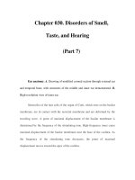
2.
Adsorption of the drug onto the red cells.
Adsorption of drugs by red cells may cause hemolytic anemia by
several different mechanisms:
a. The adsorbed drug functions as hapten and the RBC as carrier, and an immune response against the drug
ensues. The antibodies, usually IgG, are present in high titers, and may activate complement after binding to
the drug adsorbed to the red cells, inducing hemolysis (Fig. 24.3) or phagocytosis. Penicillin (when
administered in high doses by the IV route) and cephalosporins can induce this type of hemolytic anemia.
b. Some cephalosporins (such as cephalothin) have been shown to modify the red-cell membrane which
becomes able to adsorb proteins non-specifically, a fact that can lead to a positive direct Coombs test but not
to hemolytic anemia.
3.
Induction of a truly autoimmune hemolytic anemia.
The anemia induced by
α
-methyldopa
(Aldomet) is the
most frequent type of drug-induced hemolytic anemia. It is particularly interesting from the pathogenic point of
view in that it is indistinguishable from a true warm autoimmune hemolytic anemia.
Figure 24.3
Diagrammatic representation of the pathogenesis of
drug-induced hemolytic anemia as a consequence of
adsorption of a drug to the red-cell membrane.
Page 470
a. Ten to 15% of the patients receiving the drug will have a positive Coombs test, and 0.8% of the patients
develop clinically evident hemolytic anemia.
b.
α
-Methyldopa is unquestionably the trigger for this type of anemia, but the antibodies are of the IgG1
isotype and react with Rh antigens. It is believed that the drug changes the membrane of red-cell precursors,
causing the formation of antibodies reactive with a modified Rh precursor. Once formed, the anti-red-cell
antibodies will react in the absence of the drug, as true autoantibodies.
c. L-dopa, a related drug used for treatment of Parkinson's disease, can also cause autoimmune hemolytic
anemia. Both
α
-methyldopa and L-dopa also stimulate the production of antinuclear antibodies.
Case 2 Revisited
• The two most probable causes of this patient's hemolytic anemia were warm autoimmune
hemolytic anemia and drug-induced hemolytic anemia. It is important to distinguish the two
because the treatment is significantly different. The history of hypertension and treatment with
α
-
methyldopa should alert the physician toward the possibility of a drug-induced hemolytic anemia.
Laboratory tests usually do not differentiate between the two conditions because the reactivity of
the antibodies is virtually identical.
• Warm-type hemolytic anemia is an autoimmune condition, which can present itself as the only
manifestation of autoimmunity or as part of the constellation of a systemic autoimmune disease.
The autoantibodies are of broad specificity, reacting with public erythrocyte antigens expressed by
almost every individual. Drug-induced hemolytic anemia can be caused by antibodies directed to
the drug, due either to previous adsorption of the drug to the red cell or to adsorption of preformed
antigen-antibody complexes to the red cell, or (as it is the case in the hemolytic anemia associated
with
α
-methyldopa) by anti-red-cell antibodies identical to those detected in true autoimmune
hemolytic anemia. How
α
-methyldopa causes the production of these antibodies is the subject of
speculation. It is believed that the drug may alter the conformation of the Rh complex on the red-
cell membrane triggering the synthesis of antibodies that cross-
react with unchanged Rh substances.
Thus, once induced, the autoantibody recognizes the red cell rather than
α
-methyldopa, and
withdrawal of the drug may not result in immediate improvement.
• In all cases of drug-induced hemolytic anemia, it is important to stop the administration of the
drug as soon as the diagnosis is established. In the case of
α
-methyldopa-
induced hemolytic anemia,
the improvement will be gradual, because the autoantibodies react with the red cells, rather than
with the drug, but the antibody titers will decrease with time and, after a point, their concentration
may still be sufficient to cause a positive direct antiglobulin test, but not to cause significant
anemia. In contrast, treatment of true autoimmune hemolytic anemia is rather more complex,
involving administration of steroids, and in cases not responding to steroids, splenectomy and/or
administration of immunosuppressive drugs.
Page 471
SELF-EVALUATION
Questions
Choose the ONE
best
answer.
24.1 A direct Coombs test using antisera to IgG is virtually always positive in:
A. Females with circulating anti-D antibodies
B. Newborns with Rh hemolytic disease
C. Patients with cold hemagglutinin disease
D. Patients with
α
-methyldopa-induced hemolytic anemia
E. Patients with warm-type autoimmune hemolytic anemia
24.2 The pathogenesis of penicillin-induced hemolytic anemia involves:
A. Drug adsorption to red cells and reaction with antipenicillin antibodies
B. Emergence of a neoantigen on the red-cell membrane
C. Formation of soluble IC, adsorption to red-cell membranes, and complement activation or phagocytosis.
D. Nonspecific adsorption and activation of complement components
E. None of the above
24.3 Which of the following drugs induces the production of autoantibodies that react with public red-cell antigens?
A.
α
-Methyldopa
B. Cephalosporin
C. Penicillin
D. Phenacetin
E. Quinidine
24.4 The destruction of Rh-positive erythrocytes after exposure to IgG anti-D antibodies is due to:
A. Complement activation
B. Fc-mediated phagocytosis
C. C3b-mediated phagocytosis
D. C3d-mediated phagocytosis
E. A combination of Fc and C3b-mediated phagocytosis
24.5 In a patient with penicillin-induced hemolytic anemia, you should be concerned with the induction of a similar
situation if prescribing:
A. Aminoglycosides
B. Aspirin
C. Cephalosporins
D. Quinidine
E. Sulfonamides
24.6 An A, Rh-negative female is unlikely to be sensitized by a first Rh-positive baby if:
A. The baby is B, Rh positive
B. The baby is A, Rh positive
C. The baby is O, Rh positive
D. The father is A, Rh positive
E. The father is B, Rh positive
24.7 The major cross-match is used to detect antibodies in:
A. The donor's red cells
B. The donor's serum