Who Needs Emotions The Brain Meets the Robot - Fellous & Arbib Part 8 pot
Bạn đang xem bản rút gọn của tài liệu. Xem và tải ngay bản đầy đủ của tài liệu tại đây (369.11 KB, 20 trang )
124 brains
or a right turn to obtain the goal. It is in this sense that by speci-
fying goals, and not particular actions, the genes are specifying
flexible routes to action. This is in contrast to specifying a reflex
response and to stimulus–response, or habit, learning in which a
particular response to a particular stimulus is learned. It also con-
trasts with the elicitation of species-typical behavioral responses
by sign-releasing stimuli (e.g., pecking at a spot on the beak of
the parent herring gull in order to be fed; Tinbergen, 1951), where
there is inflexibility of the stimulus and the response, which can
be seen as a very limited type of brain solution to the elicitation
of behavior. The emotional route to action is flexible not only
because any action can be performed to obtain the reward or avoid
the punishment but also because the animal can learn in as little
as one trial that a reward or punishment is associated with a par-
ticular stimulus, in what is termed stimulus–reinforcer association
learning. It is because goals are specified by the genes, and not
actions, that evolution has achieved a powerful way for genes to
influence behavior without having to rather inflexibly specify
particular responses. An example of a goal might be a sweet taste
when hunger is present. We know that particular genes specify
the sweet taste receptors (Buck, 2000), and other genes must
specify that the sweet taste is rewarding only when there is a
homeostatic need state for food (Rolls, 1999a). Different goals
or rewards, including social rewards, are specified by different
genes; each type of reward must only dominate the others under
conditions that prove adaptive if it is to succeed in the pheno-
type that carries the genes.
To summarize and formalize, two processes are involved in
the actions being described. The first is stimulus–reinforcer as-
sociation learning, and the second is instrumental learning of an
operant response made to approach and obtain the reward or
to avoid or escape the punisher. Emotion is an integral part of
this, for it is the state elicited in the first stage, by stimuli which
are decoded as rewards or punishers, and this state is motivat-
ing. The motivation is to obtain the reward or avoid the pun-
isher, and animals must be built to obtain certain rewards and
avoid certain punishers. Indeed, primary or unlearned rewards
and punishers are specified by genes which effectively specify
the goals for action. This is the solution which natural selection
has found for how genes can influence behavior to promote their
fitness (as measured by reproductive success) and for how the
brain could interface sensory systems to action systems.
an evolutionary theory of emotion 125
Selecting between available rewards with their associated
costs and avoiding punishers with their associated costs is a pro-
cess which can take place both implicitly (unconsciously) and
explicitly using a language system to enable long-term plans to
be made (Rolls, 1999a). These many different brain systems,
some involving implicit evaluation of rewards and others ex-
plicit, verbal, conscious evaluation of rewards and planned long-
term goals, must all enter into the selection systems for behavior
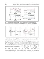
(see Fig. 5.2). These selector systems are poorly understood but
might include a process of competition between all the calls
on output and might involve structures such as the cingulate
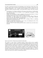
Figure 5.2. Dual routes to the initiation of action in response to rewarding and
punishing stimuli. The inputs from different sensory systems to brain structures
such as the orbitofrontal cortex and amygdala allow these brain structures to
evaluate the reward- or punishment-related value of incoming stimuli or of
remembered stimuli. The different sensory inputs enable evaluations within
the orbitofrontal cortex and amygdala based mainly on the primary (un-
learned) reinforcement value for taste, touch, and olfactory stimuli and on the
secondary (learned) reinforcement value for visual and auditory stimuli. In the
case of vision, the “association cortex,” which outputs representations of
objects to the amygdala and orbitofrontal cortex, is the inferior temporal visual
cortex. One route for the outputs from these evaluative brain structures is via
projections directly to structures such as the basal ganglia (including the
striatum and ventral striatum) to enable implicit, direct behavioral responses
based on the reward- or punishment-related evaluation of the stimuli to be
made. The second route is via the language systems of the brain, which allow
explicit (verbalizable) decisions involving multistep syntactic planning to be
implemented. (From Rolls, 1999a, Fig. 9.4.)
126 brains
cortex and basal ganglia in the brain, which receive input from
structures such as the orbitofrontal cortex and amygdala that
compute the rewards (see Fig. 5.2; Rolls, 1999a).
3. Motivation. Emotion is motivating, as just described. For exam-
ple, fear learned by stimulus–reinforcement association provides
the motivation for actions performed to avoid noxious stimuli.
Genes that specify goals for action, such as rewards, must as an
intrinsic property make the animal motivated to obtain the re-
ward; otherwise, it would not be a reward. Thus, no separate
explanation of motivation is required.
4. Communication. Monkeys, for example, may communicate their
emotional state to others by making an open-mouth threat to
indicate the extent to which they are willing to compete for
resources, and this may influence the behavior of other animals.
This aspect of emotion was emphasized by Darwin (1872/1998)
and has been studied more recently by Ekman (1982, 1993).
Ekman reviews evidence that humans can categorize facial
expressions as happy, sad, fearful, angry, surprised, and disgusted
and that this categorization may operate similarly in different
cultures. He also describes how the facial muscles produce dif-
ferent expressions. Further investigations of the degree of cross-
cultural universality of facial expression, its development in
infancy, and its role in social behavior are described by Izard
(1991) and Fridlund (1994). As shown below, there are neural
systems in the amygdala and overlying temporal cortical visual
areas which are specialized for the face-related aspects of this
processing. Many different types of gene-specified reward have
been suggested (see Table 10.1 in Rolls, 1999a) and include not
only genes for kin altruism but also genes to facilitate social
interactions that may be to the advantage of those competent
to cooperate, as in reciprocal altruism.
5. Social bonding. Examples of this are the emotions associated with
the attachment of parents to their young and the attachment of
young to their parents. The attachment of parents to each other
is also beneficial in species, such as many birds and humans,
where the offspring are more likely to survive if both parents
are involved in the care (see Chapter 8 in Rolls, 1999a).
6. The current mood state can affect the cognitive evaluation of
events or memories (see Oatley & Jenkins, 1996). This may facili-
tate continuity in the interpretation of the reinforcing value of
events in the environment. The hypothesis that backprojections
from parts of the brain involved in emotion, such as the orbito-
an evolutionary theory of emotion 127
frontal cortex and amygdala, to higher perceptual and cognitive
cortical areas is described in The Brain and Emotion, and devel-
oped in a formal model of interacting attractor networks by Rolls
and Stringer (2001). In this model, the weak backprojections
from the “mood” attractor can, because of associative connec-
tions formed when the perceptual and mood states were origi-
nally present, influence the states into which the perceptual
attractor falls.
7. Emotion may facilitate the storage of memories. One way this
occurs is that episodic memory (i.e., one’s memory of particular
episodes) is facilitated by emotional states. This may be advan-
tageous in that storing many details of the prevailing situation
when a strong reinforcer is delivered may be useful in generat-
ing appropriate behavior in situations with some similarities in
the future. This function may be implemented by the relatively
nonspecific projecting systems to the cerebral cortex and hip-
pocampus, including the cholinergic pathways in the basal
forebrain and medial septum and the ascending noradrenergic
pathways (see Rolls, 1999a; Rolls & Treves, 1998). A second
way in which emotion may affect the storage of memories is
that the current emotional state may be stored with episodic
memories, providing a mechanism for the current emotional
state to affect which memories are recalled. A third way that
emotion may affect the storage of memories is by guiding the
cerebral cortex in the representations of the world which are
established. For example, in the visual system, it may be useful
for perceptual representations or analyzers to be built which are
different from each other if they are associated with different
reinforcers and for these to be less likely to be built if they have
no association with reinforcement. Ways in which backprojec-
tions from parts of the brain important in emotion (e.g., the
amygdala) to parts of the cerebral cortex could perform this
function are discussed by Rolls and Treves (1998) and Rolls and
Stringer (2001).
8. Another function of emotion is that by enduring for minutes or
longer after a reinforcing stimulus has occurred, it may help to
produce persistent and continuing motivation and direction of
behavior, to help achieve a goal or goals.
9. Emotion may trigger the recall of memories stored in neocortical
representations. Amygdala backprojections to the cortex could
perform this for emotion in a way analogous to that in which
the hippocampus could implement the retrieval in the neocor-
128 brains
tex of recent (episodic) memories (Rolls & Treves, 1998; Rolls
& Stringer, 2001). This is one way in which the recall of memo-
ries can be biased by mood states.
REWARD, PUNISHMENT, AND EMOTION IN BRAIN
DESIGN: AN EVOLUTIONARY APPROACH
The theory of the functions of emotion is further developed in Chapter 10
of The Brain and Emotion (Rolls, 1999a). Some of the points made help to
elaborate greatly on the second function in the list above. In that chapter,
the fundamental question of why we and other animals are built to use re-
wards and punishments to guide or determine our behavior is considered.
Why are we built to have emotions as well as motivational states? Is there
any reasonable alternative around which evolution could have built com-
plex animals? In this section, I outline several types of brain design, with
differing degrees of complexity, and suggest that evolution can operate to
influence action with only some of these types of design.
Taxes
A simple design principle is to incorporate mechanisms for taxes into the
design of organisms. Taxes consist at their simplest of orientation toward
stimuli in the environment, for example, phototaxis can take the form of the
bending of a plant toward light, which results in maximum light collection
by its photosynthetic surfaces. (When just turning rather than locomotion
is possible, such responses are called tropisms.) With locomotion possible,
as in animals, taxes include movements toward sources of nutrient and away
from hazards, such as very high temperatures. The design principle here is
that animals have, through natural selection, built receptors for certain
dimensions of the wide range of stimuli in the environment and have linked
these receptors to mechanisms for particular responses in such a way that
the stimuli are approached or avoided.
Reward and Punishment
As soon as we have “approach toward stimuli” at one end of a dimension
(e.g., a source of nutrient) and “move away from stimuli” at the other end
(in this case, lack of nutrient), we can start to wonder when it is appropriate
to introduce the terms reward and punishers for the different stimuli. By
an evolutionary theory of emotion 129
convention, if the response consists of a fixed reaction to obtain the stimu-
lus (e.g., locomotion up a chemical gradient), we shall call this a “taxis,” not
a “reward.” If an arbitrary operant response can be performed by the animal
in order to approach the stimulus, then we will call this “rewarded behav-
ior” and the stimulus the animal works to obtain is a “reward.” (The operant
response can be thought of as any arbitrary action the animal will perform
to obtain the stimulus.) This criterion, of an arbitrary operant response, is
often tested by bidirectionality. For example, if a rat can be trained to either
raise or lower its tail in order to obtain a piece of food, then we can be sure
that there is no fixed relationship between the stimulus (e.g., the sight of
food) and the response, as there is in a taxis. Similarly, reflexes are arbitrary
operant actions performed to obtain a goal.
The role of natural selection in this process is to guide animals to build
sensory systems that will respond to dimensions of stimuli in the natural
environment along which actions can lead to better ability to pass genes on
to the next generation, that is, to increased fitness. Animals must be built
by such natural selection to make responses that will enable them to obtain
more rewards, that is, to work to obtain stimuli that will increase their fit-
ness. Correspondingly, animals must be built to make responses that will
enable them to escape from, or learn to avoid, stimuli that will reduce their
fitness. There are likely to be many dimensions of environmental stimuli along
which responses can alter fitness. Each of these may be a separate reward–
punishment dimension. An example of one of these dimensions might be
food reward. It increases fitness to be able to sense nutrient need, to have
sensors that respond to the taste of food, and to perform behavioral responses
to obtain such reward stimuli when in that need or motivational state. Simi-
larly, another dimension is water reward, in which the taste of water becomes
rewarding when there is body fluid depletion (see Chapter 7 of Rolls, 1999a).
Another dimension might be quite subtly specified rewards to promote, for
example, kin altruism and reciprocal altruism (e.g., a “cheat” or “defection”
detector).
With many primary (genetically encoded) reward–punishment dimen-
sions for which actions may be performed (see Table 10.1 of Rolls, 1999a,
for a nonexhaustive list!), a selection mechanism for actions performed is
needed. In this sense, rewards and punishers provide a common currency for
inputs to response selection mechanisms. Evolution must set the magnitudes
of the different reward systems so that each will be chosen for action in such
a way as to maximize overall fitness (see the next section). Food reward must
be chosen as the aim for action if a nutrient is depleted, but water reward as
a target for action must be selected if current water depletion poses a greater
threat to fitness than the current food depletion. This indicates that each
genetically specified reward must be carefully calibrated by evolution to have
130 brains
the right value in the common currency for the competitive selection pro-
cess. Other types of behavior, such as sexual behavior, must be selected
sometimes, but probably less frequently, in order to maximize fitness (as
measured by gene transmission to the next generation). Many processes
contribute to increasing the chances that a wide set of different environmental
rewards will be chosen over a period of time, including not only need-
related satiety mechanisms, which decrease the rewards within a dimension,
but also sensory-specific satiety mechanisms, which facilitate switching to
another reward stimulus (sometimes within and sometimes outside the same
main dimension), and attraction to novel stimuli. Finding novel stimuli re-
warding is one way that organisms are encouraged to explore the multidi-
mensional space in which their genes operate.
The above mechanisms can be contrasted with typical engineering design.
In the latter, the engineer defines the requisite function and then produces
special-purpose design features that enable the task to be performed. In the
case of the animal, there is a multidimensional space within which many op-
timizations to increase fitness must be performed, but the fitness function is
just how successfully genes survive into the next generation. The solution is
to evolve reward–punishment systems tuned to each dimension in the envi-
ronment which can increase fitness if the animal performs the appropriate
actions. Natural selection guides evolution to find these dimensions. That is,
the design “goal” of evolution is to maximize the survival of a gene into the
next generation, and emotion is a useful adaptive feature of this design. In con-
trast, in the engineering design of a robot arm, the robot does not need to tune
itself to find the goal to be performed. The contrast is between design by evo-
lution which is “blind” to the purpose of the animal and “seeks” to have indi-
vidual genes survive into future generations and design by a designer or engineer
who specifies the job to be performed (cf. Dawkins, 1986; Rolls & Stringer,
2000). A major distinction here is between the system designed by an engi-
neer to perform a particular purpose, for example a robot arm, and animals
designed by evolution where the “goal” of each gene is to replicate copies of
itself into the next generation. Emotion is useful in an animal because it is part
of the mechanism by which some genes seek to promote their own survival,
by specifying goals for actions. This is not usually the design brief for machines
designed by humans. Another contrast is that for the animal the space will be
high-dimensional, so that the most appropriate reward to be sought by cur-
rent behavior (taking into account the costs of obtaining each reward) needs
to be selected and the behavior (the operant response) most appropriate to
obtain that reward must consequently be selected, whereas the movement to
be made by the robot arm is usually specified by the design engineer.
The implication of this comparison is that operation by animals using
reward and punishment systems tuned to dimensions of the environment
an evolutionary theory of emotion 131
that increase fitness provides a mode of operation that can work in organ-
isms that evolve by natural selection. It is clearly a natural outcome of Dar-
winian evolution to operate using reward and punishment systems tuned to
fitness-related dimensions of the environment if arbitrary responses are to
be made by animals, rather than just preprogrammed movements, such as
taxes and reflexes. Is there any alternative to such a reward–punishment-
based system in this evolution by natural selection situation? It is not clear
that there is, if the genes are efficiently to control behavior by specifying
the goals for actions. The argument is that genes can specify actions that will
increase their fitness if they specify the goals for action. It would be very
difficult for them in general to specify in advance the particular responses
to be made to each of a myriad different stimuli. This may be why we are
built to work for rewards, to avoid punishers, and to have emotions and needs
(motivational states). This view of brain design in terms of reward and pun-
ishment systems built by genes that gain their adaptive value by being tuned
to a goal for action (Rolls, 1999a) offers, I believe, a deep insight into how
natural selection has shaped many brain systems and is a fascinating outcome
of Darwinian thought.
DUAL ROUTES TO ACTION
It is suggested (Rolls, 1999a) that there are two types of route to action
performed in relation to reward or punishment in humans. Examples of such
actions include emotional and motivational behavior.
The First Route
The first route is via the brain systems that have been present in nonhuman
primates, and, to some extent, in other mammals for millions of years. These
systems include the amygdala and, particularly well developed in primates,
the orbitofrontal cortex. (More will be said about these brain regions in the
following section.) These systems control behavior in relation to previous
associations of stimuli with reinforcement. The computation which controls
the action thus involves assessment of the reinforcement-related value of a
stimulus. This assessment may be based on a number of different factors.
One is the previous reinforcement history, which involves stimulus–
reinforcement association learning using the amygdala and its rapid updat-
ing, especially in primates, using the orbitofrontal cortex. This stimulus–
reinforcement association learning may involve quite specific information
about a stimulus, for example, the energy associated with each type of food
132 brains
by the process of conditioned appetite and satiety (Booth, 1985). A second
is the current motivational state, for example, whether hunger is present,
whether other needs are satisfied, etc. A third factor which affects the com-
puted reward value of the stimulus is whether that reward has been received
recently. If it has been received recently but in small quantity, this may in-
crease the reward value of the stimulus. This is known as incentive motiva-
tion or the salted peanut phenomenon. The adaptive value of such a process
is that this positive feedback of reward value in the early stages of working
for a particular reward tends to lock the organism onto behavior being per-
formed for that reward. This means that animals that are, for example, al-
most equally hungry and thirsty will show hysteresis in their choice of action,
rather than continually switching from eating to drinking and back with each
mouthful of water or food. This introduction of hysteresis into the reward
evaluation system makes action selection a much more efficient process in a
natural environment, for constantly switching between different types of
behavior would be very costly if all the different rewards were not available
in the same place at the same time. (For example, walking half a mile be-
tween a site where water was available and a site where food was available
after every mouthful would be very inefficient.) The amygdala is one struc-
ture that may be involved in this increase in the reward value of stimuli early
in a series of presentations; lesions of the amygdala (in rats) abolish the ex-
pression of this reward incrementing process, which is normally evident in
the increasing rate of working for a food reward early in a meal and impair
the hysteresis normally built into the food–water switching mechanism (Rolls
& Rolls, 1973). A fourth factor is the computed absolute value of the re-
ward or punishment expected or being obtained from a stimulus, for example,
the sweetness of the stimulus (set by evolution so that sweet stimuli will
tend to be rewarding because they are generally associated with energy sources)
or the pleasantness of touch (set by evolution to be pleasant according to the
extent to which it brings animals together, e.g., for sexual reproduction, ma-
ternal behavior, and grooming, and depending on the investment in time that
the partner is willing to put into making the touch pleasurable, a sign which
indicates the commitment and value for the partner of the relationship).
After the reward value of the stimulus has been assessed in these ways,
behavior is initiated based on approach toward or withdrawal from the stimu-
lus. A critical aspect of the behavior produced by this type of system is that
it is aimed directly at obtaining a sensed or expected reward, by virtue of
connections to brain systems such as the basal ganglia which are concerned
with the initiation of actions (see Fig. 5.2). The expectation may, of course,
involve behavior to obtain stimuli associated with reward, which might even
be present in a linked sequence. This expectation is built by stimulus–
reinforcement association learning in the amygdala and orbitofrontal cortex,
an evolutionary theory of emotion 133
reversed by learning in the orbitofrontal cortex, from where signals may be
sent to the dopamine system (Rolls, 1999a).
Part of the way in which the behavior is controlled with this first route
is according to the reward value of the outcome. At the same time, the ani-
mal may work for the reward only if the cost is not too high. Indeed, in the
field of behavioral ecology, animals are often thought of as performing
optimally on some cost–benefit curve (see, e.g., Krebs & Kacelnik, 1991).
This does not at all mean that the animal thinks about the rewards and per-
forms a cost–benefit analysis using thoughts about the costs, other rewards
available and their costs, etc. Instead, it should be taken to mean that in evo-
lution the system has so evolved that the way in which the reward varies
with the different energy densities or amounts of food and the delay before
it is received can be used as part of the input to a mechanism which has also
been built to track the costs of obtaining the food (e.g., energy loss in ob-
taining it, risk of predation, etc.) and to then select, given many such types
of reward and associated costs, the behavior that provides the most “net
reward.” Part of the value of having the computation expressed in this reward-
minus-cost form is that there is then a suitable “currency,” or net reward
value, to enable the animal to select the behavior with currently the most
net reward gain (or minimal aversive outcome).
The Second Route
The second route in humans involves a computation with many “if . . . then”
statements, to implement a plan to obtain a reward. In this case, the reward
may actually be deferred as part of the plan, which might involve working
first to obtain one reward and only then for a second, more highly valued
reward, if this was thought to be overall an optimal strategy in terms of re-
source usage (e.g., time). In this case, syntax is required because the many
symbols (e.g., names of people) that are part of the plan must be correctly
linked or bound. Such linking might be of the following form: “if A does
this, then B is likely to do this, and this will cause C to do this.” This implies
that an output to a language system that at least can implement syntax in
the brain is required for this type of planning (see Fig. 5.2; Rolls, 2004). Thus,
the explicit language system in humans may allow working for deferred re-
wards by enabling use of a one-off, individual plan appropriate for each situ-
ation. Another building block for such planning operations in the brain may
be the type of short-term memory in which the prefrontal cortex is involved.
For example, this short-term memory in nonhuman primates may be of
where in space a response has just been made. Development of this type of
short-term response memory system in humans enables multiple short-term
134 brains
memories to be held in place correctly, preferably with the temporal order
of the different items coded correctly. This may be another building block
for the multiple-step “if . . . then” type of computation in order to form a
multiple-step plan. Such short-term memories are implemented in the (dor-
solateral and inferior convexity) prefrontal cortex of nonhuman primates and
humans (see Goldman-Rakic, 1996; Petrides, 1996; Rolls & Deco, 2002) and
may be part of the reason why prefrontal cortex damage impairs planning
(see Shallice & Burgess, 1996; Rolls & Deco, 2002).
Of these two routes (see Fig. 5.2), it is the second, involving syntax,
which I have suggested above is related to consciousness. The hypothesis is
that consciousness is the state that arises by virtue of having the ability to
think about one’s own thoughts, which has the adaptive value of enabling
one to correct long, multistep syntactic plans. This latter system is thus the
one in which explicit, declarative processing occurs. Processing in this sys-
tem is frequently associated with reason and rationality in that many of the
consequences of possible actions can be taken into account. The actual com-
putation of how rewarding a particular stimulus or situation is or will be
probably still depends on activity in the orbitofrontal cortex and amygdala
as the reward value of stimuli is computed and represented in these regions
and verbalized expressions of the reward (or punishment) value of stimuli
are dampened by damage to these systems. (For example, damage to the
orbitofrontal cortex renders painful input still identifiable as pain but with-
out the strong affective “unpleasant” reaction to it; see Rolls, 1999a.) This
language system that enables long-term planning may be contrasted with the
first system in which behavior is directed at obtaining the stimulus (includ-
ing the remembered stimulus) that is currently the most rewarding, as com-
puted by brain structures that include the orbitofrontal cortex and amygdala.
There are outputs from this system, perhaps those directed at the basal gan-
glia, which do not pass through the language system; behavior produced in
this way is described as “implicit,” and verbal declarations cannot be made
directly about the reasons for the choice made. When verbal declarations
are made about decisions made in this first system, they may be confabula-
tions, reasonable explanations, or fabrications of reasons why the choice was
made. Reasonable explanations would be generated to be consistent with
the sense of continuity and self that is a characteristic of reasoning in the
language system.
The question then arises of how decisions are made in animals such as
humans that have both the implicit, direct, reward-based and the explicit,
rational, planning systems (see Fig. 5.2). One particular situation in which
the first, implicit, system may be especially important is when rapid reac-
tions to stimuli with reward or punishment value must be made, for then
the direct connections from structures such as the orbitofrontal cortex to
an evolutionary theory of emotion 135
the basal ganglia may allow rapid actions. Another is when there may be
too many factors to be taken into account easily by the explicit, rational,
planning system when the implicit system may be used to guide action. In
contrast, when the implicit system continually makes errors, it would be
beneficial for the organism to switch from automatic, direct action based
on obtaining what the orbitofrontal cortex system decodes as being the most
positively reinforcing choice currently available to the explicit, conscious
control system, which can evaluate with its long-term planning algorithms
what action should be performed next. Indeed, it would be adaptive for the
explicit system to regularly assess performance by the more automatic sys-
tem and to switch itself to control behavior quite frequently as otherwise
the adaptive value of having the explicit system would be less than optimal.
Another factor which may influence the balance between control by the
implicit and explicit systems is the presence of pharmacological agents such
as alcohol, which may alter the balance toward control by the implicit sys-
tem, may allow the implicit system to influence more the explanations made
by the explicit system, and may within the explicit system alter the relative
value it places on caution and restraint versus commitment to a risky action
or plan.
There may also be a flow of influence from the explicit, verbal system
to the implicit system such that the explicit system may decide on a plan of
action or strategy and exert an influence that will alter the reinforcement
evaluations made by and the signals produced by the implicit system. An
example of this might be that if a pregnant woman feels that she would like
to escape a cruel mate but is aware that she may not survive in the jungle,
then it would be adaptive if the explicit system could suppress some aspects
of her implicit behavior toward her mate so that she does not give signals
that she is displeased with her situation. (In the literature on self-deception,
it has been suggested that unconscious desires may not be made explicit in
consciousness [or actually repressed] so as not to compromise the explicit
system in what it produces; see Alexander, 1975, 1979; Trivers, 1976, 1985;
and the review by Nesse & Lloyd, 1992). Another example is that the ex-
plicit system might, because of its long-term plans, influence the implicit
system to increase its response to a positive reinforcer. One way in which
the explicit system might influence the implicit system is by setting up the
conditions in which, when a given stimulus (e.g., a person) is present, posi-
tive reinforcers are given to facilitate stimulus–reinforcement association
learning by the implicit system of the person receiving the positive reinforc-
ers. Conversely, the implicit system may influence the explicit system, for
example, by highlighting certain stimuli in the environment that are cur-
rently associated with reward, to guide the attention of the explicit system
to such stimuli.
136 brains
However, it may be expected that there is often a conflict between
these systems in that the first, implicit, system is able to guide behavior
particularly to obtain the greatest immediate reinforcement, whereas the
explicit system can potentially enable immediate rewards to be deferred
and longer-term, multistep plans to be formed. This type of conflict will
occur in animals with a syntactic planning ability (as described above), that
is, in humans and any other animals that have the ability to process a se-
ries of “if . . . then” stages of planning. This is a property of the human
language system, and the extent to which it is a property of nonhuman
primates is not yet fully clear. In any case, such conflict may be an impor-
tant aspect of the operation of at least the human mind because it is so
essential for humans to correctly decide, at every moment, whether to
invest in a relationship or a group that may offer long-term benefits or
whether to directly pursue immediate benefits (Nesse & Lloyd, 1992). As
Nesse and Lloyd (1992) describe, psychoanalysts have come to a some-
what similar position, for they hold that intrapsychic conflicts usually seem
to have two sides, with impulses on one side and inhibitions on the other.
Analysts describe the source of the impulses as the id and the modules that
inhibit the expression of impulses, because of external and internal con-
straints, as the ego and superego, respectively (Leak & Christopher, 1982;
Trivers, 1985; see Nesse & Lloyd, 1992, p. 613). The superego can be
thought of as the conscience, while the ego is the locus of executive func-
tions that balance satisfaction of impulses with anticipated internal and
external costs. A difference of the present position is that it is based on
identification of dual routes to action implemented by different systems
in the brain, each with its own selective advantage.
BRAIN SYSTEMS UNDERLYING EMOTION
Overview
Animals are built with neural systems that enable them to evaluate which
environmental stimuli, whether learned or not, are rewarding and punishing,
that is, will produce emotions and will be worked for or avoided. Sensory
stimuli are normally processed through several stages of cortical processing to
produce a sensory representation of the object before emotional valence is
decoded, and subcortical inputs to, e.g., the amygdala (LeDoux, 2000) will
be of little use when most emotions are to stimuli that require processing to
the object level (Rolls, 1999a). For example, in the taste system, taste is
analyzed in primates to provide a representation of what the taste is in the
primary taste cortex, and this representation is independent of the reward
an evolutionary theory of emotion 137
value of the taste in that it is not affected by hunger. In the secondary taste
cortex, in the orbitofrontal region (see Figs. 5.3 and 5.4), the reward value
of the taste is represented in that neurons respond to the taste only if the
primate is hungry. In another example, in the visual system, representations
of objects which are view-, position- and size-invariant are produced in the
inferior temporal visual cortex after many stages of cortical processing (see
Rolls & Deco, 2002); and these representations are independent of the emo-
tional valence of the object. Then, in structures such as the orbitofrontal
cortex and amygdala, which receive input from the inferior temporal visual
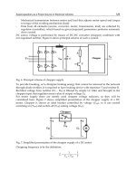
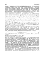
Figure 5.3. Schematic diagram showing some of the gustatory, olfactory,
visual, and somatosensory pathways to the orbitofrontal cortex and amygdala
and some of the outputs of the orbitofrontal cortex and amygdala. The
secondary taste cortex and the secondary olfactory cortex are within the
orbitofrontal cortex. V1, primary visual cortex; V2 and V4, visual cortical
areas; VP1, ventral posterolateral; VPM, ventral posterior medial.
V1 V2 V4
Thalamus
Receptors
solitary tract
VPMpc nucleus
VISION
Taste
TASTE
Bulb
Frontal operculum/Insula
visual cortex
Inferior temporal
(Primary Taste Cortex)
Nucleus of the
Amygdala Striatum
Gate
Lateral
function
by e.g. glucose utilization,
stomach distension or body
weight
Gate
Orbitofrontal
Cortex
Hypothalamus
Hunger neuron controlled
TOUCH
OLFACTION
Thalamus VPL
Olfactory
Primary somatosensory cortex (1.2.3)
Olfactory (Pyriform)
Cortex
Insula
138 brains
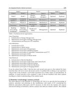
Figure 5.4. Some of the pathways involved in emotion described in the text
are shown on this lateral view of the brain of the macaque monkey. Connec-
tions from the primary taste and olfactory cortices to the orbitofrontal cortex
and amygdala are shown. Connections are also shown in the “ventral visual
system” from V1 to V2, V4, the inferior temporal visual cortex (TEO and
TE), etc., with some connections reaching the amygdala and orbitofrontal
cortex. In addition, connections from somatosensory cortical areas 1, 2, and 3
that reach the orbitofrontal cortex directly and via the insular cortex and that
reach the amygdala via the insular cortex are shown. Abbreviations: as,
arcuate sulcus; cal, calcarine sulcus; cs, central sulcus; lf, lateral (or sylvian)
fissure; lun, lunate sulcus; ps, principal sulcus; io, inferior occipital sulcus; ip,
intraparietal sulcus (which has been opened to reveal some of the areas it
contains); sts, superior temporal sulcus (which has been opened to reveal
some of the areas it contains); AIT, anterior inferior temporal cortex; FST
(fundus superior temporal) visual motion processing area; LIP, lateral
intraparietal area; MST, and MT (also called VS), are visual motion process-
ing areas; PIT, posterior inferior temporal cortex; STP, superior temporal
plane; TA, architectonic area including auditory association cortex; TE,
architectonic area including high-order visual association cortex and some of
its subareas (TEa and Tem); TG, architectonic area in the temporal pole;
V1–V4, visual areas 1–4; VIP, ventral intraparietal area; TEO, architectonic
area including posterior visual association cortex. The numerals refer to
architectonic areas and have the following approximate functional equiva-
lence: 1, 2, 3, somatosensory cortex (posterior to the central sulcus); 4,
motor cortex; 5, superior parietal lobule; 7a, inferior parietal lobule, visual
part; 7b, inferior parietal lobule, somatosensory part; 6, lateral premotor
cortex; 8, frontal eye field; 12, part of orbitofrontal cortex; 46, dorsolateral
prefrontal cortex.
an evolutionary theory of emotion 139
cortex, associations are learned between the objects and the primary rein-
forcers associated with them by the process of stimulus–reinforcement asso-
ciation learning. This is implemented by pattern association neural networks
(Rolls & Deco, 2002). In the orbitofrontal cortex and amygdala, emotional
states are thus represented. Consistent with this, electrical stimulation of
the orbitofrontal cortex and amygdala is rewarding, and damage to these
structures affects emotional behavior by affecting stimulus–reinforcement
association learning. These brain regions influence the selection of behav-
ioral actions through brain systems such as the ventral striatum and other
parts of the basal ganglia (see Fig. 5.2).
The Amygdala
The amygdala receives information about primary reinforcers (e.g., taste and
touch) and about visual and auditory stimuli from higher cortical areas (e.g.,
the inferior temporal cortex) that can be associated by learning with primary
reinforcers (Figs. 5.3 and 5.4). Bilateral removal of the amygdala in mon-
keys produces tameness; a lack of emotional responsiveness; excessive
examination of objects, often with the mouth; and eating of previously re-
jected items, such as meat (the Klüver-Bucy syndrome). In analyses of the
bases of these behavioral changes, it has been observed that there are defi-
cits in learning to associate stimuli with primary reinforcement, including
both punishments and rewards (see Rolls, 2000c). The association learning
deficit is present when the associations must be learned from a previously
neutral stimulus (e.g., the sight of an object) to a primary reinforcing stimu-
lus (e.g., the taste of food). Further evidence linking the amygdala to rein-
forcement mechanisms is that monkeys will work in order to obtain
electrical stimulation of the amygdala, that single neurons in the amygdala
are activated by brain-stimulation reward of a number of different sites,
and that some amygdala neurons respond mainly to rewarding stimuli and
others to punishing stimuli (see Rolls, 1999a, 2000c). The association learning
in the amygdala may be implemented by associatively modifiable synapses
from visual and auditory neurons onto neurons receiving inputs from taste,
olfactory, or somatosensory primary reinforcers (LeDoux, 1996; and Fellous
& LeDoux in this volume). Consistent with this, Davis (2000) found that at
least one type of associative learning in the amygdala can be blocked by local
application to the amygdala of an N-methyl-
D-aspartate receptor blocker,
which blocks long-term potentiation and is a model of the synaptic changes
that underlie learning (see Rolls & Treves, 1998). Consistent with the hy-
pothesis that the learned incentive (conditioned reinforcing) effects of pre-
viously neutral stimuli paired with rewards are mediated by the amygdala
140 brains
acting through the ventral striatum, amphetamine injections into the ven-
tral striatum enhanced the effects of a conditioned reinforcing stimulus only
if the amygdala was intact (see Everitt et al., 2000).
An interesting group of neurons in the amygdala (e.g., in the basal
accessory nucleus) responds primarily to faces. They are probably part of a
system which has evolved for the rapid and reliable identification of indi-
viduals from their faces and of facial expressions because of the importance
of this in primate social behavior. Consistent with this, activation of the
human amygdala can be produced in neuroimaging studies by some facial
expressions, and lesions of the human amygdala may cause difficulty in the
identification of some facial expressions (see Rolls, 1999a, 2000c).
The Orbitofrontal Cortex
The orbitofrontal cortex receives inputs from the inferior temporal visual
cortex, superior temporal auditory cortex, primary taste cortex, primary
olfactory (pyriform) cortex (see Figs. 5.3 and 5.4), amygdala, and midbrain
dopamine neurons. Damage to the caudal orbitofrontal cortex in the monkey
produces emotional changes. These include decreased aggression to humans
and to stimuli such as a snake and a doll and a reduced tendency to reject foods
such as meat. These changes may be related to a failure to react normally to
and learn from nonrewards in a number of different situations. This failure is
evident as a tendency to respond when responses are inappropriate, for ex-
ample, no longer rewarded. For example, monkeys with orbitofrontal dam-
age are impaired on Go/NoGo task performance (in which they should make
a response to one stimulus to obtain a reward and should not make a response
to another stimulus in order to avoid a punishment), in that they Go on the
NoGo trials. They are also impaired in an object reversal task in that they re-
spond to the object which was formerly rewarded with food. They are also
impaired in extinction in that they continue to respond to an object which is
no longer rewarded. Further, the visual discrimination learning deficit shown
by monkeys with orbitofrontal cortex damage may be due to their tendency
not to withhold responses to nonrewarded stimuli (see Rolls, 1999a, 2002).
The primate orbitofrontal cortex contains neurons which respond to the
reward value of taste (a primary reinforcer) in that they respond to the taste
of food only when hunger is present (which is when food is rewarding). It
also contains neurons which learn to respond to visual stimuli associated with
a primary reward, such as taste, and which reverse their responses to another
visual stimulus in one trial when the rewards and punishers available from those
visual stimuli reverse. Further, these visual responses reflect reward in that
feeding the monkey to satiety reduces the responses of these neurons to zero.
an evolutionary theory of emotion 141
Moreover, in part of this orbitofrontal region, some neurons combine taste
and olfactory inputs in that they are bimodal and, in 40% of cases, affected by
olfactory-to-taste association learning and by feeding the monkey to satiety,
which reduces the reward value (see Rolls, 1999a, 2000b, 2002). In addition,
some neurons in the primate orbitofrontal cortex respond to the sight of faces.
These neurons are likely to be involved in learning which emotional responses
are currently appropriate to particular individuals and in making appropriate
emotional responses given the facial expression.
Another class of neurons in the orbitofrontal cortex of the monkey responds
in certain nonreward situations. For example, some neurons responded in
extinction immediately after a lick action was not rewarded when it was made
after a visual stimulus was shown which had previously been associated with
fruit juice reward. Other neurons responded in a reversal task immediately after
the monkey had responded to the previously rewarded visual stimulus but had
obtained punishment rather than reward. Another class of orbitofrontal neu-
ron responded to particular visual stimuli only if they were associated with
reward, and these neurons showed one trial stimulus–reinforcement associa-
tion reversal (Thorpe, Rolls, & Maddison, 1983; Rolls, 1999a, 2000b, 2002).
Another class of neuron conveyed information about whether a reward had
been given, responding, for example, to the taste of sucrose or of saline.
These types of information may be represented in the responses of
orbitofrontal neurons because they are part of a mechanism which evalu-
ates whether a reward is expected and generate a mismatch (evident as a
firing of the nonreward neurons) if reward is not obtained when it is expected
(see Rolls, 1999a, 2000a,b, 2002; Kringelbach & Rolls, 2003). These neu-
ronal responses provide further evidence that the orbitofrontal cortex is in-
volved in emotional responses, particularly when these involve correcting
previously learned reinforcement contingencies, in situations which include
those usually described as involving frustration.
It is of interest and potential clinical importance that a number of the symp-
toms of frontal lobe damage in humans appear to be related to this type of
function, of altering behavior when stimulus–reinforcement associations alter,
as described next. Thus, humans with frontal lobe damage can show impair-
ments in a number of tasks in which an alteration of behavioral strategy is re-
quired in response to a change in environmental reinforcement contingencies
(Rolls, Hornak, Wade, & McGrath, 1994; Damasio, 1994; Rolls, 1999b). Some
of the personality changes that can follow frontal lobe damage may be related
to a similar type of dysfunction. For example, the euphoria, irresponsibility,
lack of affect, and lack of concern for the present or future which can follow
frontal lobe damage may also be related to a dysfunction in altering behavior
appropriately in response to a change in reinforcement contingencies. At one
time, following a report by Moniz (1936), prefrontal lobotomies or leucotomies
142 brains
(cutting white matter) were performed in humans to attempt to alleviate a
variety of problems; and although irrational anxiety or emotional outbursts were
sometimes controlled, intellectual deficits and other side effects were often
apparent (see Valenstein, 1974). Thus, these operations have been essentially
discontinued. To investigate the possible significance of face-related inputs to
orbitofrontal visual neurons described above, the responses to faces that were
made by patients with orbitofrontal damage produced by pathology or trauma
were tested. Impairments in the identification of facial and vocal emotional
expression were demonstrated in a group of patients with ventral frontal lobe
damage who had socially inappropriate behavior (Hornak, Rolls, & Wade, 1996;
Rolls, 1999b; Hornak et al., 2003a,b). The expression identification impair-
ments could occur independently of perceptual impairments in facial recogni-
tion, voice discrimination, or environmental sound recognition. Thus, the
orbitofrontal cortex in humans appears to be important not only in the rapid
relearning of stimulus–reinforcement associations but also in representing some
of the stimuli, such as facial expression, which provide reinforcing informa-
tion. Consistent with this, neuroimaging studies in humans show representa-
tions which reflect the pleasantness of the taste and smell of food and of touch,
as well as quite abstract rewards and punishers such as winning or losing money
(O’Doherty et al., 2001).
The behavioral selection system must deal with many competing re-
wards, goals, and priorities. This selection process must be capable of
responding to many different types of reward decoded in different brain
systems that have evolved at different times, even including the use in
humans of a language system to enable long-term plans to be made to obtain
goals. These many different brain systems, some involving implicit
(unconscious) evaluation of rewards and others explicit, verbal, conscious
evaluation of rewards and planned long-term goals, must all enter into the
selection of behavior. Although poorly understood, emotional feelings are
part of the much larger problem of consciousness and may involve the
capacity to have thoughts about thoughts, that is, higher-order thoughts
(see Rolls, 1999a, 2000a).
CONCLUSION
This approach leads to an appreciation that in order to understand brain
mechanisms of emotion and motivation, it is necessary to understand how
the brain decodes the reinforcement value of primary reinforcers, how it
performs stimulus–reinforcement association learning to evaluate whether
a previously neutral stimulus is associated with reward or punishment and
an evolutionary theory of emotion 143
is therefore a goal for action, and how the representations of these neutral
sensory stimuli are appropriate as input to such stimulus–reinforcement
learning mechanisms. (Some of these issues are considered in The Brain
and Emotion: emotion in Chapter 4, feeding in Chapter 2, drinking in Chap-
ter 7, and sexual behavior in Chapter 8.)
This approach also does not deny that it would be possible to imple-
ment emotions in computers and specifies what may need to be implemented
for both implicit and explicit emotions, that is, emotions with conscious
feelings. It could even be useful to implement some aspects of emotion in
computers as humans may find it more natural to then deal with com-
puters. However, I have summarized a theory of the evolutionary utility of
emotion, which is that emotion arises from the gene-based design of organ-
isms by which individual genes maximize their own survival into the next
generation by specifying the goals for flexible (arbitrary) actions. As such,
emotion arises as part of a blind search by genes to maximize their own sur-
vival, which is the “goal” of evolution. In contrast, the goal of human-
designed computers and robots is not to provide for survival of competing
genes but, instead, to achieve particular design goals specified by the engi-
neer, such as exploring new terrain and sending back pictures to earth, lift-
ing a heavy weight, or translating from one language to another.
Notes
The author has worked on some of the experiments described here with G. C.
Baylis, L. L. Baylis, M. J. Burton, H. C. Critchley, M. E. Hasselmo, J. Hornak, M.
Kringelbach, C. M. Leonard, F. Mora, J. O’Doherty, D. I. Perrett, M. K. Sanghera,
T. R. Scott, S. J. Thorpe, and F. A. W. Wilson; and their collaboration and helpful
discussions with or communications from M. Davies and M. S. Dawkins are sin-
cerely acknowledged. Some of the research described was supported by the Medi-
cal Research Council.
1. Rewards and punishers are generally external, that is, exteroceptive, stimuli,
such as the sight, smell, and taste of food when hungry. Interoceptive stimuli, even
when produced by rewards and punishers after ingesting foods and including diges-
tive processes and the reduction of the drive (hunger) state, are not good reinforc-
ers. Some of the evidence for this is that the taste of food is an excellent reinforcer,
but placing food into the stomach is not. This important distinction is described by
Rolls (1999a).
2. Part of the basis for this is that when memories are recalled, top-down con-
nections into the higher perceptual and cognitive cortical areas lead to reinstate-
ment of activity in those areas (Treves & Rolls, 1994; Rolls & Deco, 2002), which
in turn can produce emotional states via onward connections to the orbitofrontal
cortex and amygdala (Rolls, 1999a).