Báo cáo y học: "Two specific drugs, BMS-345541 and purvalanol A induce apoptosis of HTLV-1 infected cells through inhibition of the NF-kappaB and cell cycle pathways" ppsx
Bạn đang xem bản rút gọn của tài liệu. Xem và tải ngay bản đầy đủ của tài liệu tại đây (728.14 KB, 16 trang )
BioMed Central
Page 1 of 16
(page number not for citation purposes)
AIDS Research and Therapy
Open Access
Research
Two specific drugs, BMS-345541 and purvalanol A induce apoptosis
of HTLV-1 infected cells through inhibition of the NF-kappaB and
cell cycle pathways
Emmanuel Agbottah
1
, Wen-I Yeh
1
, Reem Berro
1
, Zachary Klase
1
,
Caitlin Pedati
1
, Kyelne Kehn-Hall
1
, Weilin Wu
1
and Fatah Kashanchi*
1,2,3
Address:
1
Department of Microbiology and tropical Medicine and Department of Biochemistry and Molecular Biology, The George Washington
University School of Medicine, Washington, District of Columbia 20037, USA,
2
Department of Microbiology, Institute for Proteomics Technology
and Applications, The George Washington University, Washington, District of Columbia 20037, USA and
3
The Institute for Genomic Research,
TIGR, Rockville, Maryland 20850, USA
Email: Emmanuel Agbottah - ; Wen-I Yeh - ; Reem Berro - ;
Zachary Klase - ; Caitlin Pedati - ; Kyelne Kehn-Hall - ;
Weilin Wu - ; Fatah Kashanchi* -
* Corresponding author
Abstract
Human T-cell leukemia virus type-1 (HTLV-1) induces adult T-cell leukemia/lymphoma (ATL/L), a
fatal lymphoproliferative disorder, and HTLV-1-associated myelopathy/tropical spastic paraparesis
(HAM/TSP), a chronic progressive disease of the central nervous system after a long period of
latent infection. Although the mechanism of transformation and leukemogenesis is not fully
elucidated, there is evidence to suggest that the viral oncoprotein Tax plays a crucial role in these
processes through the regulation of several pathways including NF-κB and the cell cycle pathways.
The observation that NF-κB, which is strongly induced by Tax, is indispensable for the maintenance
of the malignant phenotype of HTLV-1 by regulating the expression of various genes involved in
cell cycle regulation and inhibition of apoptosis provides a possible molecular target for these
infected cells. To develop potential new therapeutic strategies for HTLV-1 infected cells, in this
present study, we initially screened a battery of NF-κB and CDK inhibitors (total of 35 compounds)
to examine their effects on the growth and survival of infected T-cell lines. Two drugs namely BMS-
345541 and Purvalanol A exhibited higher levels of growth inhibition and apoptosis in infected cell
as compared to uninfected cells. BMS-345541 inhibited IKKβ kinase activity from HTLV-1 infected
cells with an IC
50
(the 50% of inhibitory concentration) value of 50 nM compared to 500 nM from
control cells as measured by in vitro kinase assays. The effects of Purvalanol A were associated with
suppression of CDK2/cyclin E complex activity as previously shown by us. Combination of both
BMS-345541 and Purvalanol A showed a reduced level of HTLV-1 p19 Gag production in cell
culture. The apparent apoptosis in these infected cells were associated with increased caspase-3
activity and PARP cleavage. The potent and selective apoptotic effects of these drugs suggest that
both BMS-345541 and Purvalanol A, which target both NF-κB and CDK complex and the G1/S
border, might be promising new agents in the treatment of these infected patients.
Published: 10 June 2008
AIDS Research and Therapy 2008, 5:12 doi:10.1186/1742-6405-5-12
Received: 28 November 2007
Accepted: 10 June 2008
This article is available from: />© 2008 Agbottah et al; licensee BioMed Central Ltd.
This is an Open Access article distributed under the terms of the Creative Commons Attribution License ( />),
which permits unrestricted use, distribution, and reproduction in any medium, provided the original work is properly cited.
AIDS Research and Therapy 2008, 5:12 />Page 2 of 16
(page number not for citation purposes)
Background
Human T-cell leukemia virus type 1 (HTLV-1) is associ-
ated with aggressive adult T-cell leukemia (ATL) and
HTLV-1-associated myelopathy/tropical spastic parapare-
sis (HAM/TSP) [1]. ATL arises after a long latent period of
over 50 years and involves with a multi-step mechanism
of tumorigenesis [2]. The transforming ability of HTLV-1
is primarily due to the viral oncoprotein, Tax [3]. Tax not
only transactivates viral genes by binding to CREB but
also activates cellular transcriptional factors including
nuclear factor kappa B (NF-κB), cyclic AMP responsive
element, CREB-binding protein, TATA-binding protein
and TFIIA [4-14]. Acute ATL is an aggressive leukemia with
a median survival of only 6 months and a projected 4-year
survival of about 5% [2].
NF-κB transcription factor plays a crucial roles in tumori-
genesis and tumor development [15,16]. NF-κB transcrip-
tion factor controls the expression of genes involved cell
cycle regulation and apoptosis, such as cyclin E, bcl-2, bcl-
x
L
, c-IAPs, survivin, and XIAP [16-18]. Vertebrate NF-κB
transcription complexes can be any of a variety of homo-
and heterodimers formed by the subunits p105/p50,
p100/p52, c-Rel, p65 (RelA) and RelB [19]. There are mul-
tiple pathways to activate NF-κB. The two most common
pathways are the canonical and the non-canonical path-
ways [20,21]. In the canonical pathway, proceeding the
stimulation of TNF-R, the activated IκB kinase (IKK) com-
plex containing IKKα/IKKβ/NEMO phosphorylates inhib-
itor of NF-κB (IκBα) [22,23]. The phosphorylated IκBα
(Ser32/S36) is then ubiquitinated and degraded, which
allows NF-κB (p50–p65) to enter the nucleus where it reg-
ulates the expression of specific genes [24]. In the non-
canonical pathway, the IKK complex with two IKKα subu-
nits is activated through NIK by other stimuli such as lym-
photoxin β (LTβ) and CD40 ligand, and mediates the
processing of NF-κB complex to p52/RelB [25,26]. This
IKK complex then phosphorylates p100 at C-terminal
domain and promotes the ubiquitination of p100 and the
proteasomal processing of the complex to p52/RelB [27-
29].
A number of reports have elucidated that the HTLV-1-
infected T-cells are associated with constitutively activated
NF-κB and its involvement in tumorigenesis
[25,26,30,31]. Tax is known to activate NF-κB by stimulat-
ing IKK complex in both canonical and non-canonical
pathways by interacting with NEMO [32-35]. Tax is also
reported to directly bind to and activated NF-κB [4]. The
role of various transcription factors in tumorigenesis has
previously been described [36]. NF-κB and AP-1 have
recently been implicated in cell survival and proliferation
pathways. The NF-κB pathway is activated in ATL cells that
do not express Tax, although the mechanism of activation
remains unknown [37]. One of the potential mechanisms
by which these cells could develop resistance to apoptosis
is through the activation of NF-κB [38]. From this point of
view, NF-κB has become an attractive target for therapeu-
tic intervention. Indeed, inhibition of the NF-κB pathway
by Bay 11–7082, an irreversible inhibitor of IκBα phos-
phorylation [25,39], by dehydroximethylepoxy-quin-
omicin, an inhibitor of nuclear translocation of p65, a
component of NF-κB [40-42], arsenic trioxide on NF-κB
[43,44]. and by bortezomib, a proteasome inhibitor [45],
induced apoptosis of HTLV-I-infected T-cells and ATL
cells, suggesting that inhibitors of NF-κB may be effective
targets against ATL cells in vivo.
In addition to the regulation of NF-κB pathway, viral
transactivator Tax provides some initial alternation in cell
cycle progression to the proliferation of viruses. HTLV-1
and/or Tax-expressing cells have altered expression of
some cell cycle-associated genes and accelerate cell cycle
progression in G
1
phase [46-50]. Tax targets cell cycle reg-
ulators such as p53, cyclin dependent kinases (CDKs) 4
and 6, cyclin D2, and CDK inhibitors p21
waf1
and
p16
INK4A
[51-57]. Tax expression also results in transcrip-
tional activation of cyclin E and CDK2 complex [58-61].
In addition, the cyclin E/CDK2 kinase activity is shown to
be increased in HTLV-1 infected cells [62].
Currently there is no accepted curative therapy for ATL or
HAM/TSP and the conditions, at least in the ATL, often
progresses to death with a median survival time of 13
months [63]. The prognosis of this aggressive stage
remains poor, and death is usually due to severe infection
or hypercalcemia, often associated with resistance to
intensive, combined chemotherapy. Therefore, the estab-
lishment of new therapeutic strategies for HTLV-1 infected
cells is deemed critical. Due to the presence of highly acti-
vated NF-κB pathway and tightly controlled cell cycle pro-
gression the infected cells rely on these two mechanisms
for its survival and possibly progeny formation. In an
effort to find novel inhibitors, we initially screened thirty-
five inhibitors targeting these two pathways to examine
their effect on cell growth. Two inhibitors BMS-345541
and Purvalanol A showed the best selectivity in inhibiting
HTLV-1 infected, but not uninfected, cells. Utilizing a
series of biochemical assays, we determined that BMS-
345541 inhibited IKKβ activity in vitro and induced higher
level of apoptosis in infected cells. Finally, the efficacy of
combination of both BMS-345541 and Purvalanol A in
inhibiting HTLV-1 infected cells was tested. Collectively,
understanding the inhibition mechanism, efficiency and
the combined effects of both BMS-345541 and Purvalanol
A will help gain better insights and establish novel new
therapeutic approaches for HTLV-1 infected patients.
AIDS Research and Therapy 2008, 5:12 />Page 3 of 16
(page number not for citation purposes)
Results
Screening of various inhibitors on HTLV-1 infected and
uninfected cells
Despite its tight control in normal T cells, NF-κB is consti-
tutively activated in both HTLV-I-transformed T-cell lines
and freshly isolated ATL cells suggesting that activation of
NF-κB is an important part of the oncogenic mechanism
of HTLV-I. This pathologic action may largely rely on the
viral transforming protein Tax, at least for many of the cell
lines to date that are isolated for in vitro analysis and not
necessarily are ATL samples, which also up-regulates the
expressions and activities of cyclin E/CDK2 which is
important in cell cycle transition from G
1
to S phase.
Most importantly, IKK has been established as a cellular
target of Tax and an essential component in Tax-mediated
NF-κB signaling in both canonical and non-canonical
pathways. Therefore, we reasoned that the specific target-
ing of both the NF-κB signaling and cell cycle regulators
with drugs might provide better insights into how to
inhibit HTLV-1 infected cells. We sought to identify the
targets of a range of NF-κB and CDK inhibitors in HTLV-1
infected and uninfected cells by culturing MT-2, MT-4,
C8166, c10/MJ and uninfected CEM and Jurkat T-cells
(0.5 × 10
6
cells/well) in media with inhibitor concentra-
tions ranging from 0, 0.01, 0.1, 1, and 10 μM. Cells were
treated for 48 hours and the level of growth inhibition was
estimated using trypan blue method. Results from 35
drugs that inhibit various CDKs and IKKs are shown in
Table 1 where a number of drugs inhibited HTLV-1
infected cells much more efficiently than uninfected cells.
Among the top two candidates that inhibited HTLV-1
infected cells were BMS-345541 (4(2'-aminoethyl)amino-
1,8-dimethylimidazo(1,2-a)quinoxaline) and Purvalanol
A. BMS-345541 is a selective inhibitor of IKKβ at IC
50
of
0.3 μM and to a lesser extent an inhibitor of IKKα at IC
50
of 4 μM [64,65]. All drugs were further tested at 10 μM
concentration to effectively compare these different
classes of inhibitors against one another. In Table 1, they
are ranked as high, moderate, and poor inhibitors and the
reported activities of these molecules against variety of
CDKs and IKKs are indicated in the right-hand column.
Collectively, these data indicate that initial cell based sur-
vival screening assays may be an effective tool in isolating
drugs that are more selective against HTLV-1 infected cells
as compared to control uninfected cells.
Effect of BMS-345541 on IKK
β
in infected and uninfected
cells
We next focused our attention on BMS-345541 and asked
whether this drug could inhibit the IKKβ kinase activity
on its substrate IκBα. We immunoprecipitated (IP) IKKβ
from both CEM (uninfected) and C8166 (infected) cells
and used them in an in vitro kinase assays in the presence
or absence of BMS-345541 (1.0 μM). Results are shown in
Figure 1A where C8166 cells had far stronger IKKβ kinase
activity as compared to CEM cells (compare lanes 2 to 4).
Active kinases that were incubated with BMS-345541
showed a reduction of activity from both infected and
uninfected cell extracts. However, the inhibition was
much more dramatic with kinases isolated from HTLV-1
infected cells. We next titrated various levels of BMS-
345541 for both kinases in our in vitro assay. Results are
shown in Panel B where 0.01, 0.1, and 1.0 μM of BMS-
345541 were used for a complete range of titrations. Inter-
estingly, at 0.1 μM there was a significant reduction in the
kinase activity from infected cells (lane 4 compared to
lane 9). A control drug, Purvalanol A, which is a CDK
inhibitor, did not inhibit the IKKβ kinase activity
obtained from infected cells. Collectively, these results
indicate that IKKβ from infected cells is much more sensi-
tive to BMS-345541 as compared to IKKβ from uninfected
cells.
Induction of apoptosis in HTLV-1 infected cells by BMS-
345541
Resistance to cell apoptosis is one of the mechanisms that
is important and is also required for the immortalization
of T cells [63,66]. NF-κB signaling pathway is the survival
pathway activated by HTLV-1 in order to keep the host cell
active [67,68]. BMS-345541 targets IKKβ subunit which is
responsible for activation of the NF-κB pathway [64,65].
To determine whether BMS-345541 can inhibit NF-κB
pathway and induce apoptosis in HTLV-1 infected cells,
we analyzed the level of apoptotic markers such as cas-
pase-3 and PARP in both infected and uninfected cells.
Caspase-3 is a member of cysteine protease and plays a
key role in apoptosis [69]. When apoptosis is activated,
the inactive pro-caspase-3 is processed into active large
(17 kD) and small (12 kD) subunits [70]. PARP,
poly(ADP-ribose) polymerase, is also an apoptosis
marker that is cleaved from precursor form (116 kD) into
active form (85 kD) by active caspase-3 during apoptosis
[71-73]. Results in Figure 2A are Western blots that show
titration of BMS-345541 in two infected and one unin-
fected cells. Samples were treated for 48 hours and extracts
were made for Western blotting. The top panel shows the
caspase Western and a gradual increase of p17 form in
MT-2 cells as well as C8166 cells in concentrations
between 0.5 and 1.0 μM. There was no change in the actin
levels in any of the samples treated. Panel B shows the
results of the Annexin V staining where live cells are repre-
sented at the bottom right corner box in each panel. All
three samples were treated with 0.1 μM of BMS-345541
and stained for the presence of live and apoptotic cells.
Interestingly both MT-2 and C8166 cells showed presence
of few live cells as compared to CEM cells when treated
with BMS-345541. Collectively, these data indicate that
low concentrations of IKKβ inhibitor can apoptosis HTLV-
AIDS Research and Therapy 2008, 5:12 />Page 4 of 16
(page number not for citation purposes)
Table 1: Screening of Various CDK and NFkB/IKK Inhibitors and Related Molecules for HTLV-1 Cell Killing
Selectivity Name MT-2 MT-4 C8166 C10/MJ CEM Jurkat Reported activities of
molecules (IC50 in nM)
Infected Uninfected
High BMS-345541 (10 uM) ++++* +++ +++ + - ** - IKK-1(4), IKK-2(0.3)
Purvalanol A (10 uM) ++++ ++ ++ - - - CDK1(4), CDK2(70),
CDK5(75)
Indirubin-3'-monoxime (10 uM) +++ +++ +++ +++ ++ ++ CDK1(180), CDK2(250),
CDK4(3330), CDK5(100)
Indirubin-3'-monoxime-5'-
Iodo
(10 uM) +++ +++ +++ +++ ++ ++ CDK1(25), CDK5(20)
9-Cyanopaullone (10 uM) ++++ + - - - - CDK1(24), CDK5(44)
Aloisine A (10 uM) +++ + - - - - CDK1(150), CDK2(120),
CDK5(200)
Compound 52 (10 uM) +++ + - - - - CDK1(340)
Flavopiridol (0.1 uM) ++ + - - - - CDK9(50)
Moderate r-Roscovitine (10 uM) ++ - - - - - CDK1(650), CDK2(700),
CDK5(160), CDK7(500)
Bohemine (10 uM) ++ - - - - - CDK1(1000)
s-Roscovitine (10 uM) + - - - - - CDK1(650), CDK2(700),
CDK5(160), CDK7(500)
WHI-P180 (10 uM) + N/A N/A - N/A N/A CDK2(1000)
Kenpaullone (10 uM) + - - - - - CDK1(400), CDK2(680),
CDK5(850)
2,6-Diaminopurine (10 uM) - + - - - -
Flavone (10 uM) + - - - - - CDK1(300), CDK2(100),
CDK4(400), CDK7(300)
Alsterpaullone (10 uM) ++++ ++ +++ + ++ ++ CDK1(35), CDK2(15),
CDK5(40)
CGP 74514A (10 uM) ++++ ++ +++ + ++ ++ CDK1(25)
BAY 11-7085 (10 uM) + ++++ +++ N/A ++++ ++++
BAY 11-7082 (10 uM) + ++++ +++ N/A ++++ ++++ IkBa(10000)
CAPE (10 uM) + +++ - N/A ++++ ++++
Diethylmaleate (10 uM) + +++ - N/A ++++ ++++
Parthenolide (10 uM) + +++ +++ N/A ++++ ++++
Pyrrolidinedithiocarbamic
acid
(10 uM) - +++ +++ N/A ++++ ++++
Wedelolactone (10 uM) + ++ - N/A ++ ++
Poor 6-Benzyloxypurine (10 uM) - - - - - -
5-amino alicylic acid (10 uM) - - - N/A - -
2,6-Dichloropurine (10 uM) - - - - - -
6-Dimethylaminopurine (10 uM) - - - - - - CDC
Indirubin-3'-monoxime-5'-
sulphonic acid
(10 uM) - - - - - - CDK1(5), CDK5(7)
Iso-olomoucine (10 uM) - - - - - - CDK1(>500,000),
CDK4,5(>1,000,000)
N-6-(Δ2-Isopentyl)-adenine (10 uM) - - - - - - CDK1,2,5(>50,000)
Olomoucine (10 uM) - - - - - - CDK1,2(7000), CDK5(3000)
Olomoucine, N9-isopropyl (10 uM) - - - - - - CDK1(2000)
SC-514 (10 uM) - - - - - - IKK-1(>200), IKK-2(11.2)
QNZ (10 uM) - - - N/A ++ +
* positive sign indicates level
of cell inhibition
Inhibition Percentage
** negative sign indicates no
cell inhibition
-1–5%
+25%
++ 50%
+++ 75%
++++ 90%
AIDS Research and Therapy 2008, 5:12 />Page 5 of 16
(page number not for citation purposes)
1 cells much more efficiently as compared to uninfected
cells.
Effect of BMS-345541 on inhibition of I
κ
B and p65
phosphorylation in vivo
We subsequently asked if IκB or p65 levels could be
altered in drug treated infected and uninfected cells. We
therefore Western blotted our drug treated cells with anti-
bodies against IκB, phospho IκB (ser 32), p65, phospho
p65 (ser 536), p50, p52, Tax and actin. Both ser 32 of IκB
and ser 536 of p65 are phosphorylated by IKKβ in vivo.
Results of such an experiment are shown in Figure 3 where
IκB levels essentially stayed the same in all three cell lines
except for a drop in C8166 cells at 5.0 μM. We have previ-
ously observed that cells, irrespective of infection, treated
with BMS-345541 at higher does (i.e., 10.0 μM) are toxic
and show non-specific activation of apoptotic machinery
(data not shown). There was also no change in levels of
p65 although a slight increase in C8166 cells was
observed at higher concentrations. A more interesting set
of results were observed with phosphor-IκB and phos-
phor-p65 blots. MT-2 cells treated with BMS-345541
showed a reduction of both phosphor-IκB and phosphor-
p65 levels at 0.5 μM. Similar results were also seen in
C8166 cells. Very little phosphor-IκB and phosphor-p65
were observed in CEM cells (or other control Jurkat cells,
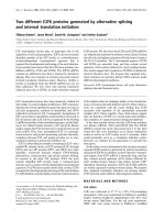
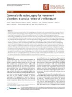
BMS-345541 inhibition of IKKβ in HTLV-1 infected cellFigure 1
BMS-345541 inhibition of IKKβ in HTLV-1 infected cell. A) BMS-345541 reduced IKKβ activity in C8166 cells. Equal
amount (1 mg) of cytoplasmic proteins was immunoprecipitated with anti-IKKβ antibody and mixed with 1 μM BMS-345541.
The IKKβ activities were examined by in vitro kinase assay using GST-IκBα as a substrate. The [γ-
32
P]-labeled IκB-α protein was
visualized by autoradiography. The IKKβ activities were quantitated by ImageQuant software. The bottom panel shows a com-
massie blue staining of GST-IκBα to show equal amount of substrate in each reaction. B) BMS-345541 inhibited IKKβ activity
in C8166 cells in dose-dependent manner; however, Purvalanol A had no effect on IKKβ. Kinase assay were performed as
described above using 0.01, 0.1, and 1 μM of BMS-345541 and 1, 10 μM of Purvalanol A. The stained gel below is a represent-
ative of the kinase reaction.
1 2 3 4 5
Kinase
Stain
GST-IκBα
α-IgG
α-IKKβ
BMS345541
+ - - - -
- + + + +
- - + - +
CEM C81
IKK Activity:
0.7K 5.2K 1.2K 95K 10.5K
A)
1 2 3 4 5 6 7 8 9 10 11 12
Kinase
Stain
GST-IκBα
BMS345541
α-IKKβ
α-IgG
C81 CEM
Purvalanol-A
-
C81
- + + + + - + + + +
+ - - - - + - - - -
+ +
IKK Activity:
2K 95K 70K 14K 7.6K 1.4K 25K 18K 17K 3K 428K 763K
B)
AIDS Research and Therapy 2008, 5:12 />Page 6 of 16
(page number not for citation purposes)
data not shown). P50, p52 levels were unchanged with
various drug concentrations and Tax levels were not
decreased at 0.5 or 1.0 μM concentration of the drug. No
changes were seen in the actin levels in any of the treated
cells. Collectively, these results indicate that inhibition of
IKKβ in HTLV-1 infected cells by BMS-345541 affects
phosphorylation of both IκB and p65 molecules, both of
which may be the hallmarks of NF-κB activation in HTLV-
1 infected cells.
Inhibition of cyclin/CDK complexes by Purvalanol A
We have previously shown that cyclin E/CDK2 kinase
activity is de-regulated in HTLV-1 infected cells and these
cells are especially susceptible to Purvalanol A treatment
[62]. Moreover, Purvalanol A, which is a purine analog
that competes with the ATP binding site in CDKs, has
been shown to inhibit cyclin E/CDK2 and cyclin A/CDK2
kinase activities with an IC
50
of 0.035 and 0.07 μM,
respectively [74-77]. We therefore treated both infected
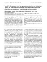
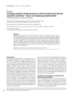
BMS-345541 induction of apoptosis in C8166Figure 2
BMS-345541 induction of apoptosis in C8166. A) BMS-345541 induced caspase-3 and PARP cleavage C8166. MT-2,
C8166, and CEM cells were treated with BMS-345541 at 0.1, 0.5, 1, and 5 μM for 48 hr. Total cell extracts were subjected to
Western blot analysis for caspase-3 and PARP. β-actin Western blot was used as internal control. The results of caspase-3
were quantitated and normalized with β-actin. The ratio of c/un PARP was calculated by dividing cleaved PARP to un-cleaved
PARP (data not shown). B) Detection of apoptosis through annexin V and PI staining. Cells were washed three times in PBS
and re-suspended in binding buffer, stained with annexin V-FITC and PI for 15 minutes at room temperature. Analysis was per-
formed on a BD FacsCalibur flow cytometer.
PARP
MT-2 C81 CEM
caspase-3 (p17)
β-actin
0 0.1 0.5 1 5
BMS-345541 (μM)
0 0.1 0.5 1 5 0 0.1 0.5 1 5
1 2 3 4 5 6 7 8 9 10 11 12 13 14 15
cleaved PARP
A)
No Treatment BMS-345541 (1 μM)
MT-2
C81
CEM
B)
AIDS Research and Therapy 2008, 5:12 />Page 7 of 16
(page number not for citation purposes)
and uninfected cells for 48 hours with Purvalanol A and
Western blotted for caspase-3 and PARP molecules.
Results in Figure 4A show that the caspase-3 p17 molecule
was present in infected cells treated with 0.1 and 0.5 μM
of Purvalanol A (lanes 3 and 7). This was important since
Purvalanol A did not significantly activate caspase-3 in
CEM (lanes 11–15) or Jurkat cells (data not shown).
There were no changes in actin (bottom panel), cyclin E,
or cyclin A expression levels when treated with Purvalanol
A (Panel B). Therefore Purvalanol A treatment induces
caspase-3 activation in HTLV-1 infected, and not in unin-
fected cells. This is consistent with our previous results
where Purvalanol A treatment of infected cells inhibited
cyclin E/CDK2 complex activity in HTLV-1 infected cells,
inhibited transcription of the LTR promoter and pro-
moted apoptosis [62]. Along these lines, we also assayed
for changes in cell cycle progression and apoptosis in
these cells using FACS analysis. Results in Figure 5 show
the titration of Purvalanol A for all three cell types. Inter-
estingly, significant apoptosis appeared in infected cells
treated at 1.0 and 5.0 μM concentrations.
Inhibition of viral replication using both drugs
We next decided to use both drugs in a viral replication
assay in MT-2 cells. MT-2 cells normally produce low lev-
els of infectious HTLV-1 virions that could be detected in
the supernatant using p19 gag ELISA. However, treatment
of these cells with TNF can produce at least 1–2 log more
virus that is shed into the supernatant. We therefore
treated MT-2 cells with TNF for 2 hours and subsequently
treated them with BMS-345541 alone (0.1 μM), Purvala-
nol A alone (0.5 μM), or a combination of both drugs.
Results in Figure 6A show that, as compared to untreated
cells, TNF treatment induced high amounts of p19 gag in
the supernatant (lanes 1 and 2). Both drugs alone reduced
p19 levels to some degree however; the best inhibition
was seen with the combination of both drugs where NF-
κB and CDK pathways were targeted in these cells. Similar
results were also obtained in 293 cells transfected with
ACH full-length infectious clone, where a combination of
both drugs inhibited p19 expression as compared to when
treated with one drug alone (Panel B). Collectively, these
results imply that low concentrations of NF-κB and CDK
inhibitors that normally do not cause cell death in unin-
fected cells are effective inhibitors against HTLV-1 infected
cells.
Discussion
In contrast with the latest progress in the understanding of
HTLV-1 infection, its pathogenesis and its mechanism of
action, more progress in developing therapies for these
infected cells is needed. There has been only very limited
improvement in the prognosis of virally associated dis-
eases (ATL and HAM/TSP) during the past several years.
However few well established pathways including NF-κB
and cell cycle progression have been shown to be tightly
regulated in HTLV-1 and Tax expressing cells and there-
fore providing viable targets for treatment [51,78,79].
Along these lines, we searched various inhibitors targeting
these two pathways using published literature and our
own search using few small libraries of compounds tested
here. We selected inhibitors with low-high IC
50
in various
cell types and identified their cell growth inhibition effi-
ciencies in HTLV-1 infected and uninfected cells. Results
in Table 1 clearly show that there are various compounds
that specifically target HTLV-1 (and Tax) producing cells.
Many of these compounds have known targets and more
importantly are not inhibitors of other viruses including
HIV-1 (more then 78% have different IC50 in HIV-1
infected cells, data not shown). Furthermore, the inhibi-
tors in high selectivity group showed higher inhibition
efficiency in MT-2 cells which normally produces some
level of full length infectious HTLV-1 particles in the
absence of any inducer. Therefore, it is interesting to note
that these inhibitors not only had specificity to inhibit Tax
expressing cells but also showed better growth inhibition
toward infected cells that produce high titer virus.
In high selectivity group, BMS-345541 and Purvalanol A
demonstrated the best selectivity to block growth of all
HTLV-1 infected cells and no blockage to control cells in
these concentrations (Table 1). Indirubin-3'-monoxime
and 5'-Indo-indirubin-3'-monoxime inhibited growth of
infected cells and also inhibited control cells. 9-Cyano-
paullone, Aloisine A, Compound 52, and Flavopiridol
showed less growth inhibition in inhibiting two out of
four infected cell lines. Consequently, we decided to focus
and study the mechanism of BMS-345541 and Purvalanol
A inhibition in HTLV-1 infected cells.
In this study, we showed that BMS-345541 inhibited IKKβ
kinase activity from HTLV-1 infected cell. IKKβ subunits
associating with canonical pathway is responsible for acti-
vating NF-κB by phosphorylating IκBα. Furthermore,
BMS-345541 induced higher level of apoptosis in C8166
and other cells (data not shown). Therefore, we specu-
lated that BMS-345541 suppressed IKKβ and further
blocked NF-κB signaling pathway, the survival pathway,
to induce apoptosis. As illustrated in our model, in the
presence of BMS-345541, the level of unphosphorylated
IκBα is expected to increase and keep NF-κB dimmers in
cytoplasm and block its transcriptional ability (Figure 7).
In addition, IKKβ activity in C8166 was dramatically
down-regulated by BMS-345541 with an IC
50
at 0.05 μM
in a dose-dependent manner, whereas the IC
50
in CEM
cell was at 0.5 μM. The HTLV-1 infected cell was at least 10
times more sensitive to BMS-345541 than control cells.
This critical difference is thought to be the related to the
NF-κB pathway in HTLV-1 infected cell. NF-κB is tightly
controlled in normal T-cells; however, HTLV-1 control of
AIDS Research and Therapy 2008, 5:12 />Page 8 of 16
(page number not for citation purposes)
the host cells depends on constitutively activated NF-κB
for quelling apoptosis. Inhibition of NF-κB in HTLV-1
infected cell is tantamount to blocking the significant sur-
vival pathway.
In infected patients, dysregulation of cell cycle regulatory
proteins is considered to promote cell cycle progression
and overcome cellular checkpoints. Tax activates the
expression of cyclin D2, cyclin E, CDK2, and CDK4 and
the kinase activity of cyclin E/CDK2 which accelerates G
1
/S transition and promotes passage through the restriction
point immediately [2,51]. Furthermore, it has been
shown that other viruses such as Epstein-Barr virus (EBV)
also accelerates viral replication by activating S-phase pro-
moting CDKs such as cyclin E/CDK2 and cyclin A/CDK2
and consequently accumulating hyperphosphorylated
non-functional Rb [80]. In this study, we identified the
CDK inhibitor with the best specificity to ATL cells to be
Purvalanol A (Table 1). This drug showed induction of
apoptosis as evident from increased caspase 3 activity.
Purvalanol A was previously shown by us to effect the in
vivo transcription of HTLV-1 promoter and inhibit viral
replication and cell growth by MTT assay [81].
An important advance in the treatment of ATL was
reported in two preliminary phase II studies with the com-
bination of an anti-retroviral agent zidovudine (AZT) and
interferon-α (IFN-α) in previously untreated, as well as in
relapsed acute ATL and ATL lymphoma [82-84]. The
phase II study showed a high response rate which has
never been previously reached with any chemotherapy
regimen [85]. Dual drugs treatment with arsenic trioxide
and IFN-α in ATL patients also had significant inhibition
and specificity in phase II trial [86]. Arsenic trioxide tar-
gets the NF-κB pathway by stabilizing IκB-α and IκB-ε
[44]. The combination drug treatment induced proteaso-
mal degradation of Tax and resulted in the reversal of NF-
κB transcription factor activation [87]. Therefore, we uti-
lized a combined treatment of HTLV-1 infected cells with
BMS-345541 and Purvalanol A. We performed similar
experiments in MT-2 cells that can produce high amounts
of virus after TNF treatment. Interestingly, combination of
both drugs at low concentration inhibited viral produc-
tion without having any toxic effects (in either infected or
uninfected cells). Although it should also be noted that
our results don't show if Purvalanol A and BMS-345541
prevent cells from HTLV-1 infection and whether possible
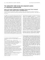
Effect of BMS-345541 on inhibition of IκB and p65 phosphorylation in vivoFigure 3
Effect of BMS-345541 on inhibition of IκB and p65 phosphorylation in vivo. MT-2, C8166, and CEM cells were
treated with BMS-345541 at 0.1, 0.5, 1, and 5 μM for 48 hr. Total cell extracts were collected and subjected to Western blot
analysis using anti-IκB, phospho IκB (ser 32), p65, phospho p65 (ser 536), p50, p52, Tax and actin. Twenty five microgram of
each extract was used to separate on a 4–20% SDS/PAGE. Levels of total IκB and p65 did not change between cell types, how-
ever there was a dramatic increase of phosphor-IκB and phosphor-p65 in HTLV-1 infected cells and their suppression by BMS-
345541 which inhibits IKKβ activity in vivo.
Tax
IκB
p52
p50
phospho-IκB (ser 32)
phospho-p65 (ser 536)
p65
0 0.1 0.5 1 5
BMS-345541 (μM)
0 0.1 0.5 1 5 0 0.1 0.5 1 5
1 2 3 4 5 6 7 8 9 10 11 12 13 14 15
MT-2 C81 CEM
β-actin
AIDS Research and Therapy 2008, 5:12 />Page 9 of 16
(page number not for citation purposes)
receptor(s) of HTLV-1 infection are altered when using
these drugs. Collectively, combination of two drugs that
can inhibit both NF-κB and CDK machineries in HTLV-1
"hyper-active" cells seem to be a viable option in inhibit-
ing infection. Future experiments are in progress to
develop second and third generation drugs, as well as
their effect in fresh ATL samples and inhibition in mouse
models.
Conclusion
Recently, unique therapeutic approaches targeting mole-
cules and/or mechanisms involved in the pathogenesis of
HTLV-1 have been explored, and some have produced
encouraging results that might lead to breakthrough ther-
apies. In this study, we have demonstrated that two drugs
(BMS-345541 and Purvalanol A) out of thirty-five drugs
studied that target NF-κB or CDK pathways had the best
specificity in inhibiting the growth of HTLV-1 infected but
not uninfected cells. The effect of BMS-345541 is through
the inhibition of IKKβ kinase activity resulting in dephos-
phorylation of IκBα and inactivation of NF-κB pathway.
The specificity of BMS-345541 with IC
50
of 50 nM in
HTLV-1 infected cell compared to IC
50
of 500 nM in unin-
fected cell therefore renders the infected cells 10 times
more sensitive to the drug than uninfected cell. The other
inhibitor, Purvalanol A induced higher level of inhibition
in MT-2 cells and the mechanism was previously shown
by us to be associated with inhibition of functional cyclin
E/CDK2 complexes. Combination of these two inhibitors
induced even higher level of p19 Gag expression in
infected cells. Therefore, treatment of HTLV-1 infected
cells with either BMS-345541, Purvalanol A or a combina-
tion of these two drugs hold promising leads in treatment
of infected cells.
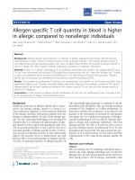
Purvalanol A induction of apoptosis in MT-2Figure 4
Purvalanol A induction of apoptosis in MT-2. A) Purvalanol A induced caspase-3 and PARP cleavage in MT-2 and C8166
cells. MT-2, C8166, and CEM were treated with Purvalanol A at various 0.1, 0.5, 1, and 5 μM for 48 hr. After 48 hr of treat-
ment, total cell extracts were collected and subjected to Western blot analysis of caspase-3 and PARP. Westeron blot of β-
actin was used as an internal control. B) Twenty five microgram of each Purvalanol A treated (5 μM for 48 hr) extracts from
both MT-2 and C8166 cells were also used for western blot against cyclin A, E and actin.
PARP
caspase-3 (p17)
β-actin
MT-2 C81 CEM
0 0.1 0.5 1 5
Purvalanol A (μM)
0 0.1 0.5 1 5 0 0.1 0.5 1 5
1 2 3 4 5 6 7 8 9 10 11 12 13 14 15
cleaved PARP
A)
B)
MT-2 C81
Purvalanol A
- + - +
Cyclin A
Cyclin E
Actin
1 2 3 4
AIDS Research and Therapy 2008, 5:12 />Page 10 of 16
(page number not for citation purposes)
Methods
Cell lines and reagents
MT-2, MT-4, C8166, and C10/MJ were all obtained from
NIH AIDS Research & Reference Reagent Program. They
are all HTLV-1 infected cell lines and some including
C8166 contain defective viruses but still express Tax. MT-
2 cells carry multiple copies of the HTLV-1 cosmopolitan
subtype and normally produce some full length infectious
HTLV-1 particles in the absence of any inducer [157]. MT-
4 cells are established from the human T cells isolated
from a patient with adult T-cell leukemia. CEM and Jurkat
cells are the uninfected control T lymphocyte cell lines. All
cell lines were cultured at 37°C up to 1 × 10
5
cells per ml
in RPMI 1640 medium containing fetal bovine serum
(10%), streptomycin, penicillin antibiotics (1%) and L-
Glutamine (1%) (Gibco/BRL). The CDK inhibitors used
were: Aloisine A (270-385-M001), Alsterpaullone (270-
275-M001), Bohemine (270-390-M001), CGP74514A
(270-391-M001), Compound 52 (270-248-M001), 9-
cyanopaullone (270-282-M001), 6-dimethylaminopu-
rine (480-050-M100), indirubin-3'-monoxime (270-271-
M001), 5-iodo-indirubin-3'-monoxime (270-424-M001),
N-6-(Δ2-Isopentenyl)-adenine (350-034-M100), Ken-
paullone (270-274-M001), Olomoucine (350-013-
M005), N9-isopropylolomoucine (270-397-M001), Pur-
valanol A (270-246-M001), (R)-Roscovitine (350-251-
M001), (S)-Roscovitine (350-293-M001) were purchased
from Alexis Inc. and 6-benzyloxypurine (387606), 2,6-
diaminopurine (247847), 2,6-dichloropurine (D73103),
Flavone (F2003) were purchase from Sigma-aldrich Inc.
Indirubin-3'-monoxime-5-sulfonic acid (402088), iso-
olomoucine (495622), WHI-P180 (681500) were pur-
chased from Calbiochem Inc. The CDK inhibitor, fla-
vopiridol was a kind gift from Dr. Ajit Kumar at the
GWUMC. The NF-κB inhibitors included BMS-345541
(401480), SC-514 (401479) were purchased from Calbi-
ochem Inc. and 5-Aminosalicylic acid (430-110-G005),
BAY 11-7082 (270-219-M010), BAY 11-7085 (270-220-
Cell cycle analysis of cells treated with or without drugsFigure 5
Cell cycle analysis of cells treated with or without drugs. For fluorescence-activated cell sorting (FACS) analysis, both
untreated and Purvalanol A treated CEM, MT-2 and C8166 cells (0.1 – 5.0 μM) were stained with a mixture of propidium
iodide buffer followed by cell sorting analysis. The acquired FACS data were analyzed by ModFit LT software (Verity Software
House, Inc.).
Untreated Pur (0.1) Pur (0.5) Pur (1.0) Pur (5.0)
Channels (FL2-H)
0 10 20 30 40 50 60 7
0
Apopto
s
Dip G1
Dip G2
Dip S
CEM
G1: 15.73 %
S: 84.27 %
G2: 0.00 %
Apop: 0.00 %
Channels (FL2-H)
0 10 20 30 40 50 60 70
Apoptos
i
Dip G1
Dip G2
Dip S
G1: 28.97 %
S: 69.88 %
G2: 1.15 %
Apop: 4.02 %
Channels (FL2-H)
0 10 20 30 40 50 60 70
Apoptosi
s
Dip G1
Dip G2
Dip S
G1: 49.13 %
S: 50.87 %
G2: 0.00 %
Apop: 0.00 %
Channels (FL2-H)
0 30 60 90 120
Apoptosi
s
Dip G1
Dip G2
Dip S
G1: 48.52 %
S: 36.90 %
G2: 14.58 %
Apop: 1.90 %
Channels (FL2-H)
0 10 20 30 40 50 60 70
Apoptosi
s
Dip G1
Dip G2
Dip S
G1: 27.13 %
S: 70.64 %
G2: 2.23 %
Apop: 12.31 %
MT-2
Channels (FL2-H)
0 30 60 90 120 150
Apoptosis
Dip G1
Dip G2
Dip S
G1: 50.73 %
S: 36.99 %
G2: 12.28 %
Apop: 0.71 %
Channels (FL2-H)
0 30 60 90 120 150
Apoptosi
s
Dip G1
Dip G2
Dip S
G1: 47.86 %
S: 39.86 %
G2: 12.28 %
Apop: 1.27 %
Channels (FL2-H)
0 30 60 90 120 150
Apoptosis
Dip G1
Dip G2
Dip S
G1: 50.22 %
S: 40.36 %
G2: 9.42 %
Apop: 1.94 %
Channels (FL2-H)
0 30 60 90 120 150
Apoptosi
s
Dip G1
Dip G2
Dip S
G1: 49.49 %
S: 30.76 %
G2: 19.76 %
Apop: 3.21%
Channels (FL2-H)
0 30 60 90 120 150
Apoptosi
s
Dip G1
Dip G2
Dip S
G1: 36.32 %
S: 57.71 %
G2: 5.97 %
Apop: 22.92%
C81
Channels (FL2-H)
0 30 60 90 120
Apoptosis
Dip G1
Dip G2
Dip S
G1: 70.21 %
S: 22.11 %
G2: 7.68 %
Apop: 4.26 %
Channels (FL2-H)
0 30 60 90 120
Apoptosis
Dip G1
Dip G2
Dip S
G1: 73.19 %
S: 19.23 %
G2: 7.59 %
Apop: 5.37 %
Channels (FL2-H)
0 30 60 90 120
Apoptosis
Dip G1
Dip G2
Dip S
G1: 64.62 %
S: 28.51 %
G2: 6.87 %
Apop: 3.96 %
Channels (FL2 -H)
0 30 60 90 120
Apoptosis
Dip G1
Dip G2
Dip S
G1: 71.94 %
S: 18.35 %
G2: 9.71 %
Apop: 6.97 %
Channels (FL2-H)
0 30 60 90 120
Apoptosis
Dip G1
Dip G2
Dip S
G1: 0.00 %
S: 3.44 %
G2: 96.56 %
Apop: 26.34 %
AIDS Research and Therapy 2008, 5:12 />Page 11 of 16
(page number not for citation purposes)
M010), caffeic acid phenylethyl ester (270-244-M010),
diethylmaleate (280-017-G005), Parthenolide (350-258-
M025), pyrrolidinedithiocarbamic acid (400-002-G005)
were purchased from Alexis Inc. and QNZ (6-amino-4-(4-
phenoxyphenylethylamino)quinazoline (EI-352),
Wedelolactone (EI-316) were purchased from Biomol Inc.
All inhibitors were prepared in 10 mM stock solution. 2,6-
Dichloropurine and diethylmaleate were dissolved in eth-
anol; Flavone was dissolved in acetone; Flavopiridol and
pyrrolidinedithiocarbamic acid were dissolved in water;
5-aminosalicylic acid was dissolved in hydrochloric acid.
The other twenty-nine inhibitors were all dissolved in
DMSO.
Drugs screening and cell counting
HTLV-1 infected cells and uninfected cells were treated
with thirty-five inhibitors at four concentrations including
0.01, 0.1, 1, and 10 μM. Forty-eight hours after treatment,
Double drugs treatment results in lower p19 Gag levels in HTLV-1 infected cellsFigure 6
Double drugs treatment results in lower p19 Gag levels in HTLV-1 infected cells. A) MT-2 cells (HTLV-1 infected)
were treated with TNF-α (10 ng/ml) for 2 h, washed, and subsequently treated with a specific NF-kB or CDK inhibitor. Acti-
vated cells were subsequently treated them with BMS-345541 alone (0.1 μM), Purvalanol A alone (0.5 μM), or a combination of
both drugs. Samples were collected after 7 days and used for detection of p19 Gag using ELISA. B) Log phase 293 cells were
transfected with ACH.pcTax (wild type HTLV-1 clone, generous gift of Dr. Lee Ratner, Washington University) using electro-
poration method. After transfection, the cells were cultured in complete medium and culture supernatants were collected at 4
days post-transfection, and virus particle production was monitored by p19 ELISA. Drug treatments (as in panle A) were 6 hrs
after transfection of the 293 cells for a total of 150 hrs.
Gag p19 (pg/ml)
TNF-α
TNF-α + BMS-345541 (0.1 uM)
TNF-α + Purv. (0.5 uM)
TNF-α + BMS-345541 + Purv.
1 2 3 4 5
None
0
200
400
600
800
1000
1200
0
500
1000
1500
2000
2500
ACH + BMS-345541(0.1 uM)
ACH + Purv. (0.5 uM)
ACH + BMS-345541 + Purv.
1 2 3 4
ACH
A) B)
Gag p19 (pg/ml)
AIDS Research and Therapy 2008, 5:12 />Page 12 of 16
(page number not for citation purposes)
cytotoxicity was primarily determined by the color of
media and cell viability by trypan blue exclusion. Cells
were counted for the number of living cells every 24–48
hrs. Subsequent focusing experiments used flow data to
check for viability and apoptosis.
Cytoplasmic extracts
Cytoplasmic extracts were prepared according to the fol-
lowing procedure. Briefly, cells were collected and washed
with PBS once and then once with 80 μl of ice-cold buffer
A (Tris-HCl (pH 7.4, 10 mM), MgCl
2
(1.5 mM), KCl (10
mM), DTT (1 mM), 0.4% NP-40, phenylmethylsulfonyl
Inhibition mechanism of BMS-345541 and Purvalanol A in HTLV-1 infected cells results in blocking canonical NF-κB signaling pathway and cell cycle progressionFigure 7
Inhibition mechanism of BMS-345541 and Purvalanol A in HTLV-1 infected cells results in blocking canonical
NF-κB signaling pathway and cell cycle progression. In the absence of drug, hyperactive IKK complex phosphorylates
IκB-α resulting in IκB-α degradation and p65/p50 translocation. The genes transcribed by p65/50 include anti-apoptotic genes
which are responsible for survival of virus infected cells. In the presence of BMS-345541, the activity of IKK complex is inhib-
ited which results in decreased IκB-α phosphorylation, therefore p65/p50 are kept in cytoplasm. Hence, the expression of anti-
apoptotic proteins are decreased which make HTLV-1 infected cells more susceptible and sensitive to the action of the drug.
Without drugs, cyclin E/CDK2 phosphorylates Rb and induces Rb degradation. The free E2F then transcribes genes which are
necessary for G1/S transition. However, Purvalanol A inhibits CDK2 (a non-essential protein in the life cycle of a cell) activity,
as previously shown by us, which results in decreased Rb phsophorylation and inactivated E2F. Therefore, the infected cells
may be blocked at the G
1
checkpoint and simultaneously have lower viral expression.
canonical NF
canonical NF
-
-
kB
kB
pathway
pathway
IkB
IkB
-
-
a
a
IkB
IkB
-
-
a
a
NF
NF
-
-
kB
kB
NF
NF
-
-
kB
kB
canonical NF
canonical NF
-
-
kB
kB
pathway
pathway
IkB
IkB
-
-
a
a
IkB
IkB
-
-
a
a
NF
NF
-
-
kB
kB
NF
NF
-
-
kB
kB
AIDS Research and Therapy 2008, 5:12 />Page 13 of 16
(page number not for citation purposes)
fluoride (1 mM), aprotinin (10 μg/ml), pepstatin (1 μM),
NaF (50 mM), and Na
3
VO
4
(1 mM)). Cells were lysed in
80 μl of buffer A by gently passing the cell suspension
through a 28-gauge needle. The cytoplasmic extracts were
collected by pelleting for 8 sec in an Eppendorf microcen-
trifuge and the supernatant was collected. The protein
concentration for each preparation was determined with a
Bio-Rad protein assay kit (Bio-Rad Laboratories, Hercules,
CA, USA).
Immunoprecipitation and in vitro kinase assay
Reaction mixtures (24 μl) contained (final concentra-
tions) 40 mM β-glycerophosphate, pH 7.4, 7.5 mM
MgCl2, 7.5 mM EGTA, 5% glycerol, [γ
32
-P]ATP (0.2 mM,
1 μCi), 50 mM NaF, 1 mM orthovanadate, and 0.1% (v/
v) β-mercaptoethanol. Phosphorylation reactions were
performed with 2 mg of cytoplasmic extract immunopre-
cipitated with appropriate antibody and washed in lysis
buffer containing 50 mM Tris-HCl (pH 7.5), 120 mM
NaCl, 5 mM EDTA, 50 mM NaF, 0.2 mM Na
3
VO
4
, 1 mM
DTT, 0.5% NP-40 and protease inhibitors (Protease inhib-
itor cocktail tablets, Boehringer Mannheim, one tablet per
50 ml) or with 1 μg of purified recombinant GST-IκBα at
37°C for 1 hour. Reactions were stopped by adding 1 vol-
ume of Laemmli sample buffer containing 5% β-mercap-
toethanol and ran on a 4–20% SDS/PAGE. Gels were
autoradiographed and bands were counted using a Molec-
ular Dynamics PhosphorImager software.
Immunoblotting
Total cellular extracts (20 μg) were separated by a 4–20%
Tris-glycine gel then transferred to a PVDF membrane
(Immobilon-P transfer membranes; Millipore Corp.) Fol-
lowing the transfer, the blots were blocked with 5% non-
fat dry milk in PBS + 0.1% Tween-20 for 2 hr and washed
three times with PBS + 0.1% Tween-20 at 4°C. The blots
were then probed with 1:200 dilution of primary anti-
body against caspase-3 (H-277; Santa Cruz Biotechnol-
ogy, sc-7148), PARP (H-250; Santa Cruz Biotechnology,
sc-7150), CDK2 (M2; Santa Cruz Biotechnology, sc-163),
cyclin A (H-432; Santa Cruz Biotechnology, sc-751), cyc-
lin E (C-19; Santa Cruz Biotechnology, sc-198), and actin
(c-11; Santa Cruz Biotechnology, sc-1615). The blots were
then probed with a 1:750 dilution of secondary antibod-
ies for 1 h at 4°C, followed by washes in PBS + 0.1%
Tween-20 and detected using SuperSignal West Dura
Extended Duration Substrate Kit (Pierce, Rockford, IL,
USA).
HTLV-1 p19 ELISA
MT-2 cells (HTLV-1 infected) were treated with TNF-α (10
ng/ml) for 2 h, washed, and subsequently treated with a
specific NF-kB or CDK inhibitor. Media from MT-2
infected cells were centrifuged to pellet the cells, and
supernatants were collected and diluted to 1:100 to
1:1,000 in RPMI 1640 prior to ELISA. Seven days later
samples were collected and used for p19 gag ELISA. The
HTLV-1 p19 core antigen ELISA kit was from Retro-Tek
(Cellular Products) and RT/PCR using HTLV-1 specific
Tax primers (data not shown).
ACH transfcetion of cells
Log phase 293 cells were transfected with 20 μg of
ACH.pcTax (wild type HTLV-1 clone) using electropora-
tion method. After transfection, the cells were cultured in
complete medium supplemented with 10% fetal calf
serum (FCS), 2 mM L-glutamine, 50 μg of penicillin/ml,
and 50 U of streptomycin/ml. Cell culture supernatants
were collected at 4 days post-transfection, and virus parti-
cle production was monitored by p19 ELISA as described
above. Drug treatment was 6 hrs after transfection of the
293 cells for a total of 150 hrs.
Flow Cytometry
For cell cycle analysis, cells treated with or without drugs
were collected by low speed centrifugation and washed
with PBS without Ca
2+
and Mg
2+
and then fixed with 70%
ethanol. For fluorescence-activated cell sorting (FACS)
analysis, cells were stained with a mixture of propidium
iodide buffer (PBS with Ca
2+
and Mg
2+
, 10 μg/ml RNase A,
0.1% Nonidet P-40, and 50 μg/ml propidium iodide) fol-
lowed by cell sorting analysis. The acquired FACS data
were analyzed by ModFit LT software (Verity Software
House, Inc.). Cells were washed twice with cold PBS with-
out Ca
2+
and Mg
2+
, resuspended in 1× binding buffer (10
mM HEPES-NaOH (pH 7.4), 140 mM NaCl, 2.5 mM
CaCl
2
) and 5 μl of propidium iodide/10
5
cells, and incu-
bated at room temperature for 15 min. Cells were
acquired and analyzed using CELLQuest software (BD
Biosciences).
Detection of apoptosis through annexin V and PI staining
was done according to the manufacturers protocol (BD
Pharmingen, San Jose, CA). In brief, cells were washed
three times in PBS and re-suspended in binding buffer at
1 × 10⁶ cells/ml. An aliquot of 1 × 10⁵ cells was stained
with annexin V-FITC and PI for 15 minutes at room tem-
perature. Analysis was performed on a BD FacsCalibur
flow cytometer. Cells were considered to be early apop-
totic if they exhibited staining for annexin V, but not PI.
The double positive population was considered to be in
the late stage of apoptosis.
Competing interests
The authors declare that they have no competing interests.
Authors' contributions
EA performed the initial drug screening assays along with
WIY, WIY carried most of the subsequent confirmation
and Western blots, RB, ZK, and CP carried out confirma-
AIDS Research and Therapy 2008, 5:12 />Page 14 of 16
(page number not for citation purposes)
tory experiments on Westerns, FACS, as well as kinase
assays, KKH and WW provided the day to day leadership
and direction for the project, FK also provided the overall
direction and the funding for the project.
Acknowledgements
Both E. Agbottah and W-I Yeh contributed equally to this work and share
first authorship. We thank Ann Richmond and Dean Ballard (Department
of Cancer Biology, Vanderbilt University) for the expression plasmids as
well as the GST-IκBα. The current research was supported by grants from
the George Washington University REF funds to Akos Vertes and FK;
McCormick Grant and NIH grants AI065236, AI043894 to FK.
References
1. Verdonck K, Gonzalez E, Van Dooren S, Vandamme AM, Vanham G,
Gotuzzo E: Human T-lymphotropic virus 1: recent knowledge
about an ancient infection. Lancet Infect Dis 2007, 7:266-281.
2. Matsuoka M, Jeang KT: Human T-cell leukaemia virus type 1
(HTLV-1) infectivity and cellular transformation. Nat Rev Can-
cer 2007, 7:270-280.
3. Peloponese JM Jr., Kinjo T, Jeang KT: Human T-cell leukemia
virus type 1 Tax and cellular transformation. Int J Hematol
2007, 86:101-106.
4. Suzuki T, Hirai H, Yoshida M: Tax protein of HTLV-1 interacts
with the Rel homology domain of NF-kappa B p65 and c-Rel
proteins bound to the NF-kappa B binding site and activates
transcription. Oncogene 1994, 9:3099-3105.
5. Yin MJ, Christerson LB, Yamamoto Y, Kwak YT, Xu S, Mercurio F,
Barbosa M, Cobb MH, Gaynor RB: HTLV-I Tax protein binds to
MEKK1 to stimulate IkappaB kinase activity and NF-kappaB
activation. Cell 1998, 93:875-884.
6. Xiao G, Cvijic ME, Fong A, Harhaj EW, Uhlik MT, Waterfield M, Sun
SC: Retroviral oncoprotein Tax induces processing of NF-
kappaB2/p100 in T cells: evidence for the involvement of
IKKalpha. Embo J 2001, 20:6805-6815.
7. Caron C, Rousset R, Beraud C, Moncollin V, Egly JM, Jalinot P: Func-
tional and biochemical interaction of the HTLV-I Tax1 trans-
activator with TBP. Embo J 1993, 12:4269-4278.
8. Clemens KE, Piras G, Radonovich MF, Choi KS, Duvall JF, DeJong J,
Roeder R, Brady JN: Interaction of the human T-cell lympho-
tropic virus type 1 tax transactivator with transcription fac-
tor IIA. Mol Cell Biol 1996, 16:4656-4664.
9. Colgin MA, Nyborg JK: The human T-cell leukemia virus type 1
oncoprotein Tax inhibits the transcriptional activity of c-Myb
through competition for the CREB binding protein. J Virol
1998, 72:9396-9399.
10. Gachon F, Thebault S, Peleraux A, Devaux C, Mesnard JM: Molecu-
lar interactions involved in the transactivation of the human
T-cell leukemia virus type 1 promoter mediated by Tax and
CREB-2 (ATF-4). Mol Cell Biol 2000, 20:3470-3481.
11. Harrod R, Tang Y, Nicot C, Lu HS, Vassilev A, Nakatani Y, Giam CZ:
An exposed KID-like domain in human T-cell lymphotropic
virus type 1 Tax is responsible for the recruitment of coacti-
vators CBP/p300. Mol Cell Biol 1998, 18:5052-5061.
12. Nicot C, Mahieux R, Opavsky R, Cereseto A, Wolff L, Brady JN, Fran-
chini G: HTLV-I Tax transrepresses the human c-Myb pro-
moter independently of its interaction with CBP or p300.
Oncogene 2000, 19:2155-2164.
13. Yin MJ, Paulssen EJ, Seeler JS, Gaynor RB: Protein domains
involved in both in vivo and in vitro interactions between
human T-cell leukemia virus type I tax and CREB. J Virol 1995,
69:3420-3432.
14. Kashanchi F, Brady JN: Transcriptional and post-transcriptional
gene regulation of HTLV-1. Oncogene 2005, 24:5938-5951.
15. Aggarwal BB: Nuclear factor-kappaB: the enemy within. Cancer
Cell 2004, 6:203-208.
16. Okamoto T, Sakurada S, Yang JP, Merin JP: Regulation of NF-
kappa B and disease control: identification of a novel serine
kinase and thioredoxin as effectors for signal transduction
pathway for NF-kappa B activation. Curr Top Cell Regul 1997,
35:149-161.
17. Basseres DS, Baldwin AS: Nuclear factor-kappaB and inhibitor
of kappaB kinase pathways in oncogenic initiation and pro-
gression. Oncogene 2006, 25:6817-6830.
18. Taylor GP, Matsuoka M: Natural history of adult T-cell leuke-
mia/lymphoma and approaches to therapy. Oncogene 2005,
24:6047-6057.
19. Ghosh S, Karin M: Missing pieces in the NF-kappaB puzzle. Cell
2002, 109 Suppl:S81-96.
20. Pikarsky E, Porat RM, Stein I, Abramovitch R, Amit S, Kasem S, Gutk-
ovich-Pyest E, Urieli-Shoval S, Galun E, Ben-Neriah Y: NF-kappaB
functions as a tumour promoter in inflammation-associated
cancer. Nature
2004, 431:461-466.
21. Gilmore TD: Introduction to NF-kappaB: players, pathways,
perspectives. Oncogene 2006, 25:6680-6684.
22. Karin M, Cao Y, Greten FR, Li ZW: NF-kappaB in cancer: from
innocent bystander to major culprit. Nat Rev Cancer 2002,
2:301-310.
23. Schesser K, Spiik AK, Dukuzumuremyi JM, Neurath MF, Pettersson S,
Wolf-Watz H: The yopJ locus is required for Yersinia-medi-
ated inhibition of NF-kappaB activation and cytokine expres-
sion: YopJ contains a eukaryotic SH2-like domain that is
essential for its repressive activity. Mol Microbiol 1998,
28:1067-1079.
24. Pomerantz JL, Baltimore D: Two pathways to NF-kappaB. Mol
Cell 2002, 10:693-695.
25. Mori N, Yamada Y, Ikeda S, Yamasaki Y, Tsukasaki K, Tanaka Y,
Tomonaga M, Yamamoto N, Fujii M: Bay 11-7082 inhibits tran-
scription factor NF-kappaB and induces apoptosis of HTLV-
I-infected T-cell lines and primary adult T-cell leukemia cells.
Blood 2002, 100:1828-1834.
26. Mori N, Fujii M, Iwai K, Ikeda S, Yamasaki Y, Hata T, Yamada Y, Tan-
aka Y, Tomonaga M, Yamamoto N: Constitutive activation of
transcription factor AP-1 in primary adult T-cell leukemia
cells. Blood 2000, 95:3915-3921.
27. Bonizzi G, Bebien M, Otero DC, Johnson-Vroom KE, Cao Y, Vu D,
Jegga AG, Aronow BJ, Ghosh G, Rickert RC, Karin M: Activation of
IKKalpha target genes depends on recognition of specific
kappaB binding sites by RelB:p52 dimers. Embo J 2004,
23:4202-4210.
28. Derudder E, Dejardin E, Pritchard LL, Green DR, Korner M, Baud V:
RelB/p50 dimers are differentially regulated by tumor necro-
sis factor-alpha and lymphotoxin-beta receptor activation:
critical roles for p100. J Biol Chem 2003, 278:23278-23284.
29. Yilmaz ZB, Weih DS, Sivakumar V, Weih F: RelB is required for
Peyer's patch development: differential regulation of p52-
RelB by lymphotoxin and TNF. Embo J 2003,
22:121-130.
30. Schwabe RF, Schnabl B, Kweon YO, Brenner DA: CD40 activates
NF-kappa B and c-Jun N-terminal kinase and enhances
chemokine secretion on activated human hepatic stellate
cells. J Immunol 2001, 166:6812-6819.
31. Hironaka N, Mochida K, Mori N, Maeda M, Yamamoto N, Yamaoka
S: Tax-independent constitutive IkappaB kinase activation in
adult T-cell leukemia cells. Neoplasia 2004, 6:266-278.
32. Chu ZL, Shin YA, Yang JM, DiDonato JA, Ballard DW: IKKgamma
mediates the interaction of cellular IkappaB kinases with the
tax transforming protein of human T cell leukemia virus
type 1. J Biol Chem 1999, 274:15297-15300.
33. Harhaj EW, Sun SC: IKKgamma serves as a docking subunit of
the IkappaB kinase (IKK) and mediates interaction of IKK
with the human T-cell leukemia virus Tax protein. J Biol Chem
1999, 274:22911-22914.
34. Jin DY, Giordano V, Kibler KV, Nakano H, Jeang KT: Role of
adapter function in oncoprotein-mediated activation of NF-
kappaB. Human T-cell leukemia virus type I Tax interacts
directly with IkappaB kinase gamma. J Biol Chem 1999,
274:17402-17405.
35. Yamaoka S, Courtois G, Bessia C, Whiteside ST, Weil R, Agou F, Kirk
HE, Kay RJ, Israel A: Complementation cloning of NEMO, a
component of the IkappaB kinase complex essential for NF-
kappaB activation. Cell 1998, 93:1231-1240.
36. Karunagaran D. ABB: Transcription factors as targets fr the
drug development. In Molecular pathomechanisms and new trends in
drug development Edited by: Keri G. TI. New York, NY, Harwood Aca-
demic Publisher; 2002:76-91.
AIDS Research and Therapy 2008, 5:12 />Page 15 of 16
(page number not for citation purposes)
37. Mori N, Fujii M, Ikeda S, Yamada Y, Tomonaga M, Ballard DW,
Yamamoto N: Constitutive activation of NF-kappaB in pri-
mary adult T-cell leukemia cells. Blood 1999, 93:2360-2368.
38. Nicot C, Mahieux R, Takemoto S, Franchini G: Bcl-X(L) is up-reg-
ulated by HTLV-I and HTLV-II in vitro and in ex vivo ATLL
samples. Blood 2000, 96:275-281.
39. Dewan MZ, Terashima K, Taruishi M, Hasegawa H, Ito M, Tanaka Y,
Mori N, Sata T, Koyanagi Y, Maeda M, Kubuki Y, Okayama A, Fujii M,
Yamamoto N: Rapid tumor formation of human T-cell leuke-
mia virus type 1-infected cell lines in novel NOD-SCID/gam-
mac(null) mice: suppression by an inhibitor against NF-
kappaB. J Virol 2003, 77:5286-5294.
40. Ohsugi T, Horie R, Kumasaka T, Ishida A, Ishida T, Yamaguchi K,
Watanabe T, Umezawa K, Urano T: In vivo antitumor activity of
the NF-kappaB inhibitor dehydroxymethylepoxyquinomicin
in a mouse model of adult T-cell leukemia. Carcinogenesis 2005,
26:1382-1388.
41. Watanabe M, Ohsugi T, Shoda M, Ishida T, Aizawa S, Maruyama-Nagai
M, Utsunomiya A, Koga S, Yamada Y, Kamihira S, Okayama A, Kikuchi
H, Uozumi K, Yamaguchi K, Higashihara M, Umezawa K, Watanabe T,
Horie R: Dual targeting of transformed and untransformed
HTLV-1-infected T cells by DHMEQ, a potent and selective
inhibitor of NF-kappaB, as a strategy for chemoprevention
and therapy of adult T-cell leukemia. Blood 2005,
106:2462-2471.
42. Ohsugi T, Kumasaka T, Ishida A, Ishida T, Horie R, Watanabe T, Ume-
zawa K, Yamaguchi K: In vitro and in vivo antitumor activity of
the NF-kappaB inhibitor DHMEQ in the human T-cell leuke-
mia virus type I-infected cell line, HUT-102. Leuk Res 2006,
30:90-97.
43. El-Sabban ME, Nasr R, Dbaibo G, Hermine O, Abboushi N, Quignon
F, Ameisen JC, Bex F, de The H, Bazarbachi A: Arsenic-interferon-
alpha-triggered apoptosis in HTLV-I transformed cells is
associated with tax down-regulation and reversal of NF-
kappa B activation. Blood 2000, 96:2849-2855.
44. Nasr R, Rosenwald A, El-Sabban ME, Arnulf B, Zalloua P, Lepelletier
Y, Bex F, Hermine O, Staudt L, de The H, Bazarbachi A: Arsenic/
interferon specifically reverses 2 distinct gene networks crit-
ical for the survival of HTLV-1-infected leukemic cells. Blood
2003, 101:4576-4582.
45. Satou Y, Nosaka K, Koya Y, Yasunaga JI, Toyokuni S, Matsuoka M:
Proteasome inhibitor, bortezomib, potently inhibits the
growth of adult T-cell leukemia cells both in vivo and in vitro.
Leukemia 2004, 18:1357-1363.
46. Lemoine FJ, Marriott SJ: Accelerated G(1) phase progression
induced by the human T cell leukemia virus type I (HTLV-I)
Tax oncoprotein. J Biol Chem 2001, 276:31851-31857.
47. Suzuki T, Kitao S, Matsushime H, Yoshida M: HTLV-1 Tax protein
interacts with cyclin-dependent kinase inhibitor p16INK4A
and counteracts its inhibitory activity towards CDK4. Embo J
1996, 15:1607-1614.
48. Parker SF, Perkins ND, Gitlin SD, Nabel GJ: A cooperative inter-
action of human T-cell leukemia virus type 1 Tax with the
p21 cyclin-dependent kinase inhibitor activates the human
immunodeficiency virus type 1 enhancer. J Virol 1996,
70:5731-5734.
49. Low KG, Dorner LF, Fernando DB, Grossman J, Jeang KT, Comb MJ:
Human T-cell leukemia virus type 1 Tax releases cell cycle
arrest induced by p16INK4a. J Virol 1997, 71:1956-1962.
50. Jeang KT, Giam CZ, Majone F, Aboud M: Life, death, and tax: role
of HTLV-I oncoprotein in genetic instability and cellular
transformation. J Biol Chem 2004, 279:31991-31994.
51. Marriott SJ, Semmes OJ: Impact of HTLV-I Tax on cell cycle
progression and the cellular DNA damage repair response.
Oncogene 2005, 24:5986-5995.
52. de La Fuente C, Santiago F, Chong SY, Deng L, Mayhood T, Fu P, Stein
D, Denny T, Coffman F, Azimi N, Mahieux R, Kashanchi F: Overex-
pression of p21(waf1) in human T-cell lymphotropic virus
type 1-infected cells and its association with cyclin A/cdk2. J
Virol 2000, 74:7270-7283.
53. Cereseto A, Diella F, Mulloy JC, Cara A, Michieli P, Grassmann R,
Franchini G, Klotman ME: p53 functional impairment and high
p21waf1/cip1 expression in human T-cell lymphotropic/
leukemia virus type I-transformed T cells. Blood 1996,
88:1551-1560.
54. Pise-Masison CA, Radonovich M, Sakaguchi K, Appella E, Brady JN:
Phosphorylation of p53: a novel pathway for p53 inactivation
in human T-cell lymphotropic virus type 1-transformed cells.
J Virol
1998, 72:6348-6355.
55. Pise-Masison CA, Mahieux R, Jiang H, Ashcroft M, Radonovich M,
Duvall J, Guillerm C, Brady JN: Inactivation of p53 by human T-
cell lymphotropic virus type 1 Tax requires activation of the
NF-kappaB pathway and is dependent on p53 phosphoryla-
tion. Mol Cell Biol 2000, 20:3377-3386.
56. Neuveut C, Low KG, Maldarelli F, Schmitt I, Majone F, Grassmann R,
Jeang KT: Human T-cell leukemia virus type 1 Tax and cell
cycle progression: role of cyclin D-cdk and p110Rb. Mol Cell
Biol 1998, 18:3620-3632.
57. Akagi T, Ono H, Shimotohno K: Expression of cell-cycle regula-
tory genes in HTLV-I infected T-cell lines: possible involve-
ment of Tax1 in the altered expression of cyclin D2, p18Ink4
and p21Waf1/Cip1/Sdi1. Oncogene 1996, 12:1645-1652.
58. de La Fuente C, Deng L, Santiago F, Arce L, Wang L, Kashanchi F:
Gene expression array of HTLV type 1-infected T cells: Up-
regulation of transcription factors and cell cycle genes. AIDS
Res Hum Retroviruses 2000, 16:1695-1700.
59. Huang Y, Ohtani K, Iwanaga R, Matsumura Y, Nakamura M: Direct
trans-activation of the human cyclin D2 gene by the onco-
gene product Tax of human T-cell leukemia virus type I.
Oncogene 2001, 20:1094-1102.
60. Santiago F, Clark E, Chong S, Molina C, Mozafari F, Mahieux R, Fujii
M, Azimi N, Kashanchi F: Transcriptional up-regulation of the
cyclin D2 gene and acquisition of new cyclin-dependent
kinase partners in human T-cell leukemia virus type 1-
infected cells. J Virol 1999, 73:9917-9927.
61. Iwanaga R, Ohtani K, Hayashi T, Nakamura M: Molecular mecha-
nism of cell cycle progression induced by the oncogene prod-
uct Tax of human T-cell leukemia virus type I. Oncogene 2001,
20:2055-2067.
62. Kehn K, Deng L, de la Fuente C, Strouss K, Wu K, Maddukuri A, Bay-
lor S, Rufner R, Pumfery A, Bottazzi ME, Kashanchi F: The role of
cyclin D2 and p21/waf1 in human T-cell leukemia virus type
1 infected cells. Retrovirology 2004, 1:6.
63. Chlichlia K, Moldenhauer G, Daniel PT, Busslinger M, Gazzolo L,
Schirrmacher V, Khazaie K: Immediate effects of reversible
HTLV-1 tax function: T-cell activation and apoptosis. Onco-
gene 1995,
10:269-277.
64. Burke JR, Pattoli MA, Gregor KR, Brassil PJ, MacMaster JF, McIntyre
KW, Yang X, Iotzova VS, Clarke W, Strnad J, Qiu Y, Zusi FC: BMS-
345541 is a highly selective inhibitor of I kappa B kinase that
binds at an allosteric site of the enzyme and blocks NF-kappa
B-dependent transcription in mice. J Biol Chem 2003,
278:1450-1456.
65. Yang J, Amiri KI, Burke JR, Schmid JA, Richmond A: BMS-345541
targets inhibitor of kappaB kinase and induces apoptosis in
melanoma: involvement of nuclear factor kappaB and mito-
chondria pathways. Clin Cancer Res 2006, 12:950-960.
66. Petropoulos CJ, Givol I, Hughes SH: Comparative analysis of the
structure and function of the chicken c-myc and v-myc
genes: v-myc is a more potent inducer of cell proliferation
and apoptosis than c-myc. Oncogene 1996, 12:2611-2621.
67. Mauxion F, Jamieson C, Yoshida M, Arai K, Sen R: Comparison of
constitutive and inducible transcriptional enhancement
mediated by kappa B-related sequences: modulation of
activity in B cells by human T-cell leukemia virus type I tax
gene. Proc Natl Acad Sci U S A 1991, 88:2141-2145.
68. Arima N, Molitor JA, Smith MR, Kim JH, Daitoku Y, Greene WC:
Human T-cell leukemia virus type I Tax induces expression
of the Rel-related family of kappa B enhancer-binding pro-
teins: evidence for a pretranslational component of regula-
tion. J Virol 1991, 65:6892-6899.
69. Nicholson DW, Ali A, Thornberry NA, Vaillancourt JP, Ding CK, Gal-
lant M, Gareau Y, Griffin PR, Labelle M, Lazebnik YA, et al.: Identifi-
cation and inhibition of the ICE/CED-3 protease necessary
for mammalian apoptosis. Nature 1995, 376:37-43.
70. Han Z, Hendrickson EA, Bremner TA, Wyche JH: A sequential
two-step mechanism for the production of the mature
p17:p12 form of caspase-3 in vitro. J Biol Chem 1997,
272:13432-13436.
71. Rice WG, Hillyer CD, Harten B, Schaeffer CA, Dorminy M, Lackey
DA 3rd, Kirsten E, Mendeleyev J, Buki KG, Hakam A, et al.: Induction
Publish with BioMed Central and every
scientist can read your work free of charge
"BioMed Central will be the most significant development for
disseminating the results of biomedical research in our lifetime."
Sir Paul Nurse, Cancer Research UK
Your research papers will be:
available free of charge to the entire biomedical community
peer reviewed and published immediately upon acceptance
cited in PubMed and archived on PubMed Central
yours — you keep the copyright
Submit your manuscript here:
/>BioMedcentral
AIDS Research and Therapy 2008, 5:12 />Page 16 of 16
(page number not for citation purposes)
of endonuclease-mediated apoptosis in tumor cells by C-
nitroso-substituted ligands of poly(ADP-ribose) polymerase.
Proc Natl Acad Sci U S A 1992, 89:7703-7707.
72. Lazebnik YA, Kaufmann SH, Desnoyers S, Poirier GG, Earnshaw WC:
Cleavage of poly(ADP-ribose) polymerase by a proteinase
with properties like ICE. Nature 1994, 371:346-347.
73. Kaufmann SH, Desnoyers S, Ottaviano Y, Davidson NE, Poirier GG:
Specific proteolytic cleavage of poly(ADP-ribose) polymer-
ase: an early marker of chemotherapy-induced apoptosis.
Cancer Res 1993, 53:3976-3985.
74. Sherr CJ, Roberts JM: CDK inhibitors: positive and negative
regulators of G1-phase progression. Genes Dev 1999,
13:1501-1512.
75. Villerbu N, Gaben AM, Redeuilh G, Mester J: Cellular effects of
purvalanol A: a specific inhibitor of cyclin-dependent kinase
activities. Int J Cancer 2002, 97:761-769.
76. Bain J, McLauchlan H, Elliott M, Cohen P: The specificities of pro-
tein kinase inhibitors: an update. Biochem J 2003, 371:199-204.
77. Gray NS, Wodicka L, Thunnissen AM, Norman TC, Kwon S, Espinoza
FH, Morgan DO, Barnes G, LeClerc S, Meijer L, Kim SH, Lockhart DJ,
Schultz PG: Exploiting chemical libraries, structure, and
genomics in the search for kinase inhibitors. Science 1998,
281:533-538.
78. Matsuoka M, Jeang KT: Human T-cell leukemia virus type I at
age 25: a progress report. Cancer Res 2005, 65:4467-4470.
79. Sun SC, Yamaoka S: Activation of NF-kappaB by HTLV-I and
implications for cell transformation. Oncogene 2005,
24:5952-5964.
80. Kudoh A, Daikoku T, Sugaya Y, Isomura H, Fujita M, Kiyono T, Nishi-
yama Y, Tsurumi T: Inhibition of S-phase cyclin-dependent
kinase activity blocks expression of Epstein-Barr virus imme-
diate-early and early genes, preventing viral lytic replication.
J Virol 2004, 78:104-115.
81. Wang L, Deng L, Wu K, de la Fuente C, Wang D, Kehn K, Maddukuri
A, Baylor S, Santiago F, Agbottah E, Trigon S, Morange M, Mahieux R,
Kashanchi F: Inhibition of HTLV-1 transcription by cyclin
dependent kinase inhibitors. Mol Cell Biochem 2002,
237:137-153.
82. Gill PS, Harrington W Jr., Kaplan MH, Ribeiro RC, Bennett JM, Lieb-
man HA, Bernstein-Singer M, Espina BM, Cabral L, Allen S, et al.:
Treatment of adult T-cell leukemia-lymphoma with a com-
bination of interferon alfa and zidovudine. N Engl J Med 1995,
332:1744-1748.
83. Hermine O, Bouscary D, Gessain A, Turlure P, Leblond V, Franck N,
Buzyn-Veil A, Rio B, Macintyre E, Dreyfus F, et al.: Brief report:
treatment of adult T-cell leukemia-lymphoma with zidovu-
dine and interferon alfa. N Engl J Med 1995, 332:1749-1751.
84. Bazarbachi A, Hermine O: Treatment with a combination of
zidovudine and alpha-interferon in naive and pretreated
adult T-cell leukemia/lymphoma patients. J Acquir Immune Defic
Syndr Hum Retrovirol 1996, 13 Suppl 1:S186-90.
85. Hermine O, Allard I, Levy V, Arnulf B, Gessain A, Bazarbachi A: A
prospective phase II clinical trial with the use of zidovudine
and interferon-alpha in the acute and lymphoma forms of
adult T-cell leukemia/lymphoma. Hematol J 2002, 3:276-282.
86. Hermine O, Dombret H, Poupon J, Arnulf B, Lefrere F, Rousselot P,
Damaj G, Delarue R, Fermand JP, Brouet JC, Degos L, Varet B, de The
H, Bazarbachi A: Phase II trial of arsenic trioxide and alpha
interferon in patients with relapsed/refractory adult T-cell
leukemia/lymphoma. Hematol J 2004, 5:130-134.
87. Kfoury Y, Nasr R, Hermine O, de The H, Bazarbachi A: Proapop-
totic regimes for HTLV-I-transformed cells: targeting Tax
and the NF-kappaB pathway. Cell Death Differ 2005, 12 Suppl
1:871-877.