Immobilizing topoisomerase I on a surface plasmon resonance biosensor chip to screen for inhibitors pptx
Bạn đang xem bản rút gọn của tài liệu. Xem và tải ngay bản đầy đủ của tài liệu tại đây (889.76 KB, 9 trang )
Tsai et al. Journal of Biomedical Science 2010, 17:49
/>Open Access
RESEARCH
© 2010 Tsai et al; licensee BioMed Central Ltd. This is an Open Access article distributed under the terms of the Creative Commons At-
tribution License ( which permits unrestricted use, distribution, and reproduction in any
medium, provided the original work is properly cited.
Research
Immobilizing topoisomerase I on a surface
plasmon resonance biosensor chip to screen for
inhibitors
Hsiang-Ping Tsai
†1,2
, Li-Wei Lin
†3
, Zhi-Yang Lai
†3
, Jui-Yu Wu
2
, Chiao-En Chen
4
, Jaulang Hwang
4
, Chien-Shu Chen
5
and
Chun-Mao Lin*
2
Abstract
Background: The topoisomerase I (TopI) reaction intermediate consists of an enzyme covalently linked to a nicked
DNA molecule, known as a TopI-DNA complex, that can be trapped by inhibitors and results in failure of re-ligation.
Attempts at new derivative designs for TopI inhibition are enthusiastically being pursued, and TopI inhibitors were
developed for a variety of applications. Surface plasmon resonance (SPR) was recently used in TopI-inhibition studies.
However, most such immobilized small molecules or short-sequence nucleotides are used as ligands onto sensor
chips, and TopI was used as the analyte that flowed through the sensor chip.
Methods: We established a sensor chip on which the TopI protein is immobilized to evaluate TopI inhibition by SPR.
Camptothecin (CPT) targeting the DNA-TopI complex was used as a representative inhibitor to validate this label-free
method.
Results: Purified recombinant human TopI was covalently coupled to the sensor chip for the SPR assay. The binding of
anti-human (h)TopI antibodies and plasmid pUC19, respectively, to the immobilized hTopI was observed with dose-
dependent increases in resonance units (RU) suggesting that the immobilized hTopI retains its DNA-binding activity.
Neither CPT nor evodiamine alone in the analyte flowing through the sensor chip showed a significant increase in RU.
The combination of pUC19 and TopI inhibitors as the analyte flowing through the sensor chip caused increases in RU.
This confirms its reliability for binding kinetic studies of DNA-TopI binders for interaction and for primary screening of
TopI inhibitors.
Conclusions: TopI immobilized on the chip retained its bioactivities of DNA binding and catalysis of intermediates of
the DNA-TopI complex. This provides DNA-TopI binders for interaction and primary screening with a label-free method.
In addition, this biochip can also ensure the reliability of binding kinetic studies of TopI.
Background
DNA topoisomerases (Tops) regulate the topological
state of DNA that is crucial for replication transcription,
recombination, and other cellular transactions. Mamma-
lian somatic cells express six Top genes: two TopI (TopI
and TopImt), two TopII (TopIIα and β), and two TopIII
genes (TopIIIα and β) [1]. TopI produces a single-strand
break in DNA, allows relaxation of DNA, and then re-
ligates it, thus restoring the DNA double strands. The
enzymatic mechanism involves two sequential transester-
ification reactions [2]. In the cleavage reaction, the active
site of tyrosine (Tyr723 in human TopI) acts as a nucleo-
phile. A phenolic oxygen attacks a DNA phosphodiester
bond, forming an intermediate in which the 3' end of the
broken strand is covalently attached to TopI tyrosine by
an O
4
-phosphodiester bond. The re-ligation step consists
of transesterification involving a nucleophilic attack by
the hydroxyl oxygen at the 5' end of the broken strand.
The equilibrium constant of the breakage and closure
reactions is close to unity, and the reaction is reversible.
Some TopI- and TopII-targeting drugs are reported to
stabilize the covalent Top-DNA complex, thereby pre-
* Correspondence:
1
Department of Biochemistry, School of Medicine, Taipei Medical University,
T
aipei, Taiwan
†
Contributed equally
Full list of author information is available at the end of the article
Tsai et al. Journal of Biomedical Science 2010, 17:49
/>Page 2 of 9
venting re-ligation [3]. The TopI reaction intermediate
consists of an enzyme covalently linked to a nicked DNA
molecule, known as a "cleavable complex". Covalently
bound TopI-DNA complexes can be trapped and purified
because enzymatic re-ligation is no longer functional.
Top inhibitors were developed for antitumor [4], antiviral
[5], antibacterial [6], anti-epileptic [7], and immunomod-
ulation [8] applications. Camptothecin (CPT) and its
derivatives are representative drugs that target DNA TopI
by trapping a covalent intermediate between TopI and
DNA, and are the only clinically approved TopI inhibitors
for treating cancers. Many derivatives were synthesized,
and some of them are in various stages of preclinical and
clinical development in recent years. There were more
than 150 patents dealing with the modification of the
CPT scaffold to obtain derivatives with an improved anti-
cancer activity [9]. Attempts at new derivative designs for
TopI inhibition continue to be actively developed. How-
ever, several limitations including chemical instability in
the blood, susceptibility to multiple drug resistance
(MDR), and severe side effects [10] have prompted the
discovery of novel TopI inhibitors ahead of CPT.
Surface plasmon resonance (SPR) biosensing is an ana-
lytical technique that requires neither radiochemical nor
fluorescent labels to provide real-time data on the affin-
ity, specificity, and interaction kinetics of protein interac-
tions [11]. This optical technique detects and quantifies
changes in the refractive index in the vicinity of the sur-
face of sensor chips onto which ligands are immobilized.
As changes in the refractive index are proportional to
changes in the adsorbed mass, the SPR technology allows
detection of analytes that interact with the ligands immo-
bilized on the sensor chip [12]. The use of SPR to mea-
sure binding parameters for interactions is widely
reported. Many applications range from purification [13],
epitope mapping, and ligand fishing to identifying small
molecules in a screening mode achieved by measuring
reaction kinetics (ka, kd), and binding constants (KD).
Directly monitoring the binding of low-molecular-mass
compounds to immobilized macromolecules has had sig-
nificant impacts on pharmaceutical discoveries [14].
Methods were developed for TopI-DNA cleavable com-
plex detection to verify TopI inhibitor activity [15,16].
SPR was recently used in TopI-inhibition studies. How-
ever, most of those immobilized small molecules or
short-sequence nucleotides were used as ligands on sen-
sor chips, and TopI was used as the analyte that flowed
through the sensor chip [17,18]. TopI protein preparation
is much more complicated than that for DNA, and large
quantities of analytes are consumed with large-scale
screening using SPR. It would be beneficial to develop an
SPR assay with TopI immobilized onto the sensor chip as
the ligand to detect TopI-DNA cleavage complexes in
response to a variety of analytes.
Methods
Reagents and antibodies
Camptothecin (CPT) and evodiamine (EVO) were pur-
chased from Sigma-Aldrich (St. Louis, MO, USA).
Enhanced chemiluminescence (ECL) reagents were pur-
chased from PerkinElmer (Waltham, MA, USA). A Plas-
mid Midiprep Kit was obtained from Promega (Madison,
WI, USA). All solvents used in this study were from
Merck (Darmstadt, Germany) or Sigma-Aldrich.
Recombinant human (h)TopI protein expression and
purification
Complementary (c)DNAs encoding full-length hTop I
were subcloned into the baculoviral expression vectors,
pFastBac HTa and pFastBac HTc. The bacmid constructs
were prepared using a Bac-to-Bac baculovirus expression
system protocol (Invitrogen, Carlsbad, CA, USA). To
express and purify the recombinant hTopI, a recombinant
baculoviral stock was used to infect 2 × 10
7
Sf-9 insect
cells per 140-mm plate. Infected cells were cultured at
27°C for 3 days. An Ni-NTA column/imidazole was used
for hTopI fractionation [19].
Western blot analysis
Purified protein samples were resolved by sodium dode-
cylsulfate polyacrylamide gel electrophoresis (SDS-
PAGE) and electrotransferred onto a polyvinylidene dif-
luoride (PVDF) membrane (ImmobilonP, Millipore, Bill-
erica, MA, USA). The membrane was incubated with a
primary rabbit antibody against hTopI or γ-H2AX,
respectively, at 4°C overnight, and then incubated with a
horseradish peroxidase (HRP)-conjugated secondary
immunoglobulin G (IgG) antibody; the immunoreactive
bands were visualized with PerkinElmer ECL reagents
[19].
Comet assay (single-cell gel electrophoresis)
The comet assay is a widely used method to analyze the
consequence of TopI inhibition of DNA integrity, since it
enables DNA strand breaks to be detected with high sen-
sitivity at the single-cell level. TopI cleavage complexes
are characterized by TopI-concealed single-strand
breaks. When TopI is digested by proteasomes, the sin-
gle-strand breaks collide with replication runoff to form
DNA double-strand breaks (DSBs) on the leading strand.
To determine the extent of DNA damage in cells, comet
assays were performed according to the Trevigen
CometAssay™ kit protocol (Trevigen, Gaithersburg, MD,
USA) with slight modifications [20]. A2780 cells were
treated with 25 μM CPT or EVO for 1 h. The final cell
density was about 15,000 cells/mL. The cell suspension
(at 50 μL) was then mixed with 500 μL of 0.5% low-melt-
ing-point agarose (Invitrogen) at 37°C and subsequently
transferred onto glass slides. Slides were then immersed
Tsai et al. Journal of Biomedical Science 2010, 17:49
/>Page 3 of 9
in prechilled lysis buffer (2.5 M NaCl, 100 mM EDTA, 10
mM Tris (pH 10), 10% DMSO, and 1% Triton X-100) for
40 min, followed by electrophoresis in 1× TBE buffer at 1
V/cm for 10 min at room temperature. After electropho-
resis, slides were dehydrated in 70% alcohol for 20 min
and air-dried. Cells were then stained with SYBR
®
Green I
(Invitrogen) for 5 min. Images were visualized under a
fluorescence microscope (IX71, Olympus, Tokyo, Japan)
and captured with a CCD camera [21]. On each slide, the
nuclei of cells were examined using a fluorescence micro-
scope (Olympus) equipped with an excitation filter of
460~490 nm for detecting DNA migration patterns. Indi-
vidual tail moments, measured by combining the amount
of DNA in the tail with the distance of migration of 50
analyzed cells, were calculated using image analysis soft-
ware (Comet Assay Software Project, http://
www.casp.of.pl/). Tail moment was calculated according
to the formula: tail moment = tail DNA% × tail length
([percent of DNA in the tail] × [tail length]). The mean ±
S.E. was obtained from at least 50 cells for each treatment
group. Statistical analysis was performed using a two-
tailed unpaired Student's t-test.
pUC19 plasmid DNA preparation
The pUC19 plasmid was amplified in Escherichia coli and
purified with the Plasmid Midiprep System (Promega,
Madison, WI) following the manufacturer's instructions.
The purity was established using the OD 260/280 ratio
determined on a NanoDrop ND-1000 spectrophotometer
(NanoDrop, Wilmington, DE, USA). Only DNA samples
with an OD260/280 ratio of 1.7~1.8 and no degradation
on the gel were used for the assays.
DNA relaxation assay
The inhibitory effect of CPT on supercoiled DNA strand
breakage caused by TopI was evaluated. pUC19 plasmid
DNA (200 ng) was incubated at 37°C for 30 min in a reac-
tion solution (40 mM Tris-acetate, 100 mM NaCl, 2.5
mM MgCl2, and 0.1 mM EDTA; pH 7.5) in the presence
or absence of 2~8 μM of an inhibitor in a final volume of
20 μl. The conversion of the covalently closed circular
double-stranded supercoiled DNA to a relaxed form was
used to evaluate DNA strand breakage induced by TopI.
Samples were loaded onto a 1% agarose gel, and electro-
phoresis was performed in TAE buffer (40 mM Tris-ace-
tate and 1 mM EDTA). The gel was stained with ethidium
bromide (0.5 μg/mL) for 5 min then photographed under
transmitted ultraviolet light [22].
hTopI ligand immobilization on a sensor chip
For immobilization of the recombinant hTopI, hTopI was
coupled to the carboxylmethylated dextran surface of a
General Layer Medium (GLM) capacity chip (Bio-Rad,
Hercules, CA) following the protocol described in the
Bio-Rad ProteOn One-Shot Kinetics Kit Instruction
Manual with slight modifications [23]. Direct binding
experiments were performed on the Bio-Rad ProteOn™
XPR 36 protein interaction array system (Bio-Rad).
Briefly, the surface was activated with 0.1 M N-hydroxy-
succinimide and 0.25 M N-ethyl-N'-(3-dimethylamino-
propyl) carbodiimide at a flow rate of 25 μL/min. hTopI
was diluted in 10 mM sodium acetate (pH 7.5) and immo-
bilized at 25°C using a flow rate of 25 μl/min for 288 s
(120 μl). Activated carboxylic groups were quenched with
an injection of 1 M ethanolamine (pH 8.0). A reference
surface was prepared in the same manner excluding
hTopI. Immobilization of hTopI was verified by an imme-
diate injection of anti-hTopI antibodies.
Analyte assay in an SPR sensor chip
Solutions of CPT and/or plasmid DNA pUC19 of known
concentrations were prepared in filtered and degassed
topo reaction buffer by serial dilutions. All binding exper-
iments were done at 25°C with a constant flow rate of 100
μl/min of Topo reaction buffer (40 mM Tris-acetate (pH
7.5), 2.5 mM MgCl
2
, 100 mM NaCl, and 1 mM EDTA). A
DMSO calibration curve was included to correct for
refractive index mismatches between the running buffer
and inhibitor dilution series. To correct for nonspecific
binding and bulk refractive index changes, a blank chan-
nel without drugs was used as a control for each experi-
ment. Sensorgrams for all binding interactions were
recorded in real time and analyzed after subtracting that
from the blank channel. After each measurement, the
surface was regenerated with 0.5 M NaCl in 0.05 M
NaOH.
Data processing and analysis
The equilibrium dissociation constants (KD) for evaluat-
ing the protein-analyte binding affinity were determined
by a steady-state affinity fitting analysis using the results
from ProteOn Manager 2.0 (Bio-Rad).
Computational molecular docking
The X-ray crystal structure of human topoisomerase I-
DNA complex [24] was retrieved from the Protein Data
Bank /> for docking studies. After
addition of hydrogen atoms, the resulting protein-DNA
complex structure was used in the docking simulations.
The 3-D structure of EVO studied was built and opti-
mized by energy minimization using the MM2 force field
and a minimum RMS gradient of 0.05 in the software
Chem3D 6.0 (CambridgeSoft, Cambridge, MA). The
docking simulations were performed using the GOLD
program (version 3.1) [25] on a Silicon Graphics Octane
workstation with dual 270 MHz MIPS R12000 proces-
sors. The GOLD program utilizes a genetic algorithm
(GA) to perform flexible ligand docking simulations. The
Tsai et al. Journal of Biomedical Science 2010, 17:49
/>Page 4 of 9
annealing parameters for hydrogen bonding and Van der
Waals interactions were set to 4.0 Å and 2.5 Å, respec-
tively. The GoldScore fitness function was applied for
scoring the docking poses using
EXTERNAL_ENERGY_WT = 1.375.
Results
Purification of hTopI
The recombinant hTopI obtained using the baculovirus
expression system was purified. The hTopI expressed by
Sf-9 cells was extracted using Triton X-100. Figure 1
shows the different purity levels of the hTopI protein sub-
jected to Ni-column affinity purification. At the final elu-
tion from the Ni-column (Fig. 1, lane 3, left panel),
purified hTopI was obtained from Sf-9 cells which
expressed hTopI. Purified hTopI was further verified by
Western blot analyses with serial dilutions (20, 10, and 5
μg/lane) using rabbit antibodies against hTopI (right).
Inhibition of TopI catalysis by CPT
TopI-DNA cleavage complexes are the key DNA lesion
induced by CPT. When single-strand breaks collide with
replication runoff, they form DNA DSBs on the leading
strand. Figure 2A shows that after treatment with CPT
(25 μM) for 1 h, nuclei of control cells presented a com-
pact round area of fluorescence, and no DNA tail was
detected. In contrast, treated cells showed DNA tailing,
indicating the increased electrophoretic mobility of the
DNA fragments, which shows the presence of strand
breaks within the nuclear DNA. The addition of CPT to
cells enhanced DNA breaks represented by the tailing
area calculation (p < 0.005, vs. Untreated cells; by Stu-
dent's t-test). An in vitro DNA relaxation assay is often
used to measure TopI activity. TopI is known to relax
supercoiled plasmid DNA to an open circular form in
vitro and in vivo. Here CPT inhibition of supercoiled
DNA relaxation in vitro was evaluated. Recombinant
hTopI's induction of supercoiled pUC19 plasmid relax-
ation was used as the assay system, and the results are
shown in Figure 2B. Because of their different densities,
supercoiled DNA migrated faster on the agarose gel than
did relaxed circular DNA shown in the control (Fig. 2B,
lanes 1 and 2). CPT treatment inhibited TopI relaxation
activity, and a greater amount of uncatalytic supercoiled
DNA was retained in a concentration-dependent manner
(Fig. 2B, lanes 3~6, 2~8 μM). The results ensure the avail-
ability of all materials, including purified recombinant
hTopI, the pUC19 plasmid, and CPT, for subsequent
assays of TopI catalysis.
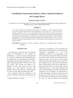
Figure 1 Purification of recombinant human topoisomerase I
(hTopI) obtained using a baculovirus expression system (lane 1,
cell lysate; lane 2, partial purified fraction; and lane 3, Ni-NTA col-
umn purified protein) (left panel). Purified hTopI was further verified
by Western blot analyses using serially diluted protein amounts (20, 10,
and 5 μg/lane), and probed with rabbit antibodies against hTopI
(right).
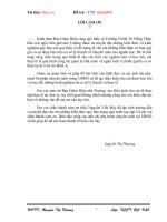
Figure 2 Inhibitory activity of camptothecin(CPT) on topoi-
somerase I (TopI). (A) CPT-induced DNA damage in A2780 ovarian
carcinoma cells. Magnification, ×200. Cells were untreated, treated
with DMSO, and CPT (25 μM) for 1 h, and were then analyzed by a neu-
tral comet assay as described in "Materials and Methods." Upper panel,
representative images. Lower panel, histogram of the tail moment
plotted against each treatment condition. p values for comparisons
(marked with *) were 0.005 as determined by two-tailed Student's t-
test. (B) CPT prevented DNA from recombinant hTop I conversion of
supercoiled DNA to relaxed closed circular DNA. pUC19 (0.2 μg) plas-
mid DNA was incubated at 37°C for 30 min with hTopI in the presence
or absence of 2~8 μM of inhibitors.
Tsai et al. Journal of Biomedical Science 2010, 17:49
/>Page 5 of 9
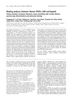
SPR assay of covalent complex formation
The SPR assay was used to measure the formation of the
DNA-TopI cleavage complex. This assay differs from the
gel assay by its high throughput, being in real time and
label-free, and directly determining the binding between
the analyte and ligand. Recombinant hTopI was cova-
lently coupled to the carboxylmethylated dextran surface
of the chip using standard amine-coupling chemistry.
The immobilization curves are shown in Figure 3A. The
highest level of immobilization was achieved at 4000 RU.
The binding of anti-hTopI antibodies to immobilized
hTopI was observed in real time after reference subtrac-
tion of the response of the hTopI-free control. The
response was proportional to the antibody concentration
(Fig. 3B, lower panel) while the signals were fairly weak in
the hTopI-free channel (upper panel). The pUC19 plas-
mid was loaded onto the hTopI-immobilized sensor chip,
and binding affinities were analyzed. The binding of the
pUC19 plasmid to immobilized hTopI was detected by
the concentration-dependent increase in RU (Fig. 3C),
which suggests that the sensor chip-immobilized hTopI
retained its DNA-binding activity. RU values of CPT
alone (0~250 nM) in the analyte flowing through the sen-
sor chip remained fairly constant (Fig. 4A, upper panel),
which indicates that CPT did not bind to hTopI without
DNA. This suggests that the binding of CPT to TopI on
the sensor chip was dependent on the DNA content
because CPT bound to hTopI at the stage of forming
intermediates of the TopI-DNA cleavage complex. To
characterize the drug-binding kinetics using the SPR sen-
sor chip, plasmid DNA (1.0 μg/mL) was included in the
analyte. The combination of pUC19 plasmid DNA and
CPT (0~250 nM) as the analyte was measured flowing
through the sensor chip, and the RU increased in a con-
centration-dependent manner (Fig. 4A, lower) with a KD
value of 4.1 × 10
-29
(Ka = 9.11 × 10
7
, Kd = 3.74 × 10
-21
)
compared to DNA only, according to the ProteOn Man-
ager 2.0 calculation. In the presence of the TopI inhibitor,
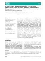
Figure 3 Surface plasmon resonance sensorgram for the immobilized recombinant human topoisomerase I (hTopI). (A) A sensorgram of hTo-
pI immobilized on the General Layer Medium sensor surface. (B) Verification of TopI immobilization using serially diluted polyclonal antibodies against
TopI. All curves of lower panel were obtained by subtracting the reference signals from the hTopI-free channel (upper panel). (C) Sensorgram of the
interaction between immobilized recombinant hTopI and pUC19 plasmid DNA. Concentrations of DNA were 0~1000 ng/mL. Data are representative
of three independent experiments.
Tsai et al. Journal of Biomedical Science 2010, 17:49
/>Page 6 of 9
CPT, re-ligation was impeded; and DNA and TopI were
trapped in a covalent cleavage complex. Similar results
were obtained with a different TopI inhibitor, EVO, with a
KD value of 5.15 × 10
-20
(Ka = 7.27 × 10
7
, Kd = 3.74 × 10
-
12
) compared to DNA only (Fig. 4B). The interaction
caused an increase in the mass of ligand immobilized on
the biosensor chip, and was reflected in a rise in RU. The
TopII inhibitor, VP-16, did not bind to the TopI-immobi-
lized chip (data not shown).
EVO binds to TopI and causes DNA damage
A 3D molecular model was created to evaluate the dock-
ing of CPT and EVO to the TopI-DNA cleavable com-
plex. From prior assays, we learned that EVO and CPT
are TopI inhibitors which exert similar mechanisms;
therefore, they would be expected to dock to the site of
the TopI-DNA complex. EVO showed weaker binding
(Fig. 5A, yellow, fitness score 67.78) than did CPT (Fig.
5A, green), consistent with the SPR assays (Fig. 4). EVO,
which bears a non-planar structure, could not completely
intercalate in spaces between DNA bases to form π-π
stacking. CPT compactly docked in spaces between DNA
bases to form π-π stacking. Results of the structure-based
molecular modeling account for the similar bindings of
CPT and EVO to the TopI-DNA complex. Figure 5B
shows that after treatment with EVO (25 μM) for 1 h,
nuclei of control cells presented a compact round area of
fluorescence, and no DNA tail was detected. In contrast,
treated cells showed DNA tailing, indicating the
increased electrophoretic mobility of the DNA frag-
ments, which shows the presence of strand breaks within
nuclear DNA. The addition of EVO to cells enhanced
DNA breaks represented by the tailing area calculation (p
< 0.005, vs. untreated cells; by Student's t-test). To further
verify the DNA-damaging effect on cells, the phosphory-
lation of histone H2AX (γ-H2AX), a biomarker for DNA
DSBs, was detected upon TopI poison treatment. An
immunoblot assay was performed to confirm the effect of
EVO on γ-H2AX levels, and the result showed that levels
Figure 4 Surface plasmon resonance sensorgram of the interaction between immobilized topoisomerase I (TopI) and TopI inhibitors. (A)
The interaction of camptothecin (CPT) (0~250 nM) with immobilized recombinant hTopI without plasmid DNA in the analytes (upper panel), and with
plasmid DNA (1000 ng/mL) in the analytes (lower). (B) The interaction of evodiamine (EVO) (0~125 nM) with immobilized recombinant hTopI without
plasmid DNA in the analytes (upper panel), and with plasmid DNA (1000 ng/mL) in the analytes (lower). Data are representative of three independent
experiments.
Tsai et al. Journal of Biomedical Science 2010, 17:49
/>Page 7 of 9
of γ-H2AX protein produced by EVO increased in a con-
centration-dependent manner after 6 h of treatment. The
relative level of γ-H2AX after treatment with 0~20 μM
EVO increased to > 3-fold versus the control (Fig. 5C). β-
Actin with constant expression was used as the internal
control.
Discussion
Small-molecule high-throughput screening of drugs
today is mainly designed for those which are dependent
upon artificial labels or reporter systems, which can
influence the effectiveness due to certain experimental
limitations. SPR is known to be a powerful tool for study-
ing biomolecular interactions in a sensitive and label-free
detection format. However, label-free methods have been
consigned to a supporting role as secondary assays due to
throughput and expense constraints. Recent improve-
ments in optical biosensor-based, automated patch clamp
and mass spectrometric technologies have enhanced
their utility for the primary screening of libraries of
small-sized compounds [26]. The major advantages of
direct-binding SPR assays compared to other biophysical
screening methods are binding kinetic information and
very low consumption of the target molecule. Yet SPR
assays need reasonably pure and active proteins, as the
detection principle is related to detection of the mass
measured as a change in the refractive index; there are
proteins which are unstable in acidic conditions which
are used in the pre-concentration step. This problem can
be minimized by mixing the target with the immobiliza-
tion buffer immediately before injection onto the sensor
chip. Antifreeze glycerol is not suitable for use in protein
preparation because it causes a severe interference in the
refractive index readout. Using DMSO as the antifreeze
in the protein preparation significantly reduced this
problem.
SPR-based biosensor technologies can directly monitor
the binding of small molecules to immobilized macro-
molecules and thus allow the study of interaction kinetics
and the evaluation of binding constants. Immobilization
Figure 5 Evodiamine (EVO) binds to topoisomerase I (TopI) and causes DNA damage. (A) Molecular modeling of camptothecin (green) and
EVO (yellow). (B) EVO-induced DNA damage in A2780 cells. Magnification, ×200. Cells were untreated, treated with DMSO, and EVO (25 μM) for 1 h,
and were then analyzed by a neutral comet assay as described in "Materials and Methods." Upper panel, representative images. Lower panel, histo-
gram of the tail moment plotted against each treatment condition. p values for comparisons (marked with *) were 0.005 as determined by two-tailed
Student's t-test. (C) γ-H2AX levels after EVO treatment in A2780 cells. Cells were treated with 0~20 μM EVO for 6 h. Cell lysates were immunoblotted
with antibody against γ-H2AX. β-Actin with constant expression was used as the internal control.
Tsai et al. Journal of Biomedical Science 2010, 17:49
/>Page 8 of 9
of DNA molecules on sensor chip for drug or protein
interactions was successfully established. Immobilization
of biotinylated linear or circular DNA on the sensor sur-
face for TopI and topII kinetic assays was performed
using an SPR analysis [27-29]. However, determining the
binding constant is complicated by multiple binding sites
of the target DNA. In addition, in some situations, each
binding site has a different intrinsic affinity for binding
independently to each binder, which causes a hindrance
to determining the affinity constant. Lin et al. provided
several modes of determining the binding constant and
stoichiometry of DNA-targeting drugs with SPR technol-
ogy [12]. No previous effort immobilizing Top proteins
on sensor chips was able to render binary protein-inhibi-
tor or ternary protein-DNA-inhibitor interaction assays.
In addition, there are no plural binding sites for immobi-
lized TopI that make it easier to determine the binding
constant. This work is the first demonstration that a
Top1-immobilized sensor chip can provide a valid assay
of DNA- and inhibitor-binding activities using SPR tech-
nology. It also enables a more-precise understanding of
the kinetics of TopI reactions.
We preliminarily reported that EVO is a TopI inhibitor
that has a variety of potential clinical applications [19]. In
the present study, we demonstrated EVO trapping on an
established TopI-immobilized sensor chip in the presence
of DNA in flow-through analytes. EVO displayed weaker
binding activity on the TopI-immobilized sensor chip
than CPT in the SPR assay, which is consistent with the
results of a DNA-relaxation assay [19]. This result
prompted further reliability verification of a new TopI
inhibitor using computer-aided molecular modeling, an
in vivo comet assay for DNA damage, and the γ-H2AX
level, a biomarker for DNA DSBs [2]. The molecular
modeling showed that EVO co-docked with the CPT in
the binding site of the TopI-DNA-cleavable complex.
EVO treatment of A2780 cells caused comet tailing sug-
gesting DNA fragmentation that is a hallmark of Top
inhibition. An early response to the induction of DNA
DSBs, which can be induced by either TopI or TopII, is
phosphorylation of the H2AX at the serine-139 residue,
in the conserved C-terminal SQEY motif, forming γ-
H2AX [30]. γ-H2AX is predominantly mediated by an
ataxia telangiectasia mutation (ATM) through continued
phosphorylation proximal to DNA breakage sites which
spreads to adjacent areas of chromatin [31]. Increasing γ-
H2AX levels in a concentration-dependent manner upon
EVO treatment in A2780 cells are consistent with the
results of the SPR and comet assays. Taken together with
our previous report [19], we concluded that EVO is able
to inhibit TopI by formation of the TopI-DNA complex
that exerts a similar mechanism as CPT. The results of
SPR for EVO were verified using a variety of methods to
ensure the reliability of the TopI-immobilized sensor
chip. This novel method will be useful for comparing the
affinities of various TopI inhibitors and selecting the most
suitable candidates for DNA-TopI trapping, as well as
facilitating in vitro screening procedures.
Conclusions
We established and validated a label-free method for
evaluating TopI inhibitors using an SPR analysis. TopI
immobilized on the chip retained its bioactivities of DNA
binding and catalysis of intermediates of the DNA-TopI
complex. This provides DNA-TopI binders for interac-
tion and primary screening. In addition, this biochip can
also ensure the reliability of binding kinetic studies of
To pI.
Competing interests
The authors declare that they have no competing interests.
Authors' contributions
HPT carried out the SPR experiments, LWL carried out the Top1 activity assay,
ZYL carried out the CPT inhibitory effects on Top1, JYW participated in the
study design of SPR, CEC carried out the Top1 expression and purification, JH
participated in the study design and coordination, CTC carried out the molecu-
lar modeling assay, and CML organized the design of the study and manuscript
preparation.
All authors read and approved the final manuscript.
Acknowledgements
This study was supported by grants from the National Science Council (NSC98-
2113-M-038-001) and Taipei Medical University Hospital (96TMU-TMUH-08).
Author Details
1
Graduate Institute of Medical Sciences, Taipei Medical University, Taipei,
Taiwan,
2
Department of Biochemistry, School of Medicine, Taipei Medical
University, Taipei, Taiwan,
3
Department of Internal Medicine, Taipei Medical
University Hospital, Taipei, Taiwan,
4
Institute of Molecular Biology, Academia
Sinica, Taipei, Taiwan and
5
School of Pharmacy, PR China Medical University,
Taichung, Taiwan
References
1. Wang JC: Cellular roles of DNA topoisomerases: a molecular
perspective. Nat Rev Mol Cell Biol 2002, 3:430-440.
2. Teicher BA: Next generation topoisomerase I inhibitors: Rationale and
biomarker strategies. Biochem Pharmacol 2008, 75:1262-1271.
3. Liu LF: DNA topoisomerase poisons as antitumor drugs. Annu Rev
Biochem 1989, 58:351-375.
4. Wethington SL, Wright JD, Herzog TJ: Key role of topoisomerase I
inhibitors in the treatment of recurrent and refractory epithelial
ovarian carcinoma. Expert Rev Anticancer Ther 2008, 8:819-831.
5. Sadaie MR, Mayner R, Doniger J: A novel approach to develop anti-HIV
drugs: adapting non-nucleoside anticancer chemotherapeutics.
Antiviral Res 2004, 61:1-18.
6. Anderson VE, Osheroff N: Type II topoisomerases as targets for
quinolone antibacterials: turning Dr. Jekyll into Mr. Hyde. Curr Pharm
Des 2001, 7:337-353.
7. Song J, Parker L, Hormozi L, Tanouye MA: DNA topoisomerase I inhibitors
ameliorate seizure-like behaviors and paralysis in a Drosophila model
of epilepsy. Neuroscience 2008, 156:722-728.
8. Verdrengh M, Tarkowski A: Impact of topoisomerase II inhibition on
cytokine and chemokine production. Inflamm Res 2003, 52:148-153.
9. Basili S, Moro S: Novel camptothecin derivatives as topoisomerase I
inhibitors. Expert Opin Ther Pat 2009, 19:555-574.
10. Pommier Y: DNA topoisomerase I inhibitors: chemistry, biology, and
interfacial inhibition. Chem Rev 2009, 109:2894-2902.
Received: 23 March 2010 Accepted: 17 June 2010
Published: 17 June 2010
This article is available from: 2010 Tsai et al; licensee BioMed Central Ltd. This is an Open Access article distributed under the terms of the Creative Commons Attribution License ( which permits unrestricted use, distribution, and reproduction in any medium, provided the original work is properly cited.Journa l of Biome dical Scie nce 2010, 17:49
Tsai et al. Journal of Biomedical Science 2010, 17:49
/>Page 9 of 9
11. Rich RL, Myszka DG: Survey of the year 2004 commercial optical
biosensor literature. J Mol Recognit 2005, 18:431-478.
12. Lin LP, Huang LS, Lin CW, Lee CK, Chen JL, Hsu SM, Lin S: Determination
of binding constant of DNA-binding drug to target DNA by surface
plasmon resonance biosensor technology. Curr Drug Targets Immune
Endocr Metabol Disord 2005, 5:61-72.
13. Lackmann M, Bucci T, Mann RJ, Kravets LA, Viney E, Smith F, Moritz RL,
Carter W, Simpson RJ, Nicola NA, Mackwell K, Nice EC, Wilks AF, Boyd AW:
Purification of a ligand for the EPH-like receptor HEK using a biosensor-
based affinity detection approach. Proc Natl Acad Sci USA 1996,
93:2523-2527.
14. Myszka DG: Survey of the 1998 optical biosensor literature. J Mol
Recognit 1999, 12:390-408.
15. Goswami A, Qiu S, Dexheimer TS, Ranganathan P, Burikhanov R, Pommier
Y, Rangnekar VM: Par-4 binds to topoisomerase 1 and attenuates its
DNA relaxation activity. Cancer Res 2008, 68:6190-6198.
16. Rao VA, Klein SR, Agama KK, Toyoda E, Adachi N, Pommier Y, Shacter EB:
The iron chelator Dp44mT causes DNA damage and selective
inhibition of topoisomerase IIalpha in breast cancer cells. Cancer Res
2009, 69:948-957.
17. Syrovets T, Buchele B, Gedig E, Slupsky JR, Simmet T: Acetyl-boswellic
acids are novel catalytic inhibitors of human topoisomerases I and
IIalpha. Mol Pharmacol 2000, 58:71-81.
18. Sikder D, Unniraman S, Bhaduri T, Nagaraja V: Functional cooperation
between topoisomerase I and single strand DNA-binding protein. J
Mol Biol 2001, 306:669-679.
19. Chan AL, Chang WS, Chen LM, Lee CM, Chen CE, Lin CM, Hwang JL:
Evodiamine stabilizes topoisomerase I-DNA cleavable complex to
inhibit topoisomerase I activity. Molecules 2009, 14:1342-1352.
20. Lin CP, Ban Y, Lyu YL, Liu LF: Proteasome-dependent processing of
topoisomerase I-DNA adducts into DNA double strand breaks at
arrested replication forks. J Biol Chem 2009, 284:28084-28092.
21. Johnson MK, Loo G: Effects of epigallocatechin gallate and quercetin on
oxidative damage to cellular DNA. Mutat Res 2000, 459:211-218.
22. Ting CY, Hsu CT, Hsu HT, Su JS, Chen TY, Tarn WY, Kuo YH, Whang-Peng J,
Liu LF, Hwang J: Isodiospyrin as a novel human DNA topoisomerase I
inhibitor. Biochem Pharmacol 2003, 66:1981-1991.
23. Bravman T, Bronner V, Lavie K, Notcovich A, Papalia GA, Myszka DG:
Exploring "one-shot" kinetics and small molecule analysis using the
ProteOn XPR36 array biosensor. Anal Biochem 2006, 358:281-288.
24. Staker BL, Hjerrild K, Feese MD, Behnke CA, Burgin AB Jr, Stewart L: The
mechanism of topoisomerase I poisoning by a camptothecin analog.
Proc Natl Acad Sci USA 2002, 99:15387-15392.
25. Jones G, Willett P, Glen RC, Leach AR, Taylor R: Development and
validation of a genetic algorithm for flexible docking. J Mol Biol 1997,
267:727-748.
26. Shiau AK, Massari ME, Ozbal CC: Back to basics: label-free technologies
for small molecule screening. Comb Chem High Throughput Screen 2008,
11:231-237.
27. Hou MH, Lu WJ, Lin HY, Yuann JM: Studies of sequence-specific DNA
binding, DNA cleavage, and topoisomerase I inhibition by the dimeric
chromomycin A3 complexed with Fe(II). Biochemistry 2008,
47:5493-5502.
28. Leontiou C, Lightowlers R, Lakey JH, Austin CA: Kinetic analysis of human
topoisomerase IIalpha and beta DNA binding by surface plasmon
resonance. FEBS Lett 2003, 554:206-210.
29. Renodon-Corniere A, Jensen LH, Nitiss JL, Jensen PB, Sehested M:
Interaction of human DNA topoisomerase II alpha with DNA:
quantification by surface plasmon resonance. Biochemistry 2002,
41:13395-13402.
30. Rogakou EP, Boon C, Redon C, Bonner WM: Megabase chromatin
domains involved in DNA double-strand breaks in vivo. J Cell Biol 1999,
146:905-916.
31. Savic V, Yin B, Maas NL, Bredemeyer AL, Carpenter AC, Helmink BA, Yang-
Iott KS, Sleckman BP, Bassing CH: Formation of dynamic gamma-H2AX
domains along broken DNA strands is distinctly regulated by ATM and
MDC1 and dependent upon H2AX densities in chromatin. Mol Cell
2009, 34:298-310.
doi: 10.1186/1423-0127-17-49
Cite this article as: Tsai et al., Immobilizing topoisomerase I on a surface
plasmon resonance biosensor chip to screen for inhibitors Journal of Biomed-
ical Science 2010, 17:49