Báo cáo y học: "Direct electrochemical analyses of human cytochromes b5 with a mutated heme pocket showed a good correlation between their midpoint and half wave potentials" doc
Bạn đang xem bản rút gọn của tài liệu. Xem và tải ngay bản đầy đủ của tài liệu tại đây (1.29 MB, 15 trang )
RESEARC H Open Access
Direct electrochemical analyses of human
cytochromes b
5
with a mutated heme pocket
showed a good correlation between their
midpoint and half wave potentials
Tomomi Aono
1†
, Yoichi Sakamoto
1†
, Masahiro Miura
1
, Fusako Takeuchi
2
, Hiroshi Hori
3
, Motonari Tsubaki
1*
Abstract
Background: Cytochrome b
5
performs central roles in various biological electron transfer reactions, where
difference in the redox potential of two reactant proteins provides the driving force. Redox potentials of
cytochromes b
5
span a very wide range of ~400 mV, in which surface charge and hydrophobicity around the
heme moie ty are proposed to have crucial roles based on previous site-directed mutagenesis analyses.
Methods: Effects of mutations at conserved hydrophobic amino acid residues consisting of the heme pocket of
cytochrome b
5
were analyzed by EPR and electrochemical methods. Cyclic voltammetry of the heme-binding
domain of human cytochrome b
5
(HLMWb
5
) and its site-directed mutants was conducted using a gold electrode
pre-treated with b-mercarptopropionic acid by inclusion of positively-charged poly-L-lysine. On the other hand,
static midpoint potenti als were measured under a similar condition.
Results: Titration of HLMWb
5
with poly-L-lysine indicated that half-wave potential up-shifted to -19.5 mV when the
concentration reached to form a complex. On the other hand, midpoint potentials of -3.2 and +16.5 mV were
obtained for HLMWb
5
in the absence and presence of poly-L-lysine, respectively, by a spectroscopic
electrochemical titration, suggesting that positive charges introduced by binding of poly-L-lysine around an
exposed heme propionate resulted in a positive shift of the potential. Analyses on the five site-specific mutan ts
showed a good correlation between the half-wave and the midpoint potentials, in which the former were 16~32
mV more negative than the latter, suggesting that both binding of poly-L-lysine and hydrophobicity around the
heme moie ty regulate the overall redox potentials.
Conclusions: Present study showed that simultaneous measurements of the midpoint and the half-wave potentials
could be a good evaluating methodology for the analyses of static and dynamic redox properties of various
hemoproteins including cytochrome b
5
. The potentials might be modulated by a gross conformational change in the
tertiary structure, by a slight change in the local structure, or by a change in the hydrophobicity around the heme
moiety as found for the interaction with poly-L-lysine. Therefore, the system consisting of cytochrome b
5
and its partner
proteins or peptides might be a good paradigm for studying the biological electron transfer reactions.
Background
Cytochromes b can be defined as electron transfer pro-
teins having heme b group(s), noncovalently bound to
the protein. b-Type cytochromes possess a wide range
of properties and functions in a large number of differ-
ent redox processes. Among them, cytochromes b
5
are
ubiquitously found in animals, plants, fungi and some
bacteria. The mi crosomal and mitochondrial (outer
membrane; OM) variants are known and are present in
a membrane-bound form. On the other hand, bacterial
and those from erythrocytes and some a nimal tissues
are water-soluble (such as for the reduction of
* Correspondence:
† Contributed equally
1
Department of Chemistry, Graduate School of Science, Kobe University, 1-1
Rokkodai-cho, Nada-ku, Kobe, Hyogo 657-8501, Japan
Full list of author information is available at the end of the article
Aono et al. Journal of Biomedical Science 2010, 17:90
/>© 2010 Aono et al; licensee BioMed Central Ltd. This is an Open Access article distributed under the terms of the Creative Commons
Attribution License ( s/by/2.0), which permits unrestrict ed use, distribu tion, and reproduction in
any medium, provided the original work is properly cited.
methemoglobin in erythrocytes and for the biosynthesis
of N-glycolylneuraminic acid [1]). A membrane-bound
(microsomal) form of cytochrome b
5
is required for
numerous biosynthetic and biotransformati on reactions,
which include cytochrome P450-dependent reactions
[2], desaturation of fatty acids [3], plasmalogen biosynth-
esis [4], and cholesterol bios ynthesis [5,6]. The role of
cytochrome b
5
in microsomal P-450-dependent mono-
oxygenase reactions ha s been studied most exten sively
[2]. In addition, a number of fusion enzymes exist in
nature containing cytochrome b
5
as a domain compo-
nent. These include mitochondrial flavocytochrome b
2
(L-lactate dehydrogenase) [7], sulfite oxidase [8], the Δ
5
-
and Δ
6
-fatty acid desaturases [9], and yeas t inositolpho-
sphorylceramide oxidase [10]. Plant and fungal nitrate
reductases are also cytochrome b
5
-containing fusion
enzymes [11].
For human cytochrome b
5
, only a few naturally occur-
ring mutations recognized as a genetic disorder have
been reported. One such example was found by Kurian
et al. [12]. They report ed that naturally occurring
human cytochrome b
5
T60A mutant [12] displayed an
impaired hydroxylamine reduction capacity. They
observed further that the expressed protein in rabbit
reticulocyte lysate system showed an enhanced suscept-
ibility to the proteol ytic degradation. Expression level in
transfected HeLa cells was also significantly lowered.
Another genetically confirmed example was previously
reported. In this case, Steggles et al. identified a homo-
zygous splice site mutation in the CYB5A gene, resulting
in premature truncation of the protein, leading to a very
high methemoglobin concentration in red blood cells of
the patient, being consistent with methemoglobinemia
type IV [13]. The patient exhibited female genitalia at
birth , but, was determined as a male pseudohermaphro-
dite, pro bably due to the low levels of androgen synth-
esis by the lack of cytochrome b
5
activity, which has
been shown to participate in 17a-hydroxylation in adre-
nal steroidogenesis [14].
Whereas more than 300 patients had been reported
with hereditary methemoglobinemia types I or II, only a
few cases of type IV had been reported. Thus, one may
attribute that the rarity of naturally occurring cyto-
chrome b
5
mutation may be due to lethality of most
type IV mutations. However, in a very recent study by
employing transgenic mice, Finn et al. found that cyto-
chrome b
5
completely null mice were viable, fertile and
produced grossly normal pups at expected Mendelian
ratios [15]. Further, the cytochrome b
5
null mice exhib-
ited a number of intriguing phenotypes, including
altered drug metabolism, methemoglobinemia, disrupted
steroid hormone biosynthesis. In addition, the cyto-
chrome b
5
null mice displayed skin defects and retarda-
tion of neonatal development. These observations
sugge sted that cytochrome b
5
might play a role control-
ling s aturated/unsaturated homeostasis of fatty acids in
higher animals including human.
The membrane-bound form of cytochrome b
5
is asso-
ciated with the endoplasmi c reticulum. It has a molecu-
lar weight of 16,700 Da and contains about 134 amino
acids in animals (Fig ure 1A). It is composed of three
domains: a hydrophilic heme-containing catalytic
domain of about 99 amino acids; a membrane-binding
hydrophobic domain containing about 30 amino acids at
the car boxy terminus of the molecule; and a membrane-
targeting region represented by the 10-amino-acid
sequence located at the carboxy-terminus of the mem-
brane-binding domain. Three-dimensional structures of
a number of cytochrome b
5
are known [16], but only
for the heme-containing hydrophilic catalytic domain
[17]. Two His residues (His44 and His68) provide the
fifth and sixth heme ligands (Figure 1A, B), and two
propionate groups of the heme b lies at the opening of
the heme-binding pocket, which is formed by highly
conserved hydrophobic amino acid residues (Figure 1A).
The roles of each amino acid were investigated by
detailed site-directed mutagenesis in the past with
employing various struct ural, spectroscopic and electro-
chemical techniques, including X-ray crystallography
[18-20], NMR [21-23], UV-visible absorption spectro-
scopy, and redox potential measurements [24].
Redox potentials of various forms of cytochrome b
5
span a range of ~400 mV. It is well documented that
several factors could regulate and induce changes in the
reduction potential of cytochrome b
5
spanning almost
ent ire rang e observed. The electrostatic contribution by
surface charges might play imp orta nt roles in adjusting
the selecti vity of the prote in-protein interaction. On the
other hand, difference in the redox potential of two
reactant proteins provides the driving force for the elec-
tron transfer reactions. Thus, the clarification of the reg-
ulatory mechanism of the redox potentials might be
essential for the understanding of the biological electron
transfer reactions.
Biological redox potential measurements were usually
conducted either by an equilibrating electrochemical
method or by employing a dynamic cyclic voltammetry.
Common features to all the past voltammetric experi-
ments involving cytochrome b
5
and electrodes pre-
treated with various thiol-contai ning aliphatic acid or
related groups are the large difference between the half-
wave potential (E
1/2
) and the midpoint potential deter-
mined by the equilibrating method [25]. In the case of
rat OM cytochrome b
5
, its midpoint potential deter-
mined by the equilibratin g method s howed as low as
-102 mV; whereas the half-wave potential was found as
+8 mV [25]. Similar large positive shifts were reported
for b ovine liver microsomal cytochrome b
5
(~+31 mV)
Aono et al. Journal of Biomedical Science 2010, 17:90
/>Page 2 of 15
(B)
(C)
(
A
)
MA AQ SD KD V KY Y T L E E I K K H NH SK ST WL I LH H K V Y D L T K F L E EH PGG E E V L R EQ AGGD A T EN F E D V GH S
MA EQ SD KA V K Y YT L E E I K K H N H SK ST WL I LH H K V Y D L T K F L ED H PGG E E V L R EQ AGGD A T E N F ED I GH S
MA EQ SD KD V K Y YT L E E I QK H KD SK ST WV I L H H KV YD L T K F L E EH PGG E E V L R EQ AGGD A T EN F ED V GH S
MA GQ SD KD V KY Y T L E E I QK H KD SK ST WV I LH H K V Y D LT K F L E EH PGG E E V L R EQ AGGD A T EN F ED V GH S
MA EQ SD EA V K Y YT L E E I QK H N H SK ST WL I LH H K V Y D L T K F L E EH PGG E E V L R EQ AGGD A T EN F E D V GH S
MA E E SS K AV KY Y T L E E I Q K H N N S K S T WL I LH Y KV YD L T K F L E EH PGG E E V L R EQ AGGD A T EN F E D V GH S
MA T A EA SG S D GK GQ E V ET S V T Y Y R L E E V A K RN S L K E LWL V I HG RV YD V T R F L N EH PGG E E V L L EQ AG V D A S E S F E D V GH S
MA T P EA SG SG RN GQ G SD PA V T Y Y R L E E V A K RN T A E E T WMV I H GR V Y D I T R F L SE H PGG E E V L L EQ AGA D A T E SF ED V GH S
MA D L KQ I T L K E I A E H N T NK SAWL V I GN K V FD V T K F L D E H P GG C E V L L EQ AG SD G T EA F ED VGH S
M P K V Y SY Q E V A E H NG P EN FW I I I DD KV YD V SQ F KD EH PGGD E I I MD L GGQ D A T E SF VD I GH S
M S V H KY T R A E V A A RD N N KQ N L I I I DN V V YD V A A F L ED H PGG T E V L V DN A G SD A SE C FH EV GH S
T D A R E L S K T F I I G E L H PD D- - - R SK L S K PM E T L I T T V D SN S SWWT-NWV I P A I SA L I V A LM Y R L Y M AD D
T D A R E L S K T F I I G E L H PD D- - - R SK I A K P V E T L I T T VD SN S SWWT-NWV I PA I SA V V VA LM Y R I Y T A ED
T D A R E L S K T Y I I G E L H PD D- - - R S K I AK P S E T L I T T V E SN S SWWT-NWV I P A I SA L V V A LMY RL Y MA ED
T D A R E L S K T Y I I G E L H PD D- - - R S K I AK P S D T L I T T V E SN S SWWT-NWV I P A I SA LA V A LM Y R L Y MA ED
T D A R EM S K T F I I G E L H PD D- - - R PK LN K P P E T L I T T I D SS S SWWT-NWV I P A I SA V A V A LM Y R L Y MA ED
T D A R E L S K T F I I G E L H PD D- - - R SK I T K P S E S I I T T I D SN P SWWT-NWL I P A I SA L F VA L I Y H LY T S EN
SD A R EM LKQ Y Y I GD I H P SD L K P E SG S KD P S QN D T C K S CWA-YW I L P I I G A V L LG F L Y R Y Y T SE SK S S
PD A R EM LKQ Y Y I GD V H PN D L K P KD GD KD P S K N N S C Q S SWA-YW I V P I VGA I L I G F LY RH F WA D S K S S
T D A RHM KD EY L I G E V V A SE R K T Y S YD K KQW K S- - T T EQ D N KQ RGG E SMQ T D N I V Y FA L L A V I V A L V Y Y L I A A
D EA L R L L KG L Y I GD V- - D K T S E R V SV EK V ST S E NQ SK G SG T L VV I L A I LM L GV AY Y L L N E
E I A I EWRN T F K V G E I-V D E E K L EV KC KQ P S A A E S A EP L T L GG L L A V Y G P P V AM A V L A Y L L YT F L FG
rabbit b5
horse b5
rat b5
mouse b5
human b5
bovine b5
human OMb5
rat OMb5
C.elegans b5
yeast b5
silkworm b5
rabbit b5
horse b5
rat b5
mouse b5
human b5
bovine b5
human OMb5
rat OMb5
C.elegans b5
yeast b5
silkworm b5
*
*
++
+
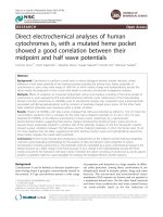
Figure 1 Alignment of amino acid sequences of cytochrome b
5
from various species (A), a close-up view of tertiary structure of
human cytochrome b
5
around the heme-pocket with three conserved hydrophobic residues (Leu51, Ala59, and Gly67) and two heme
axial ligands (His44 and His68) indicated (B), a close-up view around the heme pocket with acidic amino acid residues (C).(A) Amino
acid sequences of cytochromes b
5
from various species are aligned. Two heme axial ligands (His44 and His68) are indicated by an asterisk (*). On
the other hand, corresponding positions to three target residues (Leu51, Ala59, and Gly67) in the present study are indicated by a cross (+).
Amino acid sequences were obtained from [GenBank; NP_001164735 for rabbit b
5
, P00170 for horse b
5
, AAB67610 for rat b
5
, P56395 for mouse
b
5
, AAA35729 for human b
5
, NP_776458 for bovine b
5
, BAA23735 for human OMb
5
, AAH72535 for rat OMb
5
; CAB01732 for C.elegans b
5
, P40312
for yeast b
5
, NP_001106739 for silkworm b
5
]. (B) Human cytochrome b
5
NMR solution structure [PDB code: 2I96 model 1] is shown in a ribbon
model with a bound heme b prosthetic group. In addition, three conserved residues (Leu51, Ala59, and Gly67) and two heme axial ligands
(His44 and His68) are indicated. (C) Acidic amino acid residues located on the surface of the heme-binding domain (corresponding to LMWb
5
)
are indicated.
Aono et al. Journal of Biomedical Science 2010, 17:90
/>Page 3 of 15
[26] and chicken liver microsomal cytochrome b
5
(~+40
mV) [27].
The large positive shift (+110 mV) observed for rat
OM cytochrome b
5
were attributed to the binding of
multivalent cations, such as, poly-L-lysine, which were
used for shielding the negatively charged protein surf ace
and negat ively-charged electrode surface to facilitate the
electron transfer [25]. The difference in the potentials
was ascribed, initially, for the binding of multivalent
cations to the specific charged residues on the surface of
cytochrome b
5
, such as Glu and Asp (Figure 1C) [25],
leading to the modulation of the heme redox potential
differently from that measured by the equilibrating
method. Later, however, a carboxylate of an exposed
heme propionate group and conserved acidic residues
(Glu44, Glu48, Glu56, and Asp60) (Figure 1C) (corre-
sponding to Glu49, Glu53, Glu61, and Asp65, respec-
tively, of human cytochrome b
5
)wereproposedtobe
responsible for the specific binding of mult ivalent
cations [28]. The formation of such a complex will
result in a neutralization of the charge on the heme pro-
pionate and lowering of the dielectric of the exposed
heme microenvironment by excluding water from the
complex interface. These two factors act synergistically
to dest abilize the positive charge of the ferric heme with
respect to the neutral ferrous heme, leading to a positive
shift of the redox potential upon binding of poly-L-
lysine [28,29]. This postulation was partly verified by the
esterification of the heme propionate groups, leading to
the half-wave potential to be independent of the con-
centration of multivalent cations [28,29].
In the present study, we focused on three conserved
hydrophobic amino acid residues (Leu51, Ala59, and
Gly67) consisting of the heme-binding pocket (Figure
1A, B). These residues were not investigated previously
despite of their higher conservation among the various
members o f cytochrome b
5
protein family (Figure 1A).
Gly67 is located besides the heme axial His residue
(His68) and is near the entrance of the heme-pocket
crevice (Figure 1B). Leu51 and Ala59, on the other
hand, are located in the bottom of the heme pocket
(Figure1B).TheformerisonthesideofHis44residue,
the other heme axial ligand. The latter is on the side of
His68 residue. These two residues might be essential for
the stabilization of the heme prosthetic group in the
hydrophobic heme pocket. Therefore, we selected repla-
cing amino acid residues n ot too hazardous for the
maintenance of the heme cavity. Accordingly, we chose
Thr, Ile, Ala, Ser residues fo r the replacement of Leu51,
Ala59, and Gly67 residues. We produced and purified
site-directed mutants for these three sites, having parti-
cular interests in the changes of local structure and
hydrophobicity of the heme pocket, which may affect
the redox properties of cytochrome b
5
.Wemeasured
spectroscopic and electrochemica l properties (i.e., redox
potentials were analyzed by an equilibrating method and
a cyclic voltammetry technique) of these mutants to
clarify the structural and electrochemical importance of
the conserved residues.
Methods
Construction of the expression plasmid for wild-type and
site-directed mutants of HLMWb
5
The gene coding for a soluble domain (amin o acid resi-
dues from Met1 to Leu99; LMWb
5
) of human cyto-
chrome b
5
in pIN3/b
5
/2E1/OR plasmid [30,31] was
subcloned into pCW
ori
vector as previously described
[32]. Then, the BamH I-Hind III fragment of the pC/
LMWb
5
plasmid encoding e ntire LMWb
5
(amino acid
residues from Met1 to Leu99) was inserted into the
BamH I- Hind III site of pBluescript II KS(+) to form a
plasmid p BS/LMWb
5
for easier handling upon the site-
directed mutagenesis. The nucleotide sequence of the
pBS/LMWb
5
plasmid was confirmed with a DNA
sequencer (PRISM 3100 Genetic Analyzer, ABI).
The site-directed mutagenesis was conducted using
QuikChange Site-Directed Mutagenesis Kit (Stratagene,
La Jolla, CA, USA) according to the manufacturer’ s
manual. Following mutagenic primers were used (substi-
tuted codons are underlined): for L51I, L51I-R (5’ -
CCAGCTTGTTCCCT
GATAACTTCTTCCCCACC-3’)
and L51I-F (5’-GGTGGGGAAGAAGTT
ATCAGGGAA-
CAAGCTGG-3’ ); for L51T, L51T-R (5’ -CCAGCTT
GTTCCCT
TGTAA CTTCT TCCCCACC-3’)andL51T-F
(5’ -GGTGGGGAAGAAGTT
ACAAGGGAACAAGCT
GG-3’); for A59V, A59V-R (5’-CCTCAAAGTTCTCAG-
T
AACGTCACCTCCA GCTTG-3’) and A59V-F (5’-CAA
GCTGGAGGTGAC
GTTACTGAGAACTTTGAGG-3’);
for A59 S, A59S-R (5’ -CAAGCTGGAGGTGAC
TC-
TACTGAGAACTTTGAGG-3’ )andA59S-F(5’ -CA
AGCTGGAGGTGAC
TCTACTGAGAACTTTGAGG-
3’); for G67A, G67A-R(5’-GGCATCTGTAGAGTG
CGC-
GACATCCTCAAAGTTC-3’)andG67A-F(5’ -GAAC
TTTGAGGATGTC
GCGCACTCTACAGATGCC-3’ );
and for G67 S, G67S-R (5’ -GGCATCTGTAGAGTG
C-
GAGACATCCTCAAAGTTC-3’)andG67S-F(5’ -GAA
CTTTGAGGATGTC
TCGCACTCTACAGATGCC-3’ ).
After the si te-directed mutagenesis, transformation, and
plasmid preparation, each mutated plasmid (pBS/L51I,
pBS/L51T, pBS/A59V, pBS/A59 S, pBS/G67A, pBS/
G67S) was treated with Nde IandHind III. The each
Nde I-Hind III fragment of pBS/LMWb
5
plasmid and
the m utated plasmids was i nserted into the Nde I-Hind
III site of pET-28b(+) vector (Novagen, Merck, Darm-
stadt, Germany) to construct pET/HLMWb
5
, pET/L51I,
pET/L51T, pET/A59V, pE T/A59 S, pET/G67A, and
pET/G67 S, respectively, to achieve an efficient expres-
sion and an easier purification of a recombinant protein.
Aono et al. Journal of Biomedical Science 2010, 17:90
/>Page 4 of 15
The pET-28b(+) vector contains a 6x-His-tag moiety at
the upstream of the Nde I-Hind III site and, therefore,
gives an additional extension with a sequence of
MGSSHHHHHHSSGLVPRGSH at the NH
2
-terminus of
the LMWb
5
protein (designated as HLMWb
5
, hereafter).
Mutations were confirmed with an ABI PRISM 3100
Genetic Analyzer (Ap plied Biosystems Japan Ltd. ) for
both types of plasmids prepared from pBS and pET vec-
tors. Escherichia coli strain BL21(DE3 )pLysS was trans-
formed with pET/HLMWb
5
(or with one of the mutated
pET plasmids) and was cultivated in low-salt Luria-Ber-
tani (LB) medium containing 30 μg/ml of kanamycin
and 34 μg/ml chloramphenicol at 37°C for pre-culture.
After the pre-culture, HLMWb
5
protein (or each
mutant protein) was produced by growing the trans-
formed cells at 37°C in TB medium (12.0 g/L of tryp-
tone, 24.0 g/L yeast extract, 4 ml/L glycerol, 23.1 g/L
KH
2
PO
4
, and 125.4 g/L K
2
HPO
4
) in the presence of
30 μg/ml of kanamycin and 34 μg/ml of chlorampheni-
col. Induction of the protein expression was achieved by
addition of 200 μM (final) IPTG when the cells had
growntoanO.D.of0.6at600nm.Then,theincuba-
tion temperature was lowered to 26°C. Cells were har-
vested 48 h after the addition of IPTG and were frozen
in liquid nitrogen and stored at -80 °C until use. The
thawed cells were mixed with a lysis buffer (20 mM
Tris-HCl buffer (pH 8.0) containing 0.5 mM EDTA)
and disrupted by the treatment with lysozyme (final,
1mg/mL)andDNase(final,50μg/mL) i n the presence
of 1 mM of phenylmethylsulfonyl fluoride followed by
sonication on ice with a model 250 sonifier (Branson
Ultrasonic). The disrupted cells were centrifuged at
26,000 g for 20 min at 4 °C. The supernatant was saved
as a crude extract.
Purification of HLMWb
5
was conducted as follows.
The crude extract was loaded onto a column of DEAE-
Sepharose CL-6B previously equilibrated with 20 mM
Tris-HCl (pH 8.0) buffer containing 0.5 mM EDTA.
The HLMWb
5
was adsorbed in the column as a redd ish
band. The column was washed with the same buffer
containing 50 mM NaSCN. The adsorbed LMWb
5
was
eluted by a linear gradient of NaSCN concentration
from 50 to 300 mM in the same buffer. Main fractions
were collected based on the SDS-PAGE analysis (12%
gel) and absorbance at 414 nm and were concentrated
to about 5 mL using an Amicon concentrator and a
Millipore membrane (MWCO = 10,000). The concen-
trated HLMWb
5
was, then, subjected onto an affinity
column chromatography with Ni-NTA agarose gel
(QIAGEN) previously equilibrated with 50 mM sodium
phosphate buffer (pH 8.0) containing 10 mM imidazole
and 300 mM NaCl. T he column was washed with
50 mM sodium-phosphate buffer (pH 8.0) containing
20 mM imidazole and 300 mM NaCl. F inally, adsorbed
HLMWb
5
protein was eluted with 50 mM sodium-phos-
phate buffer (pH 8.0) containing 250 mM imidazole and
300 mM NaCl and the eluate was collected. Fractions
that showed a single protein band on SDS-PAGE were
pooled and concentrated, gel-filtrated against 50 mM
sodium phosphate buffer (pH 7.0) with PD-10 mini-
column (Amersham Bioscience). The full-le ngth form of
human cytochrome b
5
was purified according to t he
procedure as described previously [33]. Concentrations
of purified recombinant proteins were de termi ned spec-
trophotometrically from the absorbance at 423 nm in
the dithionite-reduced form using the extinction coeffi-
cient o f 163 mM
-1
cm
-1
[34]. The protein concentration
was determined with a modified Lowry method as pre-
viously described [35], in which bovine serum albumin
was used as a standard.
EPR spectroscopy
Oxidized HLMWb
5
samples (or mutants in the oxidized
form) in 50 mM potassium-phosphate buffer (pH 7.0)
were concentrated to about 200 ~500 μM with a 50-mL
Amicon concentrator fitted with a membrane filter
(Millipore PTTK04110; pore size MWCO = 10,000). For
HLMWb
5
and G67A mutant, concentrated poly-L-lysine
solution (5 mM; Sigma-Aldrich Japan K .K.; mol. wt. =
1,000~4,000; corresponding to 8~30 lysine residues) was
added to make its final concentration as 400 μM. The
samples were introduced into EPR tubes and frozen in
liquid nitrogen (77 K). EPR measurements were carried
out at X-band (9.23 GHz) microwave frequency using a
Varian E-109 EPR spectrometer with 100-kHz field
modulation. An Oxford flow cryostat (ESR-900) was
used for the measurements at 15K. The microwave fre-
quency was calibrated with a microwave frequency
counter (Takeda Riken Co., Ltd., Model TR5212). The
strength of the magnetic field was determined with an
NMR field meter (ECHO Electronics Co., Ltd., Model
EFM 2000AX). The accuracy of the g-values was
approximately +0.01.
Cyclic voltammetry
All electrochemical measurements were done as pre-
viously described [25,32] using a water-jacketed conical
cell that allowed measurements to be made at controlled
temperatures using volumes as small as 150 μL. An ALS
electrochemical analyzer (model 611A) was used for all
measurements. All sample solutions (100 μM, heme
basis, in 50 mM sodium phosphate buffer pH 7.0) were
purged with Ar gas befo re use and bl anketed with Ar
during th e electrochemical determinations. For the mea-
surements of the full-length form (1-134 aa) of human
cytochrome b
5
, 50 mM sodium-phosphate buffer (pH
7.0) containing 0.5% (v/v) Triton X-100 was used as the
buffer. The Au electrode was derivatized with 100 mM
Aono et al. Journal of Biomedical Science 2010, 17:90
/>Page 5 of 15
of 3-mercaptopropionate, as previously described
[25,32]. Poly-L-lysine was added to a final concentration
of 50~300 μM just before the measurements. Concen-
tration of poly-L-lysine solution was calculated assuming
the formal mol. wt. = 4,000. Therefore, actual conce n-
tration of poly-L-lysine in the sample solution might be
higher than the indicated values. The average of the
cathodic and anodic peak potentials was taken as the
formal potential. All potentials we re measured at 25°C
versus an Ag/AgCl electrode with an internal filling
solution of 3 M KCl saturated with AgCl and are then
converted versus the standard hydrogen potential (SHE).
Spectroscopic redox titrations
Spectroscopic redox titra tions were performed essentially
as described by Du tton [36] and Takeuchi [37], u sing a
Shimadzu UV-2400PC spectrometer equipped with a ther-
mostatted cell holder connected to a low temperature
thermobath (NCB-1200, Tokyo Rikakikai Co, Ltd, Tokyo,
Japan). A custom anaerobic cuvette (1-cm light path, 5-ml
sample volume) equipped with a combined platinum and
Ag/AgCl electrode (6860-10C, Horiba, Tokyo, Japan) and
a screw-capped side arm was used. Purified HLMWb
5
sample or its site-specific mutants (final, 15 μM) either in
the presence or absen ce of poly-L-lysine (200 μM) in
50 mM sodium-phosphate buffer (pH 7.0) was mixed with
redox medi ators (anthraquinone-2,6-disulfon ate, 20 μM;
1,2- naphthoquino ne, 20 μM; phenazine methosu lfate, 20
μM; duroquinone, 20 μM; 2-hydroxy-1,4-naphtoquinon e,
20 μM; riboflavin, 20 μM). For the redox measurements of
the full-length form of human cytochrome b
5
,50mM
sodium-phosphate buffer (pH 7.0) containing 0.5% (v/v)
Triton X-100 was used as the buffer. The sample was kept
under a flow of moistened Ar gas to exclude dioxygen and
was continuously stirred with a small magnetic stirrer
(CC-301, SCINICS, Tokyo, Japan) insid e. Reductive titra-
tion was performed at 25°C by addition of small aliquots
of sodium dithionite (4 or 16 mM) solution through a nee-
dle in the rubber septum on the side arm; for a subsequent
oxidative titration, potassium ferricyanide (4 or 16 mM)
was used as the titrant. In an appropriate interval, visible
absorption spectra and redox potentials were recorded.
The changes in absorbance (A555.0 minus A565.6; the
peak in reduced form minus isosbestic point of HLMWb
5
)
were corrected considering the dilution effect and ana-
lyzed with Igor Pro (v. 6.03A2) employing a Nernst equa-
tion with a single redox component.
Results
Purification of soluble domain of human cytochrome b
5
(HLMWb
5
) and its mutants
Purification of H LMWb
5
and its site-specific mutants
was successful except for L51T mutant. Failure of purifi-
cation for the L51T mutant was d ue to the inability to
obtain a heme-bound holo-form. We confirmed that
enough amounts o f the protein corresponding to
HLMWb
5
was produced in E. coli cells upon addition of
IPTG based o n the SDS- PAGE analysis and CBB-250
staining. Addition of excess amounts of heme solution
during the disruption of the E. coli cells to reconstitute
the holo-form was unsuccessful, suggesting that the
heme-pocket of the L51T mutant was perturbed signifi-
cantly and not suitable for the accommodation of the
heme prosthetic group, leading to the denatured form.
Thus, we did not pursue the L51T mutant further in
the present study.
Properties of soluble domain of human cytochrome
b
5
(HLMWb
5
) and its mutants
The purified HLMWb
5
showed characteristic visible
absorption spectra as a native form of cytochrome b
5
by
showing absorption peaks at 413 nm for oxidized form
and at 555, 526, and 423 nm for reduced form (spectra
not shown). Purified HLMWb
5
showed a single protein-
staining band (CBB-250 staining) upon SDS-PAGE (12%
gel) analysis with an apparent molecular size of 16.5
kDa. This va lue was, however, much la rger than the
expected value (13548.91 Da) for the NH
2
-terminal
extension (20 amino acid residues, containing the 6x-
His-tag moiety) plus the soluble domain (1-99 aa) of
human cytochrome b
5
. To clarify the biochemical nat-
ure of the HLMWb
5
, we conducted MALDI-TOF-MS
analyses. Untreated HLMWb
5
sample showed a single
peak at 13418 m/z corresponding to a mono-protonated
form. A doubly-protonat ed form showed a weak peak at
6709 m/z. This result suggested that a post-translational
modification (i.e., removal of the initial Met residue)
had occurred in HLMWb
5
.MALDI-TOF-MSanalyses
on the tryptic peptides of HLMWb
5
(data not shown)
proved that the Met residue at the initiation site was
missing. We concluded that the purified HLMWb
5
pro-
tein is a form with the sequence corresponding to 2-
119 aa of HLMWb
5
(theoretical molecular weight;
13471.72 Da).
All the purified mutants showed very similar UV-visi-
ble absorption spectra with those of HLMWb
5
, indicat-
ing that those site-specific mutations around the heme-
binding pocket (except for the L51T mutant) did not
affect signif icantly on the coordination or the electronic
structure of the heme moiety.
EPR spectroscopy of HLMWb
5
and its mutants
The EPR spectrum of oxidized HLMWb
5
measured at
15K showed g
z
=3.03,g
y
=2.22,andg
x
=1.43(Figure
2A; trace a ), very close to those reported for rat [38], rat
outer mitochondrial membrane (OM) [39] and pig [40]
cytochromes b
5
and human LMWb
5
[32] in which the
6xHis-tag sequence (20 aa) at the NH
2
-terminal region
Aono et al. Journal of Biomedical Science 2010, 17:90
/>Page 6 of 15
is not present, or human erythrocyte cytoc hrome b
5
[41]. However, it was slightly different from the report
for the recombinant human erythrocyte cytochrome b
5
(g
z
= 3.06, g
y
= 2.22, and g
x
=1.42)[42].Itmustbe
noted that there was no high-spin signals around g~6
nor the signals from adventitiously bound non-heme
iron at g = 4.3 in the spectra (spectra not shown) [38].
All the purified mutants showed very similar EPR
spectra to that of HLMWb
5
as shown in Figure 2A. Clo-
ser examinations indicated that G67A mutant showed a
slight perturbation on its heme coordination by showing
g
z
= 3.06 and g
y
= 2.20, close to the values for house fly
cytochrome b
5
[43]. These results confirmed that the
site-specific mutations introduced around the heme-
binding pocket to modulate the hydrophobicity did not
affect signif icantly on the coordination or the electronic
structure of the heme prosthetic group.
For HLMWb
5
and the G67A mutant, effec ts of the
addition o f poly-L-lysine (final concentration, 400 μM)
on the EPR spectrum were examined. However, there
was no apparent shift of their respective g-values (spec-
tra not shown).
Cyclic voltammetry of LMWb
5
and its mutants
The Au e lectrode pre-treated with 3-merca ptopropio-
nic acid gave reversible voltammetric responses for t he
HLMWb
5
solution but only in the presence of poly-L-
lysine. Without poly-L-lysine, there was no peak cur-
rent.Atleast50μM of poly-L-lysine was required to
observe a stable peak current (data not shown). In Fig-
ure 3A, a typical voltammogram for HLMW b
5
in the
presence of 200 μM of poly-L-lysine is shown. A plot
of the square root of the scan rate vs.peakcurrent
(I
pa
)(orI
pc
, result not shown) was linear for scan rates
up to and greater than 200 mV/sec (Figure 3B), indi-
cating a diffusion-controlled reaction. The half-wave
potential (corresponding to the midpoint potential)
was estimated as -19.5 mV (vs.SHE),whichwasclose
to the values for the full-length human cytochrome b
5
(a)HLMWb
5
Ma
g
netic Field (T)
(b)A59V
(c)A59S
(d)G67A
(e)G67S
(f)L51I
0.2 0.3
0.4 0.5
15
K
g
y
=
2
.
22
g
y
=2.20
g
z
=3.03
g
z
=3.04
g
z
=3.06
Figure 2 X-band EPR spectrum of oxidized HLMWb
5
measured
at 15K and effects of the mutations on the spectrum. Following
samples in oxidized form in 50 mM sodium phosphate buffer pH
7.0 were frozen at 77K and their respective EPR spectrum was
measured at 15K. HLMWb
5
(trace a, 0.50 mM); A59V (trace b, 0.12
mM); A59 S (trace c, 0.19 mM); G67A (trace d, 0.20 mM); G67 S
(trace e, 0.24 mM), and L51I (trace f, 0.27 mM). Ordinate of each
spectrum was normalized appropriately based on the concentration
for an easier visualization. Other conditions are described in the text.
The signal around g = 2 in G67A mutant (d) was due to a
contaminant from EPR tube.
600x10
-9
400
200
0
-200
Current (A)
-0.
5
-0.4-0.3-0.2-0.10.0
E (V)
HLMWb5 (100 μM)
poly-L-lysine = 200 μM
300x10
-9
250
200
150
100
50
0
Peak current I
pa
(A)
1614121086420
[
Scan rate
]
1/2
(A)
(
B)
Figure 3 Cycl ic voltammogram of HLMWb
5
in 50 mM sodium
phosphate buffer pH 7.0. (Panel A) The gold electrode was
modified with b-mercaptopropionic acid and the voltammogram of
HLMWb
5
(100 μM (final) in 50 mM sodium phosphate buffer pH 7.0)
was obtained in the presence of 200 μM of poly-L-lysine. The
potential shown is vs. an Ag/AgCl reference electrode with an
internal filling solution of 3 M KCl saturated with AgCl (E° = +197
mV vs. SHE). Scan rate = 100 mV/sec. (Panel B) Plot of the anodic
peak current I
pa
against the square root of the scan rate ν
1/2
.
Aono et al. Journal of Biomedical Science 2010, 17:90
/>Page 7 of 15
(-20.5 mV) a nd LMWb
5
without the 6xHis-tag moiety
(-21 mV) [32] and for bovine liver cytochrome b
5
(-6 mV, -14 mV) [44] measured under similar experi-
mental condi tions (Table 1). The se results indicated
that presence of 6xHis-tag moiety or COOH-terminal
hydrophobic transmembrane segment does not affect
significantly on the redox properties of t he hydrophilic
heme-binding domain of HLMWb
5
. However, it must
be noted that, in the case of full-length human
cytochrome b
5
(-20.5 mV), we observed relatively large
peak separation values and, more significantly, the p lot
of the square root of the scan rate vs.peakcurrent
was not clearly linear. This might be due to the pre-
sence of detergent Triton X-100 (0.5~1.0%), which
may interfere the smooth diffusion of cytochrome b
5
molecules at t he electrode surface by forming micelles
with the COOH-terminal hydrophobic segments
incorporated.
Table 1 Half-wave potentials of HLMWb
5
and its site-specific mutants in comparison with various animal cytochrome
b
5
and their site-specific mutants.
Samples half-wave potential (mV) (vs. SHE) Electrode references
HLMWb
5
-19.5 Au* present study
LMWb
5
-21 Au* [32]
full-length human cyt. b
5
-20.5 Au* present study
L51I -30.5 Au* present study
A59V -29 Au* present study
A59S -31.5 Au* present study
G67A -40.5 Au* present study
G67S -32 Au* present study
human erythrocyte cyt. b
5
-9 Au** [42]
rat OM cyt. b
5
(soluble domain) +8 Au* [25]
rat OM cyt. b
5
(soluble domain) -40 Au*+Mg
2+
[25]
rat OM cyt. b
5
(soluble domain) -78 Au*+Cr
3+
[25]
rat OM cyt. b
5
(soluble domain) -27 Carbon [28]
DiMe OM cyt. b
5
(soluble domain) +20 Carbon [28]
V61L/V45L -14 Carbon [28]
rat OM cyt. b
5
(soluble domain) -26 ITO [29]
DiMe OM cyt. b
5
(soluble domain) +4 ITO [29]
V61I/V45I -24 ITO [29]
rat liver cyt. b
5
(soluble domain) +16.2 Au*
2
[38]
A67V (soluble domain) -2.8 Au*
2
[38]
rat liver cyt. b
5
(soluble domain) -7 Au*
3
[47]
bovine liver cyt. b
5
(tryptic fragment)
+20mMMg
2+
-6 Au*
4
[44]
bovine liver cyt. b
5
(tryptic fragment)
+ 20 mM Cr(NH
3
)
6
3+
-14 Au*
4
[44]
bovine liver cyt. b
5
(tryptic fragment) -10 Au*
3
[18]
V61E (bovine liver, tryptic) -25 Au*
3
[18]
V61Y (bovine liver, tryptic) -33 Au*
3
[18]
V61H (bovine liver, tryptic) +11 Au*
3
[18]
V61K (bovine liver, tryptic) +17 Au*
3
[18]
V45Y -35 Au*
3
[48]
V45H +8 Au*
3
[48]
V45E -26 Au*
3
[48]
The half-wave potentials (E
1/2
) were measured from respective cyclic voltammogram using various electrodes pre-treated as indicated.
Au*, gold-electrode modified with b-mercaptopropionic acid + poly-L-lysine (200 μM) carbon, DDAB-modified glassy carbon electrode
ITO, indium-doped tin oxide electrode + poly-L-lysine (200 μ M)
Au**, gold-electrode modified with KCTCCA peptide
Au*
2
, gold-electrode modified with HO(CH
2
)
4
SH
Au*
3
, gold-electrode modified with cysteine
Au*
4
, gold-electrode modified with HSCH
2
COOH
Aono et al. Journal of Biomedical Science 2010, 17:90
/>Page 8 of 15
As noted previously, the voltammetric response of
outer mitochondrial membrane (OM) cytochrome b
5
measured by the Au electrode pre-treated with 3-mer-
captopropionic acid (or similar thiol-containing
reagents) were very dependent on the concentration of
multivalent ions in the sample solution [25]. It was pos-
tulated that multivalent cations could bind to the pro-
tein surface and to the electrode surface simultaneously
and allow the negatively charged protein to approach
the negatively charged electrode [25]. This phenomenon
was termed as “ ion gating” [45]. Therefore, we con-
ducted detailed analyses concerning the dependency of
half-wave potential (E
1/2
)ofHLMWb
5
on the concen-
tration of poly-L-lysine in a range of 50~300 μM(Fig-
ure 4). Results showed that half-wave potential (E
1/2
)
shifted in the positive direction as the concentration o f
poly-L-lysine increased and, around 200 μMofpoly-L-
lysine, it reached a plateau with a value about -20 mV
(Figure 4 line (a)).
Rivera et al. reported that the electron transfer
between the negatively charged electrode and the nega-
tively charged OM cytochrome b
5
was promoted by the
addition of Mg
2+
or Ca
2+
, i nstead of poly-L-lysine [25].
However, in the present study, we could not observe
any effects of Mg
2+
or Ca
2+
(~20 mM) to produce a
reversible cyclic voltammogram of HL MWb
5
;ratherit
caused a precipitation of the prote in in the sample sol u-
tion. Therefore, we did not pursue further on the effects
of these cations on the cyclic voltammo gram in the pre-
sent study.
We, then, measured the cyclic volatmmogram for the
five site-specific mutants (L51I, A59V, A59 S, G67A,
G67S) in the presence of poly-L-lysine in differ ent con-
centrations (50~300 μM) and the apparent half-wave
potentials (E
1/2
) were calculated (Figure 4; Table 1).
A typical result for the A59 S mutant is shown in Figure
4 line (b). In this case, half-wave p otential shifted posi-
tively as the concentration of poly-L-lysine increased
and, at 200 μM of poly-L-lysine, it reached a plateau as
observed for wild-type HLMWb
5
(Figure 4 line (a)). The
maximum value was around -30 mV. Similar concentra-
tion dependency was also observed for the G67 S and
G67A mutants (Figure 4 lines (e) and (f)), although the
G67A mutant showed a significant negative shift in its
half-wave potentials (Figure 4 line (e)). It is noteworthy
that the concentration required to reach a plateau wa s
around 200 μM in most of the samples measured in the
present study. This value was c onsistent with the pre-
vious proposal for the formation of the OM cytochrome
b
5
-poly-L-lysine complex (1:2) [25]. However, for the
L51I and A59V mutants, dependency of the half-wave
potential on the poly-L-lysine concentration was not
observed (Figure 4 lines (c) and (d)). In these two
mutants, the half-wave potential was around -30 mV
irrespective of the concentration of poly-L-lysine (Figure
4 lines (c) and (d)).
Spectroscopic electrochemical titrations of HLMWb
5
and its mutants
Spectroscopic redox behavior of HLMWb
5
(Figure 5)
showed a good agreement between the points obtained
during reductive and oxidative titrations (Figure 5; solid
circles for th e reductive ph ase and × for the oxidative
phase). The apparent midpoint potentials were esti-
mated to be around 0 mV at pH = 7.0. Least square fit-
ting analysis using the Nernst equation with a single
redox component showed the midpoint potential as -3.2
mV (Figure 5; a solid curve fitted for solid circles), con-
sistent with a previous report on human erythrocyte
cytochrome b
5
(-2 mV) determined by a similar method
[46]. We also measured the midpoint potential for the
full-length form of human cytochrome b
5
(under an
identical buffer condition but in the presence of 0.5%
(v/v) Triton X-100) and found it as -2.6 mV (data not
shown). This result confirmed that presence of 6xHis-
tag sequence (20 aa) at the NH
2
-terminal region or
COOH-terminal hydrophobic transmembrane segment
does not affect significantly on the redox properties of
the hydrophilic heme-binding domain of HLMWb
5
.
-50
-40
-30
-20
Half-wave potential (mV)
35
0
300250200150100500
pol
y
-L-l
y
sine (μM)
(a)
(b)
(c)
(d)
(f)
(e)
Figure 4 Dependency of the half-wave potential (E
1/2
)of
HLMWb
5
, A59 S, A59V, L51I, G67A, and G67 S mutants on the
concentration of poly-L-lysine. Titration was conducted using the
gold electrode modified with b-mercaptopropionic acid and the
scan rate was maintained at 100 mV/sec. The peak to peak
separation of the cyclic volatmmograms throughout the titration
was around 67 mV. Line (a), HLMWb
5
(WT); line (b), A59 S, line (c),
L51I; line (d), A59V; line (e), G67A; line (f), G67 S.
Aono et al. Journal of Biomedical Science 2010, 17:90
/>Page 9 of 15
Midpoint potentials of the site-specific mutants were
obtained similarl y. The values were tabulated in Table 2.
The lowest value was found for the L51I mutant; but all
the midpoint potentials were found within a relatively
narrow range of 7 mV difference. This fact indicated
that the site-specific mutations introduced in the pre-
sent study did not affect significantly on their static
redox properties.
In the next stage, we examined the effect of addition
of poly-L-lysine (final 200 μM) on the redox potentials
of HLMWb
5
and its site-specific mutants determined by
a static equilibrium method. In the case of HLMWb
5
,
the effect was evident (Figure 5B; solid squares for the
reductive phase and + for the oxidative phase). The
least square fitting analysis using the Nernst equation
with a single redox component showed that the addition
of poly-L-lysine caused a pos itive shift of its midpoint
potential by ~20 mV (from -3.2 mV to +16.5 mV). Simi-
lar po sitive shifts of the midpoi nt potential upon addi-
tion of poly-L-lysine were found for all the samples
examined in the present study including the full-length
cytochrome b
5
and f ive site-specific mutants (Table 2).
It is noteworthy that the shifts were close to +20 mV
except for the G67A mutant.
Discussion
Relative importance and roles of the three conserved
residues
Three conserved hydrophobic amino acid r esidues
(Leu51, Ala59, and G ly67) consisting of the heme-bind-
ing pocket of cytochrome b
5
were not investigated in
the past, despite of their relatively high conservation
among the cytochrome b
5
protein family (Figure 1A).
The most significant effect of the mutation w as
observed for the L51T mutant, in which the heme-
pocket moiety might be perturbed significantly and
would not be suitable for the accommodation of a heme
prosthetic group, leading to an apo-form (or a dena-
tured form) when expressed in E. coli cells. Introduction
of a hydrophilic Thr residue in the bottom of the hydro-
phobic heme-pocket might be too harsh to maintain the
original native structure, suggesting the critical role of
this hydrophobic residue (Figure 1B). Our computer
modeling study indicated that the L51T mutant would
have a larger cavity in the heme pocket above the heme
plane, being consistent with this view (see Fi g. S1(A and
B); additional file 1). On the other hand, introduct ion of
a Ser (or Ala) residue by replacing Gly67 residue did
not cause such an effect within the heme-pocket, indi-
cating that a hydrophilic residue at the entrance of the
pocket might be tolerable and, therefore, did not cause
significant influences (Figure 1B). Results of the compu-
ter modeling study were consistent with this view (see
Fig. S1(A and C); additional file 1). Ala59 residue resides
in the lowest bottom of the heme pocket. The computer
modeling study indicated that substitution with Ser (or
Val) did not cause a ny substantial cha nge in the heme
pocket as well. EPR spectra of the oxidized forms of
these mutants (except for the L51T) showed, indeed,
similar spectra with that of HLMWb
5
(Figure 2). How-
ever, only for the G67A mutant, its EPR spectrum indi-
cated a slight but distinct perturbation (g
z
= 3.06, g
y
=
2.20) (Figure 2), suggesting some important role(s) of
Gly67 residue as an adjacent one to the axial His68 resi-
due. As a whole, these obser vations indicated that the
three c onserved hydrophobic amino acid residues
(Leu51, Ala59, and Gly67) were not particularly
2.5
2.0
1.5
1.0
0.5
0.0
Absorbance
700650600550500450400
Wavelength (nm)
HLMWb5 (WT)
100
80
60
40
20
0
Reduced (%)
4002000-200
Redox
p
otential
(
mV
)
HLMWb5 (WT)
(A)
(B)
Figure 5 Midpoint potential measurement of HLMWb
5
with
spectroelectrochemical titration. Spectroelectrochemical titration
was conducted by recording the absorption spectrum of HLMWb
5
(15 μM in 50 mM sodium phosphate buffer pH 7.0) at various redox
potentials by the addition of sodium dithionite to the oxidized form
at 25°C in the presence of various redox mediators (for detail, see
main text). Least-square curve-fitting of the spectroelectrochemical
titration data by using the Nernst equation assuming a single redox
component. Solid circles indicate data points for the reductive
phase and + for the oxidative phase. Other conditions are indicated
in the main text.
Aono et al. Journal of Biomedical Science 2010, 17:90
/>Page 10 of 15
important in having direct interactions with the heme
prosthetic group but were very important for maintain-
ing the hydrophobic and structural ly-organized environ-
ments around the heme prosthetic group. It might be
noteworthy that naturally occurring human cytochrome
b
5
T60A mutant [12] displayed an enhanced susceptibil-
ity to proteolytic degradation, indicating the destabilized
structure around its heme pocket.
Cyclic voltammetry of cytochrome b
5
In our present study, we observed a just reverse phe-
nomenon reported for OM cytochrome b
5
[25], in
which the half-wave potential was about 110 mV higher
than the midpoint potential determined by the equili-
brating method (Table 1 &2). In our present case, the
half-wave potential of HLMWb
5
(-19.5 mV; in the pre-
sence of 200 μM of poly-L-lysine) was about 16 mV
lower than the midpoint potential measured by an eq ui-
librium method (-3.2 mV) (Table 1 &2), although the
half-wave potential itself showed a positive shift as the
concentration of poly-L-lysine was increased, as found
for OM cytochrome b
5
[25], reaching the plateau of
-17.5 mV. A similar redox behavior to our HLMWb
5
was reported previously for bovine liver cytochrome b
5
Table 2 Midpoint potentials of human and bovine cytochrome b
5
and its site-specific mutants.
Samples Midpoint potentials (mV) (vs. SHE) method References
HLMWb
5
-3.2 optical titration present study
HLMWb
5
+ poly-L-lysine (200 μM) +16.5 optical titration present study
human erythrocyte cytochrome b
5
-2 optical titration [46]
full-length human cytochrome b
5
-2.6 optical titration present study
full-length human cytochrome b
5
+ poly-L-lysine (200 μM) +8.7 optical titration present study
L51I -9.5 optical titration present study
L51I + poly-L-lysine (200 μM) +10.5 optical titration present study
A59V -7.7 optical titration present study
A59V + poly-L-lysine (200 μM) +11.7 optical titration present study
A59S -4.9 optical titration present study
A59 S + poly-L-lysine (200 μM) +9.6 optical titration present study
G67A -8.4 optical titration present study
G67A + poly-L-lysine (200 μM) -2.7 optical titration present study
G67S -7.3 optical titration present study
G67S + poly-L-lysine (200 μM) +14.2 optical titration present study
bovine liver cyt. b
5
(tryptic fragment) +5.1 OTTLE [49]
bovine liver cyt. b
5
(tryptic fragment) -1.8 OTTLE [50]
bovine liver cyt. b
5
(tryptic fragment) +5 OTTLE [44]
bovine liver cyt. b
5
(tryptic fragment)
+20mMMg
2+
+15 OTTLE [44]
bovine liver cyt. b
5
(tryptic fragment) +2 OTTLE [24]
F35L -26 OTTLE [24]
F35H -49 OTTLE [24]
F35Y -64 OTTLE [24]
rat OM cyt. b
5
(soluble domain) -102 OTTLE [25]
rat OM cyt. b
5
(soluble domain)
+ poly-L-lysine (104 μM) -70 OTTLE [28]
rat OM DiMe cyt. b
5
(soluble domain) -36 OTTLE [28]
rat OM DiMe cyt. b
5
(soluble domain)
+ poly-L-lysine (104 μM) -33 OTTLE [28]
V61L -117 OTTLE [28]
V61L/V45L -148 OTTLE [28]
V61I/V45I -63 OTTLE [29]
Midpoint potentials of human cytochrome b
5
(HLMWb
5
) and its mutants were estimated for the redox titration data obtained in the absence or presence of poly-
L-lysine by a least-square curve fitting using the Nernst equation with assuming a single redox component. For a comparative purpose, midpoint potentials for
the tryptic fragment of bovine liver cytochrome b
5
and OM cytochrome b
5
obtained by OTTLE method were presented.
OTTLE, optically-transparent-thin-layer-electrode in the presence of Ru(NH
3
)
6
as a mediator
Aono et al. Journal of Biomedical Science 2010, 17:90
/>Page 11 of 15
tryptic fragment, in which midpoint potential deter-
mined by the equilibrating method (in the presence of
20 mM Mg
2+
) showed +15 mV, whereas the half-wave
potential under a similar condition was -6 mV, leading
to a negative shift of -21 mV (Table 1 &2) [44].
The difference between the half-wave potential and
midpoint potential determined by the equilibrating
method observed for bovine liver cytochrome b
5
tryptic
fragment was ascribed to the different surface prop ert ies
of the electrodes used [44]. Following the proposal by
Wang et al. [44], our present results can be explained
reasonably. In the cyclic voltammetry, poly-L-lysine
binds simultaneously with the protein moiety and the
carboxy group of b-mercaptopropionic acid on the sur-
face of the electrode. In the spectroscopic equilibrating
method, poly-L-lysine binds only to the protein and the
electron transfer occurs directly between the electrode
and the protein. There fore, in the cyclic voltammetry, the
interaction of poly-L-lysine with the carboxylates of the
electrode-coated b-mercaptopropionic acid decreased its
effective density of positive charge and, therefore, the
half-wave potential is more negative than those measured
by the spectroscopic equilibrating method. Additionally,
dehydration of the heme edge by excluding water from
the complex interface might also contribut e significantly
on the positive shift of the half-wave potential [29].
However, the differences between the half-wav e poten-
tial and midpoint potential determi ned by the equilibrat-
ing method were so much different each other among
OM cytochrome b
5
, human cytochrome b
5
,andbovine
liver cytochrome b
5
. This fact suggested that the exact
mechanism for determining the redox potential is very
complex. Reality might exist between the two simplified
possibilities. The gross tertiary structures around the
heme moiety would be conserved well among OM cyto-
chrome b
5
, human cytochrome b
5
, and bovine liver cyto-
chrome b
5
(Figure 1B and 1C) and , therefore, the
distributions of acidic residues on the surface of the
heme domain are also well conserved (Figure 1A and
1C). Therefore, the proposed scheme for the formation
of the complex between OM cytochrome b
5
and poly-L-
lysine occurs on the protein surface of HLMWb
5
deli-
neated by the exposed heme propionate and correspond-
ing acidic residues (Glu49, Glu53, Glu61, and Asp65) as
well. Therefo re, slight confor matio nal difference s around
the heme propionate group would be a very important
factor for controlling the heme redox potentials.
Effects of site-specific mutations within the heme pocket
on the cyclic voltammetry
Other factor(s) important for the regulation of heme
redox potential is the hydrophobicity around the heme
pocket [29]. To evaluate such a hydrophobic effect
within the heme pocket on the r edox potential, we
produced five site-speci fic mutants in ex pecting to have
different mo dulations on the hydrophobicity. However,
the midpoint potentials for these mutants showed only
slight variations ranging from -5 to -9 mV. This result
might be consistent with the results of our computer
modeling study, which indicated that the sit e-specific
mutants did not cause any substantial changes in the
heme pocket except for the L51T mutant (see Fig. S1(A
and B); additional file 1).
On the other hand, the half -wave potentials for these
mutants showed a much larger variation (-29~-43 mV)
and a more negative value than that of HLMWb
5
(-19.5
mV). More interestingly, the half-wave potentials for
these mutants were categorized into two groups, one
showing clear dependency on the poly-L-lysine concen-
tration (HLMWb
5
, A59 S, G67A, a nd G67S), and the
other showing independency on the poly-L-lysine con-
centration (L51I and A59V) (Figure 4). The curvature of
the titration curves for those showing the dependency
on the poly-L-lysine concentration was somewhat simi-
lar each other (Figure 4), indicating a similar mechanism
for controlling the redo x po tential being operative
within those. Therefore, for these mutants, very similar
interactions between poly-L-lysine and the protein s ur-
face of HLMWb
5
delineated by the exposed heme pro-
pionate and the acidic residues (Glu49, Glu53, Glu61,
and Asp65) (Figure 1C) might occur, as proposed ori-
ginally for rat OM cytochrome b
5
. Following this sce-
nario, one may argue that th e large variation in the half-
wave potential might be ascribed to the difference in the
dehydration around the heme moiety upon the complex
formation with poly-L-lysine [29]. On the other hand,
the mutants showing an independency on the poly-L-
lysine concentration (i.e.,L51IandA59V)mightbe
reflecting the difference in microenvironment around
the heme propionate group itself caused by the slight
change in the heme cavity structure. Alternatively, since
both Leu51 and Ala59 locate in the bottom of the heme
cavity (Figure 1B), slight conformational change upon
the mutations might propagate to the local negative sur-
face structure around Glu49, Glu53, Glu61, and Asp65
(Figure 1C), resulting in the independency on the poly-
L-lysine concentration. However, our computer model-
ing study did not support any of these possibilities,
indicating the limitation of this kind of modeling study.
One may argue about the cause of the significant nega-
tive shift in the half-wave potential of the G67A mutant
(Figu re 4 line (e); Tab le 1). The likely explanation for t he
negative shift would be a change in the hydrophobicity
within the heme-pocket. But we should not exclude the
possibility of a slight structural change caused by the
replacement. Indeed, the G67A mutant showed a distinct
negative value compared to HLMWb
5
in the midpoint
potential measurement as well (Table 2). How ever, the
Aono et al. Journal of Biomedical Science 2010, 17:90
/>Page 12 of 15
G67 S mutant, that might be expected to cause just a
reverse of the G67A mutant, actually showed an inter-
mediate value between those of HLMW b
5
and the G67A
mutant. Therefore, the significant negative shift would be
caused not only by changes in the hydrophobicity but by
other factors incl uding changes in the heme coordination
(as evidenced by the slight shifts of g-values in its EPR
spectrum) (Figure 2 trace d). Further, the binding mode
of poly-L-lysine itself might be altered due to a slight
change in local negative surface structure, resulting in
lowering of the dehydration effect upon the complex for-
mation at the heme edge [29].
Correlations between the half-wave potential and
midpoint potential
Interestingly, when the midpoint potential me asured in
the absence of poly-L-lysine was plotted against the
half-wave potential for each of HLMWb
5
and mutants,
there was a good correlation between these two values
(Figure 6 line a), in which the former were always
16~32 mV more positive than the latter. When the mid-
point potential measured in the presence of poly-L-
lysine (200 μM) was plotted against the half-wave poten-
tial similarly, there was a good correlation as well, in
which the midpoint po tential values were further up-
shifted by 10~20 mV (F igure 6 line b). This fact sug-
gested that both the binding of poly-L-lysine and the
changes of the hydrophobicity around the heme moiety
(both within the heme-pocket and the exposed heme
edge) regulate the half-wave potential of cytochrome b
5
and that the overall redox potentials were modulated by
both factors in similar extents.
Conclusions
Present study showed that simultaneous measurements of
the midpoint potential and the half-wave potential could
be a good evaluating methodology for the analyses of static
and dynamic redox properties of various hemoproteins,
including cytochrome b
5
, if we took them with an appro-
priate precaution. In the actual biological electron transfer,
the reduction potential of cytochrome b
5
might be modu-
lated differently upon the formation of a transient complex
with a partner protein (cytochrome c, hemoglobin, or
cytochrome b
5
reductase). The modulations might be
mediated by a gross conformational change in the tertiary
structure, by a slight change(s) in the local structure
including surface charges, or by the change(s) in the
hydrophobicity around the heme moiety (both within the
heme-pocket and the exposed heme edge), as found for
the interaction with poly-L-lysine. Therefore, the system
consisting of cytochrome b
5
and its partner protein(s) or
small peptide(s) might be a good paradigm for the study
of biological electron transfer reactions.
List of abbreviations used
Abbreviations used are: LMWb
5
: human liver microsomal cytochrome b
5
soluble domain (amino acid residues from Met1 to Leu99); HLMWb
5
: human
liver microsomal cytochrome b
5
soluble domain with an additional
extension of the sequence of MGSSHHHHHHSSGLVPRGSH at the NH
2
-
terminus of the LMWb
5
protein; EPR: electron paramagnetic resonance; OM:
outer mitochondrial membrane; MALDI-TOF: matrix-assisted laser desorption
ionization-time of flight; SHE: standard hydrogen electrode.
Additional material
Additional file 1: Results of Computer Modeling Study. A computer
modeling study for the three-dimensional structure of human
cytochrome b
5
using the coordinate of an NMR solution structure (PDB
code: 2I96; model 1)
20
15
10
5
0
-5
-10
-15
Midpoint potential
(
mV
)
-45 -40 -35 -30 -25 -20 -1
5
Half-wave
p
otential (mV)
+200 μM poly-L-lysine
without poly-L-lysine
(a)
(b)
Figure 6 Correlations between the midpoint potential and
half-wave potential (E
1/2
) for HLMWb
5
and its site-specific
mutants. The half-wave potentials (E
1/2
) were measured at a gold
electrode modified with b-mercaptopropionic acid in the presence
of 200 μM of poly-L-lysine at 25°C as described in the main text.
Midpoint potentials were measured in the absence (line (a))or
presence (line (b)) of 200 μM of poly-L-lysine as described in the
main text. Lines were drawn to show the correlations assuming a
linear function (f(x) = bx + a). Calculated coefficients and standard
deviations are a = 2.945 ± 3.13, b = 0.2863 ± 0.105 for line (a) (with
Pearson product-moment correlation coefficient = 0.768 and paired
student’s t-test value, **P < 0.01), and a = 26.946 ± 8.17, b =
0.59028 ± 0.274 for line (b) (with Pearson product-moment
correlation coefficient = 0.706 and paired student’s t-test value, **P
< 0.01).
Aono et al. Journal of Biomedical Science 2010, 17:90
/>Page 13 of 15
Acknowledgements
This work was supported by Grants-in-Aid for Scientific Research on Priority
Areas (System Cell Engineering by Multi-scale Manipulation; 18048030 and
20034034 to M.T.) from the Japanese Ministry of Education, Science, Sports
and Culture and by Grant-in-Aid for Scientific Research (C) (22570142 to M.
T.) from Japan Society for the Promotion of Science. We thank Dr. Park
(Yokohama City University, Kanagawa, Japan) for helping us to perform the
computer modeling study on cytochrome b
5
mutants.
Author details
1
Department of Chemistry, Graduate School of Science, Kobe University, 1-1
Rokkodai-cho, Nada-ku, Kobe, Hyogo 657-8501, Japan.
2
Department of
Pharmacy, College of Pharmaceutical Sciences, Ritsumeikan University,
Kusatsu, Shiga 525-8577, Japan.
3
Center for Quantum Science and
Technology under Extreme Conditions, Osaka University, 1-3
Machikaneyama-cho, Toyonaka, Osaka 560-8531, Japan.
Authors’ contributions
This study was designed and supervised by FT and MT. Experiments were
performed by AT and YS. Analysis of the data was performed by AT, YS, MM
and MT. EPR experiments and the data analysis were performed by HH. MT
drafted the manuscript and all authors read and approved the final version.
Competing interests
The authors declare that they have no competing interests.
Received: 26 August 2010 Accepted: 4 December 2010
Published: 4 December 2010
References
1. Kawano T, Kozutsumi Y, Kawasaki T, Suzuki A: Biosynthesis of N-
glycolylneuraminic acid-containing glycoconjugates. Purification and
characterization of the enzyme of the cytidine monophospho- N-
acetylneuraminic acid hydroxylation system. J Biol Chem 1994,
269:9024-9029.
2. Schenkman JB, Jansson I: The many roles of cytochrome b
5
. Pharm Therap
2003, 97:139-152.
3. Cho HP, Nakamura MT, Clarke SD: Cloning, expression, and nutritional
regulation of the mammalian Δ-6 desaturase. J Biol Chem 1999,
274:471-477.
4. Paltauf F, Prough RA, Masters BSS, Johnston JM: Evidence for the
participation of cytochrome b
5
in plasmalogen biosynthesis. J Biol Chem
1974, 249:2661-2662.
5. Fukushima H, Grinstead GF, Gaylor JL: Total enzymic synthesis of
cholesterol from lanosterol. Cytochrome b
5
-dependence of 4-methyl
sterol oxidase. J Biol Chem 1981, 256:4822-4826.
6. Vergères G, Waskell L: Cytochrome b
5
, its functions, structure and
membrane topology. Biochimie 1995, 77:604-620.
7. Guiard B, Groudinsky O, Lederer F: Homology between bakers’ yeast
cytochrome b
2
and liver microsomal cytochrome b
5
. Proc Natl Acad Sci
USA 1974, 71 :2539-2543.
8. Guiard B, Lederer F: The “b
5
-like” domain from chicken-liver sulfite
oxidase: a new case of common ancestral origin with liver cytochrome
b
5
and bakers’ yeast cytochrome b
2
core. Eur J Biochem 1977, 74:181-190.
9. Napier JA, Sayanova O, Stobart AK, Shewry PR: A new class of cytochrome
b
5
fusion proteins. Biochem J 1997, 328:717-720.
10. Dunn TM, Haak D, Monaghan E, Beeler TJ: Synthesis of monohydroxylated
inositolphosphorylceramide (IPC-C) in Saccharomyces cerevisiae requires
Scs7p, a protein with both a cytochrome b
5
-like domain and a
hydroxylase/desaturase domain. Yeast 1998, 14:311-321.
11. Lu G, Lindqvist Y, Schneider G, Dwivedi U, Campbell W: Structural studies
on corn nitrate reductase: Refined structure of the cytochrome b
5
reductase fragment at 2.5 Å, its ADP complex and an active-ste mutant
and modeling of the cytochrome b domain. J Mol Biol 1995, 248:931-948.
12. Kurian JR, Longlais BJ, Trepanier LA: Discovery and characterization of a
cytochrome b
5
variant in humans with impaired hydroxylamine
reduction capacity. Pharmaco Genom 2007, 17:597-603.
13. Steggles AW, Kaftory A, Giordano SJ: The analysis of type IV
methemoglobinemia. Identification of a patient lacking cytochrome b
5
.
Am J Hum Genet 1992, 51:A177.
14. Giordano SJ, Kaftory A, Steggles AW: A splicing mutation in the
cytochrome bs gene from a patient with congenital
methemoglobinemia and pseudohermaphrodism. Hum Genet 1994,
93:568-570.
15. Finn RD, McLaughlin LA, Hughes C, Song C, Henderson CJ, Wolf CR:
Cytochrome b
5
null mouse: a new model for studying inherited skin
disorders and the role of unsaturated fatty acids in normal homeostasis.
Transgenic Res 2010.
16. Banci L, Bertini I, Ferroni F, Rosato A: Solution structure of reduced
microsomal rat cytochrome b
5
. Eur J Biochem 1997, 249:270-279.
17. Lederer F: The cytochrome b
5
-fold: An adaptable module. Biochimie 1994,
76:674-692.
18. Xue L-L, Wang Y-H, Xie Y, Yao P, Wang W-H, Qian W, Huang Z-X: Effect of
mutation at valine 61 on the three-dimensional structure, stability, and
redox potential of cytochrome b
5
. Biochemistry 1999, 38:11961-11972.
19. Yao P, Wu J, Wang Y-H, Sun B-Y, Xia Z-X, Huang Z-X: X-ray crystallography,
CD and kinetic studies revealed the essence of the abnormal behaviors
of the cytochrome b
5
Phe35– > Tyr mutant. Eur J Biochem 2002,
269:4287-4296.
20. Cao C, Zhang Q, Xue L-L, Ma J, Wang Y-H, Wu H, Huang Z-X: The solution
structure of the oxidized bovine microsomal cytochrome b
5
mutant
V61H. Biochem Biophys Res Commun 2003, 307:600-609.
21. Sarma S, Dangi B, yan C, DiGate RJ, Banville DL, Guiles RD: Characterization
of a site-directed mutant of cytochrome b
5
designed to alter axial
imidazole ligand plane orientation. Biochemistry 1997, 36:5645-5657.
22. Sun Y-L, Wang Y-H, Yan M-M, Sun B-Y, Xie Y, Huang Z-X, Jiang S-k, Wu H-M:
Structure, interaction and electron transfer between cytochrome b
5
, its
E44A and/or E56A mutants and cytochrome c. J Mol Biol 1999,
285:347-359.
23. Cao C, Zhang Q, Wang Z-Q, Wang Y-F, Wang Y-H, Wu H, Huang Z-X:
1
H
NMR studies of the effect of mutation at Valine45 on heme
microenvironment of cytochrome b
5
. Biochimie 2003, 85:1007-1016.
24. Yao P, Xie Y, Wang Y-H, Sun Y-L, Huang Z-X, Xiao G-T, Wang S-D:
Importance of a conserved phenylalanine-35 of cytochrome b
5
to the
protein’s stability and redox potential. Protein Eng 1997, 10:575-581.
25. Rivera M, Wells MA, Walker FA: Cation-promoted cyclic voltammetry of
recombinant rat outer mitochondrial membrane cytochrome b
5
at a
gold electrode modified with β-mercaptopropionic acid. Biochemistry
1994, 33:2161-2170.
26. Glenn JDH, Bowden EF: Diffusionless electrochemistry of cytochrome b
5
adsorbed on a multilayer film electrode. Chem Lett 1996, 25:399-400.
27. Bagby S, Barker PD, Di Gleria K, Hill HAO, Lowe VJ: The direct
electrochemistry of cytochrome b
5
at peptide-modified electrodes.
Biochem Soc Trans 1988, 16:958-959.
28. Rivera M, Seetharaman R, Girdhar D, Wirtz M, Zhang X, Wang X, White S:
The reduction potential of cytochrome b
5
is modulated by its exposed
heme edge. Biochemistry 1998, 37:1485-1494.
29. Wirtz M, Oganesyan V, Zhang X, Studera J, Rivera M: Modulation of redox
potential in electron transfer proteins: Effects of complex formation on
the active site microenvironment of cytochrome b
5
. Faraday Discuss 2000,
116:221-234.
30. Cooper MT, Porter TD: Cytochrome b
5
coexpression increases the
CYP2E1-dependent mutagenicity of dialkylnitrosamines in
methyltransferase-deficient strains of Salmonella typhimurium. Mutat Res
2001, 484:61-68.
31. Mokashi V, Li L, Porter TD: Cytochrome b
5
reductase and cytochrome b
5
support the CYP2E1-mediated activation of nitrosamines in a
recombinant Ames test. Arch Biochem Biophys 2003, 412:147-152.
32. Nakanishi N, Takeuchi F, Okamoto H, Tamura A, Hori H, Tsubaki M:
Characterization of heme-coordinating histidyl residues of cytochrome
b
5
based on the reactivity with diethylpyrocarbonate: A mechanism for
the opening of axial imidazole rings. J Biochem 2006, 140:561-571.
33. Hamada J, Nakanishi N, Takeuchi F, Park S-Y, Tsubaki M: Interaction of tail-
anchored proteins with liposomes in different cholesterol content; Initial
steps for the fabrication of artificial neuroendocrine vesicles. International
Symposium on Micro-NanoMechatronics and Human Science Symposium
“Micro- and Nano-Mechatronics for Information-Based Society”, The 21st
Century COE Program 2006, 1-6.
34. Marohnic CC, Barber MJ: Arginine 91 is not essential for flavin
incorporation in hepatic cytochrome b
5
reductase. Arch Biochem Biophys
2001, 389:223-233.
Aono et al. Journal of Biomedical Science 2010, 17:90
/>Page 14 of 15
35. Markwell MAK, Haas SM, Tolbert NE, Bieber LL: Protein determination in
membrane and lipoprotein sampled: Manual and automated
procedures. Methods Enzymol 1981, 72:296-303.
36. Dutton PL: Redox potentiometry: Determination of midpoint potentials
of oxidation-reduction components of biological electron-transfer
systems. Methods Enzymol 1978, 54:411-435.
37. Takeuchi F, Kobayashi K, Tagawa S, Tsubaki M: Ascorbate inhibits the
carbethoxylation of two histidyl and one tyrosyl residues indispensable
for the transmembrane electron transfer reaction of cytochrome b
561
.
Biochemistry 2001, 40:4067-4076.
38. Sarma S, DiGate RJ, Goodin DB, Miller CJ, Guiles RD: Effect of axial ligand
plane reorientation on electronic and electrochemical properties
observed in the A67V mutant of rat cytochrome b
5
. Biochemistry 1997,
36:5658-5668.
39. Rivera M, Barillas-Mury C, Christensen KA, Little JW, Wells MA, Walker FA:
Gene synthesis, bacterial expression, and
1
H NMR spectroscopic studies
of the rat outer mitochondrial membrane cytochrome b
5
. Biochemistry
1992, 31:12233-12240.
40. Ikeda M, Iizuka T, Takao H, Hagihara B: Studies on the heme environment
of oxidized cytochrome b
5
. Biochim Biophys Acta 1974, 336:15-24.
41. Passon PG, Reed DW, Hultquist DE: Soluble cytochrome b
5
from human
erythrocytes. Biochim Biophys Acta 1972, 275:51-62.
42. Lloyd E, Ferrer JC, Funk WD, Mauk MR, Mauk AG: Recombinant human
erythrocyte cytochrome b
5
. Biochemistry 1994, 33:11432-11437.
43. Guzov VM, Houston HL, Murataliev MB, Walker FA, Feyereisen R: Molecular
cloning, overexpression in Escherichia coli, structural and functional
characterization of house fly cytochrome b
5
. J Biol Chem 1996,
271:26637-26645.
44. Wang Y-H, Cui J, Sun Y-L, Yao P, Zhuang J-H, Xie Y, Huang Z-X: The direct
electrochemistry of cytochrome b
5
and its mutant proteins. J Electroanal
Chem 1997, 428:39-45.
45. Takehara K, Yamada S, Ide Y: Use of the laser-desorption technique for
the preparation of a mixed-thiol monolayer on a gold electrode. J
Electroanal Chem 1992, 333:339-344.
46. Abe K, Sugita Y: Properties of cytochrome b
5
, and methemoglobin
reduction in human erythrocytes. Eur J Biochem 1979, 101:423-428.
47. Rodgers KK, Sligar SG: Surface electrostatics, reduction potentials, and the
internal dielectric constant of proteins. J Am Chem Soc 1991,
113:9419-9421.
48. Wang Z-Q, Wang Y-H, Wang W-H, Xue L-L, Wu X-Z, Xie Y, Huang Z-X: The
effect of mutation at valine-45 on the stability and redox potentials of
trypsin-cleaved cytochrome b
5
. Biophys Chem 2000, 83:3-17.
49. Reid LS, Taniguchi VT, Gray HB, Mauk AG: Oxidation-reduction equilibrium
of cytochrome b
5
. J Am Chem Soc 1982, 104:7516-7519.
50. Walker FA, Emrick D, Rivera JE, Hanquet BJ, Buttlaire DH: Effect of heme
orientation on the reduction potential of cytochrome b
5
. J Am Chem Soc
1988, 110:6234-6240.
doi:10.1186/1423-0127-17-90
Cite this article as: Aono et al.: Direct electrochemical analyses of
human cytochromes b
5
with a mutated heme pocket showed a good
correlation between their midpoint and half wave potentials. Journal of
Biomedical Science 2010 17:90.
Submit your next manuscript to BioMed Central
and take full advantage of:
• Convenient online submission
• Thorough peer review
• No space constraints or color figure charges
• Immediate publication on acceptance
• Inclusion in PubMed, CAS, Scopus and Google Scholar
• Research which is freely available for redistribution
Submit your manuscript at
www.biomedcentral.com/submit
Aono et al. Journal of Biomedical Science 2010, 17:90
/>Page 15 of 15