Báo cáo y học: "Granulocyte-CSF induced inflammation-associated cardiac thrombosis in iron loading mouse heart and can be attenuated by statin therapy" pptx
Bạn đang xem bản rút gọn của tài liệu. Xem và tải ngay bản đầy đủ của tài liệu tại đây (8.71 MB, 15 trang )
Lian et al. Journal of Biomedical Science 2011, 18:26
/>
RESEARCH
Open Access
Granulocyte-CSF induced inflammation-associated
cardiac thrombosis in iron loading mouse heart
and can be attenuated by statin therapy
Wei S Lian2,3†, Heng Lin4†, Winston TK Cheng5, Tateki Kikuchi2 and Ching F Cheng1,2*
Abstract
Background: Granulocyte colony-stimulating factor (G-CSF), a hematopoietic cytokine, was recently used to treat
patients of acute myocardial infarction with beneficial effect. However, controversy exists as some patients
developed re-stenosis and worsened condition post G-CSF delivery. This study presents a new disease model to
study G-CSF induced cardiac thrombosis and delineate its possible mechanism. We used iron loading to mimic
condition of chronic cardiac dysfunction and apply G-CSF to mice to test our hypothesis.
Methods and Results: Eleven out of fifteen iron and G-CSF treated mice (I+G) showed thrombi formation in the
left ventricular chamber with impaired cardiac function. Histological analysis revealed endothelial fibrosis, increased
macrophage infiltration and tissue factor expression in the I+G mice hearts. Simvastatin treatment to I+G mice
attenuated their cardiac apoptosis, iron deposition, and abrogated thrombus formation by attenuating systemic
inflammation and leukocytosis, which was likely due to the activation of pAKT activation. However, thrombosis in I
+G mice could not be suppressed by platelet receptor inhibitor, tirofiban.
Conclusions: Our disease model demonstrated that G-CSF induces cardiac thrombosis through an inflammationthrombosis interaction and this can be attenuated via statin therapy. Present study provides a mechanism and
potential therapy for G-CSF induced cardiac thrombosis.
Background
Granulocyte colony-stimulating factor (G-CSF), a hematopoietic cytokine, induces mobilization of the hematopoietic stem cells from the bone marrow into the
peripheral blood circulation. In traditional bone marrow
transplantation, G-CSF is given to healthy donors for
allogenic hematopoietic cell collection [1,2]. Recently,
G-CSF has been used to treat acute myocardial infarction (AMI) patients with intention to mobilize autologous stem cells and thus to replace infarct cardiac
muscle cells. Although G-CSF treatment improved cardiac function in both clinical studies and in animal
models of AMI [3-5], this treatment remains controversial since equivocal benefits [6-8] and some AMI
patients developed re-stenosis and worsened condition
* Correspondence:
† Contributed equally
1
Department of Medical Research, Tzu Chi General Hospital and Department
of Pediatrics, Tzu Chi University, Hualien, Taiwan
Full list of author information is available at the end of the article
post G-CSF delivery [9,10]. In addition, three cases of
late stent thrombosis were reported in a cohort study of
24 patients who had undergone intra-coronary infusion
of G-CSF after primary stenting for AMI [11]. These
observations raise concerns about the clinical long-term
safety profile of G-CSF therapy for AMI patients. It is
suggested that G-CSF may induce a hyper-coagulable
state due to the combination of activated endothelial
cells and increased platelet-neutrophil complex formation [12-14]. However, the type of patients that are at
risk for thrombosis as well as the mechanism underlying
G-CSF related thrombosis is still not clear.
In the present study, a new in vivo disease model to
study G-CSF induced cardiac thrombosis in mice is presented. We assumed that patients with atherosclerosis,
diabetes, chronic heart failure, or other diseases with
chronic inflammation or vasculopathy may be at higher
risk for thrombosis after G-CSF treatment. Since
chronic iron loading increases vascular oxidative stress
and accelerate atherosclerosis [15-17]; we provided iron
© 2011 Lian et al; licensee BioMed Central Ltd. This is an Open Access article distributed under the terms of the Creative Commons
Attribution License ( which permits unrestricted use, distribution, and reproduction in
any medium, provided the original work is properly cited.
Lian et al. Journal of Biomedical Science 2011, 18:26
/>
loading and G-CSF to mice to test our hypothesis by
examining the incidence of cardiovascular thrombosis.
Interestingly, intra-cardiac thrombus formation was
observed in iron and G-CSF (I+G) treated mice. In addition, we showed that HMG-CoA reductase inhibitor, or
statin therapy, could abrogate thrombus formation in I
+G mice [18,19]. Using this novel animal disease model,
our objective was to elucidate the molecular mechanism
of post G-CSF cardiac thrombosis and to investigate
possible modalities for its treatment and prevention.
Materials and methods
Mobilization of autologous stem cells by G-CSF
In order to test whether G-CSF can mobilize autologous
stem cells, we divided male C57BL/6 mice (bw 25-30
gm) into four groups (n = 5/group) and injected them
with 50, 100, 200 μg/kg bw G-CSF or saline daily for 5
days respectively. Blood serum was then harvested for
flow analysis.
Iron loading and G-CSF administration
Male C57BL/6 mice (body weight (bw): 25-30 gm) were
divided into four experimental groups (n = 15-18/
group). (1) Iron loading and G-CSF supplement (I+G
group): 10 mg/25 gm bw/day iron dextran (SigmaAldrich Co. U.S.A.), was injected five times/week
intraperitoneally (ip) for 4 weeks, and 100 μg/kg bw
recombinant human G-CSF (Granocyte, Chugai Pharmaceutical, Co., Ltd, Tokyo, Japan), was administered
five times/week subcutaneously during the second week.
(2) G group: Dextrose (0.1 ml of 10%) instead of iron
dextran was injected five times/week for 4 weeks. GCSF was administered as in I+G group. (3) I group: 0.1
ml saline (instead of G-CSF) was administered subcutaneously five times/week during the second week and
iron dextran was injected as I+G group. (4) Control or
C group: Only 10% dextrose and saline solutions were
administered as in I+G group (Figure 1A). Mice underwent in vivo cardiac echocardiography at the end of the
second and fourth week. Similar protocols of iron loading and G-CSF supplement to mice were previously
described [3,20].
Simvastatin or tirofiban treatment to I+G mice, blood
counts and serum ELISA
The second set of male C57BL/6 mice were injected
(ip) with 10 mg/kg bw simvastatin (USP, Laucala Campus Suva, Fiji Islands) for first two weeks (days 1st, 3rd,
and 5th/week) in addition to four weeks of I+G treatment. Mice were divided into the following four
groups (n = 10/group), I+G group, I+G plus simvastatin group (I+G+St), iron only group (I), and control or
C. Protocols for iron loading and G-CSF supplement
were the same as before. A third set of male C57BL/6
Page 2 of 15
mice were injected with tirofiban (400 ug/kg, Merck &
Co., INC.) using Alzet minipumps (model 2004, Alzet)
for the first two weeks in addition to four weeks of
I+G treatment. Mice were divided into the following
three groups (n = 10/group), I+G group, I+G plus tirofiban group, and control group. Complete blood counts
and leukocyte classification were checked with the
CELL-DYN ® 3700 (Abbott Park, Illinois, U.S.A.) and
serum C-reactive protein (CRP, Immuno-Biological
Laboratories, IBL, USA), ICAM-1 and MCP-1 level
were determined with the Quantikine ® ELISA (R&D
systems, Germany) using an ELISA plate reader at
450 nm with a correction at 570 nm.
Echocardiography studies
Mice were anesthetized with pentobarbital (50 mg/kg
body weight, ip). The anterior chest was shaved and laid
in a left decubitous position with application of gel on
the chest wall for better scanhead-skin contact. The
echocardiography system (HDI 5000, Phllips, U.S.A.)
was equipped with 2D, M-mode, and pulse wave Doppler imaging. Heart rate, left-ventricle (LV) dimension
in both systolic and diastolic stages, the LV fractional
shortening/ejection fraction and mitral valvular inflow
with diastolic E and A waves in Doppler flow mapping
were measured.
Histology
Mice were perfused through the LV with 4% paraformaldehyde in 0.1 M PBS. The paraffin-embedded
cardiac cross sections (5 μm) were stained with Hematoxylin & Eosin, Masson’s trichrome and iron-specificPrussian blue. Trichrome-stained sections were used to
detect a cumulative index of myocardial damage, including fibrosis and inflammation. The cardiac coronary
artery and liver paraffin section were stained with
Hematoxylin & Eosin.
Immunohistochemistry and immunofluorescent analysis
Mice were perfused transcardially with 4% paraformaldehyde in 0.1 M PBS and post fixed with the same fixative overnight at 4°C. Coronal heart were paraffinembedded and tissue sections were cut into 5 μm thickness. After blocking deparaffinized sections and then
treated with epitope retrieval buffer (Thermal scientific,
Inc.) in 95~100°C for 30 min, and then quenched with
30% H2O2 and blocking 5% fetal bovine serum. The sections were then incubated with first antibody with rabbit
anti-tissue factor (Santa Cruz, FL-295, 1:300), mouse
anti-8-OHdG (Santa Cruz, 1:200), mouse anti-HNEJ-2
(Abcam, 1:200), mouse anti-CD45 (Thermo scientific,
1:200) and mouse anti-CD34 (Abcam, 1:150). Thereafter
treated with a 1:200 dilution of biotinylated anti-mouse
and anti-rabbit IgG antibody (KPL, Europe), followed by
Lian et al. Journal of Biomedical Science 2011, 18:26
/>
Page 3 of 15
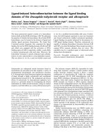
Figure 1 Protocols using G-CSF to mobilize stem cells and echocardiographic assessment of cardiac function in mice. (A) Animals were
divided into four groups for 4 weeks of iron intra- peritoneally injection (10 mg/25 gm body weight of mouse per day for 5 days/week) or
dextran injection as shown in the protocols. G-CSF (100 μg/kg/day subcutaneous injection) or saline was given for 5 days in the second week as
shown. I+G; iron plus G-CSF treatment. (B) Different dosages of G-CSF were given to mice with blood c-kit and CD45 examined by flow
cytometry analysis. (C) Representative echocardiograms of mitral-valve-flows Doppler mapping (E and A waves) in each experimental group at
end of the second and fourth week, respectively. Decreased E: A wave ratio showing diastolic dysfunction in the I+G group. E wave and A wave,
indicating LV early-filling wave and filling from atrial contraction, respectively. (D) Representative 2D echocardiogram of long axis view revealed
intra-cardiac mass (arrow) in the apex region of the left ventricle in the I+G group at 4th week exam.
Lian et al. Journal of Biomedical Science 2011, 18:26
/>
horseradish peroxidase (HRP)-conjugated streptavidinbiotin complex (Vectastain Elite ABC kit standard) for 1
hour at room temperature and then used 3,3-diaminobenzidine (DAB) as a chromogen (Vector Laboratories,
Burlingame, CA), and counterstained with Contrast
GREEN Solution (KPL, U.S.A.) for microscopic studies.
For immunofluorescent staining, sections were first
rehydrated and epitope retrieval buffer (Thermal scientific, Inc.) in 95~100°C for 30 min. Sections were then
washed and blocked with 5% fetal bovine serum for 1
hr. Sections were then double-stained with antibodies
against TF (M-20, 1:100) and CD13 (1:100) overnight at
4°C. Different Fluorescein (FITC, donkey anti goat) and
Rhodamine (TRITC, donkey anti rabbit) secondary antibodies (Jackson ImmunoResearch Lab. Inc.) were used
to obtain fluorescent colors. The stained sections were
counterstained with DAPI to visualize nuclei by ProLong antifade (Invitrogen) mounting reagent.
Flow Cytometry Analysis
Page 4 of 15
GGG CCA TCC ACA GTC TTC T-3’). The relative
expression ratio of each transcript (ICAM-1, MCP-1, tissue factor, and TNF-a) in comparison to GAPDH was
calculated as described.
Western blot analysis
Myocardium protein extracts were prepared by using a
protein extraction kit (NE-PER), and total protein concentrations was determined by BCA™ protein assay
reagent. Western Blot chemiluminescence reagents were
obtained from PIERCE (Pierce Chemical Co.). Proteins
were separated by polyacrylamide gel electrophoresis
and transferred to PVDF membranes for Western blot
analysis. Blots were incubated with either anti-p-AKT
(1:1000), anti-AKT (1:1000), anti-eNOS (1:1000) (Cell
Signaling Technology Inc.), anti-MPO (1:500) (R&D systems, Inc.) and anti-b-actin (1:2000) antibodies in nonfat dry milk in wash buffer overnight at 4°C. Blots were
then incubated with peroxidase conjugated anti-rabbit
(1:10,000) or anti-goat (1:1,000) for 1 hour at room temperature. Proteins were visualized by enhanced chemiluminescence, immunoblot signals were quantitated using
a Fujifilm Medical Systems U.S.A., Inc.
Flow cytometry analysis was performed with FACSCalibur and CellQuest Pro software (Becton Dickinson, San
Joes, CA, USA) using directly conjugated mAbs against
the following markers: CD11b-PE and Ly-6G-FITC or
CD45-PE and CD117-PE (c-kit) (BD biosciences) with
corresponding isotype matched controls. Blood samples
were washed with PBS buffer and red blood cells were
removed by RBC lysis buffer. Briefly, mAbs and cells
were incubated for 30 minutes at 4°C and unbound
reagents were removed by washing. Cells were then
resuspended in fixing buffer (PBS containing 1%formaldehyde and 1% FBS) for flow analysis.
Statistical analysis was done by SPSS for Windows (version 12.0). All data are described as means ± standard
deviation (S.D.). The two groups were compared using
the Student’s t-test. Statistical analysis was performed
with one-way ANOVA by Tukey test for multiple comparisons. The differences were considered significant at
a value of P < 0.05.
RNA isolation and real-time PCR
Results
Assays were performed using Applied Biosystems PRISM
7700 sequence detection system with cDNAs derived
from mice treated with or without G-CSF following iron
injection. Glyceraldehyde-3-phosphate dehydrogenase
(GAPDH) was used as control. Thermal cycler conditions
were as follows: hold for 2 min at 50°C and 10 min at 95°
C, followed by two-step PCR for 35 cycles of 95°C for 15
s, then 60°C for 1 min. Forward and reverse primers and
a fluorescence-labeled probe were as follows: ICAM-1
sense, 5’- CGC AAG TCC AAT TCA CAC TGA -3’, and
antisense, 5’- ATT TCA GAG TCT GCT GAG AC -3);
MCP-1 sense, 5’- CAG CCA GAT GCA GTT AAC GC
-3’, and antisense, 5’- GCC TAC TCA TTG GGA TCA
TCT TG -3’); tissue factor sense, 5’- AAG GAT GTG
ACC TGG GCC TAT GAA -3’, and antisense, 5’- ACT
GCT GAA TTA CTG GCT GTC CGA T-3’); TNF-a
sense, 5’- TAC TGA ACT TCG GGG TGA TTG GTC C
-3’, and antisense, 5’- GGT TCT CTT CAA GGG ACA
AGG CTG -3’) and GAPDH sense, 5’-GGA GCC AAA
CGG GTC ATC ATC TC-3’, and antisense, 5’-GAG
G-CSF can mobilize autologous stem cell and effect
cardiac dysfunction with intra-cardiac thrombosis in I+G
mice
Statistical analysis
We first used flow cytometry to check both c-kit(+) and
CD45(+) cells from G-CSF injected mice to confirm that
G-CSF can mobilize stem cells and leukocytes in a dosage
dependent manner in our mice model before analyzing
any phenotype (Figure 1B). Echocardiography at the end
of 4th week showed that heart functions in the I+G group
was abnormal with decrement in fractional shortening and
mild chamber dilation in the left ventricle (LV) without
affecting the heart rate (Table 1). In addition, diastolic
impairment was also found in the I+G group, with
decreased E/A ratio progressively from the 2nd to 4th week
(Figure 1C, Table 1). Interestingly, intra-cardiac thrombus
were found in the LV at the 4 th week check up in I+G
group (11/15 mice, Figure 1D). Histological examination
by Masson trichrome staining confirmed the presence of
intra-cardiac thrombus with fibrosis only in the I+G but
not in other groups (Figures 2A and 2B).
Lian et al. Journal of Biomedical Science 2011, 18:26
/>
Page 5 of 15
Table 1 Echocardiographic results at the end of 2nd and 4th week in I+G and other experimental groups
HR (bpm)
LVPWs (cm) LVIDSs (cm)
IVSs (cm)
LVPWd (cm) LVIDd (cm)
IVSd (cm)
EF (%)
FS (%)
E/A ratio
1.83 ± 0.22
2wks
C
360.5 ± 33
0.08 ± 0.01
0.22 ± 0.03
0.11 ± 0.01
0.07 ± 0.01
0.35 ± 0.03
0.06 ± 0.01
75.75 ± 5.1
37.90 ± 4.4
G
333.0 ± 40
0.10 ± 0.02
0.25 ± 0.04
0.12 ± 0.01
0.07 ± 0.01
0.37 ± 0.02
0.07 ± 0.01
70.40 ± 11
33.53 ± 8.0
1.85 ± 0.23
I
372.2 ± 45
0.08 ± 0.02
0.24 ± 0.02
0.11 ± 0.02
0.05 ± 0.01
0.36 ± 0.04
0.06 ± 0.01
69.88 ± 3.6
33.50 ± 1.5
2.07 ± 0.59
I+G
362.9 ± 12
0.08 ± 0.01
0.24 ± 0.02
0.12 ± 0.02
0.06 ± 0.01
0.36 ± 0.02
0.06 ± 0.01
71.78 ± 5.6
35.23 ± 2.1
1.94 ± 0.39
4wks
C
333.5 ± 78
0.10 ± 0.02
0.22 ± 0.04
0.11 ± 0.02
0.07 ± 0.01
0.35 ± 0.03
0.06 ± 0.01
73.68 ± 6.5
36.39 ± 5.4
1.89 ± 0.17
G
I
348.2 ± 32
325.8 ± 95
0.08 ± 0.02
0.08 ± 0.04
0.25 ± 0.02
0.23 ± 0.06
0.10 ± 0.01
0.10 ± 0.02
0.06 ± 0.01
0.06 ± 0.03
0.36 ± 0.02
0.34 ± 0.04
0.06 ± 0.01
0.06 ± 0.02
65.58 ± 4.3 30.78 ± 2.6
68.50 ± 12.7 32.94 ± 11.3
1.85 ± 0.23
1.97 ± 0.14
I+G
315.9 ± 58
0.09 ± 0.01
0.28 ± 0.02*
0.12 ± 0.01
0.06 ± 0.01
0.38 ± 0.02* 0.08 ± 0.01†
58.65 ± 4.5†
1.85 ± 0.22†
26.26 ± 2.8†
IVSd, inter-ventricular septum thickness at diastole; LVIDd, left ventricular internal diameter at diastole; LVPWd, left ventricular posterior wall thickness at diastole;
IVSs, inter-ventricular septum thickness at systole; LVIDs, left ventricular internal diameter at systole; LVPWs, left ventricular posterior wall thickness at systole; FS,
fractional shortening of left ventricle; EF, ejection fraction of left ventricle; E/A, E wave/A wave ratio at left ventricular diastolic phase;*p < 0.05, †p < 0.01 vs
control, n = 12 in each group.
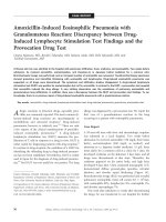
Cardiac histopathology of I+G mice
The mural thrombi found in I+G mice were mainly
located in the apex region of the LV (Figure 1D), but
also found in the chorda tendini of the LV (Figures 2B
and 2I) and in the right ventricular cavity (data not
shown). Histological analysis of the hearts from I group
and I+G groups revealed iron deposition (Figures 2C
and 2D). However, only I+G hearts revealed interstitial
fibrosis with mural thrombi, attached tightly to the
endocardium (Figures 2B and 2D). Extensive fibrosis
was observed along the border between the cardiac
endothelium and thrombi mass (Figure 2G). Macrophages with iron deposition in the cytoplasm infiltrated
into the inter-myocytic spaces of the ventricular heart
tissue (Figure 2H) and leukocytes were involved in
thrombus formation (Figure 2I). However, there are no
signs of thrombi formation in any body organs (aorta,
liver, kidney and coronary arteries) examined (see Additional file 1, Figure S1).
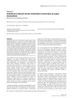
Increased expression of tissue factor in the I and I+G
hearts and its co-localization with macrophage marker
CD13
Cellular compositions of the all groups were examined
by immunohistochemistry. Tissue factor was upregulated within the myocardium where it may be
mediated by the infiltrating cells in both I and I+G
groups, with more prominent in the latter group
(Figure 3A). Confocal microscopy depicted colocalization of CD13 (a protein specific for monocytes/macrophages) with tissue factor near the endocardiummyocardium junction in the I+G heart tissue, implying
areas of prominent inflammation (Figure 3B). Here we
demonstrated that G-CSF enhances the recruitment of
monocytes/macrophages and the expression of tissue
factor in the affected heart tissue especially in the I+G
group (Figure 3C).
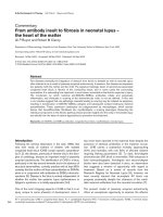
G-CSF supplement aggravates iron induced oxidative
stress, leukocyte infiltration and inflammatory profile in
heart
In order to elucidate the role of G-CSF in our I+G
model, we compared the heart tissue from both I group
and I+G group for oxidative stress, leukocyte infiltration
and inflammatory profile between them. As expected, I
+G hearts had higher levels of 4-HNE and 8-OHdG
(both are index of oxidative stress), and increased
expression of CD45 (leukocyte marker) (Figures 4A and
4B). Myeloperoxidase activity was also higher in the I+G
hearts, indicating aggravation of inflammatory profile in
the I+G hearts, as compared to the hearts from I group
(Figure 4C).
Simvastatin attenuates cardiac apoptosis, iron deposition,
and thrombosis in I+G mice in vivo
We investigated whether simvastatin, a common clinically
used HMG-CoA reductase inhibitor, can play beneficial
role in attenuating cardiac inflammation, iron deposition,
or abrogating cardiac thrombosis in I+G mice. Cardiac tissue from the I+G group, and I+G plus statin (I+G+St) and
the control group was collected at the end of 4th week and
compared. Incidence of thrombi formation were 0/10 in
the control group, 7/10 in the I+G, and 2/10 in the I+G
+St groups (p < 0.05 versus I+G group), respectively. Concomitant TUNEL assay and iron staining showed a significant decrease in apoptotic cardiomyoctes (Figures 5A and
5C) and iron deposition (Figures 5B and 5D) in the I+G
+St compared to the I+G group.
I+G mice shows leukocytosis and systemic elevation of
inflammatory profile which can be attenuated by
simvastatin but not by tirofiban treatment
To further determine if simvastatin act through its antiinflammatory effect systemically, we checked complete
blood counts and inflammatory profiles in the serum from
Lian et al. Journal of Biomedical Science 2011, 18:26
/>
Page 6 of 15
I+G and I+G+St groups. Monocytes and neutorophils
were increased in the serum from I+G mice at the end of
second week. At the 4th week recheck, leukocytosis was
aggravated in the I+G mice, but attenuated in the I+G+St
mice (Table 2). Flow cytometry analysis of CD11b and
Ly6G proteins (myeloid cells surface markers expressed
mainly on the monocytes, macrophages and granulocytes)
showed increased expression in the I+G but not in the I
+G+St group (Figure 6A). Serum inflammatory markers
MCP-1 and ICAM-1 were up-regulated in the I+G, but
not in the I+G+St group (Figure 6B). We next intended to
clarify the role of platelet in this I+G induced thrombosis
model, by giving platelet receptor inhibitor tirofiban to I
+G mice. Interesting, although number of platelets
decreased (see Additional file 1, Table S1), inflammatory
profiles (Figure 6C) and thrombus formation stayed the
same between I+G and I+G plus tirofiban groups (7/10
versus 7/10, respectively). Concomitant to the above
results, I+G group demonstrated lower cardiac CD34
expression and serum CRP level after simvastatin therapy,
but not tirofiban treatment (Figure 7). These results provide in vivo evidence that G-CSF-induced thrombosis can
only be ameliorated by simvastatin therapy, but not by tirofiban treatment, implying a significant role of inflammation association in our model.
Simvastatin also ameliorates inflammatory stage in the
heart tissue of I + G mice
Heart tissue was sampled at the end of 4 th week for
quantitative PCR analysis. Expression of ICAM-1, MCP1, TNF-a, and tissue factor increased in the I+G group
compared with the control group (Figure 8A). Interestingly, increased expression of MCP-1 and ICAM-1 were
also noted in the G-group (p < 0.05 versus control),
indicating that G-CSF alone can promote pro-inflammatory factors. Decreased expression of the above proinflammatory factors was seen in the I+G+st group
(Figure 8A). This result suggested that simvastatin attenuated the cardiac thrombus formation via down regulation of inflammatory signaling in the heart tissue.
Figure 2 Intra-cardiac thrombus formation and histopathology
of the ventricular tissue in I+G heart. (A and B) Heart crosssection at the papillary muscle level of the LV from iron (I) only (A
and C) and I+G heart (B and D) stained with Masson’s trichrome and
Prussian blue staining, respectively. Note that the formation of a
large mural thrombus in I+G heart. (E and F) Obvious fibrosis near
the endocardium was noted in the I+G heart (F), but not in the iron
only group (E). (G and H) Higher magnification of the LV from I+G
group depicted regions of prominent fibrosis between thrombus
and myocardium (G) and macrophages with cytoplasmic iron (brown
color) deposition, infiltrated into intra-cardiomyocytic spaces (H). (I)
Magnification of thrombus near the LV papillary muscle
demonstrated leukocytes (arrows) involved in thrombus formation.
Tissue section in E was stained with iron staining; tissue section in F,
G, H, and I were stained with H & E staining.
Elevated pAkt and eNOS expression in simvastatin
supplemented hearts
To elucidate the molecular pathway of statin’s antiinflammation therapy on I+G mice. Protein levels of
phosphorylated Akt (pAkt) and endothelial nitric oxide
synthase (eNOS) increased in the hearts of the G plus
statin and I+G+St groups, as compared to other groups
(Figure 8B). These results indicate that statin treatment
significantly enhanced the expression of eNOS and
phosphorylation of Akt, and that the therapeutic effect
of statin in ameliorating the thrombus formation may
act through the activation of Akt-eNOS signaling
pathway.
Lian et al. Journal of Biomedical Science 2011, 18:26
/>
Page 7 of 15
Figure 3 Immunohistochemical detection of tissue factor and its colocalization with macrophage marker (CD13) in I and I+G hearts.
(A) Immunoreactivity of tissue factor was shown in I and I+G hearts, with more prominent in the latter group. (B) Colocalization of CD13 specific
for monocytes/macrophage and tissue factor in heart tissue of I+G mice. Heart sections were stained with anti-tissue factor antibody (red in left
upper panel), anti-CD-13 antibody (green in right upper panel), merge (left lower panel), and H & E staining (right lower panel). Co-localization of
CD13 and tissue factor expression was seen in cardiac tissue near the heavy fibrosis region, implying region of prominent inflammation. Dashed
line (in sections with H & E staining) indicated region of endocardium with cardiac fibrosis seen between thrombus (left upper) and myocardium
(right lower). (C) Quantitative analysis of either tissue factor or CD13 staining positive cells in both control (C) and I+G hearts were shown in
diagrams, **P < 0.001 vs control.
Lian et al. Journal of Biomedical Science 2011, 18:26
/>
Page 8 of 15
Figure 4 G-CSF enhanced iron induced oxidative stress and leukocyte infiltration with aggravation of myeloperoxidase (MPO) activity
in heart. (A and B) Immunoreactivity of 8-OHdG, 4-HNE (both are markers for oxidative stress) and CD45 (leukocyte marker) were compared and
quantified between iron only (I) and I+G heart tissue. Representative results of three separate experiments are shown in (B). (C) MPO activities in
heart tissue from all groups and their relative expression compared with actin were shown, *p < 0.01.
Lian et al. Journal of Biomedical Science 2011, 18:26
/>
Page 9 of 15
Figure 5 Apoptosis and iron deposition/infiltration of cardiomyocytes following simvastatin treatment in I+G mice. (A and C) Apoptotic
cardiac myocytes were detected by the TUNEL assay in control group, I+G group, and I+G with simvastatin (I+G+St) treatment group
respectively. Left and right panels show the TUNEL positive (green) and nuclei (blue) fluorescence, respectively. Each histogram represents the
number of TUNEL-positive cells in each group (n = 5 animals in each group). (B and D) Iron deposition/infiltration in cardiac tissue for each
group was stained and quantified. Representative results of three separate experiments are shown. Bar = 200 μm; **p < 0.001 vs control; ††p <
0.001 vs I+G.
Discussion
Results of the present study demonstrate that G-CSF supplement on iron loading hearts can recruit neutrophils/
monocytes and up-regulate tissue factors, ICAM-1, TNFalpha, and MCP-1 thus further activating inflammatory
processes in the endo-myocardium and induce cardiac
thrombosis. Chronic iron loading can increase cardiac oxidative stress. Whereas G-CSF treatment activates serial
events of inflammation-thrombosis circuitry and that leads
to intra-cardiac thrombus formation. This inflammationassociated cardiac thrombosis in vivo can be attenuated by
simvastatin therapy, but not by tirofiban treatment. Our
results confirmed that G-CSF can induce in vivo cardiac
thrombosis through inflammation-thrombosis interaction.
Iron overload is known to accelerate arterial thrombosis through increased vascular oxidative stress and
Lian et al. Journal of Biomedical Science 2011, 18:26
/>
Page 10 of 15
Table 2 Blood count parameters (mean ± SD) acquired at end of second and fourth weeks of I+G mice with or without
statin therapy
LEUK (109/L)
ERY (1012/L)
HGB (g/dl)
NEU (109/L)
LYM (109/L)
MONO (109/L)
PLT (109/L)
2wks
C
8.79 ± 1.98
8.27 ± 0.33
13.98 ± 0.61
1.44 ± 0.13
8.89 ± 1.54
0.07 ± 0.06
1330.33 ± 45.88
I
I+G
8.20 ± 3.19
12.07 ± 0.9*
9.09 ± 0.88
8.36 ± 0.51
15.80 ± 1.18
14.28 ± 0.65
1.21 ± 0.37
2.07 ± 0.22*
6.07 ± 2.61
7.68 ± 2.16
0.71 ± 0.28†
0.72 ± 0.07†
1167.78 ± 87.37
1277.33 ± 34.08
I+G+St
8.15 ± 1.77‡
7.53 ± 0.26
13.63 ± 1.01
2.81 ± 0.87‡
5.22 ± 1.23‡
0.62 ± 0.03
1025.25 ± 420.78
C
9.93 ± 2.76
9.35 ± 0.28
16.08 ± 0.77
1.75 ± 0.18
6.46 ± 1.47
0.19 ± 0.02
1514.4 ± 76.51
I
19.1 ± 5.18†
9.36 ± 0.04
15.60 ± 0.01
11.50 ± 0.14†
7.39 ± 0.36
1.68 ± 0.56*
1455.2 ± 129.67
I+G
25.02 ± 2.53†
8.26 ± 0.27
15.46 ± 0.29
11.06 ± 1.05†
9.37 ± 1.59*
2.26 ± 0.32†
1313.8 ± 120.34*
‡
1.27 ± 0.59‡
4wks
I+G+St
‡
18.86 ± 3.45
8.40 ± 0.26
15.7 ± 0.58
‡
9.51 ± 0.61
5.88 ± 1.31
1433.7 ± 156.18
†
LEUK, leukocytes; ERY, erythrocytes; HGB, hemoglobin; NEU, neutrophil; LYM, lymphocyte; MONO, monocyte; PLT, platelet; *p < 0.05, p < 0.01 vs control;
0.05 vs I+G, n = 8 in each group.
impaired vascular reactivity [16,21] and it also impairs
cardiac function by increasing free radical production
resulting in cardiomyopathy [22,23]. However, present
study shows that iron loading alone is not sufficient to
induce intra-cardiac thrombosis as reported by others
[20]. Our results clearly indicate that G-CSF supplementation effectively initiated inflammation-thrombosis bridging thereby promoting thrombosis and recruited
subsets of hematopoietic cells, like mature neutrophils
and monocytes which bear their adhesion receptors on
the cell membrane [24]. Moreover, recent reviews also
reported a pivotal role of tissue factor in driving the
thrombosis- inflammation circuit [25,26]. This may be
responsible for accumulation of a large number of
macrophages and tissue factor expression in the affected
lesions (Figure 3B). G-CSF induced leukocyte infiltration
resulted in increased tissue factor expression with secondary thrombosis and subsequent tissue fibrosis. As
tirofiban fail to ameliorate the thrombosis, it may indicate that fibrinogen (or GPIIb/IIIa) did not have major
role in this inflammation-thrombosis process [27]. Our
in vivo mouse model could be a novel avenue for investigating inflammation and thrombosis interactions in the
cardiac endothelium, compared to previous studies that
focused mainly on the vascular endothelium [27,28].
Iron loading has multiple effects on all body tissues,
including cardiac myocytes and macrophages. For example, in a similar iron overload model (with chronic iron
treatment for 12 weeks) showed increased cardiac interstitial fibrosis in addition to inflammatory infiltration
[19]. Iron-overloaded macrophage secrete increased
levels of cytokines in response to an inflammatory stimulus and exacerbates alcoholic liver injury [29,30]. In our I
+G model, G-CSF supplementation increased ROS production and recruitment of leukocyte (Figure 4) further
aggravated inflammatory infiltration which eventually
triggered cardiac thrombosis. However, thrombosis only
‡
p<
seen in the cardiac chamber but not other organs (see
Supplementary Figure 1), may be due to the fact that
macrophage are prone to be deposited in the heart and
the liver, yet the latter organ lacks the shear stress
induced by rapid blood flow and functional impaired
endothelium unlike the heart.
Our results showing that G-CSF can promote inflammatory profiles and cardiac thrombosis that leads to cardiac dysfunction, are in contrast to previous reports
showing G-CSF therapy to be beneficial in acute myocardial infarction [3,4,31,32] and chronic cardiomyopathy induced by doxorubicin toxicity [33]. G-CSF exerts
an anti-inflammatory effect [34] as well as an angiogenic
and anti-apoptotic effect which prevents LV wall thinning and heart failure after acute myocardial infarction
[3,35]. One explanation for these disparate results could
be that chronic iron loading increases oxidative stress
and impairs endothelium-dependent vaso-relaxation
[16], a different scenario than in acute myocardial
infarction. Although G-CSF recruits hematogenic stem
cells and endothelial progenitor cells for cardiac repair,
a simultaneous induction of macrophage and tissue factor gathering “gears up” the pro-inflammatory state and
drives the inflammation-thrombosis circuit. Besides, GCSF induced leukocytosis is a well known feature that
also suggests its direct role in enhancing acute thrombosis [36].
HMG-CoA reductase inhibitors, or statins, are known
to improve cardiac dysfunction through their anti-inflammatory and anti-oxidative action. Statins also affect
endothelial function through the production of nitric
oxide [18,19]. Present study demonstrates that simvastatin can reduce the myocardial iron deposition/infiltration
score (Figure 4D) and blood leukocyte count (Table 2)
that strengthens the link between inflammation and myocardial thrombus formation. Simvastatin administration
significantly reduced the incidence of thrombus
Lian et al. Journal of Biomedical Science 2011, 18:26
/>
Page 11 of 15
Figure 6 I+G mice showed increased monocyte/neutrophil counts with elevation of inflammatory profiles which can be attenuated by
simvastatin therapy, but not by tirofiban treatment. (A) Expression of CD11b on blood serum collected from control (C), I+G, and I+G with
simvastatin treatment (I+G+St) groups respectively. Blood was labeled with PE-conjugated rat anti-mouse CD11b antibody and FITC-conjugated
Ly-6G monoclonal antibody separately, then flow cytommetry was performed on a BD FACScan flow cytometry system. Experiments were
performed twice with similar results (n = 3 mice in each group); * p < 0.05, ** p < 0.001, respectively. (B) The mouse serum was harvested and
the protein levels of MCP-1 and ICAM-1 were determined by ELISA; ** p < 0.001 vs control group; † P < 0.05, †† P < 0.01 vs I+G group,
respectively. (C) The mouse serum was collected from control, I+G, and I+G with tirofiban treatment groups respectively.
formation in the I+G heart, and expression of the proinflammatory markers ICAM-1, tissue factor, MCP-1,
and TNF-a. Furthermore, prior studies suggesting that
statin could regulate eNOS activity via post-translational
activation of phosphatidylinositol 3-kinase/protein kinase
Akt pathway (PI3K/Akt) in the endothelium [37-40].
Simvastatin treated I+G hearts in our study revealed an
elevation of both eNOS and phosphorylated Akt activity,
suggesting that simvastatin had a therapeutic effect in
ameliorating the thrombus formation in the heart.
Recently meta-analysis results from 10 clinical trials for
stem cell mobilization by G-CSF therapy for myocardial
recovery after AMI showed neither improvement of LV
function or the reduction in infarct size in patients with
AMI after reperfusion [8]. In order to effectively improve
LV contractility, future studies should focus more on the
Lian et al. Journal of Biomedical Science 2011, 18:26
/>
Page 12 of 15
Figure 7 I+G mice showed increased cardiac CD34 expression with elevation of serum c-reactive protein (CRP) levels which can be
attenuated by simvastatin therapy, but not by tirofiban treatment. (A) Immunoreactivity of CD34 were compared and quantified among
heart tissue of each experimental group as indicated. Representative results of three separate experiments are shown in (B). (C) Serum CRP levels
were examined via ELISA among each experimental group as indicated, *p < 0.05, ** p < 0.001.
autologous stem cells plus G-CSF infusion. Under such
scenario, more attention should be paid to the possible
detrimental effects of G-CSF related thrombosis. As GCSF plus stem cells might additively increase cell density
and hypercoagulable state in certain time window thus
result in re-stenosis or late thrombosis in MI patients.
Therefore, it is important to screen for high risk patients
with chronic inflammation or increased oxidative stress
like metabolic syndrome, diabetes, chronic heart failure, or
chronic atherosclerosis, before they should receive G-CSF
treatment for acute coronary heart disease. Accordingly,
present study provides an in vivo disease model to elucidate the mechanism of post G-CSF cardiac thrombosis,
which could have major clinical implication.
Lian et al. Journal of Biomedical Science 2011, 18:26
/>
Page 13 of 15
Figure 8 Cardiac mRNA analysis for inflammatory markers and protein analysis for AKT and eNOS expression in I+G mice compared
with I+G plus simvastatin treated mice. (A) Total mRNAs were prepared from whole heart tissues, and the levels of ICAM-1, MCP-1, tissue
factor, and TNF-alpha transcripts were determined by Quantitative-PCR analysis. Note that the levels of four transcripts, especially of tissue factor
and TNF-alpha reduced significantly after simvastatin administration. GADPH expression was used as a control to monitor RNA quality and
concentration; **p < 0.001. (B and C) Western blot analysis of phosphorylated AKT (pAkt), AKT, eNOS, and b-actin. Lanes from left to right
indicate heart tissues taken from the untreated control (C), G-CSF only (G), G-CSF with statin administration (G+St), I+G, and I+G with simvastatin
administration (I+G+St). Data represent results from three independent experiments. Scanning densitometry was used for semi-quantitative
analysis in compared to the Akt or b-actin levels respectively; **p < 0.001 vs control.
Lian et al. Journal of Biomedical Science 2011, 18:26
/>
Additional material
Additional file 1: Histology of I+G mice and blood parameters of I
+G mice with tirofiban treatment. A figure demonstrating histology of
other organs in I+G mice and a table listing blood parameters of I+G
mice with or without tirofiban therapy.
Page 14 of 15
7.
8.
9.
Acknowledgements
This work was supported by grants from the National Science Council (NSC
95-2314-B-303-028-MY3), Tzu Chi University (TCIRP 95007-01) and Tzu Chi
General Hospital (TCRDI 99-01 and TCRD99-49) to C.-F. Cheng, There were
no conflicts of interest for any of the authors.
10.
Author details
1
Department of Medical Research, Tzu Chi General Hospital and Department
of Pediatrics, Tzu Chi University, Hualien, Taiwan. 2Institute of Biomedical
Sciences, Academia Sinica, Taipei, Taiwan. 3Department of Animal Science
and Technology, National Taiwan University, Taiwan. 4Institute of Toxicology
and Pharmacology, Tzu Chi University, Hualien, Taiwan. 5Department of
Animal Science and Biotechnology, Tunghai University, Taichung, Taiwan.
Authors’ contributions
WSL and CFC designed the experiments and analyzed the data. WSL and HL
performed the in vivo study. HL performed the in vitro study. TK analyzed
the cardiac pathology. WTKC and TK help to coordinate this study. CFC
wrote the manuscript. All authors have read and approved the final
manuscript.
Competing interests
The authors declare that they have no competing interests.
11.
12.
13.
14.
Received: 7 December 2010 Accepted: 15 April 2011
Published: 15 April 2011
15.
References
1. Petit I, Szyper-Kravitz M, Nagler A, Lahav M, Peled A, Habler L,
Ponomaryov T, Taichman RS, Arenzana-Seisdedos F, Fujii N, Sandbank J,
Zipori D, Lapidot T: G-CSF induces stem cell mobilization by decreasing
bone marrow SDF-1 and up-regulating CXCR4. Nat Immunol 2002,
3:687-694.
2. Lévesque JP, Hendy J, Takamatsu Y, Simmons PJ, Bendall LJ: Disruption of
the CXCR4/CXCL12 chemotactic interaction during hematopoietic stem
cell mobilization induced by GCSF or cyclophosphamide. J Clin Invest
2003, 111:187-196.
3. Harada M, Qin Y, Takano H, Minamino T, Zou Y, Toko H, Ohtsuka M,
Matsuura K, Sano M, Nishi J, Iwanaga K, Akazawa H, Kunieda T, Zhu W,
Hasegawa H, Kunisada K, Nagai T, Nakaya H, Yamauchi-Takihara K, Komuro I:
G-CSF prevents cardiac remodeling after myocardial infarction by
activating the Jak-Stat pathway in cardiomyocytes. Nat Med 2005,
11:305-311.
4. Minatoguchi S, Takemura G, Chen XH, Wang N, Uno Y, Koda M, Arai M,
Misao Y, Lu C, Suzuki K, Goto K, Komada A, Takahashi T, Kosai K, Fujiwara T,
Fujiwara H: Acceleration of the healing process and myocardial
regeneration may be important as a mechanism of improvement of
cardiac function and remodeling by postinfarction granulocyte colonystimulating factor treatment. Circulation 2004, 109:2572-2580.
5. Kang HJ, Lee HY, Na SH, Chang SA, Park KW, Kim HK, Kim SY, Chang HJ,
Lee W, Kang WJ, Koo BK, Kim YJ, Lee DS, Sohn DW, Han KS, Oh BH, Park YB,
Kim HS: Differential Effect of Intracoronary Infusion of Mobilized
Peripheral Blood Stem Cells by Granulocyte Colony-Stimulating Factor on
Left Ventricular Function and Remodeling in Patients With Acute
Myocardial Infarction Versus Old Myocardial Infarction: The MAGIC Cell-3DES Randomized, Controlled Trial. Circulation 2006, 114(1 Suppl):I145-I151.
6. Ince H, Valgimigli M, Petzsch M, de Lezo JS, Kuethe F, Dunkelmann S,
Biondi-Zoccai G, Nienaber CA: Cardiovascular events and re-stenosis
following administration of G-CSF in acute myocardial infarction:
systematic review and meta-analysis. Heart 2008, 94:610-616.
16.
17.
18.
19.
20.
21.
22.
23.
24.
25.
26.
27.
28.
Abdel-Latif A, Bolli R, Zuba-Surma EK, Tleyjeh IM, Hornung CA, Dawn B:
Granulocyte colony-stimulating factor therapy for cardiac repair after
acute myocardial infarction: a systematic review and meta-analysis of
randomized controlled trials. Am Heart J 2008, 156:216-226.
Zohlnhöfer D, Dibra A, Koppara T, de Waha A, Ripa RS, Kastrup J,
Valgimigli M, Schömig A, Kastrati A: Stem cell mobilization by granulocyte
colony- stimulating factor for myocardial recovery after acute
myocardial infarction: a meta-analysis. J Am Coll Cardiol 2008,
51:1429-1437.
Kang HJ, Kim HS, Zhang SY, Park KW, Cho HJ, Koo BK, Kim YJ, Soo LD,
Sohn DW, Han KS, Oh BH, Lee MM, Park YB: Effects of intracoronary
infusion of peripheral blood stem-cells mobilised with granulocytecolony stimulating factor on left ventricular systolic function and
restenosis after coronary stenting in myocardial infarction: the MAGIC
cell randomised clinical trial. Lancet 2004, 363:751-756.
Hill JM, Syed MA, Arai AE, Powell TM, Paul JD, Zalos G, Read EJ, Khuu HM,
Leitman SF, Horne M, Csako G, Dunbar CE, Waclawiw MA, Cannon RO:
Outcomes and risks of granulocyte colony-stimulating factor in patients
with coronary artery disease. J Am Coll Cardiol 2005, 46:1643-1648.
Steinwender C, Hofmann R, Kypta A, Gabriel C, Leisch F: Late stent
thrombosis after transcoronary transplantation of granulocyte-colony
stimulating factor-mobilized peripheral blood stem cells following
primary percutaneous intervention for acute myocardial infarction. Int J
Cardiol 2007, 122:248-249.
Canales MA, Arrieta R, Gomez-Rioja R, Diez J, Jimenez-Yuste V, HernandezNavarro F: Induction of a hypercoagulability state and endothelial cell
activation by G-CSF in peripheral blood stem cell donors. J Hematother
Stem Cell Res 2002, 11:675-681.
Karadogan C, Karadogan I, Bilgin AU, Undar L: G-CSF increases the
platelet-neutrophil complex formation and neutrophil adhesion
molecule expression in volunteer granulocyte and stem cell aphaeresis
donors. Ther Apher Dia 2006, 10:180-186.
Topcuoglu P, Arat M, Dalva K, Ozcan M: Administration of granulocytecolony- stimulating factor for allogeneic hematopoietic cell collection
may induce the tissue factor-dependent pathway in healthy donors.
Bone Marrow Transplant 2004, 33:171-176.
Kurz KD, Main BW, Sandusky GE: Rat models of arterial thrombosis
induced by ferric chloride. Thromb Res 1990, 60:269-280.
Day SM, Duquaine D, Mundada LV, Menon RG, Khan BV, Rajagopalan S,
Fay WP: Chronic iron administration increases vascular oxidative stress
and accelerates arterial thrombosis. Circulation 2003, 107:2601-2606.
Russo G, Leopold JA, Loscalzo J: Vasoactive substances: nitric oxide and
endothelial dysfunction in atherosclerosis. Vasc Pharmacol 2002, 38:259-269.
Davignon J: Beneficial cardiovascular pleiotropic effects of statins.
Circulation 2004, 109(23 Suppl 1):III39-III43.
Liao JK, Laufs U: Pleiotropic effects of statins. Annu Rev Pharmacol Toxicol
2005, 45:89-118.
Oudit GY, Sun H, Trivieri MG, Koch SE, Dawood F, Ackerley C,
Yazdanpanah M, Wilson GJ, Schwartz A, Liu PP, Backx PH: L-type Ca2+
channels provide a major pathway for iron entry into cardiomyocytes in
iron-overload cardiomyopathy. Nat Med 2003, 9:1187-1194.
Araujo JA, Romano EL, Brito BE, Parthé V, Romano M, Bracho M,
Montaño RF, Cardier J: Iron overload augments the development of
atherosclerotic lesions in rabbits. Art Thromb Vasc Biol 1995, 15:1172-1180.
Kadiiska MB, Burkitt MJ, Xiang QH, Mason RP: Iron supplementation
generates hydroxyl radical in vivo: an ESR spin-trapping investigation. J
Clin Invest 1995, 96:1653-1657.
Hershko C, Link G, Cabantchik I: Pathophysiology of Iron Overload. Ann NY
Acad Sci 1998, 850:191-201.
Avalos BR: Molecular analysis of the granulocyte colony-stimulating
factor receptor. Blood 1996, 88:761-777.
Chu AJ: Tissue factor up-regulation drives a thrombosis-inflammation
circuit in relation to cardiovascular complications. Cell Biochem Funct
2006, 24:173-192.
Mackman N: Role of tissue factor in hemostasis and thrombosis. Blood
Cells Mol Dis 2006, 36:104-107.
Gawaz M, Langer H, May AE: Platelets in inflammation and atherogenesis.
J Clin Invest 2005, 115:3378-3384.
May AE, Langer H, Seizer P, Bigalke B, Lindemann S, Gawaz M: Plateletleukocyte interactions in inflammation and atherothrombosis. Semin
Thromb Hemost 2007, 33:123-127.
Lian et al. Journal of Biomedical Science 2011, 18:26
/>
Page 15 of 15
29. Wang L, Johnson EE, Shi HN, Walker WA, Wessling-Resnick M, Cherayil BJ:
Attenuated inflammatory responses in hemochromatosis reveal a role
for iron in the regulation of macrophage cytokine translation. J
Immunology 2008, 181:2723-2731.
30. Tsukamoto H, Lin M, Ohata M, Giulivi C, French SW, Brittenham G: Iron
primes hepatic macrophages for NF-kappaB activation in alcoholic liver
injury. Am J Physiol 1999, 277:G1240-1250.
31. Ohtsuka M, Takano H, Zou Y, Toko H, Akazawa H, Qin Y, Suzuki M,
Hasegawa H, Nakaya H, Komuro I: Cytokine therapy prevents left
ventricular remodeling and dysfunction after myocardial infarction
through neovascularization. FASEB J 2004, 18:851-853.
32. Deindl E, Zaruba MM, Brunner S, Huber B, Mehl U, Assmann G, Hoefer IE,
Mueller-Hoecker J, Franz WM: G-CSF administration after myocardial
infarction in mice attenuates late ischemic cardiomyopathy by
enhanced arteriogenesis. FASEB J 2006, 20:E27-E36.
33. Li L, Takemura G, Li Y, Miyata S, Esaki M, Okada H, Kanamori H, Ogino A,
Maruyama R, Nakagawa M, Minatoguchi S, Fujiwara T, Fujiwara H:
Granulocyte colony-stimulating factor improves left ventricular function
of doxorubicin- induced cardiomyopathy. Lab Invest 2007, 87:440-455.
34. Hartung T: Anti-inflammatory effects of granulocyte colony-stimulating
factor. Curr Opin Hematol 1998, 5:221-225.
35. Takano H, Qin Y, Hasegawa H, Ueda K, Niitsuma Y, Ohtsuka M, Komuro I:
Effects of G-CSF on left ventricular remodeling and heart failure after
acute myocardial infarction. J Mol Med 2006, 84:185-193.
36. Coller BS: Leukocytosis and ischemic vascular disease morbidity and
mortality: is it time to intervene? Aterioscler Thromb Vasc Biol 2005,
25:658-670.
37. Kureishi Y, Luo Z, Shiojima I, Bialik A, Fulton D, Lefer DJ, Sessa WC, Walsh K:
The HMG-CoA reductase inhibitor simvastatin activates the protein
kinase Akt and promotes angiogenesis in normocholesterolemic
animals. Nat Med 2000, 6:1004-1010.
38. Laufs U: Beyond lipid-lowering: effects of statins on endothelial nitric
oxide. Eur J Clin Pharmacol 2003, 58:719-731.
39. Fulton D, Gratton JP, McCabe TJ, Fontana J, Fujio Y, Walsh K, Franke TF,
Papapetropoulos A, Sessa WC: Regulation of endothelium-derived nitric
oxide production by the protein kinase Akt. Nature 1999, 399:597-601.
40. Dimmeler S, Fleming I, Fisslthaler B, Hermann C, Busse R, Zeiher AM:
Activation of nitric oxide synthase in endothelial cells by Akt-dependent
phosphorylation. Nature 1999, 399:601-605.
doi:10.1186/1423-0127-18-26
Cite this article as: Lian et al.: Granulocyte-CSF induced inflammationassociated cardiac thrombosis in iron loading mouse heart and can be
attenuated by statin therapy. Journal of Biomedical Science 2011 18:26.
Submit your next manuscript to BioMed Central
and take full advantage of:
• Convenient online submission
• Thorough peer review
• No space constraints or color figure charges
• Immediate publication on acceptance
• Inclusion in PubMed, CAS, Scopus and Google Scholar
• Research which is freely available for redistribution
Submit your manuscript at
www.biomedcentral.com/submit