Báo cáo Y học: Ligand-induced heterodimerization between the ligand binding domains of the Drosophila ecdysteroid receptor and ultraspiracle docx
Bạn đang xem bản rút gọn của tài liệu. Xem và tải ngay bản đầy đủ của tài liệu tại đây (257.72 KB, 9 trang )
Ligand-induced heterodimerization between the ligand binding
domains of the
Drosophila
ecdysteroid receptor and ultraspiracle
Markus Lezzi
1
, Thomas Bergman
1,
*, Vincent C. Henrich
2
, Martin Vo¨ gtli
1,†
, Christina Fro¨mel
1
,
Marco Grebe
3
, Sabina Przibilla
3
and Margarethe Spindler-Barth
3
1
Institute for Cell Biology, ETH-Ho
¨
nggerberg, Zu
¨
rich, Switzerland;
2
Department of Biology, University of North Carolina,
Greensboro, NC, USA;
3
Abteilung fu
¨
r allgemeine Zoologie und Endokrinologie, Universita
¨
t, D-89069 Ulm, Germany
The insect ecdysteroid receptor consists of a heterodimer
between EcR and the RXR-orthologue, USP. We addressed
the question of whether this heterodimer, like all other RXR
heterodimers, may be formed in the absence of ligand and
whether ligand promotes dimerization. We found that
C-terminal protein fragments that comprised the ligand
binding, but not the DNA binding domain of EcR and USP
andwhichwereequippedwiththeactivationorDNA
binding region of GAL4, respectively, exhibit a weak ability
to interact spontaneously with each other. Moreover, the
heterodimer formation is greatly enhanced upon adminis-
tration of active ecdysteroids in a dose-dependent manner.
This was shown in vivo by a yeast two-hybrid system and
in vitro by a modified electromobility shift assay. Further-
more, the EcR fragment expressed in yeast was functional
and bound radioactively labelled ecdysteroid specifically.
Ligand binding was greatly enhanced by the presence of a
USP ligand binding domain. Therefore, ecdysteroids are
capable of inducing heterodimer formation between EcR
and USP, even when the binding of these receptor proteins to
cognate DNA response elements does not occur. This
capability may be a regulated aspect of ecdysteroid action
during insect development.
Keywords: Drosophila melanogaster;yeast;two-hybrid;
ecdysone receptor; dimerization; ultraspiracle.
Ecdysteroids are widespread steroid hormones found in
invertebrates [1] and plants [2,3] that regulate a variety of
developmental, physiological, and reproductive processes
[1,3]. Among insects, these hormones regulate the expression
of genes through a highly orchestrated and coordinated
transcriptional network [4–6]. The widespread and diverse
effects of ecdysteroids on transcriptional regulation have
served as a powerful model for investigating the diverse
mechanisms by which steroid hormones, acting via nuclear
receptors, exert their effects on a variety of life processes [4,7].
The ecdysone receptor (EcR) [8], responsible for medi-
ating these responses, occupies a special position among
nuclear hormone receptors because it shows a unique
combination of characteristics [9]. Unlike the vertebrate
steroid receptors [10–12], EcR heterodimerizes with the
insect RXR orthologue, ultraspiracle (USP) [13–15]. Nev-
ertheless, while other nuclear receptors that dimerize with
RXR normally are bound to DNA response elements
already in their nonliganded state [11,16], this apparently is
not true for the EcR/USP heterodimer (see however, [17]).
Immunostaining has shown that the polytene chromosomes
of a Chironomid or Sciarid are devoid of EcR/USP signals
when prepared from developmental stages associated with
low ecdysteroid titers [18,19]. A short in vitro incubation of
the tissues with 20-hydroxyecdysone, however, is followed
by the appearance of immunostaining signals at known
ecdysteroid-responsive gene loci [18,19]. The affinity of
EcR/USP dimers for ecdysone response elements (EcREs)
clearly increases in the presence of the ecdysteroid muri-
sterone A as demonstrated by electromobility shift assays
(EMSAs) [20–23].
Neither immunostaining assay nor EMSA studies can
distinguish between three possibilities concerning how
ecdysteroids influence EcR/USP binding to DNA: (a)
ecdysteroids promote dimerizing of EcR and USP, and
enhanced DNA affinity arises as a spontaneous conse-
quence of this partnering, as indicated for the glucocorticoid
receptor [24]; (b) ecdysteroids induce binding of EcR to its
cognate EcRE half-site, analagous to the effect noted for the
thyroid hormone receptor [17]; or (c) EcR and USP
dimerize spontaneously and ecdysteroids promote the
binding of the dimer to the DNA response element [20].
Correspondence to M. Lezzi, Institute for Cell Biology,
ETH-Ho
¨
nggerberg, CH-8093 Zu
¨
rich, Switzerland.
Fax: + 41 1 633 10 69, Tel.: + 41 1 371 97 39,
E-mail:
Abbreviations: EcR, ecdysone receptor protein; USP, ultraspiracle
protein; RXR, 9-cis retinoic acid receptor; EcRE, ecdysone receptor
protein receptor response element; EMSA, electromobility shift assay;
ST-EMSA, supershift-type electromobility shift assay; LBD, ligand
binding domain; GBD, DNA binding domain of GAL4; GAD,
activation domain of GAL4; DBD, DNA binding domain of EcR.
Definition: in the present work the terms ÔmonomerÕ, Ôdimer’,
ÔhomodimerÕ,andÔheterodimerÕ are being used although the protein
complexes in question may in fact constitute multimers. Our nomen-
clature follows the common use and describes the state of interaction
of the respective nuclear receptor in a protein complex with respect to
another nuclear receptor.
*Present address: Affibody AB, Bromma, Sweden.
Present address:BankVontobelAG,Zurich,Switzerland.
(Received 16 January 2002, revised 3 May 2002,
accepted 16 May 2002)
Eur. J. Biochem. 269, 3237–3245 (2002) Ó FEBS 2002 doi:10.1046/j.1432-1033.2002.03001.x
The primary purpose of this work is to address the
possibility that EcR and USP are capable of dimerization
in the absence of a bipartite EcRE and to monitor the
potential influence of ecdysteroid on dimerization. These
studies were carried out by expressing the ligand-binding
domain (LBD) of EcR and USP on two-hybrid vectors,
and examining their ability to dimerize in the absence
and presence of ecdysteroids. The experiments presented
demonstrate that the EcR and USP LBD are capable of
dimerization in the absence of a bipartite EcRE, and that
this protein–protein interaction is dramatically enhanced
by the presence of ecdysteroids.
MATERIALS AND METHODS
Plasmids
All plasmids used in the present work were purchased from
Clontech (Palo Alto). For a description of the plasmids, see
the supplier’s protocol and the references therein. Test
plasmid pCL1 encodes for full-length wild-type GAL4 and
served to monitor nonspecific effects on the reporter
enzyme, whereas test plasmids pTD1-1 and pVA3-1 were
used to test the general conditions for two-hybrid formation
and reporter gene activation. The other plasmids used are
mentioned below.
Plasmids coding for fusion proteins with various
C-terminal fragments of EcR
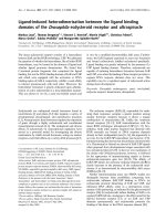
Schematic representations of the fragments are shown in
Fig. 1A.
Construct encoding fragment I
A 1.5-kb cDNA fragment of the Drosophila EcR cDNA
sequence [8] was produced by PCR using the forward
primer 5¢-CGACATATGGGCCAAGACTTTGTTAAG
AAGG-3¢ and the reverse primer 5¢-TCCCCCGGGTCTA
GACTATGCAGTCGTCGAGTGCTC-3¢.Thereby,an
NdeI site was introduced at the 5¢ end, and XbaIandSmaI
sites were introduced at the 3¢ end. The fragment was
digested with NdeI/SmaI and cloned in-frame into pAS2
giving rise to clone pAS2-EcR(375–878), coding for fusion
protein GBD–EcR(375–878) which consists of the GAL4
DNA binding domain fused to the N-terminal end of a
fragment comprising a portion of hinge region, LBD, and
the entire C-terminal domain (also called F domain) of EcR.
A 679-nucleotide portion of the EcR fragment between
AatII (nucleotide 2441) and NarI (nucleotide 3120) was
Fig. 1. Heterodimerization and ligand binding abilities of the EcR
fragments investigated in the present studies as fusion proteins with
GAL4 DNA binding domain (GBD) or GAL4 activation domain (GAD).
(A) Delineation of EcR and USP fragments tested. EcR stands for the
B1 isoform [46] of the Drosophila melanogaster ecdysone receptor [8]. It
spans amino acids 1–878. DBD: DNA binding domain (amino acids
264–329). LBD: ligand binding domain (amino acids 417–651) as
defined by [37]. Helices 10, 11 and 12 (H10, H11, H12) start at amino
acids 610, 628, and 644, respectively [37]. Fragments are numbered
I–VII and correspond to those listed in (B). The dash in fragment I
indicates the location of the spontaneous mutation L442H. The
asterisk designates the fragment which was used most frequently in the
present studies. USP designates Ultraspiracle as characterized by [14].
Delineation of its DBD and LBD as proposed by [34]. (B) Results
obtained with EcR fragments described in (A). Explanation of column
heads and signs in columns: 2-H basal, spontaneous heterodimeriza-
tion with USP fragment in vivo as determined by the two-hybrid assay
and indicated by the appearance of basal (noninduced) b-galactosidase
activity; + or – signs mean this activity to be significantly or unsig-
nificantly higher than background, respectively; 2-H rI,relative
induction of heterodimerization by muristerone A (10
)6
to 5 · 10
)5
M
)
as assayed by the two-hybrid assay (rI: induced level divided by basal
level of b-galactosidase activity): + or – signs mean rI to be signifi-
cantly or unsignificantly higher than 1, respectively; ST-EMSA,
heterodimerization induced in vitro by 10
)5
M
muristerone A and
assayed by ST-EMSA; + or – signs mean supershifted band could or
could not be detected, respectively; LB EcR, ligand binding to EcR
fragment alone; + or – signs mean specific [
3
H] ponasterone A binding
to be significantly or nonsignificantly higher than background,
respectively. LB EcR ± USP: ligand binding to EcR fragment in
presence of USP fragment; GBD- and GAD-EcR fusion proteins were
combined with GAD- or GBD-USP(172–508), respectively; meaning
of + and – signs as in LB EcR column.
3238 M. Lezzi et al. (Eur. J. Biochem. 269) Ó FEBS 2002
exchanged with the corresponding fragment of the original
cDNA clone [8].
Constructs encoding fragment II
Clone pAS2-EcR(375–652) encoding the fusion protein
GBD–EcR(375–652) between the GAL4 DNA binding
domain and a fragment comprising a large portion of hinge
plus the entire LBD of EcR was constructed in an
analogous manner to pAS2-EcR(375–878). The differences
concern the reverse primer 5¢-CGTCCCGGGTCTAGACT
AAACGTCCCAGATCTCCTCG-3¢, the length of PCR
fragment (800 nucleotides), and the exchanged fragment
(566 nucleotides from AatII at nucleotide 2441 to BglII at
nucleotide 3008). The related clone, pAS2-1-EcR(375–652),
was constructed by cutting out the NdeI–SmaIfragment
from pAS2-EcR(375–652) and recloning it into the NdeI–
SmaI site of the pAS2-1 vector. pAS2-EcR(375–652) and
pAS2-1-EcR(375–652) will not be distinguished in the
following. Clone pACT2-EcR(375–652) encodes a fusion
protein, GAD–EcR(375–652), consisting of the same EcR
fragment as pAS2-1-EcR(375–652) but combined with
GAL4 activation rather than DNA binding domain. It
was constructed by use of the forward PCR primer:
5¢-CATGCCATGGGCCAAGACTTTGTTAAGAAGG-3¢
and the reverse primer employed for constructing pAS2-
1-EcR(375–652). The NcoI–SmaI fragment encompassing
the EcR cDNA sequence nucleotide 2191–3024 was cloned
into the NcoI–SmaI site of vector pACT2.
Constructs encoding fragment III
Clone pAS2-1-EcR(375–642) encodes the same EcR-con-
taining fusion proteins as pAS2-1-EcR(375–652) except that
the C-terminal a helix 12 of the LBD in EcR is missing. It
was produced by inserting an NcoI–EcoRI restriction
fragment of a PCR product into the pAS2-1 cloning site.
For producing the PCR fragment, the same forward primer
was used as for pACT2-EcR(375–652). The reverse primer
was 5¢-CGGAATTCTCACAGTTTGCGGTTTTTGAG
CTTTAG-3¢ which generated a stop codon at nucleotide
position 2995. Clone pACT2-EcR(375–642) is analogous to
pAS2-1-EcR(375–642) and was produced by exchanging the
NcoI–EcoRI restriction fragment of pACT2-EcR(375–652)
by that of pAS2-1-EcR(375–642).
Constructs encoding fragments IV and V
The fusion proteins encoded by clones pACT2-1-EcR(375–
627) and pAS2-1-EcR(375–627) or by pACT2-1-EcR
(375–609) and pAS2-1-EcR(375–609) lack helices 11–12 or
10–12 of EcR LBD, respectively. They were produced in an
analogous manner as pAS2-1- and pACT2-EcR(375–642)
by using, however, as reverse primers 5¢-CGGAATTCTC
ACTGGTTGCCCAGCGTACGCAG-3¢ and 5¢-CGGAA
TTCTCAGACGAGGCTCATTGAGTCGCC-3¢,which
introduced stop codons at nucleotide positions 2943 and
2896, respectively.
Constructs encoding fragments VI and VII
Clones pACT2-1-EcR(403–652) and pAS2-1-EcR(403–652)
encode fusion proteins that contain a smaller piece of the
EcR hinge region than clones pACT2-1-EcR(375–652) and
pAS2-1-EcR(375–652). They were produced as described
above by introducing into the respective vectors the NcoI/
EcoRI-cut PCR fragment by use of the forward and reverse
primers 5¢-CATGCCATGGAAATAT TGGCCAAGTGT
CAAGC-3¢ and 5¢-CGGAATTCTCAAACGTCCCAGA
TCTCCTCGAG-3¢, respectively.
Clones pACT2-1-EcR(421–652) and pAS2-1-EcR
(421–652) lack the entire hinge region of EcR. They were
produced as described above using the forward primer
5¢-CATGCCATGGAGTTGGCCGTTATATACAAGTT
AATTTG-3¢.
Plasmids coding for fusion proteins with the C-terminal
fragment of USP
Clone pGAD424-USP(172–508) encodes a fusion protein
consisting of the activation domain of GAL4 and the
C-terminal part of USP including a portion of the hinge
region and the LBD. It was produced by cloning an EcoRI/
StuI-cutPCRfragmentintotheEcoRI/SmaI-cut pGAD424
vector. To generate the PCR fragment, the following
primers were used on the original USP cDNA clone of
Oro and coworkers [14] as a template: 5¢-AGGAATTCGA
AGCGGTCCAGGAGGAG-3¢ and 5¢-AAGGCCTTCTA
GACTACTCCAGTTTCATCGCCAGGCC-3¢.
Although the pGAD424 construct yielded less fusion
protein than the corresponding pACT2 construct (see
below), the same relative effects were obtained when
comparing different EcR fragments or ligands in the two-
hybrid assay.
pAS2-1-USP(172–508) was produced by cutting
pGAD424-USP(172–508) with EcoRI and SalIandthe
resulting fragment was recloned in to the respective sites of
pAS2-1. From this plasmid, the clone pACT2-USP(172–
508) was constructed by producing a NcoI–SalIfragment
that was blunted at its SalI side (filling-in reaction) and then
cloned into the NcoI–SmaIsiteofpACT2.
PCR reaction, sequence verification
For PCR amplification, standard PCR conditions were
employed. All PCR fragments and the resulting inserts were
verified by commercial sequencing (Microsynth and GEN-
terprise; Gachnang, Switzerland and Mainz, Germany,
respectively).
Yeast strains
All yeast strains (Saccharomyces cerevisiae) were purchased
from Clontech. For routine two-hybrid work, strain Y187
was used; this strain harbors the reporter gene lacZ under the
control of a GAL1UAS-GAL1TATA element. In prelimi-
nary two-hybrid studies or experiments with Ôcell-toxicÕ
fusion proteins, the low expressing strains Y153 and Y157
were also employed, which carry the same reporter gene. For
ST-EMSA and ligand binding tests, strain Y190 was
employed, which is favorable for fusion protein expression.
Although these four strains differ in their overall fusion
protein expression and/or strength of reporter gene activity
(lacZ), comparative experiments showed that the relative
effects of different EcR/USP fragment combinations or
ligand types are not influenced by the yeast strain used.
Ó FEBS 2002 Heterodimerization of EcR ligand binding domain (Eur. J. Biochem. 269) 3239
Preparation of yeast extracts for two-hybrid studies
Yeast cells were grown in YPD (1% yeast extract, 2%
Bacto-peptone, 2% glucose) at 30 °C,andthentransformed
or cotransformed by the plasmids mentioned above. The
lithium acetate procedure was used, following the manu-
facturer’s protocol (Clontech). Yeast cells were plated on
synthetic dextrose (SD) minimal medium [0.67% yeast
nitrogen base (DIFCO) and 2% glucose] lacking leucine
and tryptophan to select for cells bearing the plasmids.
Colonies were grown at 30 °C for 3 days and three colonies
were then selected for subsequent liquid culture. With
colonies that did not grow well, a colour reaction was
performed on filter lifts to monitor lacZ activity, according
to the manufacturer’s instructions, as a means to select
colonies for two-hybrid experiments and as an auxiliary
method to evaluate two-hybrid experiments. The selected
colonies were inoculated in 3 mL of an SD liquid medium,
and the culture was grown overnight with shaking. When
the D
600
of the culture reached 1.0, two 100-lL aliquots
were removed and transferred to a second tube with 1.9 mL
of SD liquid medium. One of the tubes was supplemented
with ligand (for type and final concentration, see Results)
and the other was supplemented with ethanol solvent of the
same final concentration, normally 0.25%. Triplicate sam-
ples from each liquid culture were processed according to
manufacturer’s instructions (freeze-thawing) and measured
for lacZ activity by assessing the colour change of Gal-ONp
as measured by absorbance spectrophotometry (D
420
). The
mean reading was then used to calculate the lacZ activity in
ÔMiller UnitsÕ, according to the manufacture’s protocol
(Clontech).
Ligands
Muristerone A (Sigma, Invitrogen or Alexis Biochemicals),
ponasterone A (Sigma), poststerone (a kind gift of
R. Lafont
4
, ENS, Paris), 20-hydroxyecdysone (Sigma), and
the nonsteroidal ecdysone agonist RH 5992 (a kind gift of
Rohm and Haas company, Spring House, PA, USA)
5
were
prepared as ethanol solutions (10 mgÆmL
)1
)andthen
diluted to the final concentration indicated in the text for
use in the two-hybrid experiments (for use in biochemical
analyses, see below).
Preparation of yeast extracts for biochemical analyses
For supershift-type EMSA and ligand binding studies,
growth and transformation of yeast cells were carried out as
for two-hybrid studies. Single colonies less than 4 days old
of yeast transformants expressing GAD–EcR(375–652) or
GBD–USP(172–508) were picked and cultured with sha-
king (150–200 r.p.m) at 30 °C overnight in 5 mL of SD
medium. The overnight cultures were diluted in 50 mL
YPD and grown under the same conditions until
D
600
¼ 0.6–0.8. The cells were then prepared on ice. Cells
were harvested by centrifugation (1500 g,5min,4°C) in
prechilled tubes. Pellets were washed with 50 mL ice-cold
wash buffer (20 m
M
Hepes, 20 m
M
NaCl, 20% glycerol,
1m
M
EDTA, 1 m
M
2-mercaptoethanol, pH 7.9), trans-
ferred into plastic tubes, and frozen in liquid nitrogen. The
frozen pellets were disrupted for 2 min at 2000 r.p.m.
6
using
a Mikro-Dismembrator S (B. Braun Biotech International;
Melsungen, Germany). After thawing, the homogenates
were diluted with binding buffer [wash buffer with 1 m
M
phenylmethanesulfonyl fluoride, pH 7.9] supplemented
with a mixture of protease inhibitors (aprotinin, leupeptin,
pepstatin, benzamidine, antipapain, and cymostatin; final
concentration: 2 lgÆmL
)1
each) just before use. After a
short ultrasonication the samples were centrifuged
(100 000 g,1h,4°C). Phenylmethanesulfonyl fluoride
(final concentration: 1 m
M
) was added to the supernatants.
The extracts were frozen in aliquots at )80 °C until tested.
Supershift-type electrophoretic mobility shift
assays (ST-EMSAs)
A double-stranded probe of the GAL4 binding motif was
preparedandlabelledwith[a-
32
P]dCTP, as described
previously [25]. The reaction mix contained binding buffer
[20 m
M
Hepes, pH 7.4, 100 m
M
KCl, 5% (v/v) glycerol,
2m
M
dithiothreitol, 0.1% NP-40] and yeast cell extracts
with the EcR or USP fusion proteins, 1 lg of the nonspecific
competitor poly(dIdC), 10 fmol labeled oligonucleotide,
and muristerone A at a final concentration of 10
)5
M
or as
indicated in the Results. The reaction mix was incubated at
room temperature for 30 min and separated at 10 VÆcm
)1
on a 5% nondenaturing polyacrylamide gel in Tris/borate/
EDTA (45 m
M
Tris, 45 m
M
boric acid, 0.5 m
M
EDTA
pH 8.0) for 2 h. The gel was analyzed by a
PHOSPHORIMAGER
system (Molecular Dynamics, Sunnyvale, CA, USA).
Ligand-binding assays
Yeast cell extracts were diluted with binding buffer and
supplemented with protease inhibitors immediately before
use. Ligand-binding was determined with [
3
H]ponasterone
A (specific activity 7.9 TBqÆmmol
)1
; a kind gift from.
H. Kayser, Novartis, Switzerland) using a filter assay, as
described previously [26]. Yeast extracts expressing
C-terminal EcR or USP fusion proteins were incubated
with 4–5 · 10
)9
M
[
3
H]ponasterone A for 1 h at room
temperature, either separately or after mixing. For each
sample, the nonspecific binding, determined by addition of
10
)4
M
nonlabelled 20-hydroxyecdysone, was subtracted.
The purity of the [
3
H]ponasterone A was checked routinely
by HPLC analysis before use.
RESULTS
Spontaneous heterodimerization
in vivo
The results listed in Table 1 (see also Figs 1 and 2) indicate
that EcR and USP fragments lacking their own DNA
binding domain can form heterodimers in vivo. Yeast cells
cotransfected with plasmids expressing these fragment types
in the form of fusion proteins with GAL4 activation and
DNA binding domains, respectively, exhibited b-galacto-
sidase activity above background. Neither empty vector
pairs nor combinations of empty vector with a matching
vector coding for a fusion protein were able to bring about
b-galactosidase activity above background levels (not
shown), whether or not muristerone A was included as an
inducer. This indicates that heterodimerization between the
EcR–LBD and the USP–LBD containing fusion proteins is
not the result of an interaction between the GAL4
3240 M. Lezzi et al. (Eur. J. Biochem. 269) Ó FEBS 2002
activation and DNA binding domains. Coexpression of
GAD-/GBD-fusion protein pairs containing only EcR or
USP fragments did not lead to induced b-galactosidase
activity (Table 1), even when muristerone A or the juvenile
hormone analogue methoprene (10
)5
M
)wasaddedtothe
culture (Table 1; T. Bergman, unpublished observation
7
,
respectively). This suggests, first, that these fragments do not
homodimerize and that the reported homodimerization of
full length EcR and USP [22,27] is coordinated and
established by the respective multimeric binding motifs in
DNA. Second, together with experiments in which the
GBD-fusion proteins were expressed alone (results not
shown), the EcR/EcR and USP/USP combinations indicate
that the (putative) AF-2 functions within the LBD of EcR
and USP are inactive in yeast cells, unlike in insect cells [28],
presumably because the essential coactivators are missing
[29]. As expected for a true mutual protein–protein inter-
action, reciprocal exchange of the GAL4 DNA binding and
activation domain had no impact on the reporter gene
regulation. However, the growth rate was always drastically
reduced when the yeast cells were transformed with
GBD–EcR fusion peptides encoding constructs. The phe-
nomenon of Ôcell-toxicityÕ has been encountered previously
with C-terminal progesterone receptor fragments fused to a
ubiquitin peptide [30]. It thus cannot be attributed to the
GAL4 moiety.
The EcR and USP fragments normally employed for our
studies comprised the whole ligand binding domains plus
the carboxy-terminal portion of the hinge region. In the
routine experiments, the F domain of EcR that lies on the
C-terminal side of the LBD was removed as its presence did
not affect heterodimerization appreciably (Fig. 1). Trunca-
ting helix 12 alone or helices 11 and 12 did not abolish
heterodimerization (Fig. 1). It was only after the additional
deletion of helix 10 that the EcR–LBD fragment became
incapable of interacting with USP–LBD. At the N-terminal
end, a gradual shortening of the EcR fragment resulted in a
peculiar dichotomy: GBD fusion proteins tolerated a
removal of the whole hinge region while GAD fusion
proteins failed to heterodimerize when their hinge portion
was further reduced by only 28 amino-acid residues (Fig. 1).
We interpret this negative effect as a steric hindrance of the
EcR–LBD functions through GAL4-AD when the inter-
vening region was too small or missing.
Ligand-induced heterodimerization
in vivo
Presenceofmuristerone Ainthe cultivation mediumcauseda
further increase in b-galactosidase activity of cotransfected
yeast cells (Table 1, Figs 1 and 2). As this effect was observed
neither with a plasmid (pCL1) constitutively expressing
GAL4 nor with a test two-hybrid pair (pTD1-1 and pAV3-
1), it was concluded that the ligand promotes interaction
between the EcR- and USP-LBDs rather than affecting
reporter enzyme or fusion protein stabilities. Theinductionby
ligand fully depends on the presence of helix 11 and 12 butnot
theEcRFdomain. The N-terminal portion of the hingeregion
is dispensible for ligand-induced heterodimerization. The
question of whether the same holds true for the C-terminal
region could notbeassessedbecause of the technical problems
mentioned above: fusion proteins with EcR fragments
deprived of their hinge were either toxic to the yeast cells or
did not heterodimerize, possibly because of steric hindrance.
The effect of muristerone A on heterodimerization
between EcR–LBD and USP–LBD containing fusion
Table 1. Controls to two-hybrid experiments. Betagalactosidase activity is measured as averaged ÔMiller UnitsÕ ± 95% confidence limit (number of
experiments).
12
ND, not determined.
DNA binding
construct
Activation
construct
Basal
activity
a
Induced betagalactosidase
activity (10
)6
M
)
a
Induced betagalactosidase
activity (1–5 · 10
)5
M
)
a
pCL1 pCL1
b
1244.00 ± 480.00 (5) 1021.00 ± 353.00 (3) 1508.00 (2)
pTD1-1 pVA3-1 55.10 ± 3.00 (8) 54.90 ± 2.40 (6) 59.20 ± 37.00 (3)
pAS2-1-USP(172-508) pACT2-EcR(375-652) 0.41 ± 0.07 (29) 0.71 ± 0.21 (8) 7.90 ± 1.80 (28)
pAS2-1-USP(172-508) pACT2-USP(172-508) 0.02 ± 0.02 (3) 0.06 ± 0.00 (1) 0.02 ± 0.00 (3)
pAS2-1-EcR(375-652) pACT2-USP(172-508) 0.68 ± 0.10 (12) 1.04 ± 0.19 (13) 1.77 ± 1.37 (4)
pAS2-1-EcR(375-652) pACT2-EcR(375-652) 0.03 ± 0.01 (5) 0.03 ± 0.01 (4) ND
a
Muristerone A concentration in yeast culture medium.
b
DNA binding and activation function in one protein.
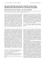
Fig. 2. Dose–response curve. Effect of increasing concentrations of
muristerone A (d, solid line) on two–hybrid interaction between GBD-
USP(172–508) and GAD-EcR(375–652). The effectof 20-hydro-
xyecdysone (.), poststerone (j), RH5992 (m), and of ponasterone A
(s) is also shown at a concentration of 2.5 or 5 · 10
)5
M
. Ligand
concentration refers to concentration of ecdysteroids or agonist in the
yeast culture medium. Where error bars (spanning 95% confidence
limits) are given, the points represent averages of at least three
experiments, each.
Ó FEBS 2002 Heterodimerization of EcR ligand binding domain (Eur. J. Biochem. 269) 3241
proteins was clearly dose-dependent (Fig. 2). A small
increase was evident at a ligand concentration of 10
)6
M
,
and the highest concentration tested (10
)4
M
murister-
one A) caused an 100-fold stimulation of reporter gene
expression compared to the noninduced state. Higher
muristerone A concentrations could not be tested because
of solubility limitations. The effect of muristerone A on
heterodimerization was ligand-specific and clearly related
to the biological activity of the compounds tested.
Besides muristerone A, only ponasterone A (for a
description of ecdysteroids; [2]) was able to promote
heterodimerization. Expectedly, the inactive ecdysteroid
poststerone [2] was ineffective. The natural ecdysteroid
20-hydroxyecdysone and the nonsteroidal ecdysone
agonist RH5992 [31] also failed to promote heterodime-
rization at the concentration tested (2.5 · 10
)5
M
)prob-
ably because of lesser activity.
Ligand-induced heterodimerization
in vitro
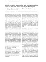
To demonstrate protein–protein interaction directly, a
modification of the electrophoretic mobility shift assay
was devised, the supershift-type (ST)-EMSA, using ex-
tracts of transfected yeast cells. Thereby, the migration of
a radioactively labeled DNA probe was further retarded
when complexed with a second protein that in turn binds
to the DNA binding protein. A GAL4 binding motif was
used as a probe to test for the GBD–USP/GAD–EcR
interaction. The investigated proteins corresponded to the
fusion proteins constructed for two-hybrid assays. The
DNA binding domain of GAL4 alone shifted the probe
to some extent (Fig. 3A), whereas its fusion with
USP(172–508) caused a further shift, which, however,
was not supershifted by the addition of muristerone A
(Fig. 3A, lanes 4 and 5). This excludes the possibility that
muristerone A would produce the supershifts seen in
Fig. 3B (lanes 2–4) by causing, for example, endogenous
yeast proteins to interact with USP or by inducing
multimerization of USP. In the absence of muristerone A,
addition of the EcR fusion protein also did not cause
supershifting (Fig. 3B). Supershifting caused by murister-
one A was noticeable at 10
)6
M
(Fig. 3B) and reached
saturation at 10
)5
M
(Fig. 3B). The effect of murister-
one A is not due to a general property of ecdysteroids as
the inactive ecdysteroid poststerone was unable to induce
supershifting (results not shown). As far as tested, all
those EcR fragments which did not react towards
muristerone A by heterodimer formation in vivo also
failed to react toward this ecdysteroid in vitro when
investigated by ST-EMSA (Fig. 1B).
Fig. 3. Heterodimer formation in vitro between GBD–USP(172–508) (GBD–USP) and GAD–EcR(375–652) (GAD–EcR) as assayed by supershift-
type (ST)-EMSA. (A) Controls. Arrows: Position of shifted bands. GAL4 (BD), DNA binding domain of GAL4. Dashed arrow: expected position
of band if supershifted. (B) Influence of various muristerone A concentrations on heterodimerization between GBD–USP(172–508) and GAD–
EcR(375–652). Arrows: position of shifted or supershifted bands, respectively.
3242 M. Lezzi et al. (Eur. J. Biochem. 269) Ó FEBS 2002
Ligand binding to EcR or USP fragments containing
fusion proteins
As heterodimerization between C-terminal fragments of
EcR and USP could be induced by the potent ecdysteroids
muristerone A or ponasterone A, we wanted to investigate
whether these fragments can bind ponasterone A in a ligand-
specific manner
8
. Therefore we conducted ligand binding
experiments with the same yeast-expressed fusion proteins
as used for the two-hybrid experiments, employing [
3
H]
ponasterone A as a probe. Extract of yeast cells expressing
only GBD–USP(172–508) did not bind [
3
H]ponasterone A
as its binding level did not differ significantly from that of
nonexpressing yeast extract (background). In contrast,
extract containing only expressed GAD–EcR(375–652)
showed a low level of [
3
H]ponasterone A binding that was
significantly higher than background. Binding to GAD–
EcR(375–652) expressing extract was specific as it could be
competed out by the active hormone 20-hydroxyecdysone.
Supplementation of this extract with GBD-USP(172–508)
increased specific [
3
H]ponasterone A binding 10-fold.
Ligand binding to the EcR fusion protein both in the
presence or absence of the USP fusion protein was
dependent on the existence of an intact helix 12. As far as
tested, those EcR fusion protein types that could not be
induced by ligand to heterodimerize also failed to bind
[
3
H]ponasterone A, in either the presence or absence of the
USP partner (Fig. 1). This suggests that ligand-induced
heterodimerization of the EcR and USP fragments is related
to the ligand binding to EcR in presence but also in absence
of USP.
DISCUSSION
The present work shows that fragments of EcR and USP
containing a complete LBD but lacking their DNA binding
domains are able to heterodimerize in vivo as well as in vitro.
This heterodimerization obviously did not depend on a
coordinated binding to a bipartite motif in DNA. Thus,
heterodimer formation may take place in their free,
nonchromosomally bound state. Moreover, the D- and
T-boxes [32,33] that are part of a weaker dimerization
interface in other nuclear receptors are not required for EcR
and USP to heterodimerize. However, binding to specific
EcREs and the presence of these boxes or other N-terminal
regions may play an important role in modulating the EcR/
USP interaction, in vivo as well as in vitro [25]. Moreover,
spontaneous heterodimerization in the absence of ligand
between EcR and USP fragments requires neither the hinge
region nor the F domain of EcR, and even helices 11 and 12
of its LBD are dispensible for a protein–protein interaction
to occur. However, helix 10 which contributes substantially
to the dimerization interface in nuclear receptors [34] must
be present (Fig. 1).
There is little doubt that the induction of b-galactosidase
activity in yeast cells cotransfected by GBD–USP(172–508)
and GAD–EcR(375–652) results from an interaction of the
two nuclear receptor fragment types rather than recruitment
of endogenous activator proteins (see controls in Table 1;
[29]). However, indirect interactions between the EcR and
USP fragments being mediated by, for example, SRC-1-like
proteins [35] cannot be excluded. Strikingly, such indirect
interactions would still depend on the presence of EcRs
helix 10. In any event, the frequency of EcR–USP hetero-
dimers in untreated cotransformants is very low (i.e. less
than 1% as inferred from the maximal reporter enzyme
activity being inducible by muristerone A), (Fig. 2).
In contrast to spontaneous heterodimerization, ligand-
induced heterodimerization requires the presence of helices
11 and 12. In vitro analyses like ST-EMSA and ligand
binding experiments substantiate this notion. This inter-
pretation conforms with the role ascribed to these helices in
the Ômouse-trapÕ mechanism that connects ligand binding to
conformational changes of EcR–LBD [34]. The requirement
of the C-terminal half of the hinge region for ligand-induced
heterodimerization could not be assessed because of tech-
nical problems. Thus, the role of the hinge region for ligand-
dependent effects and ligand binding remains a matter
of conjecture (compare [36] with the alignment in [37]).
The induction by ligand of heterodimerization between
C-terminal EcR and USP fragments is not a general effect of
steroidal or small lipophilic molecules (Fig. 2).
The concentration of muristerone A needed in the yeast
culture medium to induce heterodimerization is relatively
high. We estimated the half maximal muristerone A con-
centration (EC
50
) to be about four orders of magnitude
higher than determined with other assays [28,38]. However,
it is a general experience with yeast two-hybrid assays that
high steroid hormone concentrations must be applied to
induce dimerization [39,40]. This is because yeast cells are
equipped with efficient extrusion mechanisms to exclude
small molecules such as steroids [41] and, as recently shown,
the ecdysone agonist RH5992 [42]. The muristerone A
concentration required to induce heterodimerization as
revealed by supershifting in ST-EMSAs was about the same
(Fig. 3) as needed for demonstrating heterdimerization
when using full-length EcR and USP molecules and natural
EcREs [20–23,28]. The ponasterone A concentration used in
ligand binding experiments was about 5 · 10
)9
M
and high
enough to induce heterodimerization. This was concluded
from the fact that ligand binding was enhanced approxi-
mately 10-fold if GAD-EcR was assayed in presence of
GBD–USP which by itself does not bind [
3
H]ponaster-
one A. Thus, the two LBDs must cooperatively interact by
heterodimerization leading to an enhancement of ligand
binding to the EcR partner. A concentration of 5 · 10
)9
M
is well within the range of biological and binding activity of
ponasterone A as determined with full-length Drosophila
receptors [20,28,37,43].
Even though ligand binding to EcR–LBD was low, it was
significant and has been observed before with full-length
EcR [20,43–45]. There exists a clear positive correlation
between ligand binding and ligand inducibility of hetero-
dimerization. All those EcR fragments that did not bind
[
3
H]ponasterone A also failed to show an enhancement of
heterodimerization by muristerone A (Fig. 1). This corre-
lation is further noted in a series of point mutations in EcR–
LBD (T. Bergman, M. Lezzi, M. Grebe, W. W. Hitchcock
& V. C. Henrich, unpublished results)
9
.Furthermore,itis
striking that EcR forms that do not bind ligand in the
absence of USP cannot be induced to heterodimerize by
ligandwhenexpressedbyin vitro translation (compare EcR
of Chironomus tentans [23,45]).
The ability of Drosophila EcR to bind ligand as a
ÔmonomerÕ opens a new possibility of how ligand may act.
In a schematic model, Yao and coworkers [20] proposed
Ó FEBS 2002 Heterodimerization of EcR ligand binding domain (Eur. J. Biochem. 269) 3243
ecdysteroids to interact only with the EcR/USP hetero-
dimer, either in its free or DNA-bound state. We suggest it
may be possible that ecdysteroids interact with the EcR
monomer, rendering it more competent to heterodimerize
with USP. With such a possibility, ligand-induced dimeri-
zation could be an early step, clearly preceding the DNA
binding step of the receptor complex.
ACKNOWLEDGEMENTS
We thank Mrs Himmelberg for excellent technical assistance and
W. Schaffner and A. Barberis (University of Zurich) as well as C. Elke,
F. Thoma and J F. Mouillet (E.T.H Zurich) for technical advice and
intellectual inputs. We are grateful to R. Palli (Rohm and Haas
Company) and J M. Wurtz (University of Strasbourg) for providing us
with preprints. M. G. and S. P. were supported by fellowships of DFG
(Graduiertenkolleg: Oekotoxikologie und Umwelthygiene) and the
ÔDu
¨
sseldorf Entrepreneur FoundationÕ, respectively. This work was
financially supported by grants from ETH Zurich to M. L. and T. B. as
well as to M. V. and C. F., from the US Department of Agriculture to
V. C. H., and from DFG (Sp 350/3–1) to M. S B. Finally, V. C. H.
was a visiting professor at ETH Zurich during a research assignment
from UNC-Greensboro.
REFERENCES
1. Ka
¨
user, G. (1989) On the evolution of ecdysteroid hormones. In
Ecdysone. From Chemistry to Mode of Action. (Koolman, J., ed.)
Georg Thieme Verlag, Stuttgart, pp. 327–336.
2.Lafont,R.D.&Wilson,I.D.(1992)The Ecdysone Handbook.
Chromatographic Society Surveys. pp. 1–392. The Chromato-
graphic Society,
10
Nottingham, UK.
3. Ecker, J.R. (1997) BR1-ghtening the pathway to steroid hormone
signaling events in plants. Cell 90, 825–827.
4. Henrich, V.C., Rybczynski, R. & Gilbert, L.I. (1999) Peptide
hormones, steroid hormones and puffs: mechanisms and models in
insect development. Vitamins Hormones 55, 73–125.
5. Spindler-Barth, M. & Spindler, K D. (2000) Hormonal regulation
of larval moulting and metamorphosis – molecular aspects. In
Progress in Developmental Endocrinology (Dorn, A., ed.) pp. 117–
144, Wiley-Lyss, New York.
6. Lezzi, M. (1996) Chromosome puffing: supramolecular aspects of
ecdysone action. In Metamorphosis, Postembryonic Reprogram-
ming of Gene Expression in Amphibian and Insect Cells (Gilbert,
L.I., Tata, J.R. & Atkinson, B.G. eds) pp. 145–173, Academic
Press, San Diego.
7. Kozlova, T. & Thummel, C.S. (2000) Steroid regulation of post-
embryonic development and reproduction in Drosophila. Trends
Endocrin. Metab. 11, 276–280.
8. Koelle, M.R., Talbot, W.S., Segraves, W.A., Bender, M.T.,
Cherbas, P. & Hogness, D.S. (1991) The Drosophila EcR gene
encodes an ecdysone receptor, a new member of the steroid
receptor superfamily. Cell 67, 59–77.
9. Lezzi, M., Bergman, T., Mouillet, J F. & Henrich, V.C. (1999)
The ecdysone receptor puzzle. Arch. Insect Biochem. Physiol. 41,
99–106.
10. Mangelsdorf, D.J., Thummel, C., Beato, M., Herrlich, P., Schu
¨
tz,
G., Umensono, K., Blumberg, B., Kastner, P., Marks, M.,
Chambon, P. & Evans, R.M. (1995) The nuclear receptor super-
family: the second decade. Cell 83, 835–839.
11. Mangelsdorf, D.J. & Evans, R.M. (1995) The RXR heterodimers
and orphan receptors. Cell 83, 841–850.
12. Gehring, U. (1998) Steroid hormone receptors and heat shock
proteins. Vitamins Hormones 54, 167–205.
13. Henrich, V.C., Sliter, T.J., Lubahn, D.B., MacIntyre, A. &
Gilbert, L.I. (1990) A steroid/thyroid hormone receptor super-
family member in Drosophila melanogaster that shares extensive
sequence similarity with a mammalian homologue. Nucleic Acid
Res. 18, 4143–4148.
14. Oro, A.E., McKeown, M. & Evans, R.M. (1990) Relationship
between the product of the Drosophila ultraspiracle locus and
vertebrate retinoid X receptor. Nature 347, 298–301.
15. Shea,M.J.,King,D.L.,Conboy,M.J.,Mariani,B.D.&Kafatos,
F.C. (1990) Proteins that bind to Drosophila chorion cis-regulatory
elements: a new C2H2 zinc finger protein and a C2C2 steroid
receptor-like component. Genes Dev. 4, 1128–1140.
16. Torchia, J., Glass, C. & Rosenfeld, M.G. (1998) Coactivators and
co-repressors in the integration of transcriptional responses. Curr.
Opin. Cell Biol. 10, 373–383.
17. Cherbas, P. & Cherbas, L. (1996) Molecular aspects of ecdysteroid
hormone action. In Metamorphosis, Postembryonic Reprogram-
ming of Gene Expression in Amphibian and Insect Cells (Gilbert,
L.I., Tata, J.R. & Atkinson, B.G. eds) pp. 175–221. Academic
Press, San Diego.
18. Wegmann, I.S., Quack, S., Spindler, K D., Dorsch-Ha
¨
sler, K.,
Vo
¨
gtli, M. & Lezzi, M. (1995) Immunological studies on the
developmental and chromosomal distribution of ecdysteroid
receptor protein in Chironomus tentans. Arch. Insect Biochem.
Physiol. 30, 95–114.
19. Stocker, A.J., Amabis, J., Gorab, E., Elke, C. & Lezzi, M. (1997)
Antibodies against the
D
-domain of a Chironomus ecdysone
receptor protein react with DNA puff sites in Trichosia pubescens.
Chromosoma 106, 456–464.
20. Yao, T P., Forman, B.M., Jiang, Z., Cherbas, L., Chen, J.D.,
McKeown,M.,Cherbas,P.&Evans,R.M.(1993)Functional
ecdysone receptor is the product of EcR and Ultraspiracle genes.
Nature 366, 476–479.
21. Thomas, H.E., Stunnenberg, H.G. & Stewart, A.F. (1993)
Heterodimerization of the Drosophila ecdysone receptor with
retinoid X receptor and ultraspiracle. Nature 362, 471–475.
22. Vo
¨
gtli, M., Elke, C., Imhof, M. & Lezzi, M. (1998) High level
transactivation by the ecdysone receptor complex at the core
recognition motif. Nucleic Acids Res. 26, 2407–2414.
23. Elke, C., Rauch, P., Spindler-Barth, M. & Spindler, K D. (1999)
DNA-binding properties of the ecdysteroid receptor-complex
(EcR/USP) of the epithelial cell line from Chironomus tentans.
Arch. Insect Biochem. Physiol. 41, 124–133.
24. Reichardt, H.M., Tronche, F., Bauer, A. & Schu
¨
tz, G. (2000)
Molecular genetics of glucocorticoid signalling using the Cre/loxP
system. Biol. Chem. 38, 961–964.
25. Mouillet, J F., Henrich, V.C., Lezzi, M. & Vo
¨
gtli, M. (2001)
Differential control of gene activity by isoforms A, B1 and B2 of
the Drosophila ecdysone receptor. Eur. J. Biochem. 268, 1811–
1819.
26. Turberg, A. & Spindler, K D. (1992) Properties of nuclear and
cytosolic ecdysteroid receptors from an epithelial cell line from
Chironomus tentans. J. Insect Physiol. 38, 81–91.
27. Seibel, N.K. (1999) Identification and characterization of naturally
occuring ecdysone response elements in Chironomus tentans and
Drosophila melanogaster.PhDThesis,SwissFederalInstituteof
Technology Zurich, Switzerland.
28. Hu, X. (1998) The mechanisms of activating the functional ecdysone
receptor complex. PhD Thesis, Indiana University, USA.
29. Tran, H.T., Askari, H.B., Shaaban, S., Price, L., Palli, S.R.,
Dhadialla, T.S., Carlson, G.R. & Butt, T.R. (2001) Reconstruction
of ligand-dependent transactivation of Choristoneura umiferana
ecdysone receptor in yeast. Mol. Endocrinol. 15, 1140–1153.
30. Mak, P., McDonnell, D., Weigel, N.L., Schrader, W.T. &
O’Malley, B.W. (1989) Expression of functional chicken oviduct
progesterone receptors in yeast (Saccharomyces cerevisiae). J. Biol.
Chem. 264, 21613–21618.
31. Smagghe, G. & Degheele, D. (1994) Action of a novel nonsteroidal
ecdysteroid mimic, tebufenozide (RH-5992). Pestic. Sci. 42,85.
3244 M. Lezzi et al. (Eur. J. Biochem. 269) Ó FEBS 2002
32. Umensono, K. & Evans, R.M. (1989) Determinants of target gene
specificity for steroid/thyroid hormone receptors. Cell 57, 1139–
1146.
33. Hsie, J.C., Jurutka, P.W., Selznick, S.H., Reeder, M.C., Haussler,
C.A., Whitfield, G.K. & Haussler, M.R. (1995) The T-box near
the zinc fingers of the human vitamin D receptor is required for
heterodimeric DNA binding and transactivation. Biochem. Bio-
phys. Res. Commun. 215,1–7.
34. Wurtz, J M., Bourguet, W., Renaud, J P., Vivat, V., Chambon,
P., Moras, D. & Gronemeyer, H. (1995) A canonical structure for
the ligand-binding domain of nuclear receptors. Nat. Struct. Biol.
3, 87–94 and 206.
11
35. Westin, S., Kurokawa, R., Nolte, R.T., Wisely, G.B., MaInerney,
E.M.,Rose,D.W.,Milburn,M.V.,Rosenfeld,M.G.&Glass,C.K.
(1998) Interactions controlling the assembly of nuclear-receptor
heterodimers and co-activators. Nature 395, 199–202.
36. Perera, S.C., Sundaram, M., Krell, P.J., Retnakaran, A., Dhadi-
alla, T.S. & Palli, S.R. (1999) An analysis of ecdysone receptor
domains required for heterodimerization with ultraspiracle. Arch.
Insect Biochem. Physiol. 41, 61–70.
37. Wurtz, J M., Guillot, B., Fagart, J., Moras, D., Tietjen, K. &
Schindler, M. (2000) A new model for 20-hydroxyecdysone and
dibenzylhydrazine binding: a homology and docking approach.
Protein Sci. 9, 1073–1084.
38. Cherbas, L., Yonge, C.D., Cherbas, P. & Williams, C.M. (1980)
The morphological response of Kc-H cells to ecdysteroids: hor-
monal specificity. Wilhelm Roux’s Archives 189, 1–15.
39. Wang, H., Peters, G.A., Zeng, X., Tang, M., Ip, W. & Khan, S.A.
(1995) Yeast two-hybrid system demonstrates that estrogen
receptor dimerization is ligand-dependent in vivo. J. Biol. Chem.
270, 23322–23329.
40. Heery, D.M., Zacharewski, T., Pierrat, B., Gronemeyer, H.,
Chambon, P. & Losson, R. (1993) Efficient transactivation by
retinoid acid receptors in yeast requires retinoid X receptors. Proc.
Natl Acad. Sci. USA 90, 4281–4285.
41. Mahe, Y., Parle-McDermott, A., Nourani, A., Delahodde, A.,
Lamprecht, A. & Kuchler, K. (1996) The ATP binding cassette
transporters Pdr5 and Snq2 of Saccharomyces cerevisiae can
mediate transport of steroids in vivo. J. Biol. Chem. 271, 25167–
25172.
42. Hu, W Q., Feng, Q L., Palli, S.R., Krell, P.J., Arif, B.M. &
Retnakaran, A. (2001) The ABC transporter, PDR5 mediates the
efflux of non-steroidal ecdysone agonists in Saccharomyces cere-
visiae. Eur. J. Biochem. 268, 3416–3422.
43. Dela Cruz, F. & Mak, P. (1997) Drosophila ecdysone receptor
functions as a constitutive activator in yeast. J. Steroid Biochem.
Molec. Biol. 62, 353–359.
44. Elke, C., Vo
¨
gtli, M., Rauch, P., Spindler-Barth, M. & Lezzi, M.
(1997) Expression of EcR and USP in Escherichia coli:purifica-
tion and functional studies. Arch. Insect Biochem. Physiol. 35,
59–69.
45. Vo
¨
gtli, M., Imhof, M.O., Brown, N.E., Rauch, P., Spindler-Barth,
M., Lezzi, M. & Henrich, V.C. (1999) Functional characterization
of two Ultraspiracle forms (CtUSP-1 and CtUSP-2) from Chiro-
nomus tentans. Insect Biochem. Mol. Biol. 29, 931–942.
46. Talbot, W.S., Swyryd, E.A. & Hogness, D.S. (1993) Drosophila
tissues with different metamorphic responses to ecdysone express
different ecdysone receptor isoforms. Cell 73, 1323–1337.
Ó FEBS 2002 Heterodimerization of EcR ligand binding domain (Eur. J. Biochem. 269) 3245