ADVANCED DIGESTIVE ENDOSCOPY: ERCP - PART 2 doc
Bạn đang xem bản rút gọn của tài liệu. Xem và tải ngay bản đầy đủ của tài liệu tại đây (608.33 KB, 43 trang )
Gallbladder ERCP is not an ideal examination of the gallbladder. If the
gallbladder is filled, a delayed film of the gallbladder should be taken after 30–
45 min. This allows time for the contrast to mix with bile for better definition
of gallstones (Fig. 3.12). Failure to fill the gallbladder despite adequate filling of
the intrahepatic ducts suggests cystic duct obstruction. Stone impaction in the
CHAPTER 334
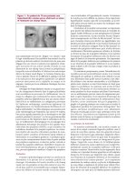
Fig. 3.12 ERCP for gallbladder stones. Gallstones may be obvious on cholangiogram. Note
aberrant duct which resembles cystic duct. Always check delayed film of gallbladder for small
stones.
This is trial version
www.adultpdf.com
cystic duct may cause edema and compression of the common hepatic duct
giving rise to Mirizzi’s syndrome.
Underfilling and delayed drainage With an adequate intrahepatic cholan-
giogram, underlying parenchymal liver diseases may be inferred from abnormal
appearance of the intrahepatic ducts. Crowding of tortuous intrahepatic ducts
may suggest liver cirrhosis. Stretching of a particular intrahepatic duct may be
seen around space-occupying lesions such as abscesses, tumors, or cysts in the
liver.
Underfilling of the bile ducts or ‘streaming effect of contrast’ may suggest an
apparent narrowing in the distal bile duct. Inadequate filling due to stricture or
obstruction may fail to detect intrahepatic pathologies such as stones in patients
with hepatolithiasis. Functional obstruction at the papilla is difficult to diag-
nose, but is suspected if there is delayed drainage of contrast (> 45 min).
The clinical diagnosis of papillary stenosis or sphincter of Oddi dysfunction
depends on the presence of abnormal liver function tests with or without a
dilated bile duct associated with right upper quadrant abdominal pain. Mano-
metric studies are necessary to confirm the diagnosis in patients without obvious
duct dilation or liver test abnormalities. Bile leaks and fistulas complicating
biliary tract surgery can be readily identified on cholangiography.
Section II: Diagnostic and therapeutic ERCP
Diagnostic ERCP
Scopes
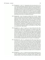
ERCP is performed using side-viewing duodenoscopes with a 2.8, 3.2, or
4.2 mm channel. All of these scopes readily accept a 5 Fr or 6 Fr catheter and
accessories. The larger channel duodenoscopes accept accessories up to 10–
11.5 Fr diameter and are used for both diagnostic and therapeutic purposes. The
larger instrument channel allows aspiration of duodenal contents even with an
accessory in place, and also permits the manipulation of two guidewires or
accessories simultaneously.
Accessories (Fig. 3.13)
The cannula or diagnostic catheter is a 6 or 7 Fr Teflon tube which tapers to a
3–5 Fr tip. It is used for injection of contrast into the ductal systems. A variety of
cannulas are available with different tip designs. A commonly used example is
the bullet tip or fluorotip catheter, which has a small metal or radiopaque tip at
FUNDAMENTALS OF ERCP 35
This is trial version
www.adultpdf.com
the end to facilitate orientation and cannulation on fluoroscopy. Other catheters
may have a tapered tip which facilitates cannulation. Some catheters have
two lumens, which allow both injection of contrast and manipulation of a
guidewire. Most allow the passage of standard (0.035 inch) guidewires.
Preparation of patient
Most ERCP examinations are performed on an outpatient basis provided that
the patient is physically fit and recovery facilities are available. Rarely, ERCP is
performed as an inpatient procedure for patients with significant comorbidities
or those in whom therapeutic procedures or surgery may be necessary.
Informed consent
ERCP is a complex procedure with significant potential hazards. It is important
that the patient understands the potential benefits, risks, limitations, and alterna-
tives. Written, informed consent should be obtained in the presence of a witness.
Fasting
The patient is instructed to fast overnight, or for at least 4 h prior to the proce-
dure. Outpatient procedures are preferably performed in the morning to allow
more time for recovery.
Antibiotics
Antibiotics are given for endocarditis prophylaxis according to local and
national guidelines. ERCP can cause clinical infection if the procedure does not
CHAPTER 336
Fig. 3.13 Accessories: cannula,
guidewire, and papillotome.
This is trial version
www.adultpdf.com
relieve the obstruction and if cleaning and disinfection regimens are not ideal.
Antibiotics are given prophylactically when difficulty in drainage is anticipated,
e.g. in patients with multiple strictures (hilar tumors or sclerosing cholangitis)
or pseudocysts. Antibiotics should also be given immediately if obstruction is
not relieved.
ERCP procedure
Intubation and examination of the stomach
When the patient is adequately sedated, a self-retaining mouth guard is placed
and the patient is supported in a left lateral/semiprone position. This position
facilitates intubation and examination of the upper GI tract with the side-
viewing duodenoscope.
With the patient in the prone position, slight left rotation of the scope is
required to correct for the change in axis. Gentle downward tip angulation
allows examination of the distal esophagus. Once in the stomach, the gastric
juice is removed by suction to minimize the risk of aspiration. The stomach is
inflated slightly to allow an adequate view of the lumen.
The endoscope is slowly advanced with the tip angled downwards looking
at the greater curve and distal stomach. With further advancement, the scope
will pass the angular incisura. The cardia can be examined by up angulation and
withdrawal of the scope.
Once past the angular incisura the tip of the scope is further angled down-
wards and the pylorus is visualized. The scope is positioned so that the pylorus
lies in the center of the field. The tip of the endoscope is then returned to the
neutral position as the pylorus disappears from the endoscopic view, the so-
called ‘sun-setting sign’.
Gentle pushing will advance the scope into the first part of the duodenum.
The scope is angled downwards again and air is insufflated to distend the duode-
num. Care must be taken to avoid overinflating the duodenum as this causes
patient discomfort and makes the procedure more difficult. Careful examina-
tion is performed to rule out any pathologies such as ulcers or duodenitis.
The scope is pushed further to the junction of the first and second part of the
duodenum.
At this point, the scope is angled to the right and upwards, and by rotating
the scope to the right and withdrawing slowly, the tip of the scope is advanced
into the second part of the duodenum. This paradoxical movement shortens
the scope using the pylorus as a pivot, bringing it into the classical ‘short scope
position’. The markings on the duodenoscope should indicate 60–65 cm at the
incisors.
FUNDAMENTALS OF ERCP 37
This is trial version
www.adultpdf.com
With the patient prone, and the scope returned to a neutral position, the
papilla can be easily visualized, in the middle of the second portion of the duode-
num. The landmark for identification of the papilla is the junction where the
horizontal folds meet the vertical fold. Duodenal diverticula may cause difficult-
ies with cannulation as the papilla may be located on the edge or rarely inside a
diverticulum.
Approaching the main papilla
A control film of the right upper abdomen is taken to look for calcification and
for air in the biliary system, prior to injection of contrast.
Cannulation is performed in the short scope position allowing better control
over angulations and tip deflection. In some difficult cases or in attempted minor
papilla cannulation, the long scope approach may be adopted. Excess bubbles in
the duodenum can be removed by injecting a diluted simethicone solution down
the channel. Duodenal contractions may be reduced with the use of antispas-
modic medication.
The presence of a periampullary diverticulum does not normally increase the
technical difficulty of cannulation, unless the papilla is displaced or located
inside the diverticulum (Fig. 3.14).
The normal papilla appears as a pinkish protruding structure and the size
may vary. Abnormalities result from previous stone passage, stone impaction,
or tumor.
Cannulation of the papilla
Cannulation is best performed in an ‘en face’ position. The cannula should be
flushed and primed with contrast to remove any air bubbles prior to insertion
into the duodenoscope. Air injected into the biliary system could mimic stones.
Flushing excess contrast in the duodenum should be avoided since hypertonic
contrast stimulates duodenal peristalsis.
A combination of 12 different maneuvers can be used for positioning the tip
of the cannula for cannulation. These include up/down and sideways angula-
tion, rotation of the endoscope, use of the elevator, and pushing in and pulling
back of the scope. Suction collapses the duodenum and pulls the papilla closer to
the endoscope. Air insufflation pushes it away. Most beginners find pancreato-
graphy easier to obtain than cholangiography. The pancreatic duct is normally
entered by inserting the cannula in a direction perpendicular to the duodenal
wall, in the 1–2 o’clock orientation (Fig. 3.15).
Fine adjustments of the position and axis of the cannula are helpful. Exces-
sive pressure in the papilla is best avoided because pushing may distort the
CHAPTER 338
This is trial version
www.adultpdf.com
papilla and increase the difficulty with cannulation. Cannulation of the CBD is
usually achieved by approaching the papilla from below, in line with the axis of
the CBD. It may be helpful to lift the roof of the papilla, and to direct the cannula
towards 11 o’clock (Fig. 3.16).
Full strength contrast should be used initially, and is injected under fluoro-
scopic control. The pancreatic duct should be filled until the tail and some side
branches are visualized. Avoid overfilling and acinarization as this increases the
risk of post-ERCP pancreatitis. When filling the CBD, start with full strength
contrast and consider switching over to dilute contrast when stones are visual-
ized. If deep cannulation is successful, aspirate bile before injecting contrast to
avoid excess contrast masking small stones in a dilated biliary system.
The left hepatic ducts usually fill before the right because they are dependent
with the patient lying prone. The gallbladder is usually filled except in cases with
cystic duct obstruction. Multiple spot films are taken during contrast injection.
It may be necessary to change the scope position to expose the portion of the
common duct hidden behind the scope.
FUNDAMENTALS OF ERCP 39
Fig. 3.14 The obscure papilla. Look for bile! Lift the overhanging fold. With prior
papillotomy, biliary orifice is often more cephalad. Note relationship of papilla to duodenal
diverticula. Probing or suction to change shape of diverticulum and axis to reveal the papilla.
This is trial version
www.adultpdf.com
CHAPTER 340
Fig. 3.15 Selective pancreatic duct cannulation. Cannula perpendicular to duodenal wall.
Aim at 1–2 o’clock position. ‘Drop’ the cannula by withdrawing tip of scope, relax
up angulation or lower elevator. Use hydrophilic guidewire.
Fig. 3.16 Selective CBD cannulation. Stay close to papilla, approach from below, lift roof of
papilla. Cannula directed at 11–12 o’clock position, use papillotome if needed.
This is trial version
www.adultpdf.com
At the end of the procedure the endoscope is withdrawn and air is suctioned
from the stomach to minimize discomfort. The patient is then turned to a supine
position and more radiographs are taken in different projections (as previously
described).
In patients with a partially filled gallbladder, immediate diagnosis of gall-
stones may be difficult due to inadequate mixing of contrast with bile. Delayed
films of the gallbladder (after about 45 min) may reveal small stones after allow-
ing time for the contrast to mix with bile.
Ease and success in cannulation
Success of diagnostic ERCP depends on the experience of the endoscopist and
the presence or absence of pathology. Successful cannulation of both ductal sys-
tems is commonly achieved in 85–90% of cases with experts achieving rates of
over 95%. The success rate is lower in patients with previous gastric surgery,
e.g. Billroth II gastrectomy.
Minor papilla cannulation
The minor papilla is located proximally and to the right of the main papilla. It
can be identified as a small protruding structure. It may not be obvious or may
appear as a slightly pinkish nipple between the duodenal folds. When promi-
nent, it can sometimes be mistaken for the main papilla; however, it does not have
a distinct longitudinal fold and the small opening usually resists cannulation.
Cannulation of the minor papilla is indicated in patients with suspected or
proven pancreas divisum and when cannulation of the pancreatic duct fails at
the main papilla. Cannulation of the minor papilla is usually best performed in
a long scope position using a 3 mm fine metal tip cannula. Bending the tip of the
cannula to form an angle facilitates cannulation.
It is important to identify the correct location of the orifice before any
attempt is made to inject contrast, as trauma from the cannula may result in
edema and bleeding and obscure the opening.
If the papilla or orifice is not obvious, it is useful to give secretin by slow IV
infusion and wait 2 min to observe the flow of pancreatic juice. During injection,
it is important to monitor the contrast filling by fluoroscopy as the tip of the can-
nula is often hidden by the endoscope in the long scope position.
Complications of diagnostic ERCP
The complication rate for diagnostic ERCP is very low in experienced hands. In
addition to the specific risks related to ERCP, the procedure also carries the risks
FUNDAMENTALS OF ERCP 41
This is trial version
www.adultpdf.com
of any endoscopic procedure including those related to sedation and scope
perforation.
Respiratory depression and other complications
Adverse drug reactions and respiratory depression due to excess medication
may occur. This complication is best prevented by giving sedation slowly in
small increments, and by assessing the overall response of the patient. Proper
monitoring of blood pressure, pulse, and oxygenation helps to avoid this com-
plication. The use of oxygen at 2 liters/min given via a nasal catheter helps to
prevent hypoxia. Glucagon may increase the blood sugar level in diabetic
patients and the anticholinergic effect of Buscopan may cause tachyarrhythmia.
These unwanted side-effects should be monitored.
Pancreatitis
Pancreatitis is the commonest serious complication of ERCP. The serum amy-
lase often increases transiently following pancreatography and may be of little
clinical significance. The incidence of clinical pancreatitis is 0.7–7%. The risk is
higher when the pancreas is overfilled, in patients with sphincter of Oddi dys-
function with manometry, and in those with pancreatic manipulation.
Cholangitis
The risk of cholangitis after ERCP is small, but may occur in patients with bile
duct obstruction due to stones or stricture, especially when biliary drainage
cannot be established. The risk of sepsis is high in patients with acute cholangitis
when the intraductal pressure is raised by excess injection of contrast. The risk
can be reduced by aspirating bile before injecting contrast.
The most common bacteria causing biliary sepsis include Gram-negative
bacteria, i.e. Escherichia coli, Klebsiella, and Enterobacter, and Gram-positive
enterococci. An improperly reprocessed duodenoscope may carry a risk of
cross-infection with other bacteria such as Pseudomonas spp.
Failed cannulation and special situations
What to do with a difficult intubation
Failure to insert the duodenoscope Side-viewing scopes are usually easier to
pass into the esophagus than standard forward-viewing scopes because of the
rounded tip. Difficulty may be encountered if the patient is anxious or struggling
CHAPTER 342
This is trial version
www.adultpdf.com
due to inadequate sedation. Careful explanation and reassurance prior to the
procedure help to alleviate the patient’s anxiety.
It is sometimes difficult for patients to swallow in the prone position.
Supporting the patient in the left lateral position during scope insertion may
help to overcome this problem. Check that the scope angulations are appropri-
ate and advance the tip of the scope over the tongue and against the posterior
pharyngeal wall; scope insertion is facilitated by asking the patient to swallow.
Do not push if resistance is encountered. It is important to synchronize your
push with the patient’s swallow. If in doubt, rule out any obstructing factors
with a forward-viewing endoscope. In rare cases, it may be necessary to guide
the scope with the left index finger in the oropharynx.
Lost in the stomach Negotiating the stomach with a side-viewing duode-
noscope is sometimes confusing. A side-viewing endoscope can function like a
forward-viewing endoscope if the tip is deflected downwards. Orientation is
easier if the patient is in the left lateral (rather than the prone) position.
Rotation of the patient into the prone position changes the axis of the
stomach, and the tip of the scope often ends up in the fundus. Air is insufflated to
distend the stomach until an adequate view of the lumen is obtained and to
locate the greater and lesser curves.
Downward angulation facilitates examination of the lumen and further pas-
sage of the endoscope. If the tip of the scope catches against the mucosa, upward
angulations will lift the tip away. It may be necessary to rotate the scope gently
to the right to align it with the axis of the stomach.
Passage of the scope is made by a series of up and down tip deflections and
pushing movement. Advance the tip until the distal antrum and pyloric opening
are seen.
Position the pyloric opening in the center of the endoscopic view and then
return the tip of the scope to the neutral position and gently push the scope
through into the duodenum. It is important to note any changes in the orientation
of the pyloric opening while changing the tip position since sideways angulations/
rotation may be necessary to compensate for a change in axis.
In a J-shaped stomach secondary to deformity, it may be necessary to deflate
the stomach and even to apply abdominal pressure to assist scope passage. If the
pyloric opening is tight or deformed, backing the tip of the scope by downward
tip deflection or, rarely, sideways angulations may help to ‘drive’ the scope into
the duodenum. Again, intubation of the pylorus is much easier in the left lateral
position.
Insufflate a small amount of air to distend the duodenum to identify the junc-
tion of the first and second part before advancing the endoscope. Passage
through a tortuous or deformed duodenum may again require downward tip
FUNDAMENTALS OF ERCP 43
This is trial version
www.adultpdf.com
deflection and checking the axis or orientation before upward tip deflection
while pushing to advance the scope.
Once the tip of the scope has passed the D1/D2 junction, return the scope to
a ‘short scope’ position by up and right angulations of the tip and rotation to the
right, while pulling back the scope gently. The patient should now be placed to
lie in a prone position. The papilla is normally seen when the scope is returned
to the neutral position after this shortening maneuver, with the markings of
65–70 cm at the incisor level in the majority of patients. If examination of the
stomach is performed with the patient in a prone position, initial rotation of the
scope to the left will compensate for a change in the axis and make the examina-
tion easier.
Failure to identify the papilla
Tip of endoscope is too proximal The tip of the scope falls short of the second
part of the duodenum. This failure to shorten into a ‘short scope’ position is
usually due to duodenal deformity caused by existing ulceration or scarring,
previous ulcer surgery, or nearby tumor. The malpositioning of the scope
is obvious on fluoroscopy. Advance the scope further by pushing gently with
downwards and sideways angulations to negotiate the bends into the third
portion of the duodenum before withdrawing the endoscope.
Rotation to the right may be necessary to maintain the scope position and
prevent it from slipping back into the stomach. Sometimes cannulation has to be
performed in a distorted and long scope position because of duodenal defor-
mity. Care should be taken while pushing the scope through a stenosed duode-
num (especially in cases with tumor infiltration) to avoid a perforation.
Tip of scope is too distal The tip of the scope is inserted into the third part of the
duodenum. This is sometimes encountered in a very short patient or as a result
of over-energetic pushing of the endoscope. Fluoroscopy is useful for checking
the position of the scope. In this situation, relax the angulations and withdraw
the scope slowly back into the second part of the duodenum, looking for the
landmarks of the papilla. In a short patient (or child), the marking on the scope
may read 50 or 55 cm and the scope may appear very straight on fluoroscopy.
It may be necessary to push in and angle the tip of the scope upwards to gain a
better position for cannulation.
Obscured papilla The papilla usually appears as a prominent structure norm-
ally located at the junction where the longitudinal mucosal fold meets the
horizontal folds in the second part of the duodenum. In rare cases the papilla
may appear as a flat and inconspicuous pinkish area. Excess fluid or bubbles in
CHAPTER 344
This is trial version
www.adultpdf.com
the duodenum sometimes obscure the papilla. Examination can be improved by
squirting anti-foam agents, such as simethicone solution, and aspiration. The
papilla may be obscured by an overhanging duodenal fold. Using the cannula to
lift up or push away the covering mucosal fold will expose the papilla.
If the papilla cannot be identified, it is useful to look for the presence of a
duodenal diverticulum in the second part of the duodenum. The papilla may lie
on the edge, or sometimes within it. Pushing on the edge of the diverticulum may
move the papilla into a more favorable position for cannulation. Excess air in
the duodenum may distend the diverticulum, thus pulling the papilla away.
Deflating the duodenum by suction helps to bring the papilla back into the
duodenal lumen or into a better axis for cannulation.
In patients with previous sphincter surgery or sphincterotomy, the biliary
orifice is usually separate from the pancreatic orifice, and is found in a more
cephalad position. A suprapapillary fistula may drain the bile duct and cannula-
tion may fail at the main orifice. It is important to check for a fistulous orifice
which may be hidden by duodenal folds.
What to do if cannulation is difficult
Abnormal papilla Cannulation may be difficult in pathological situations such
as an ampullary tumor or when severe acute pancreatitis results in local edema.
Cannulation is still possible if the orifice is seen. For an ampullary tumor, the
orifice may not be obvious if the tumor replaces the whole papilla. It is impor-
tant to avoid trauma to the tumor with the cannula since this often precipitates
bleeding which makes cannulation more difficult if not impossible. It is worth
spending a moment to observe the papilla and to identify the likely opening
before attempting cannulation. The orifice may be located in the distal or infer-
ior aspect of the papilla. Sometimes bile seen draining from the papillary orifice
helps with localization. Blindly probing the papilla may create a false passage or
result in intratumor injection of contrast or even a perforation.
Failed common duct cannulation This may result from failure to identify the
papilla or a failure to inject contrast due to poor positioning (access) or orienta-
tion (axis). Cannulation is best performed in a short scope position, which
allows better control over the tip of the duodenoscope. Avoid excess body or left
wrist movement since these may affect the scope position. It is useful to insert
the cannula and be ready for cannulation before performing fine adjustment of
the scope position. Locking the wheel that controls sideways angulations helps
to minimize movement.
Cannulation is best performed with the papilla positioned in the center of the
endoscopy field. Proper alignment is achieved by a combination of up/down and
FUNDAMENTALS OF ERCP 45
This is trial version
www.adultpdf.com
left/right angulations, rotation of the tip of the scope, and pulling back or push-
ing the tip of the scope further into the duodenum. Suction to collapse the
duodenum may pull the papilla closer to the scope. These movements, together
with lifting the cannula using the elevator, will help to align the papilla for
cannulation.
If the cannula is seen to approach the papilla from the side, adjust the right or
left angulation to put the papilla back into a central position. If the pancreatic
duct is repeatedly cannulated, the tip of the cannula should be directed upwards
towards the 11–12 o’clock position by advancing the scope further into the
second part of the duodenum, so that the tip of the cannula approaches the
papilla from below, and using the elevator to direct the cannula upwards in
the axis of the CBD. Use the cannula to lift the roof of the papilla before attempt-
ing further insertion.
Putting a curl on the tip of the cannula may facilitate cannulation. In addi-
tion, looping the cannula gently in the duodenum may help to align its tip with
the axis of the CBD. Too much pressure on the cannula may impact the tip
amongst the folds in the papilla and impede the flow of contrast. Forceful injec-
tion of contrast may result in a submucosal injection.
A metal tip cannula (bullet tip) is sometimes better than a standard Teflon
cannula. The smooth radiopaque metal tip facilitates cannulation under flu-
oroscopy. Injection of a small amount of contrast during attempted cannulation
to outline either ductal system will help in correct orientation or alignment. If
cannulation from below proves difficult because the cannula keeps sliding over
the surface of the papilla, it is useful to first angle the tip of the scope up close to
the papilla and impact the tip of the cannula against the roof of the papilla
before pushing the scope to change its axis. This so-called ‘kissing technique’
serves to align the cannula in the orifice of the bile duct before repositioning in
order to achieve deep cannulation.
If cannulation is still unsuccessful, a bowed double or triple lumen sphinc-
terotome offers additional upward lift for cannulation of the CBD. Most
endoscopists bow the sphincterotome in the duodenum before attempting can-
nulation. In this way, there is less control over the tip and cannulation is similar
to fishing for the papilla with a ‘hook’. It may be preferable to use the tip of
the sphincterotome initially like a standard cannula for cannulation. When a
change in axis is desired, the wire is then tightened (this is difficult if the wire is
still within the channel), lifting the tip of the sphincterotome in the axis of the
bile duct. In addition, the sphincterotome is gently pushed out while advancing
the tip of the scope further down into the second part of the duodenum.
Sometimes sideways angulation is necessary to achieve a correct alignment with
the axis of the bile duct. Frequent injection of small amounts of contrast during
manipulation helps to guide the sphincterotome.
CHAPTER 346
This is trial version
www.adultpdf.com
When conventional methods of deep cannulation fail, a guidewire can be
used to cannulate the bile duct. It is helpful to have contrast present in the pan-
creatic duct to guide the direction of the guidewire. We prefer to use a 0.025 or
0.035 inch hydrophilic-coated guidewire (e.g. Metro tracer wire from Wilson
Cook). The flexible tip guidewire is inserted through a catheter or a sphinctero-
tome and 5 mm of the tip is pushed gently in the direction of the CBD. It is
important that the endoscopist or an experienced assistant performs the initial
gentle probing (or exploration) at the papillary orifice with the guidewire as the
feel and control of the catheter/guidewire are important.
When the tip of the guidewire is advanced without any resistance, the catheter
is passed over the guidewire into the ductal system. Passage of the guidewire into
the pancreatic duct can be easily identified on fluoroscopy. When the guidewire
and catheter (or sphincterotome) are inserted into the bile duct, the wire is then
removed and bile is aspirated back into the catheter to confirm the position
before contrast is injected to outline the biliary system. The use of tapered tip
cannulas and precut sphincterotomy increases the risk of submucosal injection
and perforation, especially when performed by inexperienced endoscopists.
With a displaced papilla, it may sometimes be difficult to get into a correct
axis with the papilla close to the endoscope. A cannula or sphincterotome can be
positioned in the correct axis for cannulation even when the tip of the scope is
further away from the papilla in a ‘long’ position. With a bulging papilla due to
edema or an impacted stone, the orifice of the papilla may be pointing down-
wards. It is helpful to advance the tip of the scope further into the duodenum
and to approach the papilla from below in a long scope position. Using a bowed
sphincterotome passed distal to the papilla and hooking the tip into the orifice is
another way to achieve cannulation. Suction to decompress the duodenum may
also pull the papilla closer to the endoscope.
Failed pancreatic duct cannulation The most common cause is an improper
axis. The pancreatic duct is best entered by directing the cannula perpendicular
to the duodenal wall in the 1 o’clock position. It is sometimes necessary to
withdraw the tip of the scope, relaxing the upward angulation together with
adjustment of the sideways angulation and lowering the elevator to drop the
cannula. Taking a radiograph in cases with an apparent failed cannulation may
sometimes reveal a small ventral pancreas.
Pancreas divisum may account for non-visualization of the body and tail of
the pancreas which can only be demonstrated by injecting contrast through the
minor papilla. Obstruction due to carcinoma of the head of the pancreas may be
misinterpreted as a ventral pancreas. Pancreatic stones may obstruct the pancre-
atic duct and prevent proper filling. Pancreatic cannulation may be facilitated by
using a flexible tip guidewire.
FUNDAMENTALS OF ERCP 47
This is trial version
www.adultpdf.com
Pancreatic duct cannulation may fail in cases with pancreas divisum since
there may be no ventral duct.
Failed accessory (minor) papilla cannulation Identification of the accessory
or minor papilla can sometimes be difficult. The minor papilla is located in the
second part of the duodenum, to the right and proximal to the main papilla. It
may be prominent in cases with obstruction of the main pancreatic orifice or
with underlying pancreatitis. Cannulation of the minor papilla is necessary in
patients with suspected pancreas divisum to outline the dorsal pancreatic duct.
Cannulation is best performed in a long scope position and with the scope tip
angled slightly to the right. This maneuver will put the accessory papilla in the
center of the endoscopy field. In most cases, the minor papilla is not obvious and
cannulation is difficult.
It is useful to give secretin by slow IV infusion and to wait 2 min to observe
for flow of pancreatic juice from the minor papilla. Once the papilla is identified,
cannulation is attempted with a fine metal (3 mm) or needle tip cannula. Bending
the tip facilitates cannulation. It is important to avoid traumatizing the mucosa
with the tip of the cannula, as bleeding may obscure the orifice. In the long scope
position, the tip of the cannula may be hidden behind the endoscope on fluoro-
scopy but contrast is seen flowing across the spine when the dorsal duct is filled.
In difficult cases, cannulation can be attempted using a 0.018 inch flexible tip
guidewire contained in a fine tip Teflon cannula, using the tip of the guidewire to
explore the orifice. Once the guidewire is inserted into the dorsal pancreatic duct,
the cannula is advanced over the guidewire and contrast is injected through the
cannula after removal of the guidewire.
It is worth remembering that cannulation of the main pancreatic duct via the
main papilla may fail even in patients without pancreas divisum. If no obvious
flow of pancreatic juice is observed at the minor papilla after injection of
secretin, it is wise to re-examine the main papilla. A good flow of pancreatic
juice at the main papilla suggests that the patient does not have pancreas divi-
sum and further cannulation attempts should be made at the main papilla.
Failure to obtain deep CBD cannulation This usually results from a failure to
align with the correct axis of the bile duct. Pushing the tip of the cannula may
distort the papilla. The scope is adjusted so that the papilla is in the central posi-
tion. If the cannula is seen coming from below pointing towards the right or the
anterior wall of the CBD, withdraw the cannula and relax the upward angula-
tion of the scope. The direction or axis of the cannula can be altered by pulling
back the scope until the curve of the cannula is in line with the axis of the CBD.
Slight left angulation of the tip of the scope may help to slide the tip of the
cannula into the CBD.
CHAPTER 348
This is trial version
www.adultpdf.com
Manipulation is best performed with intermittent injection of contrast to
outline the direction/axis of the CBD on fluoroscopy. Using a cannula with a
metal or radiopaque tip will help in correct positioning. Care is taken to avoid
repeated injection or overfilling of the pancreatic duct. If the bile duct axis
cannot be defined, it may be necessary to use a sphincterotome as previously
described.
If the bile duct is defined, a guidewire can be used to facilitate deep cannula-
tion. The guidewire is inserted initially into the bile duct and the cannula or
sphincterotome is advanced over the guidewire. The guidewire is then removed
and bile is aspirated back into the syringe before contrast is injected to fill the
bile duct. Sometimes, stone impaction at the papilla or tumor involvement may
prevent deep cannulation of the CBD. A stiffer instrument such as a sphinctero-
tome can be used to dislodge the impacted stone.
Precut sphincterotomy to assist in CBD cannulation
Precut sphincterotomy can facilitate deep cannulation of the bile duct, and is
used when standard cannulation fails in the presence of known bile duct patho-
logy (e.g. impacted stone or tumor). Since precutting carries significant hazards,
and other safer techniques are available, it should be used only with great
caution. There should be a specific indication and a strong need to gain access
into the bile duct, such as palliation of malignant jaundice. Precut sphinctero-
tomy should not be performed for a diagnostic ERCP or as an alternative to a
good biliary cannulation technique.
Needle-knife precut technique
Precutting with the needle-knife is performed in two ways, either by inserting
the knife into the papilla and gently moving upwards, or by incising downwards
from above the papilla. Prior insertion of a stent into the pancreatic duct
protects the pancreatic orifice and may minimize the risk of pancreatitis. Precut
needle-knife sphincterotomy over a stent is also used to perform accessory
sphincterotomy for pancreas divisum.
Selective cannulation of the intrahepatic system (IHBD)
In a standard short scope position, the angulation of the scope, curvature of the
cannula, and shape of the CBD all favor cannulation of the right hepatic system.
Selective cannulation of the right hepatic system is facilitated by the use of a J-
tipped guidewire or a straight guidewire contained in a curved catheter, although
a curved cannula may sometimes lodge in the cystic duct.
FUNDAMENTALS OF ERCP 49
This is trial version
www.adultpdf.com
Cannulation of the left hepatic system is more difficult, especially if there
is stricture of the left hepatic duct. A straight tip catheter or a right angle tip
nasobiliary tube can be used to aim the guidewire. Inflating an occlusion balloon
in the mid common duct and using it as a fulcrum may help to direct the tip of a
guidewire into the respective left and right hepatic ducts.
If the axis of the CBD is straight, the tip of the catheter or nasobiliary tube
is positioned in the distal CBD pointing towards the left side, and a straight
guidewire is inserted and directed towards the origin of the left hepatic duct.
Rotation of the tip of the endoscope to the left may help to deflect the guidewire
into the left hepatic system.
If the axis of the CBD is curved, the guidewire usually ends up in the
right hepatic duct. It may be useful to try and direct the tip of the catheter or
nasobiliary tube against the wall of the common hepatic duct on the right side,
using the common hepatic duct to deflect the tip of the guidewire into the left
system. Also, unwinding a looped guidewire gently at the bifurcation may
deflect the tip, thus flipping the guidewire into the left hepatic duct.
If withdrawal of the loop and tip deflection fail, it may be helpful to continue
pushing the looped guidewire which may back itself into the left hepatic duct.
Once the tip of the guidewire is inside the left system, the guidewire is advanced
to gain a more secure position before the catheter or nasobiliary tube is ad-
vanced over the guidewire into the left hepatic duct. It is important to remember
that the distal 3 cm of a guidewire is floppy and advancing a catheter over this
portion of the guidewire may be difficult.
Pushing a stiff catheter may deflect the guidewire and thus the catheter into
the right hepatic system. It is therefore necessary to pass the guidewire further
into the desired portion of the intrahepatic system before advancing the catheter
over the stiffer portion of the guidewire. Pushing the tip of the scope further into
the duodenum may straighten the axis of the bile duct and increase the chance of
directing the guidewire into the left hepatic duct. Selective cannulation can be
performed using wires with a J or curved tip and a torque control to deflect the
wire into the respective ductal system.
Cannulation of the papilla in a Billroth II situation (Fig. 3.17)
Previous gastrectomy or gastroenterostomy changes the anatomy of the stom-
ach. The approach to the papilla is not through the usual route via the pylorus.
Instead the papilla is approached from below via the afferent loop of the
gastroenterostomy.
It is worth remembering that the orifice of the afferent loop is usually located
to the right of the anastomosis. Rotating the scope for a proper orientation, and
turning the patient to the supine position, may help facilitate passage of the
endoscope.
CHAPTER 350
This is trial version
www.adultpdf.com
In difficult cases, intubation of the gastroenterostomy is performed by backing
the scope into the correct loop. Sometimes biopsy forceps may help the passage
or advancement of the scope into the afferent loop. Passage of the scope down the
small intestine is similar to doing a colonoscopy with a side-viewing endoscope.
The presence of bile in the lumen does not always predict the afferent loop. It
is helpful to monitor the passage of the endoscope on fluoroscopy to determine
the direction and position of the scope. It is unlikely that the scope is in the affer-
ent loop if the tip is down in the pelvis on fluoroscopy. The length of the afferent
loop may vary and affect the success of reaching the papilla.
In situations where difficulty is encountered or the relevant segment is not
clearly defined, it is worth taking a biopsy close to the gastroenterostomy where
the bleeding can serve to identify the jejunal segment that has been explored. If
intubation with a side-viewing scope fails, it may be necessary to use a forward-
viewing colonoscope to examine and intubate the afferent loop. If the papilla is
successfully identified, it may be useful to place a Savary guidewire through the
colonoscope and leave it in place to guide subsequent intubation with the side-
viewing duodenoscope.
The papilla is inverted in the afferent limb and the closed off duodenum
appears as a blind stump. Cannulation of the papilla in the inverted position can
be difficult. The pancreatic duct is cannulated more readily than the bile duct
which comes down in a cephalic and steep axis. A straight cannula gives a better
axis for cannulation. For CBD cannulation it is helpful to pull back the scope
so that the tip is further away from the papilla and cannulation is performed
FUNDAMENTALS OF ERCP 51
Fig. 3.17 Billroth II cannulation. Approach via afferent loop. Straight catheter from a
distance to obtain correct axis.
This is trial version
www.adultpdf.com
from a distance. This position tends to align the tip of the cannula in the axis of
the bile duct.
In most situations the common duct is cannulated with the help of a straight
guidewire. Pushing the tip of the cannula against the duodenal wall may deflect
the tip of the guidewire in the axis of the CBD. It is useful to have contrast in the
pancreatic duct to guide the direction of the guidewire. If no contrast is present
in either system, it may be necessary to probe the papilla gently with the tip of a
guidewire (with about 1 cm of the guidewire protruding from the tip of the
catheter).
If the guidewire can be inserted deeply into the papilla without any resistance,
the catheter is advanced over the guidewire. The guidewire is then removed and
a syringe is used to suck back from the catheter to confirm its position before the
injection of contrast. Bile aspirated in the syringe indicates that the bile duct has
been cannulated. Aspirate air from the catheter before injecting contrast. When
filling the system, begin with normal contrast and inject very slowly. Part of the
residual air within the catheter may be pushed into the ductal system, which may
pose a problem if injected into the pancreas. Air bubbles injected into the bile
duct may mimic stones.
Therapeutic ERCP
Standard endoscopic sphincterotomy or papillotomy (Fig. 3.18)
Endoscopic sphincterotomy is a therapeutic application of ERCP, designed to
cut the sphincter muscle and open the terminal part of the CBD using diathermy.
It was first described in 1973, and is now widely accepted as a therapeutic
alternative to surgical management of CBD stones. Endoscopic sphincterotomy
is simple, cheap, and more acceptable to patients than surgery. The procedure
involves cutting the papilla and sphincter muscle of the distal CBD; therefore
papillotomy is an incomplete term and the term sphincterotomy is more
appropriate.
Preparation of patients The preparation of patients for sphincterotomy is the
same as for diagnostic ERCP. It can be performed as an outpatient procedure
except for patients who have coexisting cholangitis, pancreatitis, or significant
coagulopathy. Selected patients may need overnight observation in the hospital
after sphincterotomy and stone extraction.
Laboratory tests Preliminary laboratory tests including blood counts, liver bio-
chemistry, and coagulation profile should be taken prior to the procedure. Coagu-
lopathy is corrected when necessary by IV vitamin K injection or transfusion of
CHAPTER 352
This is trial version
www.adultpdf.com
fresh frozen plasma. Patients are advised to stop taking aspirin and NSAIDs
and anticoagulants are withheld for 5 days prior to elective sphincterotomy to
avoid bleeding complications. For patients who require continued anticoagula-
tion, for example those with prosthetic heart valves, admission for conversion
to intravenous heparin may be required. The procedure is performed after
withholding heparin for 4 h. Anticoagulation therapy is restarted after the
procedure.
Antibiotics may be given to patients with coexisting cholangitis and those
with significant biliary stasis.
We prefer to use the larger 4.2 mm channel endoscope for therapeutic pro-
cedures because it can accept larger accessories.
The sphincterotome (or papillotome) Sphincterotomes are available in differ-
ent designs with some specially designed for altered anatomy following gastric
surgery (e.g. Billroth II). In general, the sphincterotome is a single, double or
triple lumen Teflon catheter containing a continuous wire loop with 2–3 cm of
exposed wire close to the tip. The other end of the wire is insulated and con-
nected via an adaptor to the diathermy or electrosurgical unit. The diathermy
FUNDAMENTALS OF ERCP 53
Fig. 3.18 Standard biliary papillotomy. Single lumen papillotome. Double lumen
papillotome over a guidewire. Use blended current, stepwise cut in 11–12 o’clock direction.
Avoid excess tension on wire.
This is trial version
www.adultpdf.com
unit provides both cutting and coagulation currents, either separately or in
combination (blended mode). The power setting on the diathermy machine can
be adjusted. The early single lumen sphincterotome allowed injection of con-
trast through a single lumen, but leakage occurred around the side ports for the
wire. Double lumen sphincterotomes allow injection of contrast or passage of a
guidewire through a separate lumen and can be used for both diagnostic can-
nulation and sphincterotomy (Fig. 3.18).
More recent sphincterotomes (e.g. DASH system, Wilson Cook) have a side-
arm adaptor that allows contrast injection and insertion of a (0.025 or 0.035
inch) guidewire at the same time. The adaptor can be tightened to close an
O-ring around the guidewire to prevent spillage of contrast. The O-ring can be
loosened to allow free passage of a guidewire through the sphincterotome.
Triple lumen sphincterotomes allow both injection of contrast and passage of a
guidewire independently.
Most sphincterotome wires tend to deviate to the right when bowed or tight-
ened, potentially resulting in a deviated cut with an increased risk of complica-
tions (i.e. bleeding, perforation, and pancreatitis). It is often necessary to shape
the wire to ensure that it remains in the 12 o’clock position when bowed to
minimize the risk of complications. When a double or triple lumen sphinctero-
tome is used, it is helpful to insert a guidewire to stabilize the sphincterotome
and maintain access into the ductal system during sphincterotomy.
A diagnostic ERCP is performed to define the anatomy of the biliary system
and to confirm the presence of stones. Using standard techniques the sphinctero-
tome is inserted deeply into the CBD and its position confirmed either by
injecting contrast or wiggling the sphincterotome under fluoroscopy. This is to
prevent inadvertent cannulation and cutting of the pancreatic duct. The sphinc-
terotome is withdrawn until only one-third of the wire lies within the papilla.
The wire is then tightened so that it is in contact with the roof of the papilla.
Excess tension on the wire should be avoided to prevent an uncontrolled or
‘zipper’ cut. The position of the wire is adjusted and maintained by the elevator
bridge and up/down control of the endoscope.
Electrosurgical unit A blended (cutting and coagulation) current is passed in
short bursts to cut the roof of the papilla in a stepwise manner in the 11–1
o’clock direction. The power setting on different diathermy units varies depend-
ing on the energy output of individual units, and has to be adjusted accordingly.
For the Olympus diathermy (UES series), the power is set at 3–3.5 with a
blended current; the setting on a Valley-lab diathermy machine is 3 of cutting
and 6 of coagulation, or a power setting of 30–40 W with a blended I current.
The ERBE unit has a unique design that initially coagulates followed by cutting
the papilla; the sphincterotomy can be performed in a more controlled fashion.
CHAPTER 354
This is trial version
www.adultpdf.com
Whitening of the tissue upon passage of current is indicative of the beginning
of the cut. If the tissue does not blanch within a few seconds, it is necessary to
reduce the length of wire in contact with the papilla. It is important to avoid
increasing the power setting of the diathermy unit without adjusting or reposi-
tioning the wire.
Adequacy of sphincterotomy A gush of bile is usually seen flowing from the bile
duct when the sphincter is cut. The sphincterotomy is then completed to its full
length which is usually 1–1.5 cm. The safe length of a sphincterotomy depends
on the configuration of the distal CBD and shape of the papilla.
However, it should not go beyond the impression of the common duct on
the duodenal wall in order to avoid a perforation. The size of a sphincterotomy
can be gauged by pulling a fully tightened (bowed) knife from within the distal
bile duct to assess resistance to passage. An alternative method is to size the
sphincterotomy by pulling an inflated occlusion balloon through the opening.
Any deformity of the balloon would suggest resistance to its passage.
Wire-guided sphincterotomes An advantage of the double or triple lumen
sphincterotome is that it can be inserted over a guidewire especially in cases with
difficult cannulation. The guidewire also serves to anchor and stabilize the
sphincterotome during sphincterotomy. A properly insulated guidewire should
be used to prevent the current from jumping between the diathermy wire
and the guidewire, leading to an ineffective cut or injury to the liver. Most of
the currently available guidewires with hydrophilic coating, such as the JAG
wire (Microvasive) or Metro Tracer wire (Wilson Cook), can be used for this
purpose.
Periampullary diverticula and sphincterotomy Diverticula do not increase the
risk of sphincterotomy unless the papilla is located on the edge or inside a large
diverticulum. Cannulation may be technically more difficult and the risk of per-
foration is increased as a result of a deviated cut.
Distorted anatomy A previous Billroth II gastrectomy increases the technical
difficulty of ERCP and sphincterotomy. Although a forward-viewing scope may
facilitate entry into the afferent loop, most experts prefer to use a side-viewing
duodenoscope because of the additional elevator control. The success of sphinc-
terotomy in patients with Billroth II gastrectomy is lower than that for patients
with normal anatomy. Since the approach to the papilla is through the afferent
loop, the orientation of the papilla on endoscopy is reversed. Special sphinctero-
tomes can be employed or a needle-knife may be used to cut the papilla over a
biliary stent.
FUNDAMENTALS OF ERCP 55
This is trial version
www.adultpdf.com
Precut sphincterotomy for impacted stone
In general, deep cannulation of the CBD may fail in 5% of patients, but could be
higher because of stone impaction at the ampulla. The biliary orifice is often dis-
placed more distally because of the bulging papilla. In such cases, a precut
sphincterotomy can be performed using a needle-knife which is basically a bare
wire that protrudes for 4–5 mm at the end of a Teflon catheter. A lower power
setting on the diathermy unit is often sufficient for precut sphincterotomy.
It is relatively safe to cut directly onto the bulging intraduodenal portion of
the papilla. The needle-knife is either placed right at the orifice and the cut is
made upwards by lifting the knife, or the knife is used to cut down onto the
papilla by dropping the elevator. The risk of pancreatitis is minimal because the
impacted stone pushes the wall of the bile duct away from the pancreatic duct.
Once access to the bile duct is achieved, the sphincterotomy can be extended
with the needle-knife or using a standard sphincterotome. The impacted stone
sometimes may pass spontaneously into the duodenum after an adequate
sphincterotomy. Fine control of the needle-knife is difficult and carries an
increased risk of bleeding and perforation. It should not be undertaken lightly
by an inexperienced endoscopist or used as an alternative to good ERCP can-
nulation techniques.
Indications for sphincterotomy and results
Endoscopic sphincterotomy is useful for the removal of residual or recurrent
common duct stones in patients with a prior cholecystectomy. The success rate
of removing stones ≤ 1 cm in diameter exceeds 95% in expert hands. Patients
with large stones may require special treatment such as mechanical lithotripsy
(as discussed in a later section).
In elderly or high-risk patients with the gallbladder in situ, sphincterotomy
for CBD stone obstruction is indicated, especially in those presenting with acute
cholangitis. Interval cholecystectomy may be performed but long-term follow-
up suggests that cholecystectomy may not be necessary if gallbladder stones are
absent. Even for those with gallbladder stones the majority of patients remain
asymptomatic on long-term follow-up. Only about 10% of patients develop
subsequent biliary symptoms and require further intervention.
Urgent endoscopic drainage with sphincterotomy and/or insertion of a naso-
biliary catheter is effective in reducing the overall mortality of suppurative
cholangitis. A prospective randomized controlled study confirmed the benefits
of urgent endoscopic drainage over emergency surgery.
Sphincterotomy and removal of an impacted ampullary stone are beneficial in
patients with severe acute gallstone pancreatitis. A randomized controlled study
CHAPTER 356
This is trial version
www.adultpdf.com
demonstrated that urgent ERCP and sphincterotomy resulted in a significant
reduction in mortality and complications compared to a control group.
Precut sphincterotomy may be indicated in patients with difficult cannulation
to gain access to the bile duct for endoscopic biliary stenting. Sphincterotomy
also facilitates easier exchange of accessories and double stent placement. It is
less commonly applied to treat patients with documented papillary stenosis or
sphincter of Oddi dysfunction.
Complications of sphincterotomy
The results of sphincterotomy are operator dependent. An endoscopist must
have sufficient skill and experience with ERCP before attempting sphinctero-
tomy in order to minimize the risk of complications. Bleeding, pancreatitis, and
perforation can have serious consequences.
Postsphincterotomy bleeding Some bleeding may be observed at the time of
sphincterotomy in 2–5% of cases. Clinically significant bleeding is more likely in
cases with a deviated cut, a large sphincterotomy, and in patients with coexisting
coagulopathy. Active bleeding can be controlled by compressing the sphinctero-
tomy with a balloon inflated inside the distal bile duct against the tip of the
duodenoscope. Pure coagulation current may be applied to control the bleeding.
Injection therapy with 1:10 000 dilution of epinephrine delivered into the apex
and side of the sphincterotomy and adjacent tissue using a sclerotherapy needle
is also very effective in controlling the bleeding. Injection therapy may give rise
to tissue edema and potential biliary stasis. It is therefore necessary sometimes
to insert a nasobiliary catheter or a stent to drain the bile duct. There may be
a risk of pancreatitis if epinephrine is injected close to the pancreatic orifice.
In rare situations major hemorrhage may result from cutting an aberrant
branch of the retroduodenal artery. The resultant massive bleeding is difficult to
control with endoscopy and may require emergency surgery or radiological
embolization of the bleeding vessel. Surgical treatment for postsphincterotomy
bleeding is not straightforward since it may be difficult to identify the exact
bleeding site and the coagulated tissue does not hold sutures well. The risk of
rebleeding is high in patients with clotting disorders and these should be cor-
rected and monitored for up to 7–10 days after the sphincterotomy. Patients
should continue to withhold aspirin or NSAIDs for another 5 days to prevent
recurrent bleeding.
Pancreatitis Pancreatitis may result from inadvertent cutting of or edema
around the pancreatic orifice. It can also occur from repeated injection of con-
trast into the pancreas or excess coagulation during biliary sphincterotomy.
FUNDAMENTALS OF ERCP 57
This is trial version
www.adultpdf.com
Post-ERCP pancreatitis can be reduced by ensuring drainage of the pancreatic
duct using a temporary 3 Fr stent or a 5 Fr nasopancreatic catheter.
Cholangitis Acute cholangitis is a rare but important early complication
following sphincterotomy. This may occur when contrast is injected into an
obstructed biliary system but drainage cannot be established. Antibiotics should
be given promptly, but the risk is best minimized by ensuring drainage of the
biliary system with an indwelling stent or nasobiliary catheter.
Perforation Perforation is a rare complication of sphincterotomy and may
occur as a result of a deviated cut or excessive cutting of the papilla. Patients
complain of pain and retroperitoneal free air may be demonstrated on fluoro-
scopy. If recognized during ERCP, it may be useful to decompress the bile duct
with a nasobiliary catheter or an indwelling stent to reduce leakage and the risk
of retroduodenal abscess formation. If perforation is suspected after the proce-
dure, CT scan of the abdomen is the most sensitive test in detecting the presence
of retroduodenal air.
The patient should be kept nil by mouth with nasogastric tube decompres-
sion. Intravenous fluids and broad spectrum antibiotics are given to prevent
infection. Patients often respond to conservative management and bowel rest,
and surgical treatment is usually not necessary. However, early consultation is
wise and percutaneous drainage of retroduodenal fluid collection may be neces-
sary to prevent abscess formation.
What to do if the sphincterotomy fails to cut
Before the sphincterotomy, it is important to check that the electrosurgical or
diathermy unit is working properly, the patient’s grounding plate is connected,
and the correct adaptor is used for the sphincterotome. Poor contact of the
grounding plate can be improved using electroconducting gel or gauze soaked
with normal saline (not sterile water) placed between the patient and the
grounding plate.
If the electrical connections are correct and functional, an apparent failure to
cut may be the result of having too much wire in contact with the tissue. With-
draw the sphincterotome until only about one-third of the wire is left inside the
bile duct. Too little wire in contact with the tissue also produces an ineffective
cut. Too much coagulation current leads to formation of a coagulum adherent
to the wire and increases the resistance and difficulty in cutting the papilla. It
may be necessary to remove the sphincterotome and clean the wire before
further cutting or to insert the unbowed sphincterotome into the duct to clear
the coagulum. Poor contact between the wire and the tissue may also result in
ineffective cutting.
CHAPTER 358
This is trial version
www.adultpdf.com