ADVANCED DIGESTIVE ENDOSCOPY: ERCP - PART 7 pot
Bạn đang xem bản rút gọn của tài liệu. Xem và tải ngay bản đầy đủ của tài liệu tại đây (521.02 KB, 43 trang )
patency had improved. Clinical benefit was noted in 40 of 49 patients (82%)
during the stenting period. In 16 of these 40 patients, the stents were still in situ
at the time of the report and offered continued clinical improvement over peri-
ods ranging from 6 to 116 months. In 22 of the 40 patients, the stents were elec-
tively removed. All 22 patients experienced persistent clinical improvement
during periods ranging between 6 and 41 months (median: 28.5 months) after
stent removal. There were no demographic factors (age, sex, duration of pancre-
atitis, alcohol abuse), ERCP findings (single or multiple strictures, presence
of pancreatic duct stones, pseudocyst, or biliary stricture), or additional inter-
ventions (stricture dilation, removal of stones, drainage of pseudocyst, stenting
of bile duct stricture) that predicted the clinical outcome.
Ashby and Lo
Ashby and Lo [40], from the United States, reported results of pancreatic
stenting for strictures that differed from the European experience. Although
relief of symptoms was common (86% had significant improvement in their
symptom score), this was usually not evident until day 7. More disappointing
was the lack of long-term benefit, with recurrence of symptoms within 1 month
of stenting. This study was relatively small (21 successfully stented patients) and
included five patients with pancreatic cancer. Possible explanations for the less
favorable results were that sphincterotomy was not performed and strictures
were not dilated routinely before stent placement (to improve pancreatic duct
drainage).
Hereditary and early onset pancreatitis
Pancreatic endotherapy was evaluated in patients with hereditary pancreatitis
and idiopathic early onset chronic pancreatitis. In a report by Choudari et al.,
27 consecutive patients with hereditary chronic pancreatitis underwent endo-
scopic or surgical therapy of the pancreatic duct. Nineteen (70%) underwent
endoscopic therapy and eight (30%) underwent surgery as their primary
treatment. After a mean follow-up of 32 months, 50% of patients undergoing
endoscopic therapy were symptom free, 38% were improved, and 12% were
unchanged with respect to pain. After surgery, 38% were symptom free, 25%
were improved, and 37% were unchanged [38]. In a cohort of patients with
painful, early onset idiopathic chronic pancreatitis (aged 16–34 years) and a
dilated pancreatic duct, 11 patients underwent endoscopic therapy and were
followed for over 6 years. The median interval between onset of symptoms and
endoscopic therapy was 5 years (3–10 years). Pancreatic sphincterotomy and
stent insertion provided short-term relief in 11 patients (100%).
ENDOSCOPY IN CHRONIC PANCREATITIS 249
This is trial version
www.adultpdf.com
Complications included fever in three patients and cholecystitis in one
patient. Four patients (37%) developed recurrent pain felt to be due to recurrent
pancreatic strictures or stones, and underwent further endoscopic therapy [39].
These two patient populations of hereditary and early onset idiopathic chronic
pancreatitis illustrate the value of endoscopic therapy in affording short-term
and medium-term pain relief. Repeat endoscopic therapy is not uncommon.
Predicting the outcome of pancreatic stenting
There are few studies that have been designed to identify subgroups of patients
with chronic pancreatitis who are most likely to benefit from stenting. In a pre-
liminary report, 65 chronic pancreatitis patients with duct dilation (> or = to
6 mm), obstruction (usually a stricture with a diameter of 1 mm or less),
obstruction and dilation, or no obstruction or dilation underwent pancreatic
duct stenting for 3–6 months [37]. The presence of both obstruction and dila-
tion was a significant predictor of improvement.
Duration of stenting
The appropriate duration of pancreatic stent placement and the interval from
placement to change of the pancreatic stent are not known. Two options are
available [15]: (1) the stent can be left in place until symptoms or complications
occur; (2) the stent can be left in place for a predetermined interval (e.g. 3
months). If the patient fails to improve, the stent should be removed because
ductal hypertension is unlikely to be the cause of pain. If the patient has bene-
fited from stenting, one can remove the stent and follow the patient clinically,
continue stenting for a more prolonged period, or perform a surgical drainage
procedure. (This latter option assumes that the results of endoscopic stenting
will predict the surgical outcome.) There are limited data to support any of these
options.
In a recent preliminary report, Borel et al. [42] evaluated the effect of
definitive pancreatic duct stent placement only exchanged on demand when
symptoms recurred. In 42 patients, a single 10 Fr stent was inserted into the
main pancreatic duct following pancreatic sphincterotomy. The patients were
followed for a median of 33 months with respect to pain reduction, weight gain
or loss, and recurrence of symptoms. With recurrence of symptoms, the stent
was exchanged. Of the 42 patients, 72% had pain relief with pancreatic stenting
(pain score reduced > 50%) and 69% gained weight. Two-thirds of the patients
(n = 28) required only the single pancreatic stent placement and 12 patients
required a stent exchange after a median of 15 months. Two patients required
repeated stent exchanges for recurrence of pain. Persistence or recurrence of
CHAPTER 10250
This is trial version
www.adultpdf.com
pain was significantly associated with the development of cholestasis and con-
tinued alcohol abuse. These authors concluded that long-term pancreatic stenting
appears to be an effective, and possibly a superior, option compared to tempor-
ary stenting [42].
Does response to stenting predict the outcome of surgery?
The question may be posed: in patients with chronic pancreatitis and a dilated
pancreatic duct, will the response to pancreatic stent placement predict the
response to surgical duct decompression? In a preliminary report of a random-
ized controlled trial (n = 8), McHenry and associates evaluated the utility of
short-term (12 weeks) pancreatic duct stenting to relieve pain and to predict the
response to surgical decompression in patients with chronic pancreatitis and a
dilated main pancreatic duct [43]. Four of eight patients benefited from stenting,
while no control patient improved. Among five patients who underwent a
Puestow procedure following stent therapy, four had pain relief. Improvement
with the pancreatic stent was seen in two of four patients responding to surgery;
one patient benefited from the stent but did not improve with surgery. In
another preliminary series, reported by DuVall and colleagues [44], endoscopic
therapy predicted the outcome from surgical decompression in nine of 11
patients (82%; positive and negative predictive values were 80% and 83%,
respectively) during a 2-year postoperative follow-up interval.
Several institutions have recently reported that symptomatic improvement
may persist after pancreatic stent removal despite stricture persistence [17,23–
25]. When summarizing the results of two studies (n = 54) that evaluated the
efficacy of pancreatic duct stenting for dominant strictures, 65% of patients had
persistent symptom improvement after stent removal, although the stricture
resolved in only 33% (Table 10.4). Although these data indicate that complete
stricture resolution is not a prerequisite for symptom improvement, several
other factors may account for this outcome. First, other therapies performed at
the time of stenting (e.g. pancreatic stone removal, pancreatic sphincterotomy)
ENDOSCOPY IN CHRONIC PANCREATITIS 251
Table 10.4 Pancreatic duct stenting for dominant strictures: clinical outcome and stricture
resolution after stent removal.
Persistent improvement Median follow-up after Stricture
Reference after stent removal stent removal (months) resolution
Smits et al. 1995 [24] 23/33 (70%) 29 10/33 (20%)
Ponchon et al. 1995 [25] 12/21 (57%) 14 8/21 (38%)
Total 35/54 (65%) 23 18/54 (33%)
This is trial version
www.adultpdf.com
may contribute to patient benefit. Second, many of the unresolved strictures had
improved luminal patency (but without return of lumen diameter to normal).
Third, the pain of chronic pancreatitis tends to decrease with time and may
resolve when marked deterioration of pancreatic function occurs [40].
Long-term follow-up
In the largest multicenter trial, Rosch et al. [26] reported on the long-term follow-
up of over 1000 patients with chronic pancreatitis undergoing initial endoscopic
therapy during the period 1989–95. Some of these patients were previously
reported with shorter follow-up as noted in Table 10.3.
A total of 1211 patients from eight centers in Europe with pain and obstruc-
tive chronic pancreatitis underwent endoscopic therapy including endoscopic
pancreatic sphincterotomy, pancreatic stricture dilation, pancreatic stone
removal, pancreatic stent placement, or a combination of these methods. Over
a mean period of 4.9 years (range: 2–12 years), 1118 patients (84%) were fol-
lowed for symptomatic improvement and need for pancreatic surgery. Success
of endoscopic therapy was defined as a significant reduction or elimination of
pain and reduction in pain medication. Partial success was defined as reduction
in pain although further interventions were necessary for pain relief. Failure of
endoscopic therapy was defined as the need for pancreatic decompressive sur-
gery or patients that were lost to follow-up.
Over long-term follow-up, 69% of patients were successfully treated with
endoscopic therapy and 15% experienced a partial success. Twenty per cent
of patients required surgery with a 55% significant reduction in pain. Five per
cent of patients were lost to follow-up. The patients with the highest frequency
of completed treatment were those with stones alone (76%) as compared
to those with strictures alone (57%) and those with strictures and stones (57%)
(P < 0.001). Interestingly, the percentage of patients with no or minimal
residual pain at follow-up was similar in all groups (strictures alone 84%, stones
alone 84%, and strictures plus stones 87%) (P = 0.677). The authors of this
report concluded that endoscopic therapy of chronic pancreatitis in experienced
centers is effective in the majority of patients, and the beneficial response to
successful endoscopic therapy in chronic pancreatitis is durable and long-term
[26].
Only randomized controlled studies comparing surgical, medical, and endo-
scopic techniques will allow us to determine the true long-term efficacy of
pancreatic duct stenting for stricture therapy. There remain many unanswered
questions. Which patients are the best candidates? Is proximal pancreatic ductal
dilation a prerequisite? Does the response to stenting depend on the etiology of
the chronic pancreatitis? Finally, as noted, how does endoscopic therapy com-
pare with medical and surgical management?
CHAPTER 10252
This is trial version
www.adultpdf.com
Complications associated with pancreatic stents
True complication rates are difficult to decipher due to: (1) the simultaneous
performance of other procedures (e.g. pancreatic sphincterotomy, stricture dila-
tion); (2) the heterogeneous patient populations treated (i.e. patients with acute
or chronic pancreatitis); and (3) the lack of uniform definitions of complications
and a grading system of their severity [47]. Complications related directly to
stent therapy are listed in Table 10.5 [47,49].
Occlusion
The pathogenesis of pancreatic stent occlusion on scanning electron microscopy
mirrors biliary stent blockage with typical biofilm and microcolonies of bacteria
mixed with crystals, similar to biliary sludge. The rate of pancreatic stent occlu-
sion appears to be similar to that for biliary stents [35]. We found that 50% of
pancreatic stents (primarily 5–7 Fr) were occluded within 6 weeks of placement
and 100% of stents were occluded at more than 9 weeks when carefully evalu-
ated by water flow methods. More than 80% of these early occlusions were not
associated with adverse clinical events. In such circumstances, the stent is per-
haps serving as a dilator or a wick. Similarly, stents reported to be patent for as
long as 38 months [17] are clinically patent but would presumably be occluded
by water flow testing.
Migration
Stent migration may be upstream (i.e. into the duct) or downstream (i.e. into the
duodenum). Migration in either direction may be heralded by the return of pain
or pancreatitis. Johanson and associates [50] reported inward migration in
5.2% of patients and duodenal migration in 7.5%. These events occurred with
single intraductal and single duodenal stent flanges. Rarely, surgery is needed to
remove a proximally migrated stent. Modifications in pancreatic stent design
have greatly reduced the frequency of such occurrences. Dean and associates
[51] reported no inward migration in 112 patients stented with a four-barbed
ENDOSCOPY IN CHRONIC PANCREATITIS 253
Occlusion, which may result in pain and/or
pancreatitis
Migration into or out of duct
Duodenal erosions
Pancreatic infection
Ductal perforation
Ductal and parenchymal changes
Stone formation
Table 10.5 Complications directly
related to pancreatic duct stents.
This is trial version
www.adultpdf.com
(two internal and two external) stent. We have had no inward migration in
greater than 3000 stents with a duodenal pigtail.
Stent-induced duct changes
Although therapeutic benefit has been reported for pancreatic stenting, it is
evident that morphological changes of the pancreatic duct directly related to this
therapy occur in the majority of patients. In summarizing the results of seven
published series [52–55,57–59], new ductal changes were seen in 54% (range:
33–83%) of 297 patients. Limited observations to date indicate a tendency of
these ductal changes to improve with time following stent change and/or
removal [44,45,47,50,52,53,55,57–59].
The long-term consequences of these stent-induced ductal changes remain
uncertain. Moreover, the long-term parenchymal effects have not been studied
in humans. In a pilot study, six mongrel dogs underwent pancreatic duct stent-
ing for 2–4 months [49]. Radiographic, gross, and histological abnormalities
developed in all dogs. The radiographic findings (stenosis in the stented region
with upstream dilation) were associated with gross evidence of fibrosis, which
increased proportionally with the length of the stenting period. Histological
changes of obstructive pancreatitis were present in most experimental dogs.
Although follow-up after stent removal was short, the atrophy and fibrosis
seen were not likely to be reversible. In a recently reported study [59], paren-
chymal changes (hypoechoic area around the stent, heterogeneity, and cystic
changes) were seen on endoscopic ultrasound in 17 of 25 patients undergoing
short-term pancreatic duct stenting. Four patients who had parenchymal
changes at stent removal had a follow-up study at a mean time of 16 months.
Two patients had (new) changes suggestive of chronic pancreatitis (hetero-
geneous echotexture, echogenic foci in the parenchyma, and a thickened hyper-
echoic irregular pancreatic duct) in the stented region. While such damage in a
normal pancreas may have significant long-term consequences, the outcome in
patients with advanced chronic pancreatitis may be inconsequential.
Brief mini-stents
If brief interval stenting is needed, such as for pancreatic sphincterotomy, we
now commonly use small-diameter stents (3 or 4 Fr) with no intraductal barb
[83] (Fig. 10.1). Depending on their length, 80–90% of these stents migrate out
of the duct spontaneously. Further studies addressing issues of stent diameter as
well as composition and duration of therapy as they relate to safety and efficacy
are needed. Additionally, further evaluation of expandable stents, particularly
the coated models, is awaited.
CHAPTER 10254
This is trial version
www.adultpdf.com
Pancreatic ductal stones
Causes of pancreatic ductal stones
Worldwide, alcohol consumption appears to be the most important factor asso-
ciated with chronic calcifying pancreatitis. Although the exact mechanism of
intraductal stone formation has not been clearly elucidated, considerable pro-
gress in this area has been made [60]. Alcohol appears to be directly toxic to the
pancreas and produces a dysregulation of secretion of pancreatic enzymes (in-
cluding zymogens), citrate (a potent calcium chelator), lithostathine (pancreatic
stone protein), and calcium. These changes favor the formation of a nidus (a pro-
tein plug), followed by precipitation of calcium carbonate to form a stone [60,61].
Stones cause obstruction
The rationale for intervention is based on the premise that pancreatic stones
increase the intraductal pressure (and probably the parenchymal pressure, with
resultant pancreatic ischemia) proximal to the obstructed focus. Reports indi-
cating that endoscopic (with or without ESWL) or surgical removal of pancreatic
calculi results in improvement of symptoms support this notion [15]. Moreover,
stone impaction may cause further trauma to the pancreatic duct, with epithelial
destruction and stricture formation [53,55]. Thus, identification of pancreatic
ductal stones in a symptomatic patient warrants consideration of removal. One
or more large stones in the head with upstream asymptomatic parenchymal
atrophy probably warrant therapy also.
Endoscopic techniques for stone extraction
Pancreatic sphincterotomy
A major papilla pancreatic sphincterotomy (in patients with normal anatomy,
i.e. no pancreas divisum) is usually performed to facilitate access to the duct
prior to attempts at stone removal. There are two methods available to cut the
major pancreatic sphincter [63,64]. A standard pull-type sphincterotome (with
or without a wire guide) is inserted into the pancreatic duct and orientated along
the axis of the pancreatic duct (usually in the 12–1 o’clock position). Although
the landmarks to determine the length of incision are imprecise, authorities
recommend cutting 5–10 mm [63] (Fig. 10.4). The cutting wire should not
extend more than 6–7 mm up the duct when applying electrocautery so as to
prevent deep ductal injury. Alternatively, a needle-knife can be used to perform
the sphincterotomy over a previously placed pancreatic stent [63,64].
ENDOSCOPY IN CHRONIC PANCREATITIS 255
This is trial version
www.adultpdf.com
Biliary sphincterotomy also? Some authorities favor performing a biliary
sphincterotomy prior to the pancreatic sphincterotomy because of the high inci-
d-ence of cholangitis if this is not done [64]. Patients with alkaline phosphatase
elevation from chronic pancreatitis-induced biliary strictures are especially
at risk for cholangitis (if no biliary sphincterotomy is performed) [65]. Such
CHAPTER 10256
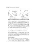
Fig. 10.4 (A) Technique of major
papilla pancreatic sphincterotomy
using a pull-type sphincterotome.
Left top: Biliary sphincterotomy is
performed using a standard pull-
type sphincterotome. Right top:
Pancreatic sphincterotomy is
performed with a pull-type
sphincterotome cutting in the 1
o’clock direction. Left bottom:
Completed biliary and pancreatic
sphincterotomy. A guidewire is in
the pancreatic duct. Right bottom:
A 6 Fr pancreatic stent is placed
following performance of the
pancreatic sphincterotomy. (B)
Technique of minor papilla
pancreatic sphincterotomy. 1.
Traction sphincterotome positioned
in minor papilla. Note the extent of
the minor papilla mound (arrows).
Duodenal juice at the minor papilla
orifice is aspirated away before
cutting to prevent heat dissipation to
juice and boiling the adjacent tissues
during the sphincterotomy. 2. Wire
is bowed taut and cut is performed
rapidly with minimal coagulation
utilizing the ERBE generator. The
optimal cut length in this setting is
unknown. The 5 mm length minor
papilla sphincterotomy is complete
without white tissue coagulum. 3.
White pancreatic stone removed
through patent sphincterotomy
orifice with balloon catheter. 4.
Excessive white coagulum at the
cut edge of the sphincterotomy in
a patient who underwent minor
papilla sphincterotomy. This may
potentially lead to restenosis of the
sphincterotomy orifice.
This is trial version
www.adultpdf.com
complications were not found by others [23,24,64,65]. Performing a biliary
sphincterotomy first, however, can expose the pancreatico-biliary septum and
allow the length of the cut to be gauged more accurately.
Pancreas divisum In patients with pancreas divisum, a minor papilla sphinc-
terotomy is usually necessary. The technique is similar to that of major papilla
sphincterotomy, except that the direction of the incision is usually in the 10–
12 o’clock position and the length of the sphincterotomy is limited to 4–8 mm.
Stone removal The ability to remove a stone by endoscopic methods alone is
dependent on the stone size and number, duct location, presence of downstream
stricture, and the degree of impaction [67,68]. Downstream strictures usually
require dilation with either catheters or hydrostatic balloons. Standard stone-
retrieval balloons and baskets are the most common accessories used to remove
stones. Passage of these instruments around a tortuous duct can be difficult, but
use of over-the-wire accessories is usually helpful. Stone removal is then per-
formed in a fashion similar to bile duct stone extraction (Fig. 10.5). Occasion-
ally, mechanical lithotripsy is necessary, particularly when the stone is larger in
diameter than the downstream duct or the stone is proximal to a stricture. A rat
tooth forceps may be helpful when a stone is located in the head of the pancreas
close to the pancreatic orifice.
Results of endoscopic treatment for stones
Sherman and colleagues Sherman and colleagues attempted to identify those
patients with predominantly main pancreatic duct stones most amenable to
endoscopic removal and to determine the effects of such removal on the pati-
ents’ clinical course [67].
Thirty-two patients with ductographic evidence of chronic pancreatitis and
pancreatic duct stones underwent attempted endoscopic removal using various
techniques, including bile duct and/or pancreatic duct sphincterotomy, stricture
dilation, pancreatic duct stenting, stone basketing, balloon extraction, and/
or flushing. Of these patients, 72% had complete or partial stone removal, and
68% had significant symptomatic improvement after endoscopic therapy.
Symptomatic improvement was most evident in the group of patients with
chronic relapsing pancreatitis (vs. those presenting with chronic continuous
pain alone; 83% vs. 46%).
Factors favoring complete stone removal included: (1) three or fewer stones;
(2) stones confined to the head or body of the pancreas; (3) absence of a down-
stream stricture; (4) stone diameter less than or equal to 10 mm; and (5) absence
of impacted stones.
ENDOSCOPY IN CHRONIC PANCREATITIS 257
This is trial version
www.adultpdf.com
CHAPTER 10258
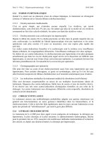
Fig. 10.5 A 40-year-old female with
alcohol-induced chronic pancreatitis
complicated by pancreatic main
duct stones. (a) Pancreatogram
revealing dilated pancreatic duct
with 5 mm diameter filling defect
consistent with a pancreatic
stone. (b) After pancreatic
sphincterotomy, a non-wire-guided
stone extraction basket was utilized.
The basket is opened fully in the
dilated pancreatic duct and the
stone is engaged. (c) Basket is
slowly closed on the stone. (d)
Stone is extracted and follow-up
pancreatogram with a balloon
catheter reveals no residual filling
defects. No further stenting was
performed.
This is trial version
www.adultpdf.com
After successful stone removal, 25% of patients had regression of the ducto-
graphic changes of chronic pancreatitis, and 42% had a decrease in the main
pancreatic duct diameter. The only complication from therapy was mild pancre-
atitis, occurring in 8%.
Smits and colleagues Smits and colleagues [68] reported the results of 53 patients
with pancreatic duct stones treated primarily by endoscopic methods alone
(eight had ESWL). Stone removal was successful in 42 patients (79%; complete
in 39 and partial in three), with initial relief of symptoms in 38 (90%). Similar
to the results reported by Sherman et al. [67], in this series, three of 11 patients
(27%) with failed stone removal had improvement in symptoms, suggesting
that some of the clinical response may be related to other therapies performed at
the time of attempted stone removal (e.g. pancreatic sphincterotomy).
During a median follow-up of 33 months, 13 patients had recurrent symp-
toms due to stone recurrence. The stones were successfully removed in 10
(77%). No factor evaluated (etiology of pancreatitis, presentation with pain or
pancreatitis, presence of single or multiple stones, location of stones, presence or
absence of a stricture) was shown to predict successful stone treatment (defined
as complete or partial removal of stones, resulting in relief of symptoms).
Cremer and colleagues Cremer and colleagues [37] reported the results of 40
patients with pancreatic duct stones who were treated by endoscopic methods
alone. Complete stone clearance was achieved in only 18 (45%). However,
immediate resolution of pain occurred in 77%. During a 3-year follow-up, 63%
remained symptom free. Clinical steatorrhea improved in 11 of 15 patients (73%).
Summary results Table 10.6 summarizes six selected series [37,67–71] report-
ing the results of pancreatic stone removal by endoscopic methods alone.
Complete stone clearance was achieved in 93 of 147 patients (63%). The major
complication rate was 9% (primarily pancreatitis), and the mortality rate was
0%. Cremer et al. [37] reported bleeding in 3% and retroperitoneal perforation
in 1.4%. Sepsis was an infrequent complication. During a 2.5-year (approxi-
mate) follow-up, 74% of patients had improvement in their symptoms.
Endoscopic therapy with ESWL
As noted, endoscopic methods alone will likely fail in the presence of large or
impacted stones and stones proximal to a stricture. ESWL can be used to frag-
ment stones and facilitate their removal (Fig. 10.6). Thus, this procedure is com-
plementary to endoscopic techniques and improves the success of non-surgical
ductal decompression.
ENDOSCOPY IN CHRONIC PANCREATITIS 259
This is trial version
www.adultpdf.com
CHAPTER 10260
Table 10.6 Selected series reporting the results of endoscopic therapy of pancreatic ductal
stones (using ERCP techniques alone).
Complete
stone Major Mean Symptom
No. of clearance complications Mortality follow-up improvement
Reference patients (%) (%) (%) (months) (%)
Schneider and Lux 3 100 0 0 N/A N/A
1985 [69]
Fuji et al. 1989 [70] 11 55 0 0 N/A N/A
Sherman et al. 1991 [67] 32 59 8 0 26 68
Kozarek et al. 1992 [71] 8 88 13 0 17 88
Cremer et al. 1993 [37] 40 45 10 0 36 63
Smits et al. 1996 [68] 53
b
74 9 0 33 81
Total 147 63 9 0 31
a
74
a
Estimate.
b
Eight also had ESWL.
N/A, not available.
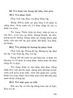
Fig. 10.6 A 41-year-old female with a
history of abdominal pain, pancreatitis,
and pancreatic calcification on CT scan.
(a) Abdominal radiograph reveals solitary
radiopaque stone in head/body region. (b)
Pancreatogram reveals an 8 mm obstructing
stone in body of pancreas pancreatic duct.
(c) A 0.018 inch diameter guidewire
was advanced beyond the stone. Further
contrast filling of duct demonstrating
upstream dilation. Following pancreatic
sphincterotomy, stone extraction with
basket was unsuccessful.
This is trial version
www.adultpdf.com
Sauerbruch and colleagues Sauerbruch and colleagues [76] were the first (in
1987) to report the successful use of ESWL in the treatment of pancreatic duct
stones. Since that time, more than 400 patients have been reported in the litera-
ture [66,74–81]. Patients with obstructing prepapillary concrement and up-
stream ductal dilation appear to be the best candidates for ESWL. In the largest
ENDOSCOPY IN CHRONIC PANCREATITIS 261
Fig. 10.6 (cont’d) (d) ESWL performed with Healthronics Lithotron spark-gap lithotriptor
at a setting of 26 kV for a total of 2500 shocks. Fragmentation of the stone demonstrated
post-ESWL. (e) Endoscopic view of small stone fragments removed from the pancreatic duct
post-ESWL.
This is trial version
www.adultpdf.com
Table 10.7 Selected series reporting the results of endoscopic therapy of pancreatic ductal stones using adjunctive ESWL.
Complete
Mean no. stone Major Mean Symptom
No. of ESWL clearance complications Mortality follow-up improvement
Reference patients sessions (%) (%) (%) (months) (%)
Neuhaus 1989 [74] 12 1.6 67 0 0 8 91
Soehendra et al. 1989 [73] 8 N/A 100 0 0 6 75
Delhaye et al. 1992 [66] 123 1.8 59 36 0 14 63
Sauerbruch et al. 1992 [76] 24 1.5 42 0 0 24 83
Schneider et al. 1994 [77] 50 2.4 60 0 0 20 90
van der Hul et al. 1994 [78] 17 1.9 41 6 0 30 65
Sherman et al. 1991 [67] 26 1.2 61 12 0 26 81
Kozarek et al. 2002 [80] 40 1.1 100 20 0 29 80
Farnbacher et al. 2002 [81] 125 2.5 51 0 0 29 93
Total 425 2.0 60 9 0 21
a
80
a
Estimate.
N/A, not available.
This is trial version
www.adultpdf.com
reported series, 123 patients with main pancreatic duct stones and proximal
dilation were treated with an electromagnetic lithotriptor, usually before pan-
creatic duct sphincterotomy [66]. Stones were successfully fragmented in 99%,
resulting in a decrease in duct dilation in 90%. The main pancreatic duct was
completely cleared of all stones in 59%. Eighty-five per cent of patients noted
pain improvement during a mean follow-up of 14 months. However, 41% of
patients had a clinical relapse due to stone migration into the main pancreatic
duct, progressive stricture, or stent occlusion.
This same center compared their results of pancreatic stone removal prior to
the availability of ESWL and after the introduction of adjunctive ESWL therapy
[37]. Stones were successfully cleared in 18 of 40 patients (45%) by endoscopic
methods alone, compared with 22 of 28 (78.6%) with ESWL. Table 10.7 sum-
marizes the results of nine selected series reporting the efficacy and safety of
adjunctive ESWL [66,67,73,74,76–78,80,81]. Complications in these series
were related primarily to the endoscopic procedure.
Although ultrasound-focused ESWL has been reported to achieve stone
fragmentation, such focusing is clearly more difficult. In the series reported by
Schneider and associates [77], stone localization was achieved in 17 of 119 ses-
sions (14%) when only ultrasonography was used to monitor the position of
the stone.
The Brussels group The Brussels group [79] studied 70 pancreatic stone pati-
ents who underwent attempts at endoscopic removal, with adjunctive ESWL
used in 41 (59%). This was a fairly homogeneous group of patients in that those
with strictures, previous pancreatic surgery, and failed pancreatic sphinctero-
tomy were excluded. The authors evaluated the immediate technical and clinical
results and reviewed the long-term outcome in patients followed for more than
2 years.
Complete (n = 35) or partial (n = 20) stone removal was achieved in 79%,
and was more frequently observed when ESWL was performed (P < 0.005) and
in the absence of a non-papillary ductal substenosis or complete main duct
obstruction (P < 0.05). Complete stone clearance was most frequently observed
with single stones or stones confined to the head (P < 0.05). In the multivariate
analysis, ESWL was the only independent factor influencing the technical results
of endoscopic management. In this series, the number of ERCPs performed per
patient was reduced from 3.4 to 2.7 after the introduction of ESWL (P < 0.01).
Of the 56 patients with pain on admission, 53 (95%) were pain free (n = 41) or
had a reduction in pain (n = 12).
In both the univariate and multivariate analyses, a significant association
was found between immediate disappearance of pain and complete or partial
main pancreatic duct clearance. During the first 2 years of follow-up after ther-
apy, 25 of 46 (54%) patients were totally pain free, whereas the frequency of
ENDOSCOPY IN CHRONIC PANCREATITIS 263
This is trial version
www.adultpdf.com
pain attacks in the remaining 21 was halved. This frequency of recurrent symp-
toms (46%) is comparable to that of surgical series [82].
Long-term pain relief was associated with: (1) earlier treatment after disease
onset (P < 0.005); (2) a low frequency of pain attacks before therapy (P < 0.05);
and (3) absence of non-papillary substenosis of the main pancreatic duct
(P < 0.05).
Interestingly, outcome was not associated with prior or continued alcohol
intake. In the multivariate analysis, pain recurrence was independently asso-
ciated with the frequency of pain attacks before therapy, the duration of disease,
and the presence of non-papillary substenosis of the main pancreatic duct. It
was suggested that such substenosis can induce ductal hypertension by blocking
migration of fragmented stones or by progressing to higher grade stenosis.
Twenty per cent underwent subsequent pancreatic surgical procedures. Of the
remaining 28 patients, there was statistically significant improvement in mean
pain scores, narcotic use, and hospitalizations when comparing intervals before
and after stone therapy [83].
Kozarek and colleagues Kozarek and colleagues performed a retrospective
review of the efficacy of ESWL as an adjunct to endoscopic therapy in 40 pati-
ents who underwent a total of 46 ESWL sessions (an average of 1.15 sessions/
patient). Eighty per cent of patients did not require surgery and had significant
pain relief, reduced number of hospitalizations, and reduced narcotic use as
compared to the pre-ESWL period over a mean 2.4-year follow-up [80].
Farnbacher and colleagues Farnbacher and colleagues retrospectively reviewed
the efficacy of pancreatic stone clearance with endoscopic and ESWL therapy.
Technical success was achieved in 85% of the 125 patients. The majority of the
patients (111 of 125) required piezoelectric ESWL for stone fragmentation.
ESWL was safe, without any serious complications. Middle-aged patients in the
early stages of chronic pancreatitis with stones in a prepapillary location were
the best candidates for successful treatment and required the least number of
ESWL treatment sessions [81].
These aforementioned studies reaffirm that ESWL as an adjunct to endo-
scopic pancreatic therapy is effective, and the results of the combined modality
may obviate the need for surgery. The results of endoscopic therapy in conjunc-
tion with ESWL for pancreatic stone disease compare favorably to the outcomes
in surgically treated patients.
Intraductal lithotripsy
Intraductal lithotripsy via mother–baby scope systems has largely failed due to
CHAPTER 10264
This is trial version
www.adultpdf.com
inability to maneuver within the relatively narrow ductal system. Results with
fluoroscopy-guided laser lithotripsy were similarly poor [71]. Pancreatoscopy
(via a ‘mother–baby’ scope system) can be used to directly visualize laser fiber
contact with the stone and fragmentation. Experience is limited to date [70,83].
Medical treatment for stones
Stone dissolution via ductal irrigation (contact dissolution) or oral agent is an
attractive endoscopic adjunct for stone removal.
Citrate Sahel and Sarles found that intraduodenal infusion of citrate in dogs
significantly increased the citrate concentration in pancreatic juice [85]. This led
to a non-randomized study of oral citrate in 18 patients with chronic pancre-
atitis, 17 of whom had pancreatic duct stones. Seven patients responded during
a mean duration of therapy of 9.5 months, with a mean stone size reduction of
21% and an improvement in symptoms [61].
Berger et al. [86] performed nasopancreatic drainage in six patients with
main pancreatic duct stones. The pancreatic duct was perfused with a mixture of
isotonic citrate and saline at 3 ml/min for 4 days. A stone-free state was achieved
in all cases.
Pancreatic pain disappeared during the perfusion, and four patients
remained free of pain during the follow-up period (1–12 months). The remain-
ing two patients had repeat therapy, which resulted in pain resolution. Pancre-
atic exocrine function was evaluated by the Lundh test in five patients before
and after therapy. An increase of 50–360% was observed in enzyme output in
three patients, while no improvement was noted in the remaining two patients.
Trimethadione Trimethadione, an epileptic agent and a weak organic acid, has
been shown in vitro to induce a concentration-dependent increase in calcium
solubility [61]. Noda et al. [87] showed promising results for trimethadione in a
dog model of pancreatic stones. Unfortunately, the doses used in the dogs, if
extrapolated to humans, could potentially be toxic. At the present time, no
rapidly effective solvent for human use is available to treat pancreatic stones.
Further trials in humans are needed to establish a role for medical therapy
(either alone or as an aid to endoscopic measures) in treating patients with
symptomatic pancreatic duct stones.
Overall results for stone treatment
These data suggest that removal of pancreatic duct stones may result in symp-
tomatic benefit. Longer follow-up is necessary to determine the stone recurrence
ENDOSCOPY IN CHRONIC PANCREATITIS 265
This is trial version
www.adultpdf.com
rate and whether endoscopic success results in long-standing clinical improve-
ment or permanent regression of the morphological changes. Overall, endo-
scopists are encouraged to remove pancreatic duct stones in symptomatic
patients when the stones are located in the main duct (in the head, body, or both)
and are thus readily accessible.
The currently available data suggest that the clinical outcome after success-
ful endoscopic removal is similar to the surgical outcome, with lower morbidity
and mortality [88]. Moreover, recurrence of symptoms due to migrated stone
fragments can be treated again by endoscopy with or without ESWL.
On the other hand, re-operation rates for recurrent pain after surgery are as
high as 20%, with a striking increase in morbidity and mortality after repeated
surgery [82]. Controlled trials comparing endoscopic, surgical, and medical
therapies are awaited.
Pancreatic pseudocysts
Pancreatic pseudocysts may complicate the course of chronic pancreatitis in
20–40% of cases [89,90]. Traditionally, surgery has been the treatment of
choice for such patients. The introduction of ultrasound- and CT-guided needle
and catheter drainage techniques provided a non-operative alternative for man-
aging patients with pseudocysts.
Endoscopic treatment for pseudocysts
More recently, an endoscopic approach has been applied for this indication. The
aim of endoscopic therapy is to create a communication between the pseudocyst
cavity and the bowel lumen. This can be done by a transpapillary and/or a trans-
mural approach. The route taken depends on the location of the pseudocyst and
whether it communicates with the pancreatic duct or compresses the gut lumen.
More than 400 cases of endoscopically managed pseudocysts have been
reported (Table 10.8) [91–100]. The results indicate that endoscopic therapy is
associated with a high technical success rate (80–95%), acceptably low complica-
tion rates (equal to or less than surgical rates), and a pseudocyst recurrence rate
of 10–20% [95].
In the largest series reported [97], 100 of 108 patients (93%) had their pseu-
docysts successfully drained. Pseudocysts recurred in 13 (13%). The presence of
chronic pancreatitis, obstructed pancreatic duct, ductal stricture, necrosis on
CT scan, and a pseudocyst greater than 10 cm in size was not predictive of
recurrent pseudocyst disease. Endoscopic therapy has also been shown to be
effective in the management of partial [100] and complete pancreatic ductal dis-
ruptions [101], pancreatico-cutaneous fistulas, infected fluid collections [102],
pancreatic ascites, pancreatic pleural effusions [9,103], and traumatic duct dis-
CHAPTER 10266
This is trial version
www.adultpdf.com
ruptions [103,104]. These studies and others [105] confirm the relative safety of
endoscopic intervention in peripancreatic fluid collections (Table 10.8).
This topic is reviewed in detail by Howell in Chapter 11.
Biliary obstruction in chronic pancreatitis
Intrapancreatic common bile duct strictures have been reported to occur in
2.7–45.6% of patients with chronic pancreatitis (Fig. 10.7). Such strictures are
a result of a fibrotic inflammatory restriction or compression by a pseudocyst
[107]. In one ERCP series, a common bile duct stricture was seen in 30% of
patients, and was associated with persistent cholestasis, jaundice, or cholangitis
in 9% [108]. Because long-standing biliary obstruction can lead to secondary
biliary cirrhosis and/or recurrent cholangitis, biliary decompression has been
recommended. Surgical therapy has been the traditional approach. Based on the
excellent outcome (with low morbidity) from endoscopic biliary stenting in
postoperative stricture [109], however, evaluation of similar techniques for bile
duct strictures complicating chronic pancreatitis was undertaken.
Standard biliary stents
Deviere and colleagues
Deviere and colleagues [108] evaluated the use of biliary stenting (one or two
plastic 10 Fr C-shaped stents) in 25 chronic pancreatitis patients with bile duct
obstruction and significant cholestasis (alkaline phosphatase > two times the
ENDOSCOPY IN CHRONIC PANCREATITIS 267
Table 10.8 Selected series reporting the results of endoscopic therapy of pseudocysts.
Method of pseudocyst
decompression
Technical No. No. No.
Reference success transpapillary ECG ECD Complications Deaths
Grimm et al. 1989 [18] 14/16 5 1 8 5 1
Cremer et al. 1989 [99] 32/33 0 11 21 3 0
Kozarek et al. 1991 [100] 12/14 12 0 0 5 0
Sahel 1991 [98] 58/67
a
26 1 31 9 1
Catalano et al. 1995 [93] 17/21 17 0 0 1 0
Smits et al. 1995 [91] 31/37
a
16 8 7 6 0
Binmoeller et al. 1995 [94] 47/53 31 6 10 6 0
Barthet et al. 1995 [92] 30/30
a
30 10 0 13 0
Howell et al. 1996 [97] 100/108 37 38 25 25 0
Total 341/379 (90%) 174 75 102 79 (20%) 2 (1%)
a
Estimate.
This is trial version
www.adultpdf.com
upper limits of normal). Nineteen patients had jaundice and seven presented
with cholangitis.
Following stent placement, cholestasis, hyperbilirubinemia, and cholangitis
resolved in all patients. Late follow-up (mean: 14 months; range: 4–72 months) of
22 patients was much less satisfactory. One patient died of acute cholecystitis and
postsurgical complications, whereas a second died of sepsis 10 months after
stenting, which was believed to be due to stent blockage or dislodgement. Stent mig-
ration occurred in 10 patients and stent occlusion in eight, resulting in cholestasis
with or without jaundice (n = 12), cholangitis (n = 4), or no symptoms (n = 2).
CHAPTER 10268
Fig. 10.7 A 38-year-old male with alcohol-induced chronic pancreatitis with recurrent bouts
of pain, cholestatic serum liver chemistries, and elevated serum amylase. CT scan revealed
enlarged head of pancreas, calcifications, and new biliary dilation. (a) Cholangiogram
revealed smooth, 3 cm long narrowing of the distal common bile duct within the head of the
pancreas, with upstream dilation typical of benign biliary stricture complicating chronic
pancreatitis. Biliary intraductal brush cytology was negative. Pancreatogram revealed
narrowing of the head of pancreas pancreatic duct, dilated secondary branches, and
calcifications. (b) A 7 Fr multiple side-hole pancreatic stent in place. Balloon dilation of the
bile duct stricture was performed with a 10 mm hydrostatic balloon.
This is trial version
www.adultpdf.com
These patients were treated with stent replacement, surgery, or both (n = 7).
Ten patients continued to have a stent in place (mean follow-up: 8 months) and
remained asymptomatic. Because of resolution of their biliary stricture, only
three patients required no further stents. The initial observation of this study is
that biliary drainage is an effective therapy for resolving cholangitis or jaundice
in patients with chronic pancreatitis and a biliary stricture. The long-term
efficacy of this treatment, however, is much less satisfactory, because stricture
resolution rarely occurs.
ENDOSCOPY IN CHRONIC PANCREATITIS 269
Fig. 10.7 (cont’d) (c) Placement of two 10 Fr polyethylene stents into bile duct and a 7 Fr
multiple side-hole pancreatic stent into pancreatic duct. Serum liver chemistries normalized
and abdominal pain improved. (d) Six months later, the patient’s daily pain was moderately
improved and ERCP was performed for possible bile duct and pancreatic stent removal.
Cholangiogram revealed persistent bile duct narrowing requiring further bile duct stenting.
Pancreatic ductal stricture in the head was improved and did not require further pancreatic
stenting.
This is trial version
www.adultpdf.com
The Amsterdam group
The Amsterdam group reported their results of placing 10 Fr biliary stents in
52 chronic pancreatitis patients with cholestasis [15]. Jaundice and cholestasis
disappeared within 2 weeks after stent insertion in all patients. During a median
follow-up duration of 32 months (range: 3 months to 10 years), 17 patients
(33%) had their stent removed without return of cholestasis. Complete resolu-
tion of the stricture was seen in 10 of the 17 patients. This suggested that com-
plete resolution of the stricture was not necessary for long-term relief of
symptoms and cholestasis.
Barthet and colleagues
Barthet and colleagues [110] also found that biliary stenting is not a definitive
therapy for chronic pancreatitis patients with a distal common bile duct stric-
ture. In their series of 19 patients (mean duration of stenting: 10 months), only
two had complete clinical (resolution of symptoms), biological (normalization
of cholestatic liver tests), and radiological (resolution of biliary stricture and
upstream dilation) recovery. Six of 10 (60%) possible clinical successes, eight
of 19 (42%) possible biological successes, and three of 19 (16%) possible
radiological successes were obtained.
Metal stents for biliary obstruction?
Because of the disappointing results with plastic stents and the concern about the
high morbidity associated with surgically performed biliary drainage proce-
dures in alcoholic (frequently debilitated) patients, the group from Brussels evalu-
ated the use of uncoated expandable metal stents for this indication [112].
Twenty patients were treated with a 34 mm long metal stent, which becomes
10 mm in diameter when fully expanded. The short length of the stent was
chosen so that surgical bypass (e.g. choledochoduodenostomy) would still be
possible if necessary. Cholestasis (n = 20), jaundice (n = 7), and cholangitis
(n = 3) resolved in all patients. Eighteen patients had no further biliary problems
during a follow-up period of 33 months (range: 24–42 months). Two patients
(10%) developed epithelial hyperplasia within the stent, resulting in recurrent
cholestasis in one and jaundice in the other. These patients were treated endo-
scopically with standard plastic stents, with one ultimately requiring surgical
drainage. The authors concluded that this therapy could be an effective alterna-
tive to surgical biliary diversion, but longer follow-up and controlled trials are
necessary to confirm these results.
CHAPTER 10270
This is trial version
www.adultpdf.com
In a recent abstract report, the Amsterdam group reported the long-term
follow-up (mean: 50 months) of a cohort of 13 patients with chronic pancreatitis-
induced biliary strictures who had undergone uncovered biliary Wallstent place-
ment. Endoscopic Wallstent was successfully placed in all patients between 1994
and 1999. Nine patients (69%) were successfully treated and four patients failed
Wallstent therapy. Of the nine patients treated successfully, four (44%) patients
required repeated endoscopic intervention (three with a second Wallstent and
one requiring cleaning with a balloon). One patient eventually required surgical
biliary diversion and three patients are continuing to need endoscopic plastic
stents through the Wallstent to maintain biliary patency [136].
Biodegradable stents
A recent exciting development in stent technology, utilizing bioabsorbable poly-
l-lactide (PLLA) polymer strands woven into the tubular mesh design similar to
the metallic stent, was reported by Haber et al. [111]. The PLLA stent is unique
in that it undergoes slow hydrolytic degradation and disintegration after 6–18
months. In the feasibility study in patients with malignant obstructive jaundice,
the endoscopic technique for placement of the bioabsorbable biliary stent was
similar to present expandable stents and was technically successful in 48 of
50 patients. The unique feature of this stent is that it may obviate the need for
follow-up endoscopy to remove/replace the stent and may potentially be an effec-
tive long-term option in benign, chronic pancreatitis-induced biliary strictures.
Stenting for biliary strictures and chronic pancreatitis: conclusion
The aforementioned studies indicate that plastic biliary stents are a useful alter-
native to surgery for short-term treatment of chronic pancreatitis-induced com-
mon bile duct strictures complicated by cholestasis, jaundice, and cholangitis.
This therapy also should be considered for high-risk surgical patients. Because
the long-term efficacy of this treatment is much less satisfactory, however, oper-
ative intervention appears to be a better long-term solution for this problem in
average-risk patients. More data on the long-term outcome, preferably in con-
trolled trials, are necessary before expandable metal stents can be advocated
for this indication. Trials of membrane-coated metal stents, bioabsorbable
stents, and removable coil spring stents are awaited.
Sphincter of Oddi dysfunction in chronic pancreatitis
Although sphincter of Oddi dysfunction (SOD) is a known cause of acute recur-
rent pancreatitis, its role in the pathogenesis of chronic pancreatitis is much less
certain [113].
ENDOSCOPY IN CHRONIC PANCREATITIS 271
This is trial version
www.adultpdf.com
Pathogenesis of SOD in chronic pancreatitis
A direct effect of alcohol on the sphincter of Oddi has been postulated [114]. In
studies performed in humans with T-tubes, it was demonstrated that intragas-
tric or intravenous [115] administration of alcohol increased the sphincter tone.
Moreover, Guelrud and colleagues [116] showed that local instillation of
alcohol on the papilla of Vater produced a significant increase in the basal pan-
creatic sphincter pressure at sphincter of Oddi manometry in both cholecystec-
tomy patients and patients with chronic pancreatitis. The authors postulated
that the increased motor activity of the sphincter of Oddi may raise the intraduc-
tular pancreatic pressure and result in disruption of small pancreatic ductules,
and back flow of pancreatic juice into the parenchyma, with subsequent injury.
Other investigators have refuted these findings by showing that intravenous
or intragastric administration of alcohol in humans results in a decrease in
sphincter of Oddi basal pressures at manometry [117].
In a preliminary study, Morita et al. showed that chronic alcohol administra-
tion in the Japanese monkey resulted in an increase in sphincter of Oddi mean basal
pressure from 9 to 20 mmHg (P < 0.01), while the phasic amplitude decreased
by 75% and the pancreatic ductal secretory rate nearly doubled [118].
Frequency of SOD in chronic pancreatitis
More recent studies using modern manometric techniques have shown a high
frequency of basal sphincter pressure abnormalities, especially the pancreatic
sphincter, in patients with established chronic pancreatitis [119]. Results of
other studies using sphincter of Oddi manometry refute these findings and have
shown no difference in the dynamics of the pancreatic sphincter in patients with
chronic pancreatitis and controls [120]. Such data suggest that the sphincter, at
times, becomes dysfunctional as part of the overall general scarring process or
has a role in the pathogenesis of chronic pancreatitis.
Surgical sphincter ablation
The surgical literature, although limited, suggests that sphincter ablation ther-
apy (both the biliary and pancreatic sphincters) alone for patients with chronic
pancreatitis and manometrically documented or suspected SOD benefits 30–
60% of patients [121,122]. Bagley and associates [123] reported a surgical series
of 67 patients with mild to moderate chronic pancreatitis undergoing empirical
biliary and pancreatic sphincterotomy (n = 33) or sphincteroplasty (n = 34).
During a 5-year follow-up, 44% of patients had pain relief. The outcome for
patients with idiopathic chronic pancreatitis was similar to that for patients
CHAPTER 10272
This is trial version
www.adultpdf.com
with alcohol-induced chronic pancreatitis. However, 92% (11/12) of patients
who stopped alcohol consumption were clinically improved, compared with
12.5% (2/16) of those who continued to drink.
Endoscopic pancreatic sphincterotomy
Because endoscopic pancreatic sphincterotomy has been performed infrequently
in most institutions, its role in the management of pancreatic sphincter stenosis
has not been defined. Kozarek et al. reported resolution of pain and clinical
episodes of pancreatitis after pancreatic sphincterotomy in six of 10 patients (1-
year follow-up) with chronic pancreatitis and suspected or manometrically docu-
mented pancreatic SOD [63]. Okolo et al. retrospectively evaluated 55 patients
who had undergone endoscopic pancreatic sphincterotomy over a 4-year period.
After a median follow-up of 16 months, 62% of patients reported improvement
of pain scores. Patients with pancreatic sphincter dysfunction (n = 15) had
significant improvement in pain (73%) compared to patients with pancreato-
graphic evidence of chronic pancreatitis (58%) [137]. The utility of endoscopic
sphincter ablation as the only therapy in patients with chronic pancreatitis
awaits further study, preferably in controlled randomized trials.
Pancreas divisum
Pancreas divisum is the most common congenital variant of pancreatic ductal
anatomy, occurring in 7% of autopsy series [124]. Most commonly, in the
setting of chronic pancreatitis, minor papilla sphincterotomy is performed to
provide access to the duct to effect stone retrieval or facilitate endoprosthesis
placement [9].
Pancreas divisum: a cause of pancreatitis?
It has been postulated that, in a subpopulation of pancreas divisum patients, the
minor papilla orifice appears to be critically small, such that excessively high
intrapancreatic dorsal duct pressures occur during active secretion [124]. This
may result in pancreatic pain or pancreatitis [125]. Although most authorities
agree that pancreas divisum is a definite cause of acute recurrent pancreatitis, its
role in the pathogenesis of chronic pancreatitis is much more controversial.
Several lines of evidence favor the association of pancreas divisum and pancre-
atitis, including: (1) the presence of pancreatographic and histological changes of
chronic pancreatitis isolated to the dorsal pancreas; (2) an increased incidence of
pancreas divisum in patients with idiopathic pancreatitis; and (3) symptomatic
benefit following dorsal duct drainage, endoscopically or surgically [124].
ENDOSCOPY IN CHRONIC PANCREATITIS 273
This is trial version
www.adultpdf.com