Báo cáo y học: " Conservative treatment of a left atrial intramural hematoma after left atrial thrombus resection and concomitant mitral valve replacement - case report" ppsx
Bạn đang xem bản rút gọn của tài liệu. Xem và tải ngay bản đầy đủ của tài liệu tại đây (930.35 KB, 5 trang )
CAS E REP O R T Open Access
Conservative treatment of a left atrial intramural
hematoma after left atrial thrombus resection and
concomitant mitral valve replacement - case report
Dirk Bruegger
1*
, Sebastian Sadoni
2
, Mikhail Primaychenko
2
, Ralf Sodian
2
, Christoph Schmitz
2
, Bruno Reichart
2
and
Daniel Chappell
1
Abstract
Left atrial intramural hematoma is a seldom cause of left atrial mass. It has been described to occur spontaneously,
after interventional procedures, after blunt chest trauma, or after aortocoronary bypass surgery. We present a case
of mitral valve replacement together with the removal of a large intraatrial space-occupying lesion. Intraoperative
transesophageal echocardiography confirmed a successful resection of this mass. Surprising ly, upon admission to
ICU, transesophageal and transthoracic echocardiography revealed a recurrence of an intramural lesion, closest
matching a hematoma, which was confirmed by contrast-enhanced computed tomography. Surgical intervention
was thoroughly discussed but a conservative management was favoured. 3 months after surgery, a reassessed
transthoracic echocardiography and computed tomography demonstrated an almost complete resolution of the
pre-existing hematoma.
Background
Atrial intramural hematomas are severe but rare complica-
tions of cardiac surgery and only few cases are described
in literature. We present a case of an unexpected intra-
mural left atrial hematoma following mitral valve replace-
ment and concomitant left atrial thrombus resection and
the beneficial role of perioperative echocardiography in
detecting and monitoring this event.
Case presentation
A 76-year-old woman with a history of intermittent atrial
fibrillation and cerebral infarction with left-sided hemipar-
esis was admitted to our hospital. Preoperative transeso-
phageal echocardiography revealed a dilated left atrium
(90 × 80 mm), mitral valve stenosis (mitral valve area
1.04 cm
2
, transmitral pressure gradient 9 mmHg), and the
presence of a large intracavitary, space-occupying lesion
attached to the left atrial roof ( Figure 1, Additional file 1).
The patient received mitral valve replacement via left
atriotomy with a biological prothesis (Perimount magna
27 mm; Edwards Lifesciences, Irvine, CA, USA), extirpa-
tion of the intracavitary mass and ligation of the left atrial
appendage. Intraoperative transesophageal echocardiogra-
phy revealed satisfactory valve function and confirmed the
successful removal of the intracavitary lesion. Histopatho-
logica l examination revealed a spherical mass which was
subsequently confirmed to be a partially calcified and
connective tissue-organized thrombus.
Surprisingly, upon admission to ICU echocardiography
revealed a reappearance of a new homogenous mass aris-
ing from the left atrial wall (Figure 2, Additional file 2).
Neither cardiac out put nor ejectio n fraction were sig nifi-
cantly influenced by the hematoma. Contrast-enhanced
computed tomography was performed and showed a
hyperdense, even bordered, intramural hematoma in the
left dorso-basal atrium subtotally obstructing the left atrial
chamber (Figure 3, Additional file 3). Surgical intervention
was thoroughly discussed interdisciplinary but in the light
of the patient’s hemodynamic stability and the high risk of
dissection of t he atrial wall at the auriculoventricular
annulus, the risk-benefit analysis favoured a conservative
managem ent. Therapeutic dose of intravenous unfractio-
nated heparin was started without a bolus 24 hours aft er
surgery with a target PTT value of 50 seconds. Heparin
* Correspondence:
1
Department of Anesthesiology, Ludwig-Maximilians-University Munich,
Marchioninistrasse 15, 81377 Munich, Germany
Full list of author information is available at the end of the article
Bruegger et al. Journal of Cardiothoracic Surgery 2011, 6:50
/>© 2011 Bruegger et al; licensee BioMed Central Ltd. This is an Open Access article distrib uted under the terms of the Creative
Commons Attribution License (http://cr eativecommons.org/licenses/by/2 .0), which permits unrestricted use, distribution, and
reproduction in any medium, provided the original work is properly cited.
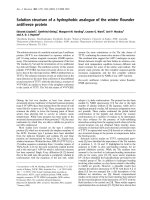
Figure 1 Preoperative transesophageal echocardiogram demonstrating a voluminous intracavitary mass (58 × 45 mm) attached to the
left atrial roof. T: thrombus; LA: left atrium; LV: left ventricle; RA: right atrium.
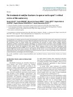
Figure 2 Postoperative transthoracic echocardiogram, apical 4-chamber view, showing development of an echogenic mass (54 × 40 mm)
compatible with an intramural left atrial hematoma. RV: right ventricle; LV: left ventricle; RA: right atrium; LA: left atrium; H: intramural hematoma.
Bruegger et al. Journal of Cardiothoracic Surgery 2011, 6:50
/>Page 2 of 5
was continued until an oral anticoagulation therapy with
warfarin was in the target INR range of 2.0 to 3.0.
In-house follow up serial echocardiography demon-
strated a stable-to-improving left atrial hematoma. A
transthoracic echocardiography and contrast-enhanced
computed tomography 3 months af ter surgery revealed
an almost complete resolution of the pre-existing hema-
toma (Figures 4 and 5, Additional files 4 and 5).
Discussion
Intramural left atrial hematomas are a very uncommon
entity, but have been described to occur spontaneously
[1-4], after aortocoronary bypass surgery [5], after percu-
taneous coronary interventions [6,7], radiofrequenc y
catheter ablations [8,9] and after blunt chest trauma [10].
In our case, routine postoperative transesophageal
echocardiography revealed an acute 5 cm diameter intra-
mural hematoma in the left atrium, which could have
remained undetected as our patient was asymptomatic.
Pre- [11] and intraoperative [12] echocardiography of the
heart is an indispensible examination for patients under-
going cardiac valve surgery. Whereas transthoracal echo-
cardiography is less invasive and more convenient to the
patient, several st udies have demonstrated transesopha-
geal echocardiography to be superior for assessing
Figure 3 Postoperative contrast-enhanced computed
tomography scan confirming development of a large
intramural hematoma almost obliteratin g the left atri al
chamber. RA: right atrium; RV: righ t ventricle; LA: left atrium; LV:
left ventricle; H: intramural hematoma; A: descending aorta.
Figure 4 Transthoracic echocardiogram with apical 4-chamber view performed three months following surgery showing a small residual
hematoma (21 × 16 mm) in the left atrium. RV: right ventricle; LV: left ventricle; RA: right atrium; LA: left atrium; H: intramural hematoma.
Bruegger et al. Journal of Cardiothoracic Surgery 2011, 6:50
/>Page 3 of 5
possible sources of cardiac embolism [13], such as intra-
cardiac thrombi o r intramural lesions [14]. As i n our
case, latter can ensure a quick and accurate diagnosis in
combination with a computed tomography.
Judging by the spontaneous regression, a conservative
approach seemed justified, despite anticoagulation.
Conclusion
A conservative approach with close-meshed serial echo-
car diographic examinations in a hemodynamically stable
and asymptomatic patient with a left atrial intramural
hematoma seems to be an appropriate strategy. This case
demonstrates the usefulness and necessity o f periopera-
tive echocardiographic imaging in the de tection and
monitoring of this unexpected event.
Consent
Written informed consent was obtained from the patient
for publication of this case report and any accompany-
ing images. A copy of the written c onsent is available
for review by the Editor-in-Chief of this journal.
Additional material
Additional file 1: Preoperative transesophageal echocardiogram
demonstrating a voluminous intracavitary mass attached to the left
atrial roof.
Additional file 2: Postoperative transthoracic echocardiogram
showing development of an echogenic mass arising from the left
atrial wall.
Additional file 3: Postoperative axial CT images.
Additional file 4: Transthoracic echocardiogram three months
following surgery revealing an almost complete resolution of the
pre-existing hematoma.
Additional file 5: Axial CT images three months following surgery.
Author details
1
Department of Anesthesiology, Ludwig-Maximilians-University Munich,
Marchioninistrasse 15, 81377 Munich, Germany.
2
Department of Cardiac
Surgery, Ludwig-Maximilians-University Munich, Marchioninistrasse 15, 81377
Munich, Germany.
Authors’ contributions
DB reviewed the case, conducted a review of the literature and drafted the
manuscript. SS and MP performed the echocardiographic studies and
participated in the design of the case report. RS and CS performed the
operation described. BR and DC confirmed the patient’s diagnosis and
revised the manuscript, contributing important intellectual content. All
authors read and approved the final manuscript.
Competing interests
The authors declare that they have no competing interests.
Received: 30 November 2010 Accepted: 13 April 2011
Published: 13 April 2011
References
1. Lombardo A, Luciani N, Rizzello V, Natale L, Pennestrí F, Ricci R,
Bonomo L, Possati GF, Crea F: Images in cardiovascular medici ne.
Spontaneous left atrial dissection and hematoma mimicking a cardiac
tumor: findings from echocardiography, cardiac computed
tomography, magnet ic resonance imaging, and pathology. Circulation
2006, 114:e249-250.
2. Lanfranchi A, Gelpi G, Rossi RS, Lemma M: A fast-growing obstructive left
atrial intramural hematoma causing acute prolonged chest pain. Interact
Cardiovasc Thorac Surg 2009, 9:363-365.
3. Shaikh N, Rehman NU, Salazar MF, Grodman RS: Spontaneous intramural
atrial hematoma presenting as a left atrial mass. J Am Soc Echocardiogr
1999, 12:1101-1103.
4. Watanabe K, Miguel B, Kemeny JL, Citron B, Camilleri LF: Spontaneous
intramural left atrial hematoma associated with systemic amyloidosis.
Ann Thorac Surg 2001, 72:2132-2134.
5. Musat I, Hieber C, Kepka A, Novotny P, Poslussny P, Schwarz S,
Fitzgerald RD: Intramural left atrial hematoma after aortocoronary artery
surgery. Anesth Analg 2003, 97:1605-1607.
6. Tavano D, Carlino M, Pisani M, Colombo A: Images in cardiovascular
medicine Conservative treatment of a left atrial hematoma and a
localized tamponade occurring during treatment of coronary total
occlusion. Circulation 2007, 115:e603-606.
7. Solzbach U, Beuter M, Haas H: Left atrial intramural hematoma after
percutaneous coronary intervention. Int J Cardiol 2010, 141:e37-38.
8. Sah R, Epstein LM, Kwong RY: Images in cardiovascular medicine.
Intramural atrial hematoma after catheter ablation for atrial
tachyarrhythmias. Circulation 2007, 115:e446-447.
9. Kurek C, Gwechenberger M, Richter B, Binder T, Loewe C, Gössinger H:
Intramural left atrial haematoma mimicking cardiac tamponade after
catheter ablation of atrial fibrillation. Europace 2009, 11:667-668.
10. Rowe SK, Porter CB: Atrial septal hematoma: two-dimensional
echocardiographic findings after blunt chest trauma. Am Heart J 1987,
114:650-652.
11. Germing A, Mugge A: What the cardiac surgeon needs to know prior to
aortic valve surgery: impact of echocardiography. Eur J Cardiothorac Surg
2009, 35:960-964.
Figure 5 Contrast-enhanced computed tomography scan three
months following surgery revealing an almost complete
absorption of the intramural hematoma. RA: right atrium; RV:
right ventricle; LA: left atrium; LV: left ventricle; H: intramural
hematoma; A: descending aorta.
Bruegger et al. Journal of Cardiothoracic Surgery 2011, 6:50
/>Page 4 of 5
12. Klein AA, Snell A, Nashef SA, Hall RM, Kneeshaw JD, Arrowsmith JE: The
impact of intra-operative transoesophageal echocardiography on cardiac
surgical practice. Anaesthesia 2009, 64:947-952.
13. De Bruijn SF, Agema WR, Lammers GJ, van der Wall EE, Wolterbeek R,
Holman ER, Bollen EL, Bax JJ: Transesophageal echocardiography is
superior to transthoracic echocardiography in management of patients
of any age with transient ischemic attack or stroke. Stroke 2006,
37:2531-2534.
14. Peters PJ, Reinhardt S: The echocardiographic evaluation of intracardiac
masses: a review. J Am Soc Echocardiogr 2006, 19:230-240.
doi:10.1186/1749-8090-6-50
Cite this article as: Bruegger et al.: Conservative treatment of a left atrial
intramural hematoma after left atrial thrombus resection and concomitant
mitral valve replacement - case report. Journal of Cardiothoracic Surgery
2011 6:50.
Submit your next manuscript to BioMed Central
and take full advantage of:
• Convenient online submission
• Thorough peer review
• No space constraints or color figure charges
• Immediate publication on acceptance
• Inclusion in PubMed, CAS, Scopus and Google Scholar
• Research which is freely available for redistribution
Submit your manuscript at
www.biomedcentral.com/submit
Bruegger et al. Journal of Cardiothoracic Surgery 2011, 6:50
/>Page 5 of 5