Báo cáo y học: "omplex pleural empyema can be safely treated with vacuum-assisted closure" pps
Bạn đang xem bản rút gọn của tài liệu. Xem và tải ngay bản đầy đủ của tài liệu tại đây (861.28 KB, 6 trang )
RESEARCH ARTIC LE Open Access
Complex pleural empyema can be safely treated
with vacuum-assisted closure
Zsolt Sziklavari
1*
, Christian Grosser
1
, Reiner Neu
2
, Rudolf Schemm
1
, Ariane Kortner
2
, Tamas Szöke
1
and
Hans-Stefan Hofmann
1,2
Abstract
Objective: For patients with postoperative pleural empyema, open window thoracostomy (OWT) is often necessary
to prevent sepsis. Vacuum-assisted closure (VAC) is a well-known therapeutic option in wound treatment. The
efficacy and safety of intrathoracal VAC therapy, especially in patients with pleural empyema with bronchial stump
insufficiency or remain lung, has not yet been in vestigated.
Methods: Between October 2009 and July 2010, eight consecutive patients (mean age of 66.1 years) with
multimorbidity received an OWT with VAC for the treatment of postoperative or recurrent pleural empyema. Two
of them had a bronchial stump insufficien cy (BPF).
Results: VAC therapy ensured local control of the empyema and control of sepsis. The continuous suction up to
125 mm Hg cleaned the wound and thoracic cavi ty and supported the rapid healing. Additionally, installation of a
stable vacuum was possible in the two patients with BPF. The smaller bronchus stump fistula closed spontaneously
due to the VAC therapy, but the larger remained open.
The direct contact of the VAC sponge did not create any air leak or bleeding from the lung or the mediastinal
structures. The VAC therapy allowed a better re-expansion of remaining lung.
One patient died in the late postoperative period (day 47 p.o.) of multiorgan failure. In three cases, VAC therapy
was continued in an outpatient service, and in four patients, the OWT was treated with conventional wound care.
After a mean time of three months, the chest wall was closed in five of seven cases. However, two patients
rejected the closure of the OWT. After a follow-up at 7.7 months, neither recurrent pleural empyema nor BPF was
observed.
Conclusion: VAC therapy was effective and safe in the treatment of complicated pleural empyema. The presence
of smaller bronchial stump fistula and of residual lung tissue are not a contraindication for VAC therapy.
1. Introduction
Thoracic empyema, the inflammatory process in a pre-
formed anatomical space, defined by the visceral and
parietal pleura, was one of the first recognised thoracic
pathological entities that had therapeutic challenge: “Ubi
pus, ibi evacua”. As a paradoxical result of increased life
expectancy, improved survival of malignant diseases and
extended operability criteria within and outside t he
scope of thoracic surgery, the pool of potential candi-
dates for pleural empyema is expanding [1]. In ad dition,
antibiotic abuse has l ed to increased numbers o f
therapy-resistant cases. Despite significant advances in
the treatment of thoracic infections, empyemas remain a
problem in modern thoracic surgery. The overall mor-
tality after postoperative pleural empyema can reach
26% [2].
For many patients, especially with postpneumonect-
omy empyema or BPF, chest tube insertion or thoraco-
scopic/open debridement fails to control the infection
and ends in sepsis. In these cases, open window thora-
costomy (OWT) should be offered [3]. Marsupialisat ion
of the cavity via rib(s) resection and open drainage is a
well-established method with low risk [4]. It can be
applied either as a definite treatment with intent to
cure, a preliminary procedure prior to definite treatment
* Correspondence:
1
Department of Thoracic Surgery, Hospital Barmherzige Brüder Regensburg,
Prüfeningerstraße 86, 93049 Regensburg, Germany
Full list of author information is available at the end of the article
Sziklavari et al. Journal of Cardiothoracic Surgery 2011, 6:130
/>© 2011 Sziklavari et al; licensee BioMed Cent ral Ltd. This is an Open Acce ss article distributed unde r the terms of the Creativ e
Commons Attribution License ( which permits unrestricted use, distribution, and
reproduction in any me dium, provided the original work is properly cited.
or as a last resort procedure when others have failed to
achieve a relatively stable disease state [1]
Since the introduction of vacuum assisted closure
therapy (VAC therapy), increasing indications for the
treatment of acute or chronic wound infections can be
found [5]. Thoracic application, especially in patients
with poststernotomy infections, is also well accepted [6].
The first reports of intrapleural VAC therapy were pub-
lished in 2006 [7]
We have reviewed our experience concerning the
management of pleural empyema with VAC therapy
after performing an OWT . In particular, the question of
VAC application in patients with BPF or remaining lung
tissue was of specific interest.
2. Patients and Methods
2.1. Study sample
In this retrosp ective study we investigated eight patients
with multimorbidity (Karnofsky index < 50%), treated
for a postoperative or recurrent pleural empyema
between October 2009 a nd July 2010. We excluded
patients who received VAC therapy for mediastinitis
after cardiac surgery or for chest wall abscesses not
involving the pleural space. The Ethics Commission at
the Krankenhaus der Barmherzigen Brüder Regensburg
approved the study.
2.2 Patient demographics
Of 414 operated patients, six patients developed post-
operative empyema (incidence: 1.5%) between October
2009 and July 2010. One patient had a recurrent post-
pneumonic empyema, the remaining patient was
referred from an outside institution.
All patients were men with a mean age of 66.1 years
and a range of 53 to 76 years. Patient demographics and
lung pathologies are summarised in Table 1. Four
patients had lung cancer and two of them received
induction chemotherapy, specifically radio-chemother-
apy. The resection of the tumour included one pneumo-
nectomy, two lobectomies and one lower bilobectomy.
After primary resection, the pathologist demonstrated
three R0 and one R1 resection. The patie nt with R1
resection received subsequent restpneumonectomy
because of BPF.
The other postoperative empyemas resulted after one
chest wall reconstruction with rib resection (fracture)
and one lung volume reduction (emphysema). Two dec-
ortications were performed (one atelectasis, one
empyema).
Five patients presente d an early/acute (≤ 30 days after
primary thoracotomy, with a mean of 24.7 days) and
three patients a late/chronic pleural empyema (> 30
days, with a mean of 68 days). Only two p atients (25%)
had detectable BPF due to bronchial stump dehiscence.
In five of eight patients, an initial intervention for treat-
ment of the detected empyema was performed (Table
1.). Independent from the time of empyema, Staphylo-
coccus, Streptococcus, and anaerobic species were the
most frequentl y isolated organisms. Additionally, Asper-
gillus fumigatus was found in two patients.
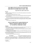
2.3 Surgical procedure (OWT and VAC therapy)
The operation for OWT and VAC included the resec-
tion of 2-4 ribs, pus evacuation, debridement, flushing
the cavity with ringer solution and 10% Betaisodona
(Povidon-Iod, Mundipharma) solution (Figure 1.).
Table 1 Demographics of patients
Variable P1 P2 P3 P4 P5 P6 P7 P8
Age 66♂ 71♂ 67♂ 76♂ 74♂ 69♂ 53♂ 53♂
Karnofsky Index < 50% Yes Yes Yes Yes Yes Yes Yes Yes
Diagnosis NSCLC
Stage
II a
Chronic
rib fracture
NSCLC
Stage
y III a
Atelectasis Postpneumonic
empyema
Emphysema NSCLC
Stage
III a
NSCLC
Stage
yIIb
Neoadjuvant Therapy No No Radiochemo. No No No No Chemo.
Primary Operation Lobectomy
R0
Chest wall
Stabilisation
Lobectomy
R0
Decort. Decort.
(thoracoscopic)
Volume
Reduction
Bilobectomy
R1
Pneumectomy
R0
Pathophys. of Empyema Postop. Postop. Postop. Postop. Recurrent Postop. Postop. Postop.
Onset Acute Chronic Acute Chronic Chronic Acute Acute Acute
Bronchopleural
Fistula
Yes No No No No No Yes No
Number of Interventions
before OWT and VAC
21100 110
Art of Intervention Restpneum.
Débridement
Débridement Chest
Tube
- - Chest Tube Restpneu. -
Microbiological Infection Strep.
Staph.
Staph. Staph. Staph.
Pseudo.
Strep. Enterobac.
Asperg.
Staph.
Asperg.
Staph.
P: Patient, NSCLC: Non-small cell lung cancer, Decort.: Decortication, BPF: Bronchopleural Fistula, Multimorbid.: Multimorbidity, Strep.: Streptococcus, Staph.:
Staphylococcus, Asperg.: Aspergillosis, Acute Empyema: < 30 days, Chronic Empyema > 30 days., Restpneum.: Restpneumectomy, Pathophys.:Pathophysiology
Sziklavari et al. Journal of Cardiothoracic Surgery 2011, 6:130
/>Page 2 of 6
Suturing the skin flaps on the margins of the OWT con-
stituted the thoracostoma. The VAC sponges (black
GranuFoam Standard Dressings, 400 - 600 microns)
were inserted in the residual pleural cavity through the
thoracostoma (Figure 1.) to fill the entire pleural space.
The sponges covered the leakage directly; no mem-
branes were used for the BPF or the remaining lung.
For the proce dure, we worked with a vacuum system
from KCI Medical (Wiesbaden, Germany). Suction was
setto-100mmHgfromthestart(maximumsuction
-125 mmHg), but in two patients with pneumonectomy,
the initial suction was -75 mmHg. The sponges were
changed once or twice a week, depending on the incor-
poration of the granulation tissue into the sponges.
Only a small amount of debridement was required at
each sponge change.
3. Results
3.1 Time of OWT and VAC
The indication for OWT and VAC intervention was
acute sepsis, failed primary surgical intervention (e.g.,
tube insertion) or complications of primary interven-
tions. The mean time between primary thoracotomy and
OWT was 52 days (range 21 days to 126 days).
In five patients, either chest tube drainage or reth ora-
cotomy with restpneumectomy/debridement initiated
the empyema treatment (Table 2.). Four patients under-
went one initial intervention before the fenestration and
vacuum closure, and one patient had two interventions.
In two patients, a detectable BPF was dissected, directly
closed by stitc hes and covered by a pericardial flap dur-
ing the first intervention. All five patients received the
OWT and VAC secondarily because of failed initial
empyema treatment. Direct creation of OWT with VAC
therapy was performed in three patients.
The mean time between first intervention and OWT
with VAC therapy was 18.4 days for directly treated
patients and 33.5 days for patients with delayed OWT
with VAC therapy.
Figure 1 Intrathoracic vacuum closure.
Table 2 VAC and outcomes
Variable P1 P2 P3 P4 P5 P6 P7 P8
Immediate/delayed Creation of
OWT
Delayed Delayed Delayed Immediate Immediate Delayed Delayed Immediate
Number of Interventions before
OWT and VAC
21100110
Art of Intervention Restpneum.
Débridement
Débridement Chest
Tube
- - Chest Tube Restpneu. -
Indication of OWT+VAC Sepsis Bleeding
Fistula
Failed
primary Th.
Osteomyelitis Fistula Failed
primary Th.
Sepsis Muscle
necrosis
P.o. mechanical ventilation after
VAC
Yes No No No No Yes Yes No
Number of VAC Changes in OR 4 2 2 1 0 5 3 0
Max. Suction mm Hg - 75 - 125 - 125 - 125 - 100 - 100 - 75 - 125
Hospitalization in days after VAC 22 45 17 15 14 38 47
(exitus)
8
Antibiotic Therapy, in days 10 12 7 6 7 19 47 6
Clinical outpatient VAC No No No Yes Yes No - Yes
Outcome Healed Healed Healed Healed Healed Healed Died of
Sepsis
Healed
Closing planned Yes Yes Yes Yes Yes Yes - Yes
Chest wall closed No* No* Yes Yes Yes Yes - Yes
OWT Duration, in days not closed not closed 51 39 31 164 - 59
P: Patient number, P.o.: postoperative, Max.: maximal, OR.: Operation room, *: closing was planned, but patient rejected it.
Sziklavari et al. Journal of Cardiothoracic Surgery 2011, 6:130
/>Page 3 of 6
3.2 Course of VAC therapy
Local control of the infection and control of sepsis
was satisfactory in seven of the eight patients treated
by OWT and VAC therapy. The patients tolerated a
suction of 75-125 mm Hg and did not reacted with
arrhythmia or haemodynamic complications due to
thetractiononthemediastinumduringattemptsto
increase the suction. Membranes for the protection of
the lung parenchyma were not necessary. Further-
more, the suction used did not create any air leak or
bleeding from the lung or the mediastinal structures.
At the time of OWT and VAC installation, three
patients were in severe clinical conditions with acute
respiratory insufficiency with mechanical ventilation.
One patient was resucitated. After implementing VAC
therapy, two patients could be weaned from ventilla-
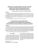
tory support after one and five days. In patients with
residual lung tissue, VAC therapy allowed improved
re-expansion of the residual lung. This expansion
couldbewellradiologicdemonstrated.(Figure2.)
In both patients with detectable BPFs, these fistulas
remained following the first intervention. At this time,
the recurrent BPFs were one millimetre and eight
millimetres, and closing was not possible in either
case. However, both patients with BPF underwent suc-
cessful local treatment of pleural empyema with suffi-
cient suction. The smaller bronchus stump fistula
closed spontaneous from VAC therapy, but the larger
remained ope n.
In the beginning of the VAC therapy, dressing
changes were performed under anaesthesia in the
operating theatre, with a mean rate of 2.1 changes and
a range of 0 to 5 changes. Additional changes were set
individually and performed without analgesic two or
three times a week. Antibiotic therapy was stopped
when the microbiological culture did not show any
further pathogenic bacteria colonisation (mean antibio-
tic therapy: 16.3 days).
3.3 Outcome of VAC-therapy
Seven of the eight patients (87.7%) were successfully
treated by OWT and VAC therapy. One patie nt died in
the late postoperative period (day 47 p.o.) of fulminant
aspergillum sepsis-related multiorgan failure. Although
he was the patient with the persistent eight millimetres
BPF, the thoracic cavity of this patient was sterile during
VAC treatment and his death was due to other factors.
The success of VAC therapy was defined by dischar-
ging the patients in good health with a K arnofsky Index
of 70% and with a non-infected pleural cavity. In most
cases the dimension of the pleural cavity was also
decrease d by OWT and VAC therapy. The mean hospi-
tal stay af ter OWT and VAC installation was 22. 7 days.
Four patients left our hospital without VAC, and the
cavity was filled with dry dressing material. Three
patients were transferred with VAC to the outpatient
service. Despite ambulant VAC therapy, these patients
had a good quality of life and excellent mobility.
In all patients, the closing of the OWT was planned,
and after a mean time of three months (97.5 ± 66.5
days), the chest wall was closed in five patients. The sur-
gical closure was performed after obliteration of the
pleural cavity with muscle transposition (M. pectoralis
N = 2, M. serratus anterior N = 1). In two patients, the
secondary clo sure was performed without tho racoplasty
because of maximal contraction of the pleural cavity.
Two patients subsequently rejected the closure of the
OWT, the last follow-up (after 15 respectively 18
months) did not show sign of recurrent infection.
After follow-up at an average of 7.7 months (range of
4 to 12 months), neither pleural empyema nor BPF
recurred in any of the seven surviving patients. All of
these patients reported a very good quality of life in an
outpatient interview.
4. Discussion
The often-cited Latin aphorism “Ubi pus, ibi evacua”
suggests that clinicians should open infected cavities.
We showed that the combination of traditional OWT
with the new intrathoracic VAC therapy fulfilled the cri-
teria of this old knowledge, especially in debilitated
patients with complicated empyema.
In regards to VAC therapy for open wound manage-
ment, this new techniq ue is often discussed as a reserve
treatment when there are no other options. In one VAC
group reported by Palmen and colleagues [8], the OWT
was delayed 58 ± 119 days after the diagnosis of the
empyema. Once treatment commenced, the total dura-
tion of OWT with VAC therapy was 31 ± 19 days. In
the present study, for comparison, patients with delayed
OWT and VAC therapy left our hospital after 31 ± 14
days and one pati ent died. In patients with initial fenes-
tration, however, the ho spital stay was only 11.5 ± 3.5
Figure 2 Radiologic demonstration; VAC dressing could help
expand dystelectatic lung.
Sziklavari et al. Journal of Cardiothoracic Surgery 2011, 6:130
/>Page 4 of 6
days. This finding was consistent with Massera and col-
leagues [9], who concluded that immediate creation of
OWT is a significant predictor of successful thoracost-
omy closure. We subscribed to this opinion and
extended early OWT installation to combined VAC
therapy. In our opinion, the alternative treatment of
OWT and VAC therapy should be discussed as soon as
possible, especially for postoperative or chronic pleural
empyema and in patients with increased risk for
impaired wound healing (e.g., diabetes, obesity, steroids).
The presence of BPF or remaining lung tissue is not a
contraindication for VAC therapy. Groetzner and collea-
gues [10], as well as Palmen and colleagues [8], defined
patients with BPF as not qualified for VAC therapy.
This recommendation led to Aru and colleagues [11].
closing all of the BPFs before application of the VAC
system . The closure of a BPF is the best precondition of
empyema treatment, but sometimes the second closure
is not possible. We treated two BPF patients with VAC
and in all the installation of vacuum was possible. In
onepatientwithaonemmfistula,theBPFwassuffi-
ciently closed after VAC therapy. The other BPF, with a
diameter of eight millimetres, could not be closed by
VAC, which was not a problem in the VAC treatment.
Future studies should investigate the diameter of BPF
that can be closed by negative pressure in VAC therapy.
VAC therapy seems to have a benef icial effect on the
re-expansion of the remaining lung in patients (Figure
2.). For example, two patients with respiratory insuffi-
ciency were quickly removed from their respirators after
VAC therapy.
Similar to other reports [5,8,10,11], we applied a maxi-
mum suction of -125 mmHg directly to the pulmonary
tissue using the V.A.C. GranuFoams. Starting with a
lower suction (-75 mmHg) was useful in patients with
prior pneumonectomy. In addition, membranes for tis-
sue protection were not necessary and no major compli-
cations related to vacuum-assisted management were
observed.
The frequency and the location of intrathoracic VAC
varies, as this part of the surgical treatment is not
defined. For example, Palmen and colleagues [ 8]. chan-
ged the system in the surgica l ward wit hout anaesthesia
every 3
rd
to 5
th
day, or more depending on purulent
secret ion or increased infection. However, Aru and col-
leagues [11]. performed all sponge changes under gen-
eral anaesthesia. For comparison, our patients
underwent two debridements and VAC changes in the
operation room, and additional changes were performed
every 3
rd
to 5
th
day in the ward.
In most cases, VA C therapy res ulted in the rapid era-
dication of local infection. We therefore withdrew anti-
bioticswhentherewerenosignsofsepsisandthe
thoracic cavity became sterile (mean time of 16.3 days).
However,theroleofsimultaneous antibiotics flushing
(e.g., V.A.C. Instill) has not yet been investigated.
After treatment of sepsis and local control of the
empyema, often with reduction of the pleural cavity,
patients could be discharged to an outpatient service with
initial daily wound care by specialized nurse technicians. It
was occasionally useful to continue the VAC therapy in
this ambulant sector with the aim of further reduction of
the pleural cavity (in the present study, N = 3). T horacic
surgeons should perform this outpatient treatment weekly.
In follow-up visits, the indication for closure of the
OWT should be periodically evaluated. We closed our
OWT after a mean time of three months, but two
patients rejected this procedure. For compar ison, Matzi
and colleagues [12]. performed closure of the thoracic
cavity after VAC therapy in all cases between the 9th
and 48th day (mean of 22 days). Additionally, Groetzner
and colleagues [10] . used the VAC system as a bridge to
reconstructive surgery and removed it after a mean per-
iod of 64 +/- 45 days (range of 7 to 134 days) in all
patients. These patients underwent direct surgical
wound closure, and complete healing without recur-
rence was achieved in 11/13 (85%) patients.
Data from the literature show that the interval
between installation and closure of the OWT is consid-
erable longer in patients without additional VAC ther-
apy [8,13]. The avera ge du ration of OWT without VAC
therapy at the Maastricht University Medical Centre was
933 ± 1422 days [8]. Maruyama and colleagues reported
an OWT interva l from 128 +/- 32, 1 to 365, 8 +/- 201
days, depending on indication [13]. In our patients with
VAC therapy the chest wall was closed after a mean
time of three months (97.5 ± 66.5 days). In the non-
VAC group of Palmen and colleagues [8]. six of the
eight patients could be discharged home. In only two of
them the OWT was closed by muscular flap. Four
patients died during follow-up because of OWT-related
complications (massive bleeding n = 1, recurrent infec-
tions of the thoracic cavity n = 3).
The rate of successful empyema treatment and closure
of OWT by reconstructive surgery is in our study as
well as in other s tudies with VAC therapy [10,12]. sub-
stantial higher in correlation to groups w ith only OWT
treatment.
In our opinion, the closure of the OWT depends on
the patient’s individual situation (e.g., general condition
of the patient, planned rehab ilitation). As a final step,
the closure of the chest guarantees full mobilisation and
a good quality of life, with only a very low risk of recur-
rent infections.
4.1. Study Limitations
We were only able to recruit eight patients who had
required an OWT and only five patients who had
Sziklavari et al. Journal of Cardiothoracic Surgery 2011, 6:130
/>Page 5 of 6
residual pulmonary parenchyma in the past year.
Because of these small numbers of patients, this study is
a series of case studies and not a randomised trial.
5. Conclusion
Patients with complicated empyema were successfully
treated with OWT and VAC therapy, s o the use of this
procedure should be discussed early. The most impor-
tant advantages of the OWT with VAC were fast treat-
ment of sepsis and local control of the pleural cavity.
Suction therap y could also improve pulmonary function
(re-expansion). In addition, the presence of bronchial
stump fistulas or residual lung tissue is not a contraindi-
cation for vacuum-assisted closure. Furthermore, the
length of hospitalization was shorter in patients with
immediate OWT and VAC-therapy installation, and
outpatient treatment with VAC-therapy is possible.
Author details
1
Department of Thoracic Surgery, Hospital Barmherzige Brüder Regensburg,
Prüfeningerstraße 86, 93049 Regensburg, Germany.
2
Department of Thoracic
Surgery, University Regensburg, Franz-Josef-Strauss-Allee 11, 93053
Regensburg, Germany.
Authors’ contributions
CG, RS, RN and AK participated in the design of the study. TS participated in
the sequence alignment and drafted the manuscript. ZS and HH conceived
of the study and participated in its design and coordination. All authors read
and approved the final manuscript.
Competing interests
The authors declare that they have no competing interests.
Received: 30 June 2011 Accepted: 6 October 2011
Published: 6 October 2011
References
1. Molnar TF: Current surgical treatment of thoracic empyema in adults. Eur
J Cardiothorac Surg 2007, 32:422-30.
2. Lemmer JH, Botham MJ, Orringer MB: Modern management of adult
thoracic empyema. J Thorac Cardiovasc Surg 1985, 90:849-55.
3. Light RW: A new classification of parapneumonic effusions and
empyema. Chest 1995, 108:299-301.
4. Deslauriers J, Jacques LF, Gregoire J: Role of Eloesser flap and
thoracoplasty in the third millennium. Chest Surg Clin N Am 2002,
12:605-23.
5. Renner C, Reschke S, Richter W: Thoracic empyema after
pneumonectomy: intrathoracic application of vacuum-assisted closure
therapy. Ann Thorac Surg 2010, 89:603-4.
6. Sjogren J, Malmsjo M, Gustafsson R, Ingemansson R: Poststernotomy
mediastinitis: a review of conventional surgical treatments, vacuum-
assisted closure therapy and presentation of the Lund University
Hospital mediastinitis algorithm. Eur J Cardiothorac Surg 2006, 30:898-905.
7. Varker KA, Ng T: Management of empyema cavity with the vacuum-
assisted closure device. Ann Thorac Surg 2006, 81:723-5.
8. Palmen M, van Breugel HN, Geskes GG, van Belle A, Swennen JM,
Drijkoningen AH, et al: Open window thoracostomy treatment of
empyema is accelerated by vacuum-assisted closure. Ann Thorac Surg
2009, 88:1131-6.
9. Massera F, Robustellini M, Pona CD, Rossi G, Rizzi A, Rocco G: Predictors of
successful closure of open window thoracostomy for
postpneumonectomy empyema. Ann Thorac Surg 2006, 82:288-92.
10. Groetzner J, Holzer M, Stockhausen D, Tchashin I, Altmayer M, Graba M:
Intrathoracic application of vacuum wound therapy following thoracic
surgery. Thorac Cardiovasc Surg 2009, 57:417-20.
11. Giorgio M, Aru MD, Nicholas B, Jew Curtis G, Tribble MD, Walter H,
Merrill MD: Intrathoracic Vacuum-Assisted Management of Persistent and
Infected Pleural Spaces. Ann Thorac Surg 2010, 90:266-71.
12. Matzi V, Lindenmann J, Porubsky C, Mujkic D, Maier A, Smolle-Juttner FM: V.
A.C treatment: a new approach to the management of septic
complications in thoracic surgery. Zentralbl Chir 2006, 131(Suppl 1):
S139-40.
13. Maruyama Riichiroh, Ondo Kaoru, Mikami Koji, Ueda Hitoshi, Motohiro Akira:
Clinical Course and Management of Patients Undergoing Open Window
Thoracostomy for Thoracic Empyema. Respiration 2001, 68:606-610.
doi:10.1186/1749-8090-6-130
Cite this article as: Sziklavari et al.: Complex pleural empyema can be
safely treated with vacuum-assisted closure. Journal of Cardiothoracic
Surgery 2011 6:130.
Submit your next manuscript to BioMed Central
and take full advantage of:
• Convenient online submission
• Thorough peer review
• No space constraints or color figure charges
• Immediate publication on acceptance
• Inclusion in PubMed, CAS, Scopus and Google Scholar
• Research which is freely available for redistribution
Submit your manuscript at
www.biomedcentral.com/submit
Sziklavari et al. Journal of Cardiothoracic Surgery 2011, 6:130
/>Page 6 of 6