Báo cáo y học: "Prosthetic valve endocarditis caused by Staphylococcus capitis: report of 4 cases" doc
Bạn đang xem bản rút gọn của tài liệu. Xem và tải ngay bản đầy đủ của tài liệu tại đây (596.27 KB, 6 trang )
CASE REP O R T Open Access
Prosthetic valve endocarditis caused by
Staphylococcus capitis: report of 4 cases
Tamaki Takano
*
, Yoshinori Ohtsu, Takamitsu Terasaki, Yuko Wada and Jun Amano
Abstract
Although Staphylococcus capitis is considered to be a rare causative organism for prosthetic valve endocarditis, we
report 4 such cases that were encountered at our hospital over the past 2 years. Case 1 was a 79-year-old woman
who underwent aortic valve replacement with a bioprosthetic valve and presented with fever 24 days later.
Transesophageal echocardiography revealed an annular abscess in the aorto-mitral continuity and mild perivalvular
regurgitation. We perfo rmed emergency surgery 5 days after the diagnosis of prosthetic valve endocarditis was
made. Case 2 was a 79-year-old woman presenting with fever 40 days after aortic valve replacement with a
bioprosthesis. Transesophageal echocardiography showed vegetati on on the valve, and she underwent urg ent
surgery 2 days after prosthetic valve endocarditis was diagnosed. In case 3, a 76-year-old man presented with fever
53 days after aortic valve replacement with a bioprosthesis. Vegetation on the prosthetic leaflet could be seen by
transesophageal echocardiography. He underwent emergency surgery 2 days after the diagnosis of prosthetic valve
endocarditis was made. Case 4 was a 68-year-old woman who collapsed at her home 106 days after aortic and
mitral valve replacement with bioprosthetic valves. Percu taneous cardiopulmonary support was started immediately
after massive mitral regurgitation due to prosthetic valve detachment was revealed by transesophageal
echocardiography. She was transferred to our hospital by helicopter and received surgery immediately on arrival. In
all cases, we re-implanted another bioprosthesis after removal of the infected valve and annular debridement. All
patients recovered without severe complications after 2 months of antibiotic treatment, and none experienced re-
infection during 163 to 630 days of observation. Since the time interva l between diagnosis of prosthetic valve
endocarditis and valve re-replacement ranged from 0 to 5 days, early surgical removal of the infected prosthesis
and an appropriate course of antibiotics were attributed to good clinical outcomes in our cases.
Keywords: Prosthetic valve endocarditis, Staphylococcus Capitis, Early surgery, Antibiotics
Background
Staphylococcus capitis (S. capitis) is considered to be a
rare causative organism of prosthetic valve endocarditis
(PVE) since only 4 cases of PVE caused by S. capitis
have been reported to date [1-3]. This bacterium is a
subty pe of coagulase-negative staphyloco cci (CoNS) and
thus produc es biofilm, which confers tolerance to disin-
fectants during surgery. Unlike most CoNS, however,
the adhesion ability of S. capitis to foreign body surfaces
is low [4,5]. Nonetheless, we report here 4 cases of PVE
caused by S. capitis that were encountered at our hospi-
tal over the past 2 years.
Case 1
A 79-year-old woman underwent aortic valve replace-
ment with a Carpentier-Edwards Magna bioprosthetic
valve (Edwards Lifesciences, Irvine, CA) for aortic steno-
sis. She presented with a feve r of over 38°C 24 days
aftertheprocedure(Table1).Thefirstbloodculture
showed no evidence of bacterial growth, but S. capitis
was detected in the second examination. Intravenous
administration of gentamicin (GM) was started, which
was later changed to abekamicin due to its susceptibility
(Table 2). Transesophageal echocardiography (TEE)
revea led an annular abscess in the aorto-m itral continu-
ity and mild perivalvular regurgitation. We performed
emergency surgery 5 days after the diagnosis of PVE
was made. The aortic bioprosthesis was fully covered
with a yellowish-white film, and vegetation was seen on
* Correspondence:
Shinshu University School of Medicine, Department of Cardiovascular
Surgery, 3-1-1 Asahi, Matsumoto 390-8621, Japan
Takano et al. Journal of Cardiothoracic Surgery 2011, 6:131
/>© 2011 Takano et al; licensee BioMed Central Ltd. This is an Open Access article distributed under the terms of the Creative Comm ons
Attribu tion License (http://creativecom mons.org/licenses/by/2.0), which permits unrestricted use, distribution, and reproduction in
any me dium, provided t he original work is properly cited.
the right coronary cusp. Valve dehiscence had occurred
around the commissure between the left and non-coron-
ary cusps (Figure 1). The prosthetic valve was removed
and the aortic annulus debrided. The intimal defect
around the commissure was repaired after debridement
with an autologous pericardial patch (Figure 2). A Med-
tronic Mosaic po rcine valve (21 mm) (Medtronic, Min-
neapolis, MN) was implanted in a supra-annular fashion
with horizontal mattress sutures from the left ventricle
to the ascending aorta, except for 5 sutures that were
passed through the aortic wall and pericardial patch at
the intimal defect. Cardiopulmonary bypass was weaned
off without difficulty and post-operative course was
uneventful. Intravenous vancomycin (VCM) and oral
minomycin (MINO) were administered for 2 months
after the aortic valve re-replacement (Table 3). We
directly contacted with the patients by phone, and no
signs of infection were seen during 630 days of follow-
up.
Case 2
A 79-year-old woman suffering from aortic stenosis
underwent aortic valve replacement with a Carpentier-
Edwards Magna bioprosthetic valve. She had a fever of
over 38°C and complained of chills 40 days after the
procedure (Table 1). All three blood cultures that were
taken revealed S. capitis, and so intravenous administra-
tion of VCM and rifampicin was commenced (Table 2).
TEE revealed vegetation on the bioprosthesis, which gra-
dually increased in size. Aortic valve re-replacement was
performed 2 days after the diagnosis of PVE was made
and 14 days after fever onset. A yellowish-white film
covered the whole bioprosthetic valve, and vegetation
was found on the stent and prosthet ic leaflet at a maxi-
mum size of 20 mm in diameter (Figure 3). Neither
valve dehiscence nor annular abscess was observed. A
Medtronic Mosaic porcine valve (19 mm) was inserted
after the Magna valve and biofilm were removed. She
presented with transient dysarthria after the surgery, but
recovered fully within a month. Intravenous VCM was
continued for 2 mo nths after the re-replacement, and
MINO was given orally after hospital discharge (Table
3). There were no signs of infection were observ ed dur-
ing 332 days of follow-up.
Case 3
A 76-year-old man was hospitalized for a fever of over
38°C and general malaise 53 days after aortic valve
replacement with a Ca rpentier-Edwards Magna bio-
prosthesis (Table 1). TEE revealed a t hickened biopros-
thetic leaflet covered with vegetation. S. capitis was
identified by a blood culture, and a pseudoaneurysm at
the edge of the aortotomy closure was seen in a chest
CT scan. We performed emergency surgery 2 days after
the diagnosis of PVE was made and 8 days after fever
onset. As the Magna bioprosthesis had detached from
Table 1 Patient characteristics
Age
(y.o.)
Sex PVE onset from
the first operation
(Days)
First
valve
operation
Surgical
Indication
Fever at
admission
Heart
failure
Embolic
Event
Re-operation from
the PVE diagnosis
(days)
Re-operation from
the fever onset
(days)
Case 1 79 F 24 AVR
(Biological)
Annular
abscess
Regurgitation
+ 5 12
Case 2 79 F 40 AVR
(Biological)
Vegetation + - - 2 14
Case 3 76 M 53 AVR
(Biological)
Vegetation + - - 2 8
Case 4 68 F 106 MVR
(Biological)
AVR
(Biological)
Regurgitation
Shock
++- 0 8
Mean 76 ± 5.2 56 ± 36 2.3 ± 2.1 10.5 ± 3
PVE; prosthetic valve endocarditis, F; female, M; male AVR; aortic valve replacement, MVR; mitral valve replacement
Table 2 Antibiotics Susceptibility
PCG MPIPC CEZ IPM GM ABK EM CLDM MINO LVFX FOM VCM ST TEIC LZD
Case 1 > = 0.5 > = 4 < = 4 < = 1 8 < = 1 < = 0.25 < = 0.25 < = 0. 5 0.5 > = 128 < = 1 < = 10 < = 0.5 N/A
Case 2 >8 >2 >16 2 >8 <=1 0.5 <=0.5 <=1 1 >16 <=2 <=2 <=2 <=2
Case 3 > = 0.5 > = 4 8 < 1 8 < = 1 < = 0.25 < = 0.25 < = 0. 5 1 > = 128 1 N/A < = 0.5 2
Case 4 > 8 > 2 > 16 4 > 8 < = 1 < = 0.25 < = 0.5 < = 1 < = 0.5 > 16 < = 2 < = 2 < = 2 < = 2
PCG; penicillin G, MPIPC; oxacillin, CEZ; cephazolin, IPM; imipenem, GM; gentamicin, ABK; arbekacin, EM; erythromycin, CLDM; clindamycin, MINO; minocycline,
LVFX; levofloxacin, FOM; fosfomycin, VCM; vancomycin, ST; sulfamethoxaz ole-trimethoprim, TEIC; teicoplanin, LZD; linezolid, N/A; not available
Takano et al. Journal of Cardiothoracic Surgery 2011, 6:131
/>Page 2 of 6
the annulus (Figure 4), the valve was removed and the
annular abscess debrided. A Medtronic Mosaic porcine
valve (21 mm) was implanted after an aort ic wall defect
in the aortic annulus caused by the debridement was
repaired with a Gelweave graft patch (Terumo Corpora-
tion, Tokyo, Japan) (Figure 5). The pseudoaneurysm of
the ascending aorta was resected and re-constructed
with a Gelweave graft. He wa s sequentially administered
intravenous teicoplanin, VCM, and linezolid (LZD) due
to liver dysfunction (Table 3), and was discharged from
the hospital 2 months after the re-replacement without
any complications . He was followed up at the outpatient
clinic, and no signs of infection were seen during 224
days after the surgery.
Case 4
A 68-year-old woman collapsed in her home 106 days
after aortic and mitral valve replacement with a Carpen-
tier-Edwards bioprosthesis (Edwards Lifesciences, Irvine,
CA)(Table1).Shewasfoundinashock-likestatewith
a systolic blood pressure of 60 mmHg, immeasurable
blood oxygen saturation, and decrease d consciousness.
As TTE showed massive mitral regurgitation due to
prosthetic valve detachment, percutaneous cardiopul-
monary support was started immediately. She was air-
lifted to our hospital, and an emergency surgery was
performed. A yellowish-white film covered the whole
mitral valve, which had become detached at 1/3 of the
annulus. An abscess was found in the remaining annulus
(Figure 6). Mitral valve re-implantation was performed
with a Medtronic Mosaic bioprosthesis (25 mm) after
entire annular debridement and partial reconstruction of
the annular defect with bovine pericardium. Intravenous
VCM and GM were administered for 2 months after the
valve re-implantation, and the patient underwent chole-
cystectomy for cholecystolithiasis that had been diag-
nosed before the initial valve replacement (Table 3). She
was discharged from the hospital without any neural
deficits and is leading a normal daily life. We directly
contacted with the patients by phone, and no signs of
infection were seen during 163 days of follow-up.
Discussion
Although several reports of native valve endocarditis
caused by S. capitis [6,7] exist, there have been only 4
published cases of S. capitis causing PVE since 1996 [2].
This paper describes 4 additional cases of PVE caused
by S. capitis encountered at our hospital that revealed
several important clinical findings.
S.capitisisasubtypeofCoNSthatischaracteristi-
cally novobiocin-sensitive, aerobic, and hemolysis-posi-
tive. However, this bacterium lacks alkaline
phosphatase activity, which differentiates it f rom S.
epidermidis [8]. S. capitis did not account for any inci-
dences of bacteremia, intravenous catheter-associated
infection, prosthetic valve infection, cerebrospinal fluid
infection, or peritonitis, although it was detected in
blood or intravenous catheters in 4% of patients with-
out these conditions [4]. The most common causative
organism of PVE is Staphylococcus aureus, followed by
the CoNS Enterococcus and Streptococcus viridans,
according to a recent report [9].
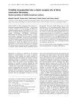
Figure 1 Operative Findings in Case 1. Yellowish-white film
covered the entire Carpentier-Edwards Magna bioprosthesis, and
vegetation located on the right coronary cusp. Valve dehiscence
was found at the commissure between the left and non-coronary
cusp.
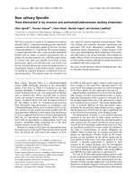
Figure 2 Operative Findings in Case 1.Intimaldefectwas
repaired with autologous pericardial patch after the debridement.
Takano et al. Journal of Cardiothoracic Surgery 2011, 6:131
/>Page 3 of 6
The ability of surface-adherent growth on prosthetic
devices is considered to be po tentially important in
causing disease [4,10]. S. capitis is known to have weak
adhesion to smooth surfaces, unlike most other CoNS,
such as S. epidermidis [4,5]. The virulence of CoNS is
mainly attributed to their adhesion ability to smooth
surfaces, biofilm production, and secretion of exoen-
zymes. An annular abscess was found in 3 of 4 cases
and prosthetic valve dehiscence occurred 2 of 4 cases in
the present report (Table 3). These findings demonstrate
that S. capitis may still cause fatal destruction of the
prosthetic valve annulus despite its relatively weak adhe-
sion ability to foreign body surfaces.
Treatment of PVE remains a challenge. The in-hospi-
tal mortality rate of PVE is 21-28.4% [9,11,12], even
with correct evaluation of the prosthetic valve by TEE in
suspected patients. It is believed that preoperative status
and complications are strongly related to the early mor-
tality in PVE [11]; preoperative catecholamine, dialysis,
pulmonary edema, ventilation, and renal insufficiency
are all predictors for 30-day mortality. In the present
series, all patients survived and none experienced re-
infection during 163 to 630 days of observation (Table
3). Urgent surgery is recommended for patients with
complicating PVE [12]. We performed re-operations
from 0 to 5 days after PVE diagnosis for a mean time
interval between fever onset and surgery of 10.5 ± 3
days, which was considerably shorter than the 15 days
reported elsewhere [12]. This early surgical intervention
Table 3 Re-operation procedure, Antibiotics and Re-infection
Annular
abscess
Valve
dehiscence
Re-operation
Procedure
Prosthesis Intravenous
antibiotics
Oral
antibiotics
Observation period
(days)
Survive
Re-
infection
Case
1
+
-
Abscess isolation
AVR
Biological
®Biological
AMK® LZD®TEIC MINO 630 +
-
Case
2
-
-
AVR Biological
®Biological
VCM MINO 332 +
-
Case
3
+
+
Valve annuls
reconstruction
AVR
Biological
®Biological
TEIC®VCM®LZD LVFX 224 +
-
Case
4
+
+
Valve annuls
reconstruction
MVR
Biological
®Biological
VCM+GM LVFX 163 +
-
AVR; aortic valve replacement, AMK; Amoxicillin, LZD; linezolid, TEIC; teicoplanin, MINO; minocycline, VCM; vancomycin, LVFX; levofloxacin, MVR; mitral valve
replacement, GM; gentamicin.
Figure 3 Operative Findings in Case 2. Carpentier-Edwards
Magna valve was covered by white and yellowish thin film, and
vegetations were attached on the stent and prosthetic leaflet.
Figure 4 Operative Findings in Case 3. The bioprosthetic valve
was totally detached from the annulus.
Takano et al. Journal of Cardiothoracic Surgery 2011, 6:131
/>Page 4 of 6
maybeconsideredtobeattributedtogoodclinical
outcome.
It is important to disti nguish causative organism from
skin flora because S. capitis may colonize on skin like
other CoNS. We repeated blood culture at least three
times and detected only S. capitis in the each case. We
therefore considered S. capitis as the causative organism
for PVE. Infection route of S. capitis could not be clearly
known because 1 of 4 ca ses were late PVE and any pre-
disposing factor was not observed in the previous
reports [1-3] although 3 of 4 presenting cases were early
PVE, which might speculate contamination during the
initial valve replacement.
S. capitis was successfully treated in all cases with
similar susceptibility and sensitivity to VCM, TEIC, and
LZD (Table 2), although d ecreased susceptibility of
CoNS to VCM and TEIC has been reported [13]. Thus,
the antibiotic treatment course used in our patients may
be useful for future cases of PVE caused by S. capitis, as
well as for other culture-negative bacterial PVE, which
accounts for 11.2% of all cases [9], whereas CEZ was
used as perioperative prophylaxis in the initial valve
replacement of all the presenting cases. Our findings
also indicate a need to reassess the virulent nature of S.
capitis, especially with regard to bioprostheses.
Conclusion
We experienced 4 cases of PVE caused by S. capitis.
Early surgical removal of the infected prosthesis and
administration of appropriate antibiotics may play
important roles in successful PVE treatment.
Consent
Written informed consent was obtained from the
patients for publication of this Case report. Copies of
the written consent forms are available for review by the
Editor-in-Chief of this journal.
List of abbreviatio ns
CoNS: coagulase-negative staphylococci; GM: gentamicin; LZD: linezolid;
MINO: minomycin; PVE: prosthetic valve endocarditis; S. capitis:
Staphylococcus capitis; TEE: Transesophageal echocardiography; VCM:
vancomycin.
Authors’ contributions
TT presented design of the case report and completed the manuscript. YO,
TT and YW are in charge of patient care. JA directed all the work. All authors
read and approved the final manuscript.
Competing interests
All the authors have read the manuscript and have approved of its
submission. The authors report no conflicts of interest.
Received: 30 June 2011 Accepted: 7 October 2011
Published: 7 October 2011
References
1. Nalmas S, Bishburg E, Meurillio J, Khoobiar S, Cohen M: Staphylococcus
capitis prosthetic valve endocarditis: Report of two rare cases and
review of literature. Heart and Lung 2008, 37:380-384.
2. Terada Y, Mitsui T, Enomoto Y: Prosthetic valve endocarditis caused by
Staphylococcus capitis. Ann ThoracSurg 1996, 62:324.
3. Domínguez Rodríguez A, García González MJ, Lara Padrón A, Laynez
Cerdeña I, Barragán Acea A, Miralles Ibarra JM, Lacalzada Almeida J, Bosa
Ojeda F, Marrero Rodríguez F, de Armas Trujillo D: Infectious endocarditis
of prosthetic valves due to Staphylococcus capitis: a new case. Rev Esp
Cardiol 1999, 52:277-278.
Figure 5 Operati ve Findings in Case 3. The defect of the aortic
annulus and aortic wall was repaired with Gelweave graft patch
after annular abscess debridement.
Figure 6 Operative Findings in Case 4. Yellowish white film
covered the whole mitral valve, and valve detachment had
occurred at the 1/3 of the annulus.
Takano et al. Journal of Cardiothoracic Surgery 2011, 6:131
/>Page 5 of 6
4. Needham CA, Stempsey W: Incidence, adherence and antibiotic
resistance of coagulase-nagative staphylococcus species causing human
diseases. Diggn Microbiol Infect Dis 1984, 2:293-299.
5. Hebert GA, Cooksey RC, Clark NC, Hill BC, Jarvis WR, Thornsberry C:
Biotyping coagulase-negative staphylococci. Journal of Clinical
Microbiology 1998, 26:1950-1956.
6. Sandoe JA, Kerr KG, Reynolds GW, Jain S: Staphylococcus capitis
endocarditis: two cases and review of literature. Heart 1999, 82:e1.
7. Kamalesh M, Aslam S: Aortic valve endocarditis due to staphylococcus
capitis. Echocardiography 2000, 17:685-687.
8. Bannerman TL, Kloos WE: Staphylococcus capitis subsp. ureolyticus subsp.
nov.from human skin. Int J Syst Bacteriol 1991, 41:144-147.
9. Wang A, Athan E, Pappas PA, Fowler VG Jr, Olaison L, Paré C, Almirante B,
Muñoz P, Rizzi M, Naber C, Logar M, Tattevin P, Iarussi DL, Selton-Suty C,
Jones SB, Casabé J, Morris A, Corey GR, Cabell CH, International
Collaboration on Endocarditis-Prospective Cohort Study Investigators:
Contemporary clinical profile and outcome of prosthetic valve
endocarditis. JAMA 2007, 297:1354-1461.
10. Longauerova A: Coagulase negative staphylococci and thir participation
in pathogenesis of human infections. Bratisl Lek Listy 2006, 107:448-452.
11. Habib G, Tribouilloy C, Thuny F, Giorgi R, Brahim A, Amazouz M, Remadi JP,
Nadji G, Casalta JP, Coviaux F, Avierinos JF, Lescure X, Riberi A, Weiller PJ,
Metras D, Raoult D: Prosthetic valve endocarditis: who needs surgery? A
multicentre study of 104 cases. Heart 2005, 91:954-959.
12. Musci M, Hübler M, Amiri A, Stein J, Kosky S, Meyer R, Weng Y, Hetzer R:
Surgical treatment for active infective prosthetic valve endocarditis: 22-
year single-centre experience. Eur J Cardiothorac Surg 2010, 38:528-538.
13. Cremniter J, Slassi A, Quincampoix JC, Sivadon-Tardy V, Bauer T, Porcher R,
Lortat-Jacob A, Piriou P, Judet T, Herrmann JL, Gaillard JL, Rottman M:
Decreased susceptibility to teicoplanin and vancomycin in coagulase-
negative Staphylococci isolated from orthopedic-device-associated
infections. J Clin Microbiol 2010, 48:1428-1431.
doi:10.1186/1749-8090-6-131
Cite this article as: Takano et al .: Prosthetic valve endocarditis caused
by Staphylococcus capitis: report of 4 cases. Journal of Cardiothoracic
Surgery 2011 6:131.
Submit your next manuscript to BioMed Central
and take full advantage of:
• Convenient online submission
• Thorough peer review
• No space constraints or color figure charges
• Immediate publication on acceptance
• Inclusion in PubMed, CAS, Scopus and Google Scholar
• Research which is freely available for redistribution
Submit your manuscript at
www.biomedcentral.com/submit
Takano et al. Journal of Cardiothoracic Surgery 2011, 6:131
/>Page 6 of 6