Báo cáo y học: "Remnant of a non-patent ductus arteriosus mimicking traumatic thoracic aorta transection: a case report" ppt
Bạn đang xem bản rút gọn của tài liệu. Xem và tải ngay bản đầy đủ của tài liệu tại đây (919.8 KB, 3 trang )
CAS E REP O R T Open Access
Remnant of a non-patent ductus arteriosus
mimicking traumatic thoracic aorta transection:
a case report
Efstratios E Apostolakis
1
, Nikolaos G Baikoussis
1*
, Christina Kalogeropoulou
2
, Efstratios Koletsis
1
, Ioanna Koniari
1
,
Dimitrios Karnabatidis
2
, Menelaos Karanikolas
3
Abstract
We present an interesting case of a 53-year-old man with a non-patent ductus arteriosus erroneously diagnosed as
acute thoracic aorta transection after a car accident. The aortography revealed a “rupture” of the linear inner curve
of the aorta in the isthmus area, as well as a protrusion of the aortic lumen in the corresponding area. During the
followed thoracotomy an intact thoracic aorta and the remnant of a non-patent ligamentum arteriosum were
found. It is the first reported case and we review all the possible entities which may give a false-positive image of
traumatic aortic transection.
Background
Aortography was for many years the “gold standard” in
diagnosis of acute traumatic aortic rupture against the
two other methods of diagnostic imaging: CT-angiogra-
phy and transesophageal ECHO [1]. Its sensitivity and
specificity in experienced hands approaches 100% [2].
However, in rare cases a false- posit ive or false-negativ e
imaging may be observed. For the false positive images
of traumatic rupture the most common causes are local
atherosclerotic lesions of the aortic wall, ductal diverti-
cula [3], remnant of non-patent ductus arteriosus or
pre-existent aneurysm of the isthmus area [4]. We
describe herein a case of an injured patient with high-
suspicion index of traumatic aortic rupture, which was
based on a false-positive aortography.
Case presentation
A 53 year-old man was transported from another hospital
with the high suspicion of a traumatic aortic rupture after
acute blunt thoracic trauma. Following a high speed car
accident he was admitted in another hospital with inju-
ries in the chest and fractur e of the left femur. A thorax-
CT scan was performed without contrast medium
because of a known chronic renal failure (creatinine
levels = 2.2 mMol/L). It showed hemothorax on the left,
minimal left lung contusi ons (of the posterior segments),
rib fractures and a periaortic hematoma at the level of
the isthmus area (figure 1). Because of a high-suspicion
indexofthoracicaorticrupture,wedecidedtodoan
emergency aortography. It revealed an interruption of the
normal contour of the thoracic aorta in the aortic isth-
mus area. A protrusion of t he aortic lumen in the corre-
sponding inner curve of the aorta supported our
suspicion for the disruption of the intima and the initia-
tion of a pseudoaneurysm’s process (figure 2) . Therefore,
an emergency operation (the interventional management
was abandoned because of technical reasons) by using
partial right femoro-femoral bypass for aortic isthmus
repair was decided. Surprisingly, and after a postero-lat-
eral thoracotomy at the 4
th
intercostals space, we
inspected an “intact” outer thoracic aortic wall, without
haematoma or related pathology at the aortic isthmus
are a. However, becaus e we did not totally exclude a pos-
sible limited disruption of the intima, or even another
pathology (see discussion), we decided to check from
inside the thoracic aorta. Following proximal and distal
dissection of the aorta, a partial cardiopulmonary bypass
was initiat ed with flow level 2- 2.6 L/min to restore a dis-
tal aortic pressure of >55-60 mm. After double clamping
and vert ical opening of the aorta wall, an intact endothe-
lium was observed. In the inner curve of the aortic
* Correspondence:
1
Cardiothoracic Surgery Department. University Hospital, Patras School of
Medicine. Patras, Greece
Apostolakis et al. Journal of Cardiothoracic Surgery 2010, 5:24
/>© 2010 Apostolakis et al; licensee BioMed Central Ltd. This is an Open Access article distributed under t he terms of the Creative
Commons Attribution License (http://creat ivecommons.org/licenses/by/2.0), which permits unrestricted use, distribution, and
reprodu ction in any medium, provided the original work is properly cited.
isthmus area and in the site of occluded ligamentum
arteriosum, a local vestigial dilatation 0.5 × 0.8 cm with
normal endothelium lining was observed. Two stitches of
prolene 4-0 reinforced with Teflon felt was used to oblit-
erate this remnant. The aortotomy was then closed, the
cardiopulmonary bypass was interrupted and the rest of
operation was as usually. The patient wa s extubated after
8 hours and his postoperative course was uneventful. The
patient underwent successfully on the 9
th
postoperative
day the surgical management of his right femur fracture
and was discharged from the hospital on the 17
th
post-
operative day in good condition.
Conclusions
In every case of suspicion of traumatic aortic
transection, the imaging diagnosis is based on spiral
Figure 1 Thorax-CT of the patient indicating left he mothorax,
left lung contusion in its posterior segments and a diffusing
periaortic hematoma in the aortic isthmus area.
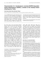
Figure 2 Aortography showed an interruption (the so called “ linear tear” ) of the normal contour of the thoracic aorta in the
corresponding area. A protrusion of the aortic lumen in the inner curve of the aorta is indicating the disruption of the intima and beginning
of a pseudoaneurysm. The preoperative evaluation of imaging was: “findings indicating a traumatic rupture of aortic isthmus”.
Apostolakis et al. Journal of Cardiothoracic Surgery 2010, 5:24
/>Page 2 of 3
CT-angiography or transesophageal echocardiography
(TEE), and rarely on the conventional aorto graphy.
Aortography is considered as the exam with the higher
specificity and sensibility approaching the 100% [2].
However, rare preexistent pathological conditions may
obscure the clearness of these i maging examinations.
Indeed, these conditions may mimic an aortic rupture
and in this way give false-positive results. Therefore, it
should be taken under consideration by the operator of
the angio-CT, or of the TEE, to avoid any pitfall for the
final diagnosis. The four rare entities which may give
false-positive imaging of aortic rup ture in the region of
theisthmusarethefollowing.A. Remnant of a non-
patent ductus arteriosus. T his vestigial may appear as a
local protrus ion of the aortic extremity of t he ductus-as
in our case- or as a scarry remnant which on the CT
ang iography creates a transformati on and an angulation
with compression between aorta and pulmonary artery
(scarry remnant forming the “corne r point” of a com-
pression between aorta and pulmonary artery) [5]. On
this remnant of the ductus arteri osus may be developed
later in the adult life, infective endocarditis [6].
B. Aneurysm of a non-patent ductus arteriosus. They
usually aris e from the a ortic extreme of the ductus and
may compress the nearest organs like trachea and eso-
phagus, giving related symptoms [4,5]. C. Aortic diverti-
culum. I t is commonly thought to be a remnant of the
closed ligamentum or ductus arteriosus. However some
authors support the hypothesis that it is a remnant of
the right dorsal aortic root [7]. It is described in thoracic
aortography as a large bulg e on the lesser curvat ure of
the aortic isthmus, in patients with a left aortic arch and
normal origin of the brachiocephalic arteries.
D. Calcification of the ligamentum arteriosum and/or
of the aortic wall in the aortic isthmus area. This calcifi-
cation in the adults may be in several patterns such as
curvilinear, punctate or clumped, and in incidence up to
65% [8]. In our case, we chose the surgical instead of
the endovascular-intervention, for the following two rea-
sons. First, because an endovascular graft was not in
time available, and second, there we re no contraindica-
tions for surgical intervention (brain injury, coagulation’s
abnormaliti es, etc). Despite of absence of signs of aortic
transection during the inspection of the thora cic aorta
(intramural hematoma, periaortic infiltration, etc), t he
image of aortography posed us in a dilemma, taken in
consideration our experience and the bibliographic data;
there is not traumatic aortic rupture without haematic
infiltration. A ccording these data, we decided open the
aorta to elucidate the differential diagnosis about the
given image of aortography.
Consent
Written informed consent was obtained from the patient
for publication o f this case report and accompanying
images. A copy of the written consent is available for
review by the Editor-in-Chief of this journal.
Author details
1
Cardiothoracic Surgery Department. University Hospital, Patras School of
Medicine. Patras, Greece.
2
Department of Interventional Radiology. University
Hospital, Patras School of Medicine. Patras, Greece.
3
Department of
Anaesthesiology and Critical Care Medicine. University Hospital of Patras.
Patras, Greece.
Authors’ contributions
All authors: 1. have made substantial contributions to conception and
design, or acquisition of data, or analysis and interpretation of data; 2. have
been involved in drafting the manuscript or revisiting it critically for
important intellectual content; 3. have given final approval of the version to
be published.
Competing interests
The authors declare that they have no competing interests.
Received: 8 November 2009 Accepted: 9 April 2010
Published: 9 April 2010
References
1. Martinez D, Johnson S, Miller O, Calhoon J: Acute traumatic aortic
transaction. Mastery of Cardiothoracic Surgery Lippincott Williams and
WilkinsKaiser L, Kron I, Spray T , 2 2007, 569.
2. Sturm J, Hankins D, Young G: Thoracic aortography following blunt chest
trauma. Am J Emerg Med 1990, 8:92-96.
3. Gleason T, Bavaria J: Trauma to the Great Vessels. Cardiac Surgery in the
Adult MacGraw Hill MedicalCohn L , 3 2008, 1139.
4. Myojin K, Ishibashi Y, Ishii K, Itoh M, Watanabe T, Kunishige H: Aneurysm of
the monpatent ductus arteriosus in the adult. A report of the case and
review of the literature. Jpn J Thorac Cardiovasc Surg 1998, 46:882-88.
5. Sebening C, Jacob H, Tochtermann U, Lange R, Vahl CF, Bodegom P,
Szabo G, Fleischer F, Schmidt K, Zilow E, Springer W, Ulmer HE, Hagl S:
Vascular tracheobronchial compression syndromes–experience in
surgical treatment and literature review. Thorac Cardiovasc Surg 2000,
48:164-74.
6. Flapper W, Dixit A, Murton M: Infective aortitis associated with the
nonpatent remnant of a ductus arteriosus. Ann Thorac Surg 2003,
76:931-33.
7. Grollman J: The aortic diverticulum: a remnant of the partially involuted
dorsal aortic root. Cardiovasc Intervent Radiol 1989, 12:14-17.
8. Wimpfheimer O, Haramati L, Haramati N: Calcification of the ligamentum
arteriosum in adults: CT features. J Comput Assist Tomogr 1996, 20:34-37.
doi:10.1186/1749-8090-5-24
Cite this article as: Apostolakis et al.: Remnant of a non-patent ductus
arteriosus mimicking traumatic thoracic aorta transection: a case report.
Journal of Cardiothoracic Surgery 2010 5:24.
Apostolakis et al. Journal of Cardiothoracic Surgery 2010, 5:24
/>Page 3 of 3