Báo cáo y học: "Thoracoscopic resection of a paraaortic bronchogenic cyst" pps
Bạn đang xem bản rút gọn của tài liệu. Xem và tải ngay bản đầy đủ của tài liệu tại đây (898.14 KB, 4 trang )
CAS E REP O R T Open Access
Thoracoscopic resection of a paraaortic
bronchogenic cyst
Baldassare Mondello, Salvatore Lentini
*
, Dario Familiari, Pietro Barresi, Francesco Monaco, Michele Sibilio,
Annunziata La Rocca, Vincenzo Micali, Ignazio Eduardo Acri, Mario Barone, Maurizio Monaco
Abstract
Bronchogenic me diastinal cysts (BMC) represent 18% of primitive mediastinal tumors and the most frequent cystic
lesions in this area. Nowadays, BMC are usually treated by VATS. However, the presence of major adhesions to vital
structures is often considered as an unfavourable condition for thoracoscopic treatment. The authors report the
thoracoscopic treatment of a BMC having dense adhesions to the aortic arch. Diagnosis and surgical treatment is
described. Rev iew of the literature and surgical options on this topic are discussed.
Background
Bronch ogenic mediastinal cysts (BMC) represent 18% of
primitive mediastinal tumors, and are the most frequent
cystic lesions in this anatomic region [1,2]. Surgical
resection is recommended. Video assisted thoracic sur-
gery (VATS) has been reported for the resection of
these lesions. However, the presence of major adhesions
to vital structures is considered by some authors as an
unfavorable condition for BMC treatment by VATS. We
report a surgica l approach by VATS for a BMC with
adhesion to the aortic arch. Diagnosis and treatment of
the specific case is reported with literature review and
therapeutic options.
Case Presentation
A 50 year old asym ptomatic woman was referred to our
out-patient clinic following occasional detection of a
mediastinal mass. On routine chest x-ray performed
before orthopaedic surgery, the suspicion arose of a
mediastinal mass. A computed tomography (CT) scan
showed a cystic mass in the posterior medi astinum
between the aortic arch and the vertebral bodies (Figure 1).
The cyst extended from the 3
rd
thoracic vertebral body
to the tracheal carina plane, with a length of 4 cm and
a transversal diame ter of 2.5 cm. The lesion appeared
cystic with a well defined capsule and lacking enhance-
ment after intravenous contrast injection. Surgical
treatment was decided upon. Preoperative broncho-
scopy excluded any communication b etween the cyst
and the tracheobronchial tree. After double lumen
intubation, the patient was placed in a right lateral
position on the operating table. Three trocars were
used: one on the fifth intercostal space al ong the ante-
rior axillary line; one on the fifth intercostal space
along the posterior axillary line; and the last one on
the 7
th
intercostal space along the midaxillary line. The
cyst was visualized by thoracoscopy, appearing with a
maj or adhesion on the distal portion of the aortic arch
(Figure 2). To facilitate surgical dissection of the cystic
lesion from the aorta, fluid aspiration was performed
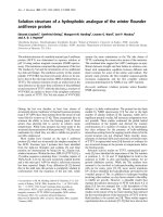
(Figure 3). Once the cyst was empty, complete resec-
tion from the adherent a orta was easily completed (Fig-
ure 4a). However, despite total lesion excision, we
completed the surgical procedure by pass ing the electro-
cautery on the pleural area where the cyst was adherent
(Figure 4b). The procedure was completed with insertion
of a chest tube. Histology examination confirmed the
diagnosis of benign bronchogenic cyst with the typical
feat ure of a ciliated columnar epithelial lining. The post-
operative (PO) course was uneventful and the patient
was discharged home on the 5
th
PO day. At 12 months
follow-up the patient remains well with no recurrence
on control CT scan.
Discussion
Bronchogenic mediastinal cysts (BMC) are a rare pathol-
ogy, accounting for 18% of all primitive mediastinal
* Correspondence:
Thoracic Surgery Unit, Cardiovascular and Thoracic Department, Policlinic
University Hospital, University of Messina, Italy
Mondello et al. Journal of Cardiothoracic Surgery 2010, 5:82
/>© 2010 Mondello et al; licensee BioMed Central Ltd. This is an Open Access article distributed under the terms of the Creative
Commons Attribution License ( which permits unrestricted use, distribution, and
reproduction in any medium, provided the original work is properly cited.
tumors and represent the most frequent cystic lesions in
this anatomic region [1-3].
They represent congenital malformations arising from
an abnormal division of the tracheo bronchial tree. In
relation to the time of separ ation from the main tra-
cheobronchial tree, the cysts may localize into the lung
parenchyma or in the mediastinum, with percentages of
33% and 66%, respectively [4-6].
They are usually unilocular, rarely multilocular. Their
wall is represen ted by a ciliated co lumnar epithelium lin-
ing, cartilage structure and occasionally may contain a
mucinoid filling. BMC are usually asymptomatic, and
often casually diagnosed. When p resent, symptoms are
usually related to the area of occurrence and include chest
pain, cough, dyspnoea, dysphagia, or emoptysis [7,8].
Complications may occur, including infection, emopty-
sis, trachea or superior vena cava compression, intracystic
haemorrhage, rupture, bronchial fistula, pneumothorax,
and malignant changes, which have all been reported
[9-13]. For this reason once the diagnosis of MBC is done,
even if asymptomatic, surgical resection may be recom-
mended. Complete resection represents the therapeutic
gold standard, minimizing the recurrence incidence. Stan-
dard treatment has been usually by thoracothomy [14].
VATS treatment gradually became the first option also for
BMC [15-17]. However, the presence of BMC with major
adhesion to vital structures has been considered as an
unfavorable condition for VATS treatment [14]. In our
case, we treated the BMC by VATS despite the important
adhesion on the aortic arch. We believe that cautious dis-
section of the cystic lesion after needle aspiration may
prove useful in this setting. Intraoperative cyst aspiration
may help in the handling of the lesion, reducing the risk of
rupture. The advantage of thoracoscopy treatment is evi-
denced by reduced discomfort for the patient. The
decreased postoperative pain is a result of the lack of inter-
costal incisions. Hospital stay and chest tube duration are
lower as compared to open thoracothomy [17]. A relevant
reason for conversion to open surgery would be major
pleural adhesions [9,14]. Aspiration of the cyst fluid has
been recommended with the aim of facilitating cyst pre-
paration [18]. We used cyst aspiration during surgical
Figure 1 CT scan showing a cystic lesion.CTscanshowinga
cystic lesion (white arrow) located between the aortic arch and the
thoracic spine.
Figure 2 Thoracoscopic finding. T horacoscopic finding: large
cystic lesion with adhesion to the aortic arch. BMC: Bronchogenic
mediastinal cyst. Ao: Aorta.
Figure 3 Intraoperative steps.Intraoperativesteps:Needle
aspiration of cystic fluid. BMC: Bronchogenic mediastinal cyst. Ao:
Aorta.
Mondello et al. Journal of Cardiothoracic Surgery 2010, 5:82
/>Page 2 of 4
dissection in o rder to better separate the cystic structure
from the underlying aorta. When the cyst has adhesion to
vital structures, surgical removal may be somewhat hazar-
dous and incomplete removal may predispose to recur-
rence. In those cases, the use of diathermy may be useful
in completing the surgical exci sion [8,19]. In our case,
even after total excision, we completed the surgical
approach with the use of electrocautery to the area of cyst
attachment. Late recurrences have been reported, even
after 25 years [20]. In any case, incomplete cyst excision
has been reported not only for VATS but also for open
surgery [5,8-10,14]. Cyst rupture during preparation does
not prevent the procedure being completed by VATS [21].
Accurate preoperative imaging studies have been recom-
mended to better plan the operative strategy. Computed
tomography (CT) scan and magnetic resonance imaging
(MRI) are considered the best methodologies for preo-
perative diagnosis, with 100% reported accuracy for MRI
[8]. Transesophageal ultrasonography may be useful,
especially if an esophageal duplication cyst with communi-
cation to the esophagus is suspected [22].
Transthoracic and transbronchial needle aspiration has
been used both diagnostically and therapeutically
[23-25]. H owever, complete resection still represents the
gold standard treatment.
Conclusion
In conclusion, we believe a b ronchogenic cyst should be
treated by complete surgical resection. As previously
reported by others, in relation to the advantages of the
minimally invasive approach, we believe VATS treatment
should be considered as the gold standard therapy. This
approach may prove useful also in cases where there are
adhesions to vital structures such as the aortic arch.
Consent
Written informed consent was obtained from the patient
for publication of this case report and accompanying
images. A copy of the written consent is available for
review by the Editor in chief of this journal.
Authors’ contributions
All authors: 1. have made substantial contributions to conception and
design, or acquisition of data, or analysis and interpretation of data; 2. have
been involved in drafting the manuscript or revisiting it critically for
important intellectual content; 3. have given final approval of the version to
be published.
Competing interests
The authors declare that they have not competing interests .
Received: 4 August 2010 Accepted: 15 October 2010
Published: 15 October 2010
References
1. Wychulis AR, Payne WS, Clagett OT, Woolner LB: Surgical treatment of
mediastinal tumors. A 40 year experience. J Thorac Cardiovasc Surg 1971,
62:379-92.
2. Bolton JW, Shahian DM: Asymptomatic bronchogenic cysts: what is the
best treatment? Ann Thorac Surg 1992, 53:1134-7.
3. Takeda S, Miyoshi S, Minami M, Ohta M, Masaoka A, Matsuda H: Clinical
spectrum of mediastinal cysts. Chest 2003, 124:125-32.
4. O’Rahilly R, Müller F: Respiratory and alimentary relations in staged
human embryos. New embryological data and congenital anomalies.
Ann Otol Rhinol Laryngol 1984, 93:421-9.
5. St-Georges R, Deslauriers J, Duranceau A, Vaillancourt R, Deschamps C,
Beauchamp G, Pagé A, Brisson J: Clinical spectrum of bronchogenic cysts
of the mediastinum and lung in the adult. Ann Thorac Surg 1991, 52:6-13.
6. McAdams HP, Kirejczyk , Rosado de Christenson ML, Matsumoto S:
Bronchogenic cyst: imaging features with clinical and histopathologic
correlation. Radiology 2000, 56:441-6.
7. Patel SR, Meeker DP, Biscotti CV, Kirby TJ, Rice TW: Presentation and
management of bronchogenic cysts in the adult. Chest 1994, 106:79-85.
8. Kanemitsu Y, Nakayama H, Asamura H, Kondo H, Tsuchiya R, Naruke T:
Clinical features and management of bronchogenic cysts: report of 17
cases. Surg Today 1999, 29:1201-5.
9. Ribet ME, Copin MC, Gosselin B: Bronchogenic cysts of the mediastinum.
J Thorac Cardiovasc Surg 1995, 109:1003-10.
10. Aktogu S, Yuncu G, Halilcolar H, Ermete S, Budunelli T: Bronchogenic cysts:
clinicopathological presentation and treatment. Eur Respir J 1996,
9:2017-21.
Figure 4 Final surgical steps. Final surgical steps showing: a) The
cyst has been completely removed. b) Diathermy burning of the
parietal pleura where cyst adhesions were present.
Mondello et al. Journal of Cardiothoracic Surgery 2010, 5:82
/>Page 3 of 4
11. Miralles Lozano F, Gonzalez-Maritez B, Luna More S, Valencia Rodriguez A:
Carcinoma arising in a calcified bronchogenic cyst. Respiration 1981,
42:135-7.
12. Endo C, Imai T, Nakagawa H, Ebina A, Kaimori M: Bronchioloalveolar
carcinoma arising in bronchogenic cyst. Ann Thorac Surg 2000, 69:933-5.
13. De Perrot M, Pache JC, Spilipoulos A: Carcinoma arising in congenital lung
cyst. Thorac Cardiovasc Surg 2001, 49:184-5.
14. Martinoid E, Pons F, Azorin J, Mouroux J, Dahan M, Faillon JM, Dujon A,
Lajos PS, Riquet M, Jancovici R: Thoracoscopi excision of mediastinal
bronchogenic cysts: results in 20 cases. Ann Thorac Surg 2000, 69:1525-8.
15. Mouroux J, Bourgeon A, Benchimol D, Bernard JL, Chazai M, Padovani B,
Richelme H: Bronchogenic cysts of the esophagus. Classic surgery or
videosurgery? Chirurgie 1991, 117:564-8.
16. Weber T, Roth CT, Bshay M, Herrmann P, Stein R, Schmid RA: Videoassisted
thoracoscopic surgery of mediastinal bronchogenic cysts in adults: a
single-center experience. Ann Thorac Surg 2004, 78:987-91.
17. Tölg C, Abelin K, Laudenbach V, de Heaulme O, Dorgeret S, Lipsyc ES,
Aigrain Y, de Lagausie P: Open vs Thoracoscopic surgical management of
bronchogenic cysts. Surg Endosc 2005, 19:77-80.
18. Hazelrigg SR, Landreneau RJ, Mack MJ, Acuff TE: Thoracoscopic resection
of mediastinal cysts. Ann Thorac Surg 1993, 56:659-60.
19. Lewis RJ, Caccavale RJ, Sisler GE: Imaged thoracoscopic surgery: a new
thoracic technique for resection of mediastinal cysts. Ann Thorac Surg
1992, 53:318-20.
20. Read CA, Moront M, Carangelo R, Holt RW, Richardson M: Recurrent
bronchogenic cysts. An argument for complete surgical excision. Arch
Surg 1991, 126:1306-8.
21. De Giacomo T, Diso D, Anile M, Venuta F, Rolla M, Ricella C, Coloni GF:
Thoracoscopic resection of mediastinal bronchogenic cysts in adults. Eur
J Cardioth Surg 2009, 36:357-59.
22. Van Dam J, Rice TW, Sivak MV Jr: Endoscopic ultrasonography and
endoscopically giuded needle aspiration for the diagnosis of upper
gastrointestinal tract foregut cysts. Am J Gastroeneterol 1992, 87:762-5.
23. McDougall JC, Fromme GA: Transcarinal aspiration of a mediastinal cyst
to facilitate anesthetic management. Chest 1990, 97(6):1490-2.
24. Kuhlman JE, Fishman EK, Wang KP, Zerhouni EA, Siegelman SS: Mediastinal
cyst: diagnosis by CT and needle aspiration. AJR Am J Roentgenol 1988,
150(1):75-8.
25. Galluccio G, Lucantoni G: Mediastinal bronchogenic cyst’s recurrence
treated with EBUS-FNA with a long-term follow-up. Eur J Cardiothorac
Surg 2006, 29:627-9.
doi:10.1186/1749-8090-5-82
Cite this article as: Mondello et al.: Thoracoscopic resection of a
paraaortic bronchogenic cyst. Journal of Cardiothoracic Surgery 2010 5:82.
Submit your next manuscript to BioMed Central
and take full advantage of:
• Convenient online submission
• Thorough peer review
• No space constraints or color figure charges
• Immediate publication on acceptance
• Inclusion in PubMed, CAS, Scopus and Google Scholar
• Research which is freely available for redistribution
Submit your manuscript at
www.biomedcentral.com/submit
Mondello et al. Journal of Cardiothoracic Surgery 2010, 5:82
/>Page 4 of 4