Báo cáo y học: "Computerized tomography myelography with coronal and oblique coronal view for diagnosis of nerve root avulsion in brachial plexus injury" potx
Bạn đang xem bản rút gọn của tài liệu. Xem và tải ngay bản đầy đủ của tài liệu tại đây (464.03 KB, 5 trang )
BioMed Central
Page 1 of 5
(page number not for citation purposes)
Journal of Brachial Plexus and
Peripheral Nerve Injury
Open Access
Research article
Computerized tomography myelography with coronal and oblique
coronal view for diagnosis of nerve root avulsion in brachial plexus
injury
Hiroshi Yamazaki*
1
, Kazuteru Doi
2
, Yasunori Hattori
2
and
Sotetsu Sakamoto
2
Address:
1
Advanced Emergency and Critical Care Center, Shinsyu University Hospital, Matsumoto, Nagano, Japan and
2
Department of Orthopedic
Surgery, Ogori Daiichi General Hospital, Ogori, Yumaguchi, Japan
Email: Hiroshi Yamazaki* - ; Kazuteru Doi - ; Yasunori Hattori - ;
Sotetsu Sakamoto -
* Corresponding author
Abstract
Background: The authors describe a new computerized tomography (CT) myelography
technique with coronal and oblique coronal view to demonstrate the status of the cervical nerve
rootlets involved in brachial plexus injury. They discuss the value of this technique for diagnosis of
nerve root avulsion compared with CT myelography with axial view.
Methods: CT myelography was performed with penetration of the cervical subarachnoid space by
the contrast medium. Then the coronal and oblique coronal reconstructions were created. The
results of CT myelography were evaluated and classified with presence of pseudomeningocele,
intradural ventral nerve rootlets, and intradural dorsal nerve rootlets. The diagnosis was by
extraspinal surgical exploration with or without spinal evoked potential measurements and choline
acetyl transferase activity measurement in 25 patients and recovery by a natural course in 3
patients. Its diagnostic accuracy was compared with that of CT myelography with axial view,
correlated with surgical findings or a natural course in 57 cervical roots in 28 patients.
Results: Coronal and oblique coronal views were superior to axial views in visualization of the
rootlets and orientation of the exact level of the root. Sensitivity and specificity for coronal and
oblique coronal views of unrecognition of intradural ventral and dorsal nerve root shadow without
pseudomeningocele in determining pre-ganglionic injury were 100% and 96%, respectively. There
was no statistically significant difference between coronal and oblique coronal views and axial views.
Conclusion: The information by the coronal and oblique coronal slice CT myelography enabled
the authors to assess the rootlets of the brachial plexus and provided valuable data for helping to
decide whether to proceed with exploration, nerve repair, primary reconstruction.
Published: 25 July 2007
Journal of Brachial Plexus and Peripheral Nerve Injury 2007, 2:16 doi:10.1186/1749-7221-2-
16
Received: 22 April 2007
Accepted: 25 July 2007
This article is available from: />© 2007 Yamazaki et al; licensee BioMed Central Ltd.
This is an Open Access article distributed under the terms of the Creative Commons Attribution License ( />),
which permits unrestricted use, distribution, and reproduction in any medium, provided the original work is properly cited.
Journal of Brachial Plexus and Peripheral Nerve Injury 2007, 2:16 />Page 2 of 5
(page number not for citation purposes)
Background
Diagnostic imaging of brachial plexus injuries is impor-
tant to locate the level of the injury, as prognosis and treat-
ment planning depend on differentiating nerve root
avulsion from lesions distal to the sensory ganglion. Pre-
operative imaging has previously been performed using
conventional myelography, computerized tomography
(CT) myelography, and magnetic resonance imaging
(MRI). Sufficient contrast between the subarachnoid
space and neural structure is not achieved with conven-
tional MRI. It includes artifacts due to cerebrospinal fluid
pulsation and movement by the patient [1,2]. Doi et al.
[3]reported the overlapping coronal-oblique slices MRI
technique, which provide clear image of the rootlets and
ganglia. Accuracy of this technique is same as that of mye-
lography/CT myelography. This technique, however,
require special skill to obtain good-quality images and
evaluate the images. Despite the advent of MRI, which has
replaced other imaging techniques for evaluation of
almost all disease of the spine, conventional myelography
and CT myelography are still considered the first-choice
examinations in the evaluation of brachial plexus injury
[4].
Reconstructions of CT images have been applied for sev-
eral assessment of disease. However, the axial CT images
still remain the standard reference of the pre-operative sit-
uations of the cervical nerve roots involved in brachial
plexus injury. We describe a new CT myelography tech-
nique with coronal and oblique coronal view, focusing on
the shadows of the rootlets. And we discuss the diagnostic
value of this technique for diagnosis of nerve root avul-
sion compared with traditional CT myelography with
axial view.
Methods
Patients
Between March 2004 and December 2006, 28 patients
with traumatic brachial plexus injury were examined at
our institution. The group comprised 24 men and 4
women, ranging in age 15 to 56 years (mean, 29 years). 21
patients had a complete brachial plexus palsy, one had
subtotal brachial plexus palsy, and four had upper bra-
chial plexus palsy.
Myelography was performed by cervical puncture employ-
ing 10 ml of water-soluble contrast medium using a con-
centration of 240 mg/ml Iotrolan (Isovist
(R)
Inj. 240.,
Bayer Yakuhin, Ltd., Osaka, Japan). We prefer lateral C1-2
interval puncture because of our experience that details of
root were better visualized than lumber puncture. Myelog-
raphy was successful in all but two patients, for whom
slight subdural injection degraded the quality of the CT
myelography. CT myelography was performed within 10
minutes following myelography in all patients. It was per-
formed on a 16-slice helical CT scanner (Aquilion 16,
Toshiba Medical Systems Co., Ltd., Tokyo, Japan) with the
following scanning protocol: Scanning parameters con-
sisted of 16 slices with 0.5-mm x-ray beam collimation,
0.75 s of rotation time, pitch factor P = 0.938, and table
feed of 10 mm·s
-1
and a reconstruction interval of 0.5
mm. The computed tomography dose index was 54.1
mGy. The patient was positioned supine with a small pil-
low placed beneath the head to flex the cervical spine. This
position aligns lordotic curvature of the cervical spine in a
straight line, which is very important to gain the good-
quality CT myelography with coronal view. Helical
images were transferred from the scanner to a worksta-
tion, Ziosoft M900 Quadra, version 3.10f (Ziosoft Inc.,
Tokyo, Japan). The transverse (axial) sequence was
acquired to determine the direction of the ventral and dor-
sal roots. Coronal views (Fig. 1) were then reconstructed
based on transverse slice. Oblique coronal views (Fig. 2)
were by cutting parallel to the neural foramen. The best
views for evaluating the dorsal root sleeves and nerve
roots were the 20° to 30° anterior oblique projection.
Reconstructions were successfully generated for all the
patients.
In good quality CT myelogram on axial view, the ventral
root and the dorsal root were clearly demonstrated in a
single image. The presence of the roots was aided by com-
parison with the contralateral intact root. When the root
of the intact side could not be identified, the affected root
was not diagnosed. In some instances, the roots and the
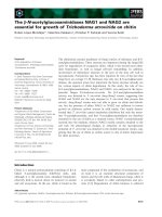
Coronal view of computerized tomography myelography vis-ualizing the ventral rootletsFigure 1
Coronal view of computerized tomography myelography vis-
ualizing the ventral rootlets. The number or size of rootlets
and the connection with the cord are well visualized.
Journal of Brachial Plexus and Peripheral Nerve Injury 2007, 2:16 />Page 3 of 5
(page number not for citation purposes)
menigocele were not visualized because of epidural punc-
ture. These images were excluded from the study.
CT myelographic diagnosis of root avulsion was based on
the either both ventral and dorsal roots and the presence
of a menigocele as follows: A(+); ventral root can be rec-
ognized, A(-); ventral root cannot be recognized, P(+);
dorsal can be recognized, P(-);dorsal root cannot be rec-
ognized, M(+); menigocele can be recognized, M(-);
menigocele cannot be recognized. When the image iden-
tify the healthy both ventral and dorsal roots without a
menigocele, the findings was classified A(+)P(+)M(-). A
nerve root was considered avulsed from the spinal cord
when either ventral or dorsal roots were unrecognizable
on axial view. On coronal and oblique coronal view,
nerve roots were considered avulsed when the number or
size of rootlets was decreased or the roots was absent.
Image criteria for the diagnosis was based on the presence
of the ventral and dorsal roots but was not the absence of
either or both roots. If the findings was classified
A(+)P(+)M(-), the roots were diagnosed as repairable.
The images were reviewed independently and blindly by
two observers without knowledge of clinical or surgical
finding. Discrepancies between the two observers were
resolved by consensus. The inter-observer reliability was
assessed.
The image findings were compared with the diagnosis for
57 cervical roots in 28 patients. Diagnosis was based on
intraoperative findings in 25 patients and clinical findings
of recovery without surgery in 3 patients. Intraoperative
findings include with direct observation of the nerve
roots, evoked spinal cord potentials from each nerve root,
and choline acetyltransferase activity measurement [5].
The sensitivity, specificity, and diagnostic accuracy in the
evaluation of the root avulsion were calculated for the 57
cervical roots in the 28 patients.
We used the Yates' chi-square test to compare the sensitiv-
ity, specificity, and diagnostic accuracy between the axial
CT images and the coronal and oblique coronal CT
images. The Cohen Kappa analysis was used for inter-
observer reliability. The level of significance was estab-
lished at p < 0.05.
Results
Good-quality CT myelographic examinations were
obtained in 49 (86%) of the 57 roots on axial view. Image
quality was degraded by epidural puncture in the other 5
roots and by unrecognition of the contralateral intact root
in the other 3 roots on axial view. On coronal and oblique
coronal view, they were obtained in 54 (95%) roots, and
image quality was degraded by epidural puncture in the
other 3 roots (no statistically significant difference). These
nerve roots with poor-quality image were excluded from
the analysis. The kappa value for the inter-observer relia-
bility of the axial view and the coronal and oblique coro-
nal view was 0.91 and 0.89, respectively.
The findings with axial view were classified as repairable
in 24 roots and non-repairable in 24. They showed 96%
sensitivity, 83% specificity, and 90% diagnostic accuracy,
with 23 true-positive findings, 20 true-negative findings,
one false-positive findings, and four false-negative find-
ings for diagnosing root avulsion.
The findings with coronal and oblique coronal view were
classified as repairable in 28 roots and non-repairable in
26. They showed 100% sensitivity, 96% specificity, and
98% diagnostic accuracy, with 26 true-positive findings,
27 true-negative findings, none false-positive findings,
and one false-negative findings for diagnosing root avul-
sion.
There was no statistically significant difference in sensitiv-
ity, specificity, and diagnostic accuracy between the two
imaging technique.
Discussion
MRI has many advantages without considerable exposure
to radiation, a possible adverse reaction to contrast mate-
rial, and the risk of lumber puncture. The most common
findings with nerve root avulsion are traumatic menin-
goceles. MRI is superior to conventional myelography and
CT myelography in visualizing small meningoceles,
which do not fill with contrast medium in a presence of a
dural scar [6]. Nerve root avulsions with no dural abnor-
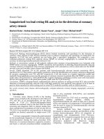
Oblique coronal view of computerized tomography myelog-raphy visualizing the dorsal rootletsFigure 2
Oblique coronal view of computerized tomography myelog-
raphy visualizing the dorsal rootlets.
Journal of Brachial Plexus and Peripheral Nerve Injury 2007, 2:16 />Page 4 of 5
(page number not for citation purposes)
malities and traumatic meningoceles without nerve root
avulsion, however, have been reported [7]. Avulsion
injury may be necessary to be evaluated on nerve rootlets.
Conventional myelography provide good anatomical
depiction of root sleeves and nerve roots. But the shadows
of the root are sometimes misjudged, if the concentration
of the contrast medium is low. It is reported to be unreli-
able at the level of the fifth and sixth cervical nerve roots
[7]. CT myelography is superior to conventional myelog-
raphy in visualizing the nerve rootlets. It is, however,
sometimes difficult to determine the exact level of the root
with axial imaging, because the roots run obliquely [1]. It
is difficult to detect the entire extent of root injuries with
single axial slice of the images.
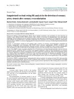
CT myelography with axial view allows demonstration of
the rootlets and also differentiation between the ventral
and dorsal rootlets (Fig. 3). A particular difficulty for diag-
nosis with axial view, although, is assessment of the root-
lets. As the spinal nerve rootlets run in oblique direction,
the continuity of some nerve rootlets from the cord to the
exit foramen can not be identified in axial view. Coronal
and oblique coronal view was superior to conventional
axial view in visualization of the number or size of root-
lets and the connection with the cord, and in orientation
of the exact level of the root. Coronal view visualized the
whole image of the ventral rootlets, and oblique coronal
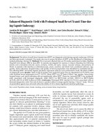
view visualized the dorsal rootlets. In the case with
decreased number of the rootlets or redundant rootlets
(Fig. 4), intraoperative diagnosis was pre-ganglionic
injury with considerable frequency. The major advantage
that CT myelography with coronal and oblique coronal
view adds to a good quality myelogram is the ability to
identified partial injury of ventral and dorsal rootlets. We
believe this technique to be useful for determining the sta-
tus of the nerve rootlets and detecting nerve root avulsion,
although diagnostic utility was not significant different.
In this study, we reviewed twenty-three of C5 root, seven-
teen of C6 root, seven of C7 root, five of C8 root, and one
of Th1 root. Exploration of the all roots was not routinely
performed, since the nerve graft is not effective in the
lower roots. Brachial plexus exploration cannot reveal
intraforaminal rootlet lesions unless laminectomy is per-
formed. Intraoperative nerve action potentials obtained at
the proximal cervical root attempt to evaluate the intrasp-
inal status of the roots extraspinally. However, nerve
action potential studies asses only the dorsal rootlets.
Therefore, even a positive nerve action potential does not
exclude the intradural avulsion of the ventral rootlet,
because the ventral rootlets are more vulnerable than the
dorsal rootlets. Choline acetyltransferase activity measure-
ment has been applied clinically to distinguish the availa-
bility of the proximal nerve stump as a donor motor nerve
during brachial plexus surgery [5]. We use choline acetyl-
transferase activity measurement for intraoperative diag-
nosis of the root avulsion in the case with discrepancies
between the nerve action potential studies and the clinical
or imaging diagnosis.
Conclusion
The development of reconstructed CT myelography with
coronal and oblique coronal view has provided important
advantages over axial view with regard to the rootlets
shadows, although diagnostic utility was not significant
different. CT myelography, in spite of its invasiveness, is
still indispensable for preoperative evaluation of cervical
In the coronal view, decreased number or redundant of the C5 rootlets (black arrow) are well recognizedFigure 4
In the coronal view, decreased number or redundant of the
C5 rootlets (black arrow) are well recognized.
Axial view of computerized tomography myelography visual-izing only a part of the rootletsFigure 3
Axial view of computerized tomography myelography visual-
izing only a part of the rootlets.
Publish with BioMed Central and every
scientist can read your work free of charge
"BioMed Central will be the most significant development for
disseminating the results of biomedical research in our lifetime."
Sir Paul Nurse, Cancer Research UK
Your research papers will be:
available free of charge to the entire biomedical community
peer reviewed and published immediately upon acceptance
cited in PubMed and archived on PubMed Central
yours — you keep the copyright
Submit your manuscript here:
/>BioMedcentral
Journal of Brachial Plexus and Peripheral Nerve Injury 2007, 2:16 />Page 5 of 5
(page number not for citation purposes)
nerve root avulsion of brachial plexus injury because of its
precise delineation of nerve rootlets shadows.
Competing interests
The author(s) declare that they have no competing inter-
ests.
Authors' contributions
HY designed the study, reviewed the images, performed
myelography, helped perform surgeries, and drafted the
manuscript. KD conceived the study and performed sur-
geries. YH reviewed the images, performed myelography,
and helped perform surgeries. SS helped perform surger-
ies. All authors read and approved the final manuscript.
References
1. Tavakkolizadeh A, Saifuddin A, Birch R: Imaging of adult brachial
plexus traction injuries. J Hand Surg [Br] 2001, 26:183-191.
2. Nakamura T, Yabe Y, Horiuchi Y, Takayama S: Magnetic resonance
myelography in brachial plexus injury. J Bone Joint Surg Br 1997,
79:764-769.
3. Doi K, Otsuka K, Okamoto Y, Fujii H, Hattori Y, Baliarsing AS: Cer-
vical nerve root avulsion in brachial plexus injuries: magnetic
resonance imaging classification and comparison with mye-
lography and computerized tomography myelography. J
Neurosurg 2002, 96:277-284.
4. Carvalho GA, Nikkhah G, Matthies C, Penkert G, Samii M: Diagnosis
of root avulsions in traumatic brachial plexus injuries: value
of computerized tomography myelography and magnetic
resonance imaging. J Neurosurg 1997, 86:69-76.
5. Hattori Y, Doi K, Dhawan V, Ikeda K, Kaneko K, Ohi R: Choline
acetyltransferase activity and evoked spinal cord potentials
for diagnosis of brachial plexus injury. J Bone Joint Surg Br 2004,
86:70-73.
6. Gasparotti R, Ferraresi S, Pinelli L, Crispino M, Pavia M, Bonetti M,
Garozzo D, Manara O, Chiesa A: Three-dimensional MR myelog-
raphy of traumatic injuries of the brachial plexus. AJNR Am J
Neuroradiol 1997, 18:1733-1742.
7. Nagano A, Ochiai N, Sugioka H, Hara T, Tsuyama N: Usefulness of
myelography in brachial plexus injuries. J Hand Surg [Br] 1989,
14:59-64.