Báo cáo y học: "Intraneural hemangioma of the median nerve: A case report" pot
Bạn đang xem bản rút gọn của tài liệu. Xem và tải ngay bản đầy đủ của tài liệu tại đây (414.37 KB, 5 trang )
BioMed Central
Page 1 of 5
(page number not for citation purposes)
Journal of Brachial Plexus and
Peripheral Nerve Injury
Open Access
Case report
Intraneural hemangioma of the median nerve: A case report
Yunus Doğramacı*, Aydıner Kalacı, Teoman Toni Sevinç and
Ahmet Nedim Yanat
Address: Dept. of Orthopaedics and Traumatology, Mustafa Kemal University Faculty of Medicine, Hatay, Turkey
Email: Yunus Doğramacı* - ; Aydıner Kalacı - ; Teoman Toni Sevinç - ;
Ahmet Nedim Yanat -
* Corresponding author
Abstract
Hemangiomas of the median nerve are very rare and, so far, only ten cases of intraneural
hemangioma of this nerve have been reported in the literature. We present a case of 14-year-old
girl who had a soft tissue mass in the region of the left wrist with signs and symptoms of carpal
tunnel syndrome. Total removal of the mass was achieved using microsurgical epineural and
interfasicular dissection. The symptoms were relieved completely, after this procedure, without
any neurologic deficit. On follow-up two years later, no recurrence was observed. Whenever a
child or young adult patient presents with CTS the possibility of a hemangioma involving the median
nerve should be kept in mind in the differential diagnosis.
Introduction
The carpal tunnel syndrome (CTS) is the most common
neuropathy due to compression seen in adults. There are
very few cases in the literature referring to patients of pae-
diatric age [1]. Most of these young patients had a meta-
bolic disorder mucopolysaccharidosis or mucolipidosis.
Other unusual causes of CTS in children are fibrolipomas
of the median nerve or intraneural perineuroma or hae-
mangioma, haemophilia (secondary to local bleeding),
musculotendinous malformation, Klippel-Trenaunay
syndrome, Poland's syndrome, scleroderma, benign local-
ised form of gigantism, intensive sports practice, and pri-
mary familial CTS [1]. Very rarely Schwannomas of the
median nerve can be mistakenly diagnosed and present as
carpal tunnel syndrome [2-4]. Lipofibromatous hamar-
toma of the median nerve at the wrist was reported, and
caused macrodactyly of the digits, and also resulted in
symptoms of carpal tunnel syndrome [5-7]. Again epithe-
lioid sarcoma of the median nerve may present with
symptoms and signs of carpal tunnel syndrome [8]. An
isolated malignant peripheral nerve sheath tumor of mild
type has also been reported to present with symptoms and
signs of carpal tunnel syndrome [9].
Posttraumatic neuroma-in-continuity of the median
nerve causing median nerve compression is rare [10].
Damage to the median nerve after vascular graft place-
ment as a result of an occult mass has been documented
in a single case [11].
Intraneural hemangioma of the median nerve is a rare
condition and only ten cases have been described in the
literature [12-20]. Due to mechanical compression, carpal
tunnel syndrome (CTS) is the main presenting feature
[12-18]. Raynaud's phenomenon may be an associated
complaint [16].
Published: 22 February 2008
Journal of Brachial Plexus and Peripheral Nerve Injury 2008, 3:5 doi:10.1186/1749-7221-3-
5
Received: 16 December 2007
Accepted: 22 February 2008
This article is available from: />© 2008 Doğramacı et al; licensee BioMed Central Ltd.
This is an Open Access article distributed under the terms of the Creative Commons Attribution License ( />),
which permits unrestricted use, distribution, and reproduction in any medium, provided the original work is properly cited.
Journal of Brachial Plexus and Peripheral Nerve Injury 2008, 3:5 />Page 2 of 5
(page number not for citation purposes)
Here we present a case of intraneural hemangioma of the
median nerve of a 14-year-old female removed surgically
by combined interfasicular and epineural resection, no
recurrence observed during the two years of postoperative
follow-up period.
Case presentation
A 14-year-old female student presented to our outpatient
clinic with painful swelling in the volar surface of the right
wrist of 3 years duration; associated with tingling and
numbness in the thumb, index, middle and radial half of
the ring fingers, difficulty in writing long paragraphs.
There was no history of trauma and relevant medical con-
dition.
Physical examination revealed a tender, soft mass, 3 × 5 ×
2 cm in dimension in the volar aspect of the right wrist.
Tinel sign was positive.
Radiographic examination revealed no bony lesion. Ultra-
sonographic examination done to exclude any vascular
lesion of the radial artery, revealed non pulsatile, cystic
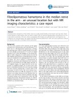
mass consistent with ganglion. An MR image obtained in
another institution revealed a 3 × 2 × 1.5 cm ovoid rapidly
enhancing mass in the volar surface of the right wrist
region (Fig. 1).
EMG examination was planned for this patient, but the
patient refused to cooperate during the test and the test
was not completed successfully.
After preoperative assessment, the patient was admitted
for surgical treatment under the diagnosis of volar gan-
glion causing CTS.
The operation was done under general anaesthesia, using
a pneumatic tourniquet. Exploration revealed a yellowish
brown soft tissue mass with areas of hemorrage and
dimensions of approximately 4 × 3 × 1.5 cm, originating
from the volar surface of the median nerve with intraneu-
ral extension and adhesions to the surrounding tissues
(Fig. 2). The mass was removed totally by interfasicular
and epineural microsurgical resection technique, without
structurally damaging the nerve fibbers.
Histopathologic and microscopic evaluation revealed
dilated and congested vascular structures in a fibrocolla-
genous stroma with areas of bleedings, consistent with
histopathologic findings of hemangioma (Fig 3).
The symptoms were relieved in the first three weeks after
the operation. On clinical and ultrasound examination,
no recurrence was observed in the first two years following
the operation.
Discussion
Benign intraneural hemangioma originating from periph-
eral nerves is rare. Most patients present in with a painful,
soft mass along the path of a nerve with signs and symp-
toms of nerve compression and entrapment.
A thorough search through the literature revealed ten
cases of hemangioma of the median nerve [12-19]. In all
the described cases CTS is the presenting feature and in
one case Raynaud's phenomenon was an associated pre-
senting feature.
The tumor may not be easily recognised until it becomes
painful and it is rarely diagnosed before surgery. In the
differential diagnosis, lipoma, lipofibroma, hamartoma
and intraneuronal Schwannoma must be considered
[20,21].
Ultrasonography may give useful information about the
nerve's dynamic relation to the surrounding musculo-
tendinous structures [22] and nerve conduction studies
may reveal non specific features of compressive neuropa-
thies [23]. For appropriate planning of surgical therapy
and preoperative diagnosis, MRI is essential and gives use-
ful information regarding tumor location, size, extent and
relationship of peripheral nerve.
Hemangioma shows a hyperintense signal on T1- and T2-
weighted images with fat suppression sequences. Flow
voids are usually apparent and feeding vessels may be vis-
ualized; these lesions are also noted to enhance after Gd-
addition. On angiography an early and persistent tumoral
blush is demonstrated [20].
Schwannoma is a slightly hypodense, solid tumor with no
vascular contrast enhancement on CT. MRI shows inter-
mediate signals on. T1-W, and T2-W imaging shows high
signal intensity with some heterogenity [24]. Lipomas
exhibit signal characteristics consistent with those of nor-
mal adipose tissue: homogeneous hyperintensity on T1-
and T2-weighted sequences [25]. MR imaging findings of
lipofibromatous hamartoma are pathognomonic which
consist of serpiginous T1- and T2-weighted low-intensity
structures containing and surrounded by fat (hyperin-
tense on T1- and hypointense on T2-weighted fat suppres-
sion sequences), giving the lesion a spaghetti-like
appearance on sagittal images, and a "coaxial cable-like"
appearance on coronal images [26].
No certain protocol has been established to manage this
difficult condition, however conservative treatment usu-
ally fails and surgery is the treatment of choice. When pos-
sible total resection of intraneural hemangiomas is
curative, partial resection may relieve symptoms but
Journal of Brachial Plexus and Peripheral Nerve Injury 2008, 3:5 />Page 3 of 5
(page number not for citation purposes)
Magnetic resonance image of intraneural hemangiomaFigure 1
Magnetic resonance image of intraneural hemangioma. (A) Sagittal T1 (B) T2 (C) axial T1 and (D) T2 (E) fat suppres-
sion images demonstrating an 3 × 2 × 1.5 cm lesion in the volar aspect of the right wrist.
Journal of Brachial Plexus and Peripheral Nerve Injury 2008, 3:5 />Page 4 of 5
(page number not for citation purposes)
recurrence may occur which may require en-bloc nerve
resection and repair with nerve graft [14].
The longest period of follow-up without recurrence has
been reported by Oztekin et al. [18]. They reported a case
of CTS due to a cavernous hemangioma of the median
nerve, which was successfully removed by epineural resec-
tion, and no recurrence was observed over a 6 year follow-
up period. Patel et al. [14] reported two cases of hemangi-
oma of the median nerve which they treated by partial
excision and resulted in recurrence in the third year, one
of the recurred case managed by resection of median nerve
and nerve grafting without recurrence, four years after sur-
gery.
Chatillon et al. [20] reported the first case of using radio-
therapy in the treatment of intraneural hemangioma. Pre-
operative embolization and postoperative radiotherapy
combined with partial resection were beneficial in a case
of intraneural hemangioma involving inferior trunk of
brachial plexus and resulted in symptomatic relief and
radiologic shrinkage in the size of the mass seen on serial
follow-up MRI images, with a follow-up period of two
years.
In our case, total resection of the hemangioma was
achieved by combined epineural resection and interfasic-
ular dissection with microsurgical resection technique, no
neurologic complications observed postoperatively and
no recurrence observed in the two year follow-up period.
The type of microsurgical dissection and resection should
be decided at the time of surgery and careful preoperative
planning using MRI, and if needed angiography, is essen-
tial for cystic lesions of the volar side of wrist. Excision of
the affected nerve and grafting should be the last choice
and should only be used in complicated cases and when
there are frequent recurrences.
Conclusion
Whenever a child or young adult patient presents with
CTS the possibility of a hemangioma involving the
median nerve should be kept in mind in the differential
diagnosis.
Acknowledgements
Written informed consent was obtained from the patient for publication of
this Case report and accompanying images. A copy of the written consent
is available for review by the Editor-in-Chief of this journal.
References
1. Van Meir N, De Smet L: Carpal tunnel syndrome in children.
Acta Orthop Belg 2003, 69:387-395.
2. Aslam N, Kerr G: Multiple Schwannomas of the median nerve:
a case report and literature review. Hand Surg 2003, 8:249-252.
3. Padua L, Pazzaglia C, Insola A, Aprile I, Caliandro P, Rampoldi M, Ber-
tolini C, Tonali P: Schwannoma of the median nerve (even out-
side the wrist) may mimic carpal tunnel syndrome. Neurol Sci
2006, 26:430-434.
4. Aydin MD, Kotan D, Keles M: Acute median nerve palsy due to
hemorrhaged schwannoma: case Report. JBPPNI 2007, 2:19.
5. Bagatur AE: Lipofibromatous hamartoma of the median
nerve. Acta Orthop Traumatol Turc 2002, 36:172-176.
6. Lorenzoni PJ, Lange MC, Kay CS, Silvado CE, Scola RH, Werneck LC:
Fibrolipomatous hamartoma of the median nerve: case
report. Arq Neuropsiquiatr 2005, 63(B):881-884.
7. Bains R, Kotwal A, Saeed W: Recurrent carpal tunnel syndrome
in a child due to fibrolipomatous hamartoma of the median
nerve successfully treated by limited excision and decom-
pression. J Plast Reconstr Aesthet Surg 2006, 59:1394-1397.
8. Harish S, Saifuddin A, Fajinmi M: Epithelioid sarcoma of the
median nerve mimicking a peripheral nerve sheath tumour.
Australas Radiol 2007, 51:71-74.
Microscopic view of hemangioma showing vascular struc-tures in a fibrocollagenous stroma with areas of bleedingsFigure 3
Microscopic view of hemangioma showing vascular
structures in a fibrocollagenous stroma with areas of
bleedings. (Hematoxylin-Eosin, ×100).
Macroscopic view of the lesion, intraneural and fasicular involvement is obviousFigure 2
Macroscopic view of the lesion, intraneural and fasic-
ular involvement is obvious.
Publish with BioMed Central and every
scientist can read your work free of charge
"BioMed Central will be the most significant development for
disseminating the results of biomedical research in our lifetime."
Sir Paul Nurse, Cancer Research UK
Your research papers will be:
available free of charge to the entire biomedical community
peer reviewed and published immediately upon acceptance
cited in PubMed and archived on PubMed Central
yours — you keep the copyright
Submit your manuscript here:
/>BioMedcentral
Journal of Brachial Plexus and Peripheral Nerve Injury 2008, 3:5 />Page 5 of 5
(page number not for citation purposes)
9. Ochsner F, Baumann RP, Kuntzer T: Carpal tunnel syndrome
with an unusual cause: a malignant nerve sheath tumor of
the median nerve. Rev Neurol (Paris) 2001, 157:1547-1549.
10. Martinelli P, Poppi M, Gaist G, Padovani R, Pozzati E: Posttraumatic
neuroma of the median nerve: a cause of carpal tunnel syn-
drome. Eur Neurol 1985, 24:13-15.
11. Goldstein LJ, Helfend LK, Kordestani RK: Postoperative edema
after vascular access causing nerve compression secondary
to the presence of a perineuronal lipoma: case report. Neu-
rosurgery 2002, 50:412-414.
12. Coessens B, De Mey A, Lacotte B, Vandenbroeck D: Carpal tunnel
syndrome due to an haemangioma of the median nerve in a
12-year-old child. Ann Chir Main Memb Super 1991, 10:255-257.
13. Kojima T, Ide Y, Marumo E, Ishikawa E, Yamashita H: Haemangi-
oma of median nerve causing carpal tunnel syndrome. Hand
1976, 8:62-65.
14. Patel CB, Tsai TM, Kleinert HE: Hemangioma of the median
nerve: a report of two cases. J Hand Surg [Am] 1986, 11:76-79.
15. Peled I, Iosipovich Z, Rousso M, Wexler MR: Hemangioma of the
median nerve. J Hand Surg [Am] 1980, 5:363-365.
16. Prosser AJ, Burke FD: Haemangioma of the median nerve asso-
ciated with Raynaud's phenomenon. J Hand Surg [Br] 1987,
12:227-228.
17. Wood MB: Intraneural hemangioma: report of a case. Plast
Reconstr Surg 1980, 65:74-76.
18. Oztekin HH, Karaarslan AA: Carpal tunnel syndrome due to a
cavernous hemangioma of the median nerve. Acta Orthop Trau-
matol Turc 2003, 37:170-172.
19. Petrovici V: Cavernous hemangioma of the palm with symp-
toms resembling carpal tunnel syndrome. Z Plast Chir 1980,
4:40-47.
20. Chatillon CE, Guiot MC, Jacques L: Lipomatous, vascular, and
chondromatous benign tumors of the peripheral nerves:
Representative cases and review of the literature. Neurosurg
Focus 2007, 22:E18.
21. Louis D: Peripheral nerve tumors of the upper extremity.
Hand Clin 1987, 3:311-318.
22. Kuo YL, Yao WJ, Chiu HY: Role of sonography in the preopera-
tive assessment of neurilemmoma. J Clin Ultrasound 2005,
33:87-89.
23. MacDonell RAL, Schwartz MS, Swash M: Carpal Tunnel Syn-
drome: Which finger should be tested? An analysis sensory
conduction in digital branches of the median nerve. Muscle
Nerve 1990, 13:601-606.
24. Hems TE, Burge PD, Wilson DJ: The role of magnetic resonance
imaging in the management of peripheral nerve tumours. J
Hand Surg 1997, 22(1):57-60.
25. Chiao HC, Marks KE, Bauer TW, Pflanze W: Intraneural lipoma of
the sciatic nerve. Clin Orthop Relat Res 1987, 221:267-271.
26. Marom EM, Helms CA: Fibrolipomatous hamartoma: pathog-
nomonic on MR imaging. Skeletal Radiol 1999, 28:260-264.