Báo cáo y học: " Is distortion of the bioprosthesis ring a risk factor for early calcificatio" docx
Bạn đang xem bản rút gọn của tài liệu. Xem và tải ngay bản đầy đủ của tài liệu tại đây (632.74 KB, 3 trang )
RESEA R C H ART I C L E Open Access
Is distortion of the bioprosthesis ring a risk factor
for early calcification?
Jose Manuel Martinez Cereijo
1
, Jose Rubio Alvarez
1*
, Juan Sierra Quiroga
1
, Anxo Martinez de Alegria
2
,
Jose Manuel Suarez Peñaranda
3
Abstract
Background: As the population ages, bioprosthesis are increasingly being used in cardiac valve replacem ent.
Pericardial bioprosthesis combine an excellent hemodynamic performance with low thrombogenicity, but valve
failure associated with calcification rema ins a concern with these valves. We descr ibe distortion of the bioprosthesis
ring as a risk factor for early calcification.
Methods: A total of 510 patients over the age of 70 years underwent isolated aortic valve repla cement with the
Mitroflow (A12) pericardial bioprosthesis. Thirty two patients (6,2%) have undergone a second aortic valve
replacement due to structural valve dysfunct ion resulting from valve calcification. In all patients a chest radiography
and coronary angiography was performed before reoperation. A 64 Multidetector Computed Tomography (MDCT)
with retrospective ECG gating study was performed in four patients to evaluate the aortic bioprosthesis.
Results: Chest radiography showed in all patients an irregular bioprosthesis ring. At preoperative coronary
angiography a distorted bioprosthesis ring was detected in all patients. Macroscopic findings of the explanted
bioprostheses included extensive calcification in all specimens.
Conclusion: There was a possible relationship between early bioprosthetic calcification and radiologic distortion of
the bioprosthesis ring.
As the population ages, bioprosthesis are increasingly
being used in cardiac valve replacement. Pericardial bio-
prosthesis combine an excellent hemodynamic perfor-
mance [1] with low thro mbogenicity [2], but valve
failure associated with calcification remains a concern
with these valves [3]. We describe distortio n of the bio-
prosthesis ring as a risk factor for early calcification.
Materials and methods
A total of 510 patients over the a ge of 70 years under-
went isolated aortic valve replacement with the Mitro-
flow (A12) pericardial bioprosthesis at our hospital.
Sixty-five percent of patients undergoing aortic valve
replacement received a 21 mm valve. Demographic data
at time of reoperation are listed in Table 1 and the size of
the bioprosthesis in Table 2 To date 32 patients (6,2%)
have undergone a second aortic valve replacement due to
structural valve dysfunction (SVD) resulting from valve
calcification. The mean time between the first operation
and reoperation was 70 months (range 36 to 98 months).
In all patients a chest radiography and coronary angio-
graphy was performed before reoperation.
A 64 Multidetector Computed Tomography (MDCT)
with retrospective ECG gating study was performed in
four patients to evaluate the aortic bioprosthesis. In two
patients the study was performed three weeks after aor-
tic valve replacement with the Mitroflow A12 pericardial
bioprosthesis. Both bioprosthesis were normal by echo-
cardiographic study. Another two patients had bio-
prosthesis with SVD.
A His topath olog ic analysis of the removed bioprosth-
esis was performed by one experienced pathologist.
Results
Macroscopic findings of the explanted bioprostheses
included extensive calcification in all specimens. In all
cases, one leaflet was more calcified than the others; in
* Correspondence:
1
Department of Cardiovascular Surgery, Santiago de Compostela University
Hospital. La Choupana, Santiago de Compostela 15706, Spain
Full list of author information is available at the end of the article
Cereijo et al. Journal of Cardiothoracic Surgery 2010, 5:77
/>© 2010 Cereijo et al; licensee BioMed Centr al Ltd. This is an Open Access article distributed under the terms of the Creative Commons
Attribution License ( which permits unrestricted use, distribution, and reproduction in
any medium, provided the original work is properly cited.
our experience, the right coronary l eaflet was commonly
the most calcified (19/32. 59,3%).
In all patients, serum levels of cholesterol, triglycerides,
lipoprotein A and calcium were within the normal range.
No patients were treated with calcium supplementation.
Chest radiography (Figure 1) showed in all patients an
irregular, non- circular bioprosthesis ring. At preopera-
tive coronary angiography a distorted bioprosthesis ring
was detected in all patients (Figure 2). An explanted bio-
prosthesis with leaflet distortion and right coronary leaf-
let calcification is shown in Figure 3.
In two patients with normal bioprosthesis, the morphol-
ogy of the leaflets and the degree of excursion were nor-
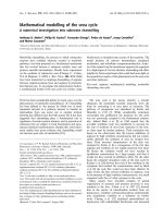
mal at MDCT, but a leaflet had a wave movement. The
distorted ring was observed in these patients (Figure 4)
and the same picture was observed in two patients with
SVD.
The percentage of distortion of the bioprosthesis ring
in patients without SVD was 76%, but no all patients
had the same degree of distortion.
Discussion
Bioprosthetic calcification is a multifactorial process;
contributing factors include the type of implant used,
mechanical stre ss, preservation method, patient age and
technical correctness of the implantation procedure [4].
The rate of structural valve dysfunction is lower in
elderly patients and early cal cificati on in this group of
patients is an uncommon event.
There is a strong relationship between mechanical
stress and calcification in bioprosthesis [4,5]; in our study
we noted that early calcification tended to develop in
patients with radiologic distortion of the bioprosthesis
ring. Liao and colleagues [4] observed similar results with
bioprostheses removed from left ventricular assist
devices. Distortion of the bioprosthesis ring may be due
to technical problems at implantation [6] or compression
Table 1 Demographic data
Age (range) 77 ± 4 (71 - 86)
Female/Male 18/14
Aortic Stenosis 28 (87,5%)
Renal insufficiency 0
Hypertension 14 (43,7%)
Diabetes 2 (6,25%)
Table 2 Bioprosthesis size
Valve Size (mm) At reoperation
19 1
21 20
23 10
25 1
Figure 1 Radiographic evaluation showed a misshapen
bioprosthesis ring.
Figure 2 Preoperative coronary angiography showed a
misshapen bioprosthesis ring.
Figure 3 Explanted bioprosthesis with leafl et distorted and
calcification.
Cereijo et al. Journal of Cardiothoracic Surgery 2010, 5:77
/>Page 2 of 3
around the ring. However we speculate that Dacron ring
traction from the annular stitch may distort the normal
planar geometry of Mitroflow pericardial bioprosthesis,
leading to distortion of the pericardial leaflet mounted
outside the stent and fixed to the Dacron ring, resulting
in a higher mechanical stress.
In our patients other casual relationship was no found.
Conclusion
In our experience there was a possible relationship
between bioprosthetic calcification and radiologic distor-
tion of the bioprosthesis ring.
Author details
1
Department of Cardiovascular Surgery, Santiago de Compostela University
Hospital. La Choupana, Santiago de Compostela 15706, Spain.
2
Department
of Radiology, Santiago de Compostela University Hospital. La Choupana,
Santiago de Compostela 15706, Spain.
3
Department of Pathology, Santiago
de Compostela University Hospital. La Choupana, Santiago de Compostela
15706, Spain.
Authors’ contributions
JMMC drafted the manuscript and did the patients follow-up.
JRA conceived of the study and participated in its design and coordination.
JSQ participated in the design of the study.
AMA carried out the radiologic studies.
JMSP carried out the pathologic studies.
All authors read and approved the final manuscript.
Competing interests
The authors declare that they have no competing interests.
Received: 13 April 2010 Accepted: 7 October 2010
Published: 7 October 2010
References
1. Bengochea JG, Sierra J, Gonzalez - Juanatey JR, Rubio J, Vega M,
Fernandez AL, Sanchez D: Left ventricular mass regressión after aortic
valve replacement with the new Mitroflow 12 A pericardial
bioprosthesis. J Heart Valve Dis 2006, 15:446-452.
2. Corbineau H, De la Tour B, Verhoye JP, Langanay Th, Lelong B, Leguerrier A:
Carpentier-Edwards supraannular porcine bioprosthesis in aortic
position: 16-year experience. Ann Thorac Surg 2001, 71:S228-S231.
3. Rubio J, Sierra J, Vega M, Adrio B, Martinez Comendador JM, Gude F,
Martinez Cereijo JM, Garcia J: Early calcification of the aortic Mitroflow
pericardial bioprosthesis in the elderly. Interact Cardio Vasc Thorac Surg
2009, 9:842-846.
4. Liao KK, Li X, Ranjit J, Amayta DM, Joyce LD, Park SJ, Bianco R, Bolman RM:
Mechanical stress: An independent determinant of early bioprosthetic
calcification in humans. Ann Thorac Surg 2008, 86:491-495.
5. Bernacca GM, Fisher AC, Wilkinson R, Mackay TG, Wheatley D: Calcification
and stress distribution in bovine pericardial heart valves. J Biomed Mater
Res 1992, 26:959-966.
6. Saunders PC, Grossi EA, Esposito RA, Bizekis CS, Strong MD, Colvin SB:
Failure of four bovine pericardial mitral prostheses. J Thorac Cardiovasc
Surg 2004, 127:267-268.
doi:10.1186/1749-8090-5-77
Cite this article as: Cereij o et al.: Is distortion of the bioprosthesis ring a
risk factor for early calcification?. Journal of Cardiothoracic Surgery 2010
5:77.
Submit your next manuscript to BioMed Central
and take full advantage of:
• Convenient online submission
• Thorough peer review
• No space constraints or color figure charges
• Immediate publication on acceptance
• Inclusion in PubMed, CAS, Scopus and Google Scholar
• Research which is freely available for redistribution
Submit your manuscript at
www.biomedcentral.com/submit
Figure 4 Three different views of the bioprosthesis ring at MDCT.
Cereijo et al. Journal of Cardiothoracic Surgery 2010, 5:77
/>Page 3 of 3