Báo cáo y học: "Functional outcome of nerve transfer for restoration of shoulder and elbow function in upper brachial plexus injury" potx
Bạn đang xem bản rút gọn của tài liệu. Xem và tải ngay bản đầy đủ của tài liệu tại đây (1.43 MB, 9 trang )
BioMed Central
Page 1 of 9
(page number not for citation purposes)
Journal of Brachial Plexus and
Peripheral Nerve Injury
Open Access
Research article
Functional outcome of nerve transfer for restoration of shoulder
and elbow function in upper brachial plexus injury
Hari Venkatramani*, Praveen Bhardwaj, Sajedur Reza Faruquee and S
Raja Sabapathy
Address: Department of Plastic, Hand and Reconstructive Microsurgery, Ganga Hospital, Mettupalayam road, Coimbatore, India
Email: Hari Venkatramani* - ; Praveen Bhardwaj - ; Sajedur Reza Faruquee - ; S
Raja Sabapathy -
* Corresponding author
Abstract
Background: Purpose of this study was to evaluate the functional outcome of spinal accessory to
suprascapular nerve transfer (XI-SSN) done for restoration of shoulder function and partial transfer of
ulnar nerve to the motor branch to the biceps muscle for the recovery of elbow flexion (Oberlin transfer).
Methods: This is a prospective study involving 15 consecutive cases of upper plexus injury seen between
January 2004 and December 2005. The average age of patients was 35.6 yrs (15–52 yrs). The injury-surgery
interval was between 2–6 months. All underwent XI-SSN and Oberlin nerve transfer. The coaptation was
done close to the biceps muscle to ensure early recovery. The average follow up was 15 months (range
12–36 months). The functional outcome was assessed by measuring range of movements and also on the
grading scale proposed by Narakas for shoulder function and Waikakul for elbow function.
Results: Good/Excellent results were seen in 13/15 patients with respect to elbow function and 8/15 for
shoulder function. The time required for the first sign of clinical reinnervation of biceps was 3 months 9
days (range 1 month 25 days to 4 months) and for the recovery of antigravity elbow flexion was 5 months
(range 3 1/2 months to 8 months). 13 had M4 and two M3 power. On evaluating shoulder function 8/15
regained active abduction, five had M3 and three M4 shoulder abduction. The average range of abduction
in these eight patients was 66 degrees (range 45–90). Eight had recovered active external rotation, average
44 degrees (range 15–95). The motor recovery of external rotation was M3 in 5 and M4 in 3. 7/15 had no
active abduction/external rotation, but they felt that their shoulder was more stable. Comparable results
were observed in both below and above 40 age groups and those with injury to surgery interval less than
3 or 3–6 months.
Conclusion: Transfer of ulnar nerve fascicle to the motor branch of biceps close to the muscle
consistently results in early and good recovery of elbow flexion. Shoulder abduction and external rotation
show modest but useful recovery and about half can be expected to have active movements. Two patients
in early fifties also achieved good results and hence this procedure should be offered to this age group also.
Surgery done earlier to 6 months gives consistently good results.
Published: 27 May 2008
Journal of Brachial Plexus and Peripheral Nerve Injury 2008, 3:15 doi:10.1186/1749-7221-3-15
Received: 7 January 2008
Accepted: 27 May 2008
This article is available from: />© 2008 Venkatramani et al; licensee BioMed Central Ltd.
This is an Open Access article distributed under the terms of the Creative Commons Attribution License ( />),
which permits unrestricted use, distribution, and reproduction in any medium, provided the original work is properly cited.
Journal of Brachial Plexus and Peripheral Nerve Injury 2008, 3:15 />Page 2 of 9
(page number not for citation purposes)
Background
In upper brachial plexus avulsion injuries loss of abduc-
tion and external rotation at shoulder and flexion at
elbow are the main functional deficits. Spinal accessory
nerve has been the most commonly used donor for resto-
ration of shoulder abduction and external rotation with
varying results in different centers. [1-9]. On average, the
results have been modest [10]. Many donors have been
tried for restoration of elbow flexion with overall good
results [1-5,11-14]. Oberlin et al [15] described the partial
transfer of ulnar nerve to the motor branch of biceps mus-
cle. This procedure has consistently shown good results
[16-19]. Since the coaptation is done very near to the mus-
cle and without nerve grafts early recovery is possible. No
significant donor deficits have been reported [15-19]. We
present our experience with fifteen consecutive cases of
upper plexus injury treated by this set of nerve transfers,
involving spinal accessory to suprascapular (XI-SSN) and
partial transfer of ulnar nerve to the biceps motor branch
(UNF-BrBi – Oberlin transfer).
Recent literature [20] and our experience also suggest that
additional transfer of median nerve fascicle to the motor
branch to brachialis gives even better results. This proto-
col is followed from December 2005. The results pre-
sented in this series are of transfer of only the ulnar nerve
fascicles (Oberlin procedure).
Methods
This was a prospective study of 15 consecutive cases of
upper plexus injury with good hand function who pre-
sented to us within 6 months of injury during the period of
January 2004 and December 2005. The average age of
patients was 35.6 yrs (15–52 yrs). There were 14 males and
one female. In all the cases mode of injury was road traffic
accident. All the patients were right hand dominant and 14
had injury to the dominant side. The surgery was done
between 2 and 6 months after the accident. The average fol-
low up was 15 months (range 12 to 36 months). Spinal
accessory to suprascapular nerve transfer (XI-SSN) and
transfer of ulnar nerve fascicle to the motor branch to biceps
(UNF-BrBi – Oberlin transfer) was done in all of them.
A detailed preoperative assessment was done in all the
case and documented as the base line. Elbow flexion,
shoulder abduction, and external rotation range of
motion were 0 degree in all the cases. Deltoid, teres
minor, supraspinatus, infraspinatus, biceps, and brachio-
radialis muscle were all paralyzed and scored M0 on Med-
ical Research Council (MRC) scoring. In two cases triceps
scored M0 (case no. 5 and 6), others had M4–M5 score.
Trapezius muscle was scored M5 and grip and pinch
strength in the hand was normal in all the cases.
With a minimum follow up of 12 months, all of them
were evaluated for range of movements at shoulder and
elbow; motor power and functional improvement.
Outcome assessment
Strength of muscle was graded using MRC scoring and
range of movements was recorder with Goniometry. The
range of elbow flexion was measured as the angle formed
between the long axis of the arm and the forearm. The
range of abduction was recorded by measuring the angle
formed between the arm axis and parallel to the spinal
cord axis. External rotation was measured with the patient
standing with the shoulder fully internally rotated and
forearm placed transversally over the abdomen. Any rota-
tion from this position was measured and noted as the
range of external rotation.
Shoulder and elbow function were graded using the scale
proposed by Narakas [2] and Waikakul et al [11] with
minimal modification as per Table 1 and Table 2 respec-
tively.
Table 2: Grading of elbow function (Waikakul et al [11] modified):
Grade Functional status
Excellent Ability to lift 2 Kg weight from 0 to 90 degrees of elbow flexion more than 30 times successively.
Good Ability to lift 2 Kg weight from 0 to 90 degrees of elbow flexion, but less than 30 repetitions successively.
Fair Motor power more than M3 power but unable to lift a 2 Kg weight.
Poor Motor power less than M3.
Table 1: Grading of shoulder function (Narakas [2] modified):
Grade Functional status
Poor No abduction movement and feeling of weightlessness in the limb (MRC 0)
Fair Stable shoulder without any subluxation but no active movement (MRC I)
Good Active abduction of < 60 degrees (MRC III) and active external rotation of < 30 degrees
Excellent Active abduction of > 60 degrees (MRC IV) and active external rotation of > 30 degrees.
Journal of Brachial Plexus and Peripheral Nerve Injury 2008, 3:15 />Page 3 of 9
(page number not for citation purposes)
Surgical technique
All patients were operated under general anesthesia in the
supine position with head and trunk turned to the oppo-
site side. The supraclavicular part of the plexus was
exposed through a transverse incision 2 cm above the
clavicle at the root of the neck and the C5 and C6 roots
were identified. Avulsion of the C5–6 roots and absence
of usable stump proximally was confirmed intra-opera-
tively. Nerve transfer was performed in the following
manner.
Spinal accessory nerve to suprascapular nerve
The spinal accessory nerve was identified along the supe-
rior border of the trapezius and confirmed with electrical
stimulation. Suprascapular nerve was then identified as it
emerged from the roots in the scar. The proximal branches
of the spinal accessory to the upper part of the trapezius
were preserved and the terminal branch was dissected and
divided as far distally as possible. It was then transposed
and coapted to the suprascapular nerve under microscopic
magnification with 10-0 Ethilon (Figure 1).
Oberlin transfer
A 10 cm incision was made over the anteromedial aspect
of the arm, starting about 4 cm distal to the pectoralis
major lateral border. The musculocutaneous nerve was
identified between the biceps and the coracobrachialis
muscles. It was traced distally to expose the motor branch
to the biceps muscle. The motor branch of the biceps was
dissected free and divided for about 2 cm from entry to the
muscle. The ulnar nerve was identified through the same
incision and confirmed by electrical stimulation. Further
dissection was done under microscopic magnification.
The epineurium of the ulnar nerve was incised and the fas-
cicles were dissected out. One fascicle of the ulnar nerve
was completely isolated and stimulated at low intensity of
0.02–0.04mA to identify the motor fascicles. We insert a
piece of glove under this fascicle to completely isolate it
from the surrounding fascicles to avoid mass stimulation
and false results. It is recommended to take the fascicles in
the anterior and medial part of the ulnar nerve which is
supposed to contain fibers predominantly to flexor carpi
ulnaris. In our experience we found any fascicle of ulnar
nerve, irrespective of its anatomical location when stimu-
lated shows contraction in most of the muscles. Hence we
choose a fascicle of appropriate size to match the size of
the nerve to biceps irrespective of the location in the ulnar
nerve. The chosen fascicle is separated from the rest of the
nerve. It must be divided 3 cm distal to the level of the
possible coaptation to turn laterally to meet the nerve to
biceps (Figure 2A). The fascicle is turned laterally and
superiorly towards the motor branch of biceps and
coapted with 10-0 Ethilon without tension with the ulnar
nerve in its usual anatomical position. The wound is
closed with a drain placed away from the nerve repair site.
The limb is strapped to the chest keeping the shoulder in
adducted and internally rotated position and elbow in
about 100 degree of flexion for 3 weeks.
Physiotherapy protocol
Stretching exercises are started at three weeks and electri-
cal stimulation is started after 6 weeks.
Results
All the patients experienced some improvement at their
shoulder function. Eight of the 15 patients had recovered
active abduction. Five patients had M3 recovery of shoul-
der abduction and three had M4 recovery. Among the
patients who had recovered active movements (eight)
average range of abduction was 66 degrees (range 45–90).
Figures 3, 4 show a patient with excellent recovery of
shoulder abduction. Eight of 15 patients had recovered
active external rotation. Among this average external rota-
tion was 44 degrees (range 15–95). The motor recovery of
external rotation was M3 in 5 cases and M4 in 3 cases. Fig-
ures 3, 4 shows clinical photograph of patient with good
recovery of external rotation at the shoulder. 7 never
recovered any active abduction or external rotation, but
all 7 felt that their shoulder was more stable and devel-
oped some control of the limb.
All regained active flexion at the elbow. 13 of 15 patients
recovered full flexion (140 degrees); one had 90 degree
and other 100 degree of anti-gravity flexion. The average
time required for clinical reinnervation of biceps (flicker
of movement) was 3 months 9 days (range 1 month 25
Spinal accessory to suprascapular nerve transferFigure 1
Spinal accessory to suprascapular nerve transfer. The
proximal branches of the spinal accessory to the upper part
of the trapezius are preserved (yellow arrow) and terminal
branch is divided and coapted to the suprascapular nerve
(black arrow).
Journal of Brachial Plexus and Peripheral Nerve Injury 2008, 3:15 />Page 4 of 9
(page number not for citation purposes)
days to 4 months). 13 patients had M4 power and 2 had
M3+. The average time taken for the recovery of antigrav-
ity elbow flexion was 5 months (range 3 1/2 months to 8
months). Figures 3, 4, 5 show clinical photograph of
patient with good recovery of flexion at the elbow.
The details of the patients and the final grading of their
elbow and shoulder function are detailed in Table 3. Table
4 details the motor grade of the recovered muscles and the
final range of movement achieved. Table 5 shows the
results with respect to age. Comparable results were
achieved in patients below and above 40 years. Table 6
shows the results with respect to the time of surgery since
the injury. There was no significant difference between the
results in these two groups. Good to excellent results were
seen in 13/15 (87%) patients with respect to elbow func-
Clinical photographs of case noFigure 3
Clinical photographs of case no. 2 showing excellent results
for elbow and shoulder function- patient could easily do
more than 30 repetitions of elbow flexion with 2 Kg weight
and had 95 degree of external rotation and 80 degree of
abduction at the shoulder.
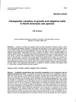
Oberlin procedureFigure 2
Oberlin procedure. The motor fascicles are separated
from the rest of the nerve over a distance of 2–3 cm (arrow).
The fascicles are turned laterally and superiorly towards the
biceps motor branch and coapted with it (arrow).
Journal of Brachial Plexus and Peripheral Nerve Injury 2008, 3:15 />Page 5 of 9
(page number not for citation purposes)
tion but only 8/15 (53%) had good to excellent results for
shoulder function.
Discussion
The incidence and severity of brachial plexus injury has
been increasing world wide mainly because of rapidly
growing number of motor cycle accidents. Most of these
injuries are high velocity injuries resulting in root avul-
sions. Although Carlstedl et al [21] have tried repairing
roots into the ventral spinal cord and Bertelli & Ghizoni
[22] have reported the direct replantation of the nerve
graft into the spinal cord with some promising results, the
surgical treatment of choice for brachial plexus root avul-
sion is nerve transfer.
Nerve transfer by reinnervating most functionally impor-
tant nerves using intact neighboring nerves has become
widely accepted since it was reported by Seddon in 1963
[23]. Since then variety of donor nerves have been used
for restoring various vital functions. Nerve transfer with
various donors has radically improved the prospects for
recovery of elbow and shoulder function, especially in
patients with irreparable lesion of upper roots. The order
of priorities when managing a case of brachial plexus
injury is to restore: Full range and power of elbow flexion;
shoulder stability; restoration of active abduction and
some external rotation. This can be obtained by reinner-
vation of musculocutaneous and suprascapular nerve. The
highest priority in nerve repair is reinnervation of the
Clinical photograph of case noFigure 5
Clinical photograph of case no. 12 showing good results for
elbow function but patient had only fair results for shoulder
function.
Clinical photographs of case noFigure 4
Clinical photographs of case no. 8 showing excellent results
for shoulder and elbow function. He had full range of elbow
flexion with good power and 90 degree of external rotation
and abduction at the shoulder.
Journal of Brachial Plexus and Peripheral Nerve Injury 2008, 3:15 />Page 6 of 9
(page number not for citation purposes)
musculocutaneous nerve to reinnervate biceps [2,13,16].
Biceps also contributes to shoulder stability [13].
Nerve transfer gives good results in restoring simple func-
tion like flexion of elbow but results are not as good when
done for complex function like shoulder abduction and
finger flexion [13]. Allieu et al [3,4], Narakas [1,2] success-
fully used spinal accessory neurotization for various recip-
ients. Shoulder stability and motion are vital to normal
use of the upper extremity. It improves the working space
of the hand and makes the hand more useful. Shoulder
stability and abduction are frequently accomplished by
arthrodesis of shoulder joint. However, according to Nar-
akas [1,2], good suprascapular neurotization will result in
twice the range of motion achieved by fusion of shoulder.
We agree with Chaung [9] that voluntary control of the
shoulder abduction produces more satisfied patients than
fusion. Moreover, the reinnervated suprascapular nerve
stabilizes the humeral heads and prevents internal rota-
tion of the humerus in patients with reactivated isolated
biceps muscle. Merrell et al [10] in their meta-analysis of
results of nerve transfer for restoration of shoulder func-
tion found that 73% of the patients who underwent nerve
transfer for restoration of shoulder abduction achieved
M3 power or more but only 26% could achieve M4 or
more. The most commonly used donor nerve was spinal
accessory (41%). The spinal accessory nerve can be
expected to provide a M3 or more strength in about 98%
of cases. Also, significantly better results were achieved by
reinnervating the suprascapular nerve (92%) than the
Table 4: Details of the recovery pattern observed.
S. No. Range of flexion at elbow (degrees) &
Motor grading
Range of abduction at shoulder (degrees) &
Motor grading
Range of external rotation at shoulder (degrees)
& Motor grading
1. 140/M4 60/M3 30/M3
2. 140/M4 80/M4 95/M4
3. 140/M4 0 0
4. 140/M4 60/M3 30/M3
5. 140/M4 50/M3 15/M3
6. 140/M4 45/M3 15/M3
7. 140/M4 0 0
8. 140/M4 90/M4 90/M4
9. 100/M3 0 0
10. 140/M4 0 0
11. 140/M4 50/M3 20/M3
12. 140/M4 0 0
13. 140/M4 0 0
14 140/M4 90/M4 60/M4
15. 90/M3 0 0
Table 3: Details of the 15 cases in the reported series
No. Age/Sex Injury Procedure Time since
injury (month)
Elbow Flexion grade
(Waikakul [11] mod.)
Shoulder Function grade
(Narakas [2] mod.)
Follow Up (Months)
1 50/M C5,6 XI-SSN* + UNF-BrBi** 6 Good Good 36
2 48/M C5,6 XI-SSN + UNF-BrBi 3 Excellent Excellent 34
3 27/M C5,6 XI-SSN + UNF-BrBi 4 Good Fair 30
4 42/M C5,6 XI-SSN + UNF-BrBi 4 Good Good 26
5 52/M C5,6,7 XI-SSN + UNF-BrBi 2 Good Good 25
6 20/M C5,6,7 XI-SSN + UNF-BrBi 3 Good Good 23
7 47/M C5,6 XI-SSN + UNF-BrBi 3 Good Fair 22
8 45/M C5,6 XI-SSN + UNF-BrBi 2 Excellent Excellent 20
9 38/M C5,6 XI-SSN + UNF-BrBi 4 Fair Fair 20
10 15/F C5,6 XI-SSN + UNF-BrBi 5 Good Fair 18
11 43/M C5,6 XI-SSN + UNF-BrBi 2 Good Good 17
12 35/M C5,6 XI-SSN + UNF-BrBi 4 Good Fair 15
13 22/M C5,6 XI-SSN + UNF-BrBi 4 Good Fair 14
14 24/M C5,6 XI-SSN + UNF-BrBi 3 Excellent Excellent 13
15 26/M C5,6 XI-SSN + UNF-BrBi 3 Fair Fair 12
*XI-SSN: Spinal accessory to Suprascapular nerve transfer
**UNF-BrBi: Partial nerve transfer of fascicles of ulnar nerve to motor branch of musculocutaneous to biceps muscle.
Journal of Brachial Plexus and Peripheral Nerve Injury 2008, 3:15 />Page 7 of 9
(page number not for citation purposes)
axillary nerve (69%). They concluded that, shoulder
reconstruction should focus on either a spinal accessory
nerve to suprascapular nerve transfer or a dual nerve trans-
fer to both suprascapular and axillary nerves. Spinal acces-
sory is suitable for nerve transfer as it is uninjured and lies
in close proximity making the transfer easy. It is a large
nerve having average of 2000 motor fibers [13], there in
no risk of axonal mixing as it is a pure motor nerve, it is
present in the same operative field and if divided after the
proximal branches to trapezius do not result in significant
weakness of trapezius muscle. Reinnervation of supras-
capular nerve with spinal accessory has been reported by
many authors [1-9]. Shoulder functional recovery after
spinal accessory to suprascapular nerve transfer has been
modest [10]. The recovery of external rotation is reported
to be very less. This can be explained by Narakas hypoth-
esis [24] i.e. the first muscle to be innervated attracts
majority of the axons, in this case supraspinatus muscle
which is only an abductor attracts all the axons and
reduces the potential of reinnervation of the external rota-
tors. The other explanation is, the antagonistic muscle- for
instance, the subscapularis muscle- which is needed for
humeral head stabilisation remain denervated [7]. Allieu
and Cenac [4], Narakas and Hentz [1], Thomeer and
Malessy [8] have reported satisfactory function in 0%,
36% and 50% respectively. Chuang et al [9] have pro-
posed dual nerve transfer using simultaneous phrenic
nerve to suprascapular nerve and spinal accessory to axil-
lary nerve transfer and achieved shoulder abduction of 20
– 90 degree (mean 55) in all their 21 patients. Using spi-
nal accessory nerve transfer to suprascapular nerve Bertelli
et al [7] reported average recovery of 30% of normal range
of abduction but there was no external rotation recovery
in any of the patients. They found that when only spinal
accessory was transferred none of the patients had recov-
ered external rotation. When spinal accessory transfer was
supplemented with the transfer of a motor branch of the
triceps muscle to teres minor it resulted in good recovery
of external rotation with average of 75% of the normal
range. They felt that increasing the number of regenerat-
ing axons improves the regeneration rate, because in the
cases where dual transfer (spinal accessory and long tho-
racic nerves) to suprascapular nerve was done external
rotation was restored. We agree that external rotation is
not restored in all the cases but some patients do recover
useful external rotation. In our series eight patients had
some amount of external rotation (44 degree) and three
had more than 60 degree of active external rotation. Our
results are much better in contrast to the Bertelli's obser-
vation probably because they have selectively applied this
transfer to the patients with global brachial plexus palsy
but we have used it is patients with upper plexus injury.
Suzuki et al [6] have reported long term results of spinal
accessory to suprascapular nerve transfer in 12 cases, aver-
age abduction of 77.1 degree and external rotation of 16.7
degree was achieved at the shoulder. They also noted that
among the patients having functioning serratus anterior
muscle 102 degree of abduction and 32.5 degrees of exter-
nal rotation could be achieved. They suggested that
patients with serratus anterior paralysis cannot regain suf-
ficient range of motion by neurotisation of spinal acces-
sory to suprascapular nerve alone because of the
consequent instability of scapulothoracic joint. They have
recommended repair of long thoracic nerve or stabiliza-
tion of scapulothoracic joint as part of reconstructive pro-
cedure [6]. None of our cases had a parlayed serratus
anterior.
Success rate of intercostals to musculocutaneous reported
in literature is 33–87% [12]. The success rate depends on
the level of the intercostal nerve transaction, the number
of nerves anatomosed, and use of nerve graft. El-Gammal
& Fathi [12] reported good results in 89.5% probably
because three nerves were used and they were directly
coapted to the musculocutaneous nerve. Phrenic to mus-
culocutaneous has a reported success rate of about 75%
but involuntary movements with respiration and cough
persist for about two years. Samardzic et al [13] reported
65% recovery rate with spinal accessory to musculocuta-
neous nerve transfer and Waikakul et al [11] reported
good recovery in 83% of their cases. But since this transfer
necessitates use of nerve graft the reinnervation takes long
time, in Waikakul's [11] series the electromyographic evi-
dence of reinnervation was first seen at an average of 11.5
months.
In 1994 Oberlin introduced a new technique for restora-
tion of elbow flexion [15]. They transferred about 10% of
Table 6: Assessment of results with respect to the age of the
patients.
Age (yrs) Excellent/Good results Fair/Poor results Total
Less than 40 6 2 8
Greater than 40 7 0 7
Table 5: Assessment of results with respect to the injury to surgery interval.
Injury to surgery interval Excellent/Good results Fair/Poor results Total
Less than 3 months 7 1 8
More than 3 months 6 1 7
Journal of Brachial Plexus and Peripheral Nerve Injury 2008, 3:15 />Page 8 of 9
(page number not for citation purposes)
the fascicles of ulnar nerve to the motor branch of the
biceps. Presence of interfascicular connection prevents
any deficit in the ulnar nerve distribution following this
procedure [15]. Bertelli et al [16], Loy et al [17],
Leechavengvongs et al [18] and Sungpet et al [19] have
used this method in 10, 18, 32 and 36 cases with consist-
ent good results. In our series good and excellent results
were seen in 86.67% which is in accordance with the pub-
lished studies [16-18]. Loy et al [17] found it to be highly
successful in C5–C6 avulsion. They found good results if
the procedure is done within months of injury. Antigrav-
ity flexion was regularly obtained in less than 6 months
without any objective or subjective sequelae of the hand.
The greatest advantage of this procedure is early recovery
as nerve coaptation is done close to the target muscle
without any interposing graft. In our cases the site of coap-
tation was about 2 cm from the biceps muscle. We
observed the clinical evidence of reinnervation as early as
1 month 25 days (average 3 months 9 days). Using a nerve
graft always has a great disadvantage as the regeneration
of nerve has to cross two barriers. Recent literature [20]
and our experience also suggests that technique of double
nerve transfer which involves partial transfer of ulna nerve
to the biceps motor branch and partial transfer of median
nerve to the motor branch to brachialis, gives even better
results.
Delay in the surgery is known to result in poor results in
brachial plexus surgeries. In our study all the cases were
operated within 6 months of injury. When dividing the
patient in two groups, less than 3 months and more than
3 months of injury to surgery duration, no significant dif-
ference in the results was noted in the present study (Table
5). Although results of nerve surgery are reported to be
inversely proportional to the age of the patient, in the
present study in the age range of 15 and 52 no significant
difference in recovery pattern was noted (Table 6). Func-
tional improvement of arm abduction is better for
patients with successful reinnervation of the biceps; eight
of our 15 patients who had good or excellent results at
shoulder function also had similar improvement at elbow
function.
Conclusion
• Nerve transfer is an effective treatment option for resto-
ration of elbow and shoulder function in brachial plexus
injury and multiple nerve transfers help in early restora-
tion of function
• Nerve transfer close to target muscle without any inter-
vening nerve graft allows faster and better recovery.
• Use of ulnar nerve fascicles to restore elbow flexion is
reliable technique and the ulnar nerve function is not
downgraded.
• Although the functional improvement in shoulder is not
as dramatic as elbow, patient satisfaction is phenomenal.
• Age of the patient shall not be criteria to deny the proce-
dure especially till late fifties, and if done within 6 months
good results are regularly obtained.
Competing interests
The authors declare that they have no competing interests.
Authors' contributions
HR and SRS were the main operating surgeons and
designed the study, PB and SRJ performed data collection
and analysis of the results, HR and PB were involved in
sequence alignment and drafting of the manuscript, SRS
edited the manuscript. All the authors have read and final
manuscript
Consent section
Written informed consent was obtained from the patients
for publication of this case report and accompanying
images. A copy of the written consent is available for
review by the Editor-in-Chief of this journal.
References
1. Narakas A, Hentz V: Neurotization in brachial plexus injuries-
Indication and results. Clin Orthop 1988, 237:43-56.
2. Narakas AO: Neurotization in the treatment of brachial
plexus injuries. In Operative Nerve Repair and Reconstruction Edited
by: Gelberman R. Philadelphia: Lippincott Williams and Wilkins Com-
pany; 1991.
3. Allieu Y, Privat JM, Bonnel F: Paralysis in root avulsion of the bra-
chial plexus: Neurotization by the spinal accessory nerve, in
Terzis JK: Microreconstruction of nerve injuries. Philadelphia,
W.B. Saunders Co; 1987:415-423.
4. Allieu Y, Cenac P: Neurotisation via the spinal accessory nerve
in complete paralysis due to multiple avulsion injuries of the
brachial plexus. Clin Orthop 1988, 237:67.
5. Millesie H: Brachial plexus injuries: Management and results, in Terzis JK:
Microreconstruction of nerve injuries Philadelphia, W.B. Saunders Co;
1987:347-360.
6. Suzuki K, Doi K, Hattori Y, Pagsaligan JM: Long term results of spi-
nal accessory nerve transfer to the suprascapular nerve in
upper type paralysis of brachial plexus injury. J Reconstr Micro-
surg 2007, 23:295-300.
7. Bertelli JA, Ghizoni MF: Transfer of the accessory nerve to the
suprascapular nerve in brachial plexus reconstruction. J
Hand Surg [Am] 2007, 32A(7):989-998.
8. Thommer RTWM, Malessy MJA: Surgical repair of brachial
plexus injury. Clin Neurol Neurosurg 1993, 95:65-72.
9. Chuang DC, Lee GW, Hashem F: Restoration of shoulder abduc-
tion by nerve transfer in avulsion of brachial plexus injury:
evaluation of 99 patients with various nerve transfers. Plast
Reconstr Surg 1995, 96:122-126.
10. Merrell GA, Barrie KB, Katz DL, Wolfe SW, Haven N: Results of
nerve transfer techniques for restoration of shoulder and
elbow function in the context of a meta-analysis of the Eng-
lish literature. J Hand Surg [Am]. 2001, 26A(2):303-314.
11. Waikakul S, Wongtragul S, Vandurongwan V: Restoration of elbow
flexion in brachial plexus avulsion injury- comparing spinal
accessory nerve transfer with intercostals nerve transfer. J
Hand Surg [Am]
1999, 24A(3):571-576.
12. El-Gammal TA, Fathi NA: Outcome of surgical treatment of
brachial plexus injuries using nerve grafting and nerve trans-
fers. J Reconstr Microsurg 2002, 18(1):7-15.
Publish with BioMed Central and every
scientist can read your work free of charge
"BioMed Central will be the most significant development for
disseminating the results of biomedical research in our lifetime."
Sir Paul Nurse, Cancer Research UK
Your research papers will be:
available free of charge to the entire biomedical community
peer reviewed and published immediately upon acceptance
cited in PubMed and archived on PubMed Central
yours — you keep the copyright
Submit your manuscript here:
/>BioMedcentral
Journal of Brachial Plexus and Peripheral Nerve Injury 2008, 3:15 />Page 9 of 9
(page number not for citation purposes)
13. Samardzic M, Rasulic L, Grujicic D, Milicic B: Results of nerve
transfer to the musculocutaneous and axillary nerves. Neuro-
surgery 2000, 46:93-103.
14. Songcharoen P, Maharsavanya B, Chotigavanich C: Spinal accessory
neurotization for restoration of elbow flexion in avulsion
injuries of the brachial plexus. J Hand Surg [Am]. 1996,
21(3):387-390.
15. Oberlin C, Beal D, Leechavengvongs S, Salon A, Dauge MC, Sarry JJ:
Nerve transfer to biceps muscle using part of ulnar nerve for
C5–C6 avulsion of the brachial plexus: anatomical study and
report of four cases. J Hand Surg [Am]. 1994, 19(2):232-237.
16. Bertelli JA, Ghizoni MF: Reconstruction of C5–C6 brachial
plexus avulsion injury by multiple nerve transfers: XI to
suprascapular, ulnar fascicle to biceps branch, and triceps
long or lateral head branch to axillary nerve. J Hand Surg [Am]
2004, 29A(1):131-139.
17. Loy S, Bhatia A, Asfazadourian H, Oberlin C: Ulnar nerve fascicle
transfer onto to the biceps muscle nerve in C5–C6 or C5–
C6–C7 avulsions of the brachial plexus. Eighteen cases. Ann
Chir Main Memb Super 1997, 16(4):275-84.
18. Leechavengvongs S, Witoonchart K, Uerpairojkit C, Thuvasethkul P,
Ketmalasiri W: Nerve transfer to biceps muscle using part of
the ulnar nerve in brachial plexus injury (upper arm type): a
report of 32 cases. J Hand Surg [Am]. 1998, 23A(4):711-716.
19. Sungpet A, Suphachatwong C, Kawinwonggowith V, Patradul A:
Transfer of a single fascicle from the ulnar nerve to the
biceps muscle after avulsions of upper roots of the brachial
plexus. J Hand Surg [Br] 2000, 25B(4):325-328.
20. Goubier J, Teboul F: Technique of the double nerve transfer to
recover elbow flexion in C5, C6 or C5–C7 brachial plexus
palsy. Techniques in Hand & Upper Extremity Surgery 2007,
11(1):15-17.
21. Carlstedt , Grane P, Hallin RG, Noren G: Return of function after
spinal cord implantation of avulsed spinal nerve roots. Lancet
1995, 346:1323-1325.
22. Bertelli JA, Ghizoni MF: Brachial plexus avulsion injury repair
with nerve transfer and nerve grafts directly implanted into
the spinal cord yield partial recovery of shoulder and elbow
movements.
Neurosurgery 2003, 52:1385-1390.
23. Seddon H: Nerve grafting. J Bone Joint Surg 1963, 45B:447-461.
24. Narakas AO: Brachial plexus lesions. Microsurgery in ortho-
paedic practice. Edited by: Leung PC, Gu YD, Ikuta Y, Narakas A,
Landi A, Weiland AJ. Singapore: World Scientific; 1995:188-254.