Báo cáo y học: "Compression of the lower trunk of the brachial plexus by a cervical rib in two adolescent girls: case reports and surgical treatment." pot
Bạn đang xem bản rút gọn của tài liệu. Xem và tải ngay bản đầy đủ của tài liệu tại đây (1.6 MB, 6 trang )
BioMed Central
Page 1 of 6
(page number not for citation purposes)
Journal of Brachial Plexus and
Peripheral Nerve Injury
Open Access
Case report
Compression of the lower trunk of the brachial plexus by a cervical
rib in two adolescent girls: case reports and surgical treatment
Lars B Dahlin*
1,2
, Clas Backman
3
, Henrik Düppe
4
, Harukazu Saito
5
,
Anette Chemnitz
2
, Kasim Abul-Kasim
6
and Pavel Maly
6
Address:
1
Hand Surgery, Department of Clinical Sciences in Malmö, Lund University, Malmö, Sweden,
2
Department of Hand Surgery, Malmö
University Hospital, Malmö, Sweden,
3
Department of Hand Surgery, Norrland University Hospital, Umeå, Sweden,
4
Department of Orthopaedic
Surgery, Malmö University Hospital, Malmö, Sweden,
5
Department of Orthopaedic Surgery, Murayama Medical Center, National Hospital
Organization, Tokyo, Japan and
6
Department of Radiology, Malmö University Hospital, Malmö, Sweden
Email: Lars B Dahlin* - ; Clas Backman - ; Henrik Düppe - ;
Harukazu Saito - ; Anette Chemnitz - ; Kasim Abul-Kasim - ;
Pavel Maly -
* Corresponding author
Abstract
Presence of a cervical rib in children is extremely rare, particularly when symptoms of compression
of the lower trunk of the brachial plexus occur. We present two cases with such a condition, where
two young girls, 11 and 16 years of age were treated by resection of the cervical rib after a
supraclavicular exploration of the lower trunk of the brachial plexus. The procedure led to
successful results, objectively verified with tests in a work simulator, at one year follow-up.
Background
A cervical rib, articulating into the first rib is typically an
asymptomatic condition that is even discovered inciden-
tally. Clinical symptoms from the lower trunk of the bra-
chial plexus by the cervical rib are less frequent. In a
pediatric population, a cervical rib with neurogenic symp-
toms is an extremely rare condition with only single cases
treated and reported [1-3]. In the published case reports,
resection of the first rib and the attached cervical rib has
been done through an axillary or a supraclavicular
approach with successful postoperative result at one
month after surgery, but long-term results are not availa-
ble. We present two cases with compression of the lower
trunk of the brachial plexus by a cervical rib in two young
girls, 11 and 16 years old. The condition was successfully
treated by resection of the cervical rib through a supracla-
vicular approach. At one year follow-up, both patients
remained free of recurrent symptoms.
Case one
An 11 year old right-handed girl with a history of a bilat-
eral tumour in the neck was referred to our hospital for a
second opinion. She had previously been examined at
another hospital due to a tumour on the right side. Diag-
nosis was based on a conventional X-ray and a biopsy
which showed bone tissue. No further treatment was
done. We had no information available of the diagnostic
and treatment considerations from that hospital. The girl
also had symptoms such as paraesthesia and pain in the
middle ring and little fingers, particularly on the right
side, often during night time. The history of the patient
included fatigue and pain while writing and working on a
computer. She had problems carrying things in the hands,
especially when the arm was pulled in the axial direction.
Lifting the arms above the shoulder plane elicited similar
symptoms in the fingers on the right side. She experienced
intolerance to cold. Range of motion in the shoulder,
Published: 6 September 2009
Journal of Brachial Plexus and Peripheral Nerve Injury 2009, 4:14 doi:10.1186/1749-7221-4-14
Received: 26 June 2009
Accepted: 6 September 2009
This article is available from: />© 2009 Dahlin et al; licensee BioMed Central Ltd.
This is an Open Access article distributed under the terms of the Creative Commons Attribution License ( />),
which permits unrestricted use, distribution, and reproduction in any medium, provided the original work is properly cited.
Journal of Brachial Plexus and Peripheral Nerve Injury 2009, 4:14 />Page 2 of 6
(page number not for citation purposes)
elbow, wrist and fingers was normal, but she expressed
pain in the three ulnar fingers during abduction above 90
degrees. She had impaired internal rotation/adduction/
extension ("hand on the back") on the right side. Exami-
nation showed palpable cervical ribs bilaterally, where
percussion in the area elicited symptoms in the three
ulnar fingers. Subjectively, she expressed a somewhat
impaired sensibility in the little fingers, particularly on the
right side. The strength of the first dorsal interosseous
muscle and the other ulnar nerve innervated muscles was
equal (no atrophy in the extremity) to the contralateral
side, but she had a positive Froment's sign. Two-point dis-
crimination (2-PD) was 2-3 mm in all fingers. A normal
pulse in the radial artery was noted even with the arm
lifted. Assisted hand assessment (AHA) showed no abnor-
mality. Isometric and dynamic tests of the right hand in a
work stimulator (BTE Primus) showed 8-10% lower val-
ues than in the left hand. Electrophysiological investiga-
tion showed no abnormalities except a slightly increased
F-wave (latency 18.9 ms; upper border 18.1). No EMG
recordings were done from individual intrinsic muscles of
the hand. Radiographs and CT of the cervical spine
showed bilateral cervical ribs articulating against a bone
prominence on the cranial surface of the first rib (Fig. 1).
The cervical rib with the "pseudoarthrotic" bony forma-
tion slightly dislocated the lower part of the brachial
plexus ventrally. On MRI performed with the arms lifted,
the space between the cervical rib, the bone formation and
the clavicle decreased (Fig. 2). MRI also showed fibrous
tissue formation around the pseudoarthrotic bone forma-
tion. There were no similar findings of the brachial plexus
on the left side despite the presence of a cervical rib.
When the patient was 12 years old, the cervical rib and the
brachial plexus on the right side was explored supraclavic-
ularly. The inferior trunk was riding over the cervical rib
while the subclavian artery was located ventral to the cer-
vical rib and the bone formation (Fig. 3). The artery was
not affected. The entire cervical rib including periosteum
and fibrotic bands was resected. Thereafter, no anatomical
structures disturbed the lower trunk. The postoperative
events were uncomplicated, except initial pain during
deep breath (conventional X-ray of the lungs showed no
pathological findings). She was treated with the anti-
inflammatory drug diclofenac to theoretically reduce new
bone formation.
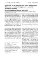
Case 1: (A) Plain radiograph oblique view showing the right cervical rib (arrow)Figure 1
Case 1: (A) Plain radiograph oblique view showing
the right cervical rib (arrow). (B-D) CT sagittal, coronal
and 3D-reconstructed images showing the pseudoarthrotic
bony formation (arrows) between the cervical rib and the
first rib.
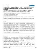
Case 1: (A-B) MRI T1-weighted coronal images showing that the space between the cervical rib (arrow) and the first rib (arrow head) diminishes upon lifting the upper arm with sub-sequent impingement of the brachial plexus in image BFigure 2
Case 1: (A-B) MRI T1-weighted coronal images show-
ing that the space between the cervical rib (arrow)
and the first rib (arrow head) diminishes upon lifting
the upper arm with subsequent impingement of the
brachial plexus in image B.
Journal of Brachial Plexus and Peripheral Nerve Injury 2009, 4:14 />Page 3 of 6
(page number not for citation purposes)
At regular follow-up at 1, 3, 6 and 12 months, she had no
remaining symptoms from the lower trunk of the brachial
plexus, except a slight allodynia around the scar during
the first six months. She had no symptoms during full
abduction. Cold intolerance was markedly reduced (none
or insignificant) and a Froment's sign was not found. At
one year follow-up, she had full range of motion and no
impairment of strength compared to the contralateral
side. Endurance, isometric and dynamic grip strength
showed 9-18% higher values than on the left side. The girl
was pleased with the surgical procedure. She continued
her leisure activities in gymnastics.
Case two
A 16-year old right-handed girl with paraesthesia in the
left arm, initially occurring periodically and later more fre-
quent, since the age of 12 was referred to our hospital due
to these symptoms. X-ray showed a cervical rib on the left
side and a minor one on the right side (no symptoms on
right side; Fig. 4). She had similar symptoms as in Case
One, such as paraesthesia and numbness in the three
ulnar fingers of the left hand when carrying things in the
hand, when a pressure was applied supraclavicularly (e.g.
carrying a backpack) or when working with the hands
above the plane of the shoulder. Percussion of the area of
the palpable cervical rib on the left side elicited symptoms
in the three ulnar fingers and "hands up tests" exaggerated
the symptoms in the same fingers. The radial pulse was
normal in all positions of the arm. She had good strength
in all muscles of the upper extremity and a normal sensi-
bility in the hand. Isometric test and endurance of grip
showed 32% and 62%, respectively and weakness in the
left hand compared to the right side (BTE Primus work
simulator). Isometric test of the flexion in the left shoul-
der and endurance showed 16% and 54%, respectively
lower values, compared to the right side. Electrophysio-
logical examination showed no abnormalities. MRI
showed a 6 cm long cervical rib from C7 on the left side,
which articulated against a cranially oriented bony proc-
ess from the first rib where the articulation was bulky (Fig.
4). The left brachial plexus was slightly lifted up by the
skeletal abnormality. On the asymptomatic right side a
2.5 cm long cervical rib was found, which had no contact
with the brachial plexus.
The brachial plexus and the cervical rib of the patient were
explored when the girl was 17 years. The brachial plexus
was distorted at and adhered to the ventral edge of the cer-
vical rib and the bony process from the first rib (Fig. 5).
The main part of the cervical rib including the bone proc-
ess from the first rib was resected after the lower trunk was
lifted up (Fig. 5). The subclavian artery was not impinged
by the bone formation. The direct postoperative events
were without problems, but later she was investigated at
the Department of Infectious Diseases due to fever of
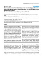
Surgical exposure and resection of the cervical rib on the right side of the 12 year old girl (Case 1)Figure 3
Surgical exposure and resection of the cervical rib on
the right side of the 12 year old girl (Case 1). The bra-
chial plexus was explored via a supraclavicular approach
(arrow lower trunk; A), revealing the cervical rib (arrow; B),
which was resected. The resected bone surface was con-
cealed with bone wax (arrow; C). After exploration, the bra-
chial plexus, particularly the lower trunk was no longer riding
above the cervical rib (D). The resected cervical rib is shown
in E.
Case 2: (A) Frontal radiograph showing bilateral cervical ribs, shorter on the right sideFigure 4
Case 2: (A) Frontal radiograph showing bilateral cer-
vical ribs, shorter on the right side. The lateral end of
the cervical ribs is marked with arrows. (B) MRI T1-weighted
coronal image showing the cervical rib (arrow head, B) with
its pseudoarthrotic bony formation that lifts up the brachial
plexus (long arrow).
Journal of Brachial Plexus and Peripheral Nerve Injury 2009, 4:14 />Page 4 of 6
(page number not for citation purposes)
unknown origin. No cause of the fever was found and
later she recovered completely. She was followed regularly
as with Case One.
At one year follow-up she had no symptoms in the hand.
The preoperative symptoms had disappeared although
she still experienced a feeling of impaired strength in the
left arm. She had full range of motion and it was not pos-
sible to provoke any paraesthesia. Tests of fine motor
activity in the hand (Crawford pins and sleeve and Minne-
sota picking test) showed improved values. Tests in the
work simulator showed improvement [isometric test 5%
weakness (preoperatively 32%), endurance 54% weaker
(preoperatively 62%), isometric test of extension with ele-
vated arm 4% weaker than the right side (preoperatively
16%), endurance of flexion/extension with elevated arm
similar value on the right side (preoperative 54%
weaker)]. MRI follow-up 11 months after surgery revealed
no occurrence of the resected cervical rib. There were no
differences compared to the two CT-scans done at three
and six months after surgery (done for other reasons; fever
investigation and a fall from a horse). The patient was
pleased with the surgery. She continued with her previous
studies and leisure activities without restriction.
Discussion
Our patients had cervical ribs bilaterally, but mainly expe-
rienced unilateral symptoms, where resection of only the
symptomatic cervical rib through a supraclavicular
approach was successfully done in both cases. Both girls
had symptoms and a history, including pain at night time
with a clear suspicion that the lower trunk of the brachial
plexus was affected since carrying heavy things and lifting
the arm above the shoulder and other activities elicited
paraesthesia and numbness particularly in the ulnar part
of the hand. Objectively, the impaired function in the arm
and the hand was clearly demonstrated with the various
tests using a work simulator, indicating the usefulness of
such novel investigation pre- and postoperatively in
patients with compression of the brachial plexus. The
symptoms of the patients corresponded to the findings in
the clinical examination and the MRI, indicating the value
of MRI. Preoperatively, neurography and EMG did not
reveal any specific impairment of nerve function, except
an increased F-wave in Case One. However, MRI showed
a clear affection of the brachial plexus from the cervical rib
in both cases when imaging was done with the arm
abducted. This indicates that MRI should be done in the
positions that elicit symptoms. The MRI findings were ver-
ified when the lower trunk of the brachial plexus was
explored. In both cases the nerve structures were riding
over the cervical rib with fibrous bands approaching the
lower trunk.
Compression of one or more of the neurovascular struc-
tures traversing the superior aperture of the chest is gener-
ally referred as thoracic outlet syndrome (TOS). This
syndrome has been the focus in a large number of articles
including description of neurophysiologic examinations,
surgical techniques and results, see for example [4-14].
But only a few papers have focused on children and ado-
lescents [15], and on the importance of cervical rib for irri-
tation of the brachial plexus and the subclavian artery [9].
A thorough history should be taken and appropriate
investigations should be undertaken in patients with a
suspected TOS to define the cause of symptom and
exclude other diagnoses [4].
In contrast to a previous report [2], our patients did not
have any muscular wasting, but only sensory symptoms,
probably explaining the lack of electrophysiological alter-
ations. In both cases, there was a successful relief of symp-
toms with a complete recovery in the younger girl and
with just minor remaining intermittent symptoms in the
older girl, at the one-year follow- up. In addition, the pre-
operative tests performed at our hand rehabilitation unit
demonstrated a clear improvement of the results at the
Exploration of the brachial plexus through a supraclavicular approach on the left side of the 17 year old girlFigure 5
Exploration of the brachial plexus through a supra-
clavicular approach on the left side of the 17 year old
girl. After skin incision and incision of the fascia the brachial
plexus (arrow; A) was located very superficially riding on the
cervical rib (arrow; B) and with a distorted anatomy of the
brachial plexus rather twisted and horizontally located on
top of the cervical rib. The cervical rib was resected in pieces
(large arrow cervical rib; small arrow fibrous tissue; C) and
the surface of the remaining exposed bone was covered with
bone wax. After resection of a cervical rib, the brachial
plexus was no longer distorted by any structures and the
subclavian artery could be observed (arrow in D). Photos
taken from below with the left arm to the right and the head
to the left.
Journal of Brachial Plexus and Peripheral Nerve Injury 2009, 4:14 />Page 5 of 6
(page number not for citation purposes)
regular follow-up at 3, 6 and 12 months. In addition, we
could objectively demonstrate improvement by examina-
tion of various tasks using the work simulator, indicating
its usefulness in pre- and postoperative investigations,
which has not been previously utilised. Electrophysiolog-
ical criteria for neurogenic thoracic outlet syndrome have
previously been suggested, such as low amplitude of the
median compound muscle action potentials, low or rela-
tively low ulnar sensory nerve action potentials, relatively
low amplitude or normal ulnar compound muscle action
potential and normal-amplitude median sensory nerve
action potential [16]. We found that the electrophysiolog-
ical investigation showed no abnormalities, which maybe
due to the fact that the lower trunk was affected to a lim-
ited extent in contrast to other published cases [2]. Elec-
trodiagnostic procedures have previously been discussed
in the literature [4,9,12]. The brachial plexus in Case Two
had a distorted (rotated; a horizontal rather than a vertical
plane) direction caused by the cervical rib and the bony
formation. We could not observe any signs that the sub-
clavian artery was compressed between the rib and the
fibrous bands even if it has been reported that a cervical
rib of more than 5.5 cm long tends to lift up and kink the
subclavian artery [3].
We decided to explore the lower trunk through a supracla-
vicular approach to be able to explore the impact of the
cervical rib on the lower trunk due to the disturbing,
mainly sensory, symptoms in the patients. Advantages of
a supraclavicular exploration for thoracic outlet syndrome
have been presented earlier with few reported complica-
tions after such approach as compared to a transaxillary
resection of the first rib [9,17,18], but conflicting opin-
ions exist about the best approach [10]. The presence of a
cervical rib and fibrous band form a barrier over which
particularly the lower trunk of the brachial plexus enters
the arm with a potential microtrauma to the trunk by
stretching and compression [19,20]. Interestingly, even if
our present cases had similar cervical ribs bilaterally (just
a short one on the right side in the older girl), symptoms
only occurred on one side. In the contralateral side of the
younger girl the symptoms were extremely rare and there-
fore no indication for exploration. In the older girl, symp-
toms occurred on the side where the cervical rib was more
prominent; thus, only a rudimentary cervical rib was pre-
sented on the asymptomatic side.
Conclusion
We suggest that the presence of a cervical rib even in chil-
dren may induce true nerve compression, where the
symptoms vary with position of the arm causing mainly
sensory symptoms in the distribution of the lower trunk.
These patients should be carefully examined and investi-
gated, including MRI and various tests in work simulator.
The possibility of surgical exploration with resection of
the cervical rib should be considered in appropriate cases.
We advocate a supraclavicular approach with a careful
exploration of the lower trunk and resection of the cervi-
cal rib, the bony formation from the first rib and fibrous
bands.
Consent
Informed consent was obtained from the patients and
their parents for publication of this case report and any
accompanying images. A copy of the written consent is
available for review by the Editor-in-Chief of this journal.
Competing interests
The authors declare that they have no competing interests.
Authors' contributions
LD, CB, HD, AC and HS operated the patients. The radio-
logical examinations were performed by KAK and PM. All
authors contributed to the creation of the manuscript and
have read/approved the manuscript.
Acknowledgements
The research on nerve injury and repair done by the authors are supported
by grants from the Swedish Research Council (Medicine), Region Skåne and
Funds from the University Hospital Malmö, Sweden. We thank Marianne
Neving and Pernilla Vikström at department of Hand Surgery for help with
pre- and postoperative examinations at the Hand Rehabilitation unit.
References
1. Cagli K, Ozcakar L, Beyazit M, Sirmali M: Thoracic outlet syn-
drome in an adolescent with bilateral bifid ribs. Clin Anat 2006,
19:558-560.
2. Tilki HE, Stalberg E, Incesu L, Basoglu A: Bilateral neurogenic tho-
racic outlet syndrome. Muscle Nerve 2004, 29:147-150.
3. Rayan GM: Lower trunk brachial plexus compression neurop-
athy due to cervical rib in young athletes. Am J Sports Med 1988,
16:77-79.
4. Urschel HC Jr, Razzuk MA: Neurovascular compression in the
thoracic outlet: changing management over 50 years. Ann
Surg 1998, 228:609-617.
5. Jamieson WG, Chinnick B: Thoracic outlet syndrome: fact or
fancy? A review of 409 consecutive patients who underwent
operation. Can J Surg 1996, 39:321-326.
6. Hug U, Jung FJ, Guggenheim M, Wedler V, Burg D, Kunzi W: ["True
neurologic thoracic outlet syndrome" anatomical features
and electrophysiological long-term follow-up of lateral the-
nar atrophy]. Handchir Mikrochir Plast Chir 2006, 38:42-45.
7. Ros DB: Overview of thoracic outlet syndromes. In Vascular dis-
orders of the upper extremity Edited by: Machleder HI. New York: Mt
Kisco; 1989:155-177.
8. Balci AE, Balci TA, Cakir O, Eren S, Eren MN: Surgical treatment
of thoracic outlet syndrome: effect and results of surgery.
Ann Thorac Surg 2003, 75:1091-1096. discussion 1096.
9. Sanders RJ, Hammond SL: Management of cervical ribs and
anomalous first ribs causing neurogenic thoracic outlet syn-
drome. J Vasc Surg 2002, 36:51-56.
10. Sheth RN, Campbell JN: Surgical treatment of thoracic outlet
syndrome: a randomized trial comparing two operations. J
Neurosurg Spine 2005, 3:355-363.
11. Ide J, Kataoka Y, Yamaga M, Kitamura T, Takagi K: Compression
and stretching of the brachial plexus in thoracic outlet syn-
drome: correlation between neuroradiographic findings and
symptoms and signs produced by provocation manoeuvres.
J Hand Surg Br 2003, 28:
218-223.
Publish with BioMed Central and every
scientist can read your work free of charge
"BioMed Central will be the most significant development for
disseminating the results of biomedical research in our lifetime."
Sir Paul Nurse, Cancer Research UK
Your research papers will be:
available free of charge to the entire biomedical community
peer reviewed and published immediately upon acceptance
cited in PubMed and archived on PubMed Central
yours — you keep the copyright
Submit your manuscript here:
/>BioMedcentral
Journal of Brachial Plexus and Peripheral Nerve Injury 2009, 4:14 />Page 6 of 6
(page number not for citation purposes)
12. Ryding E, Ribbe E, Rosen I, Norgren L: A neurophysiologic inves-
tigation of thoracic outlet syndrome. Acta Chir Scand 1985,
151:327-331.
13. Sanders RJ, Hammond SL, Rao NM: Diagnosis of thoracic outlet
syndrome. J Vasc Surg 2007, 46:601-604.
14. Huang JH, Zager EL: Thoracic outlet syndrome. Neurosurgery
2004, 55:897-902. discussion 902-893.
15. Yang J, Letts M: Thoracic outlet syndrome in children. J Pediatr
Orthop 1996, 16:514-517.
16. Gilliatt RW, Willison RG, Dietz V, Williams IR: Peripheral nerve
conduction in patients with a cervical rib and band. Ann Neurol
1978, 4:124-129.
17. Cikrit DF, Haefner R, Nichols WK, Silver D: Transaxillary or supr-
aclavicular decompression for the thoracic outlet syndrome.
A comparison of the risks and benefits. Am Surg 1989,
55:347-352.
18. Weigel G, Schmidt M, Gradl B, Girsch W: TOS-surgery via a sin-
gle supraclavicular incision. Acta Neurochir Suppl 2007,
100:141-143.
19. Sunderland S: Brachial plexus lesions due to abnormal ribs: the
"cervical rib" syndrome. In Nerves and nerve injuries New York:
Churchill Livingstone; 1978:920-942.
20. Bahm J: Critical review of pathophysiologic mechanisms in
thoracic outlet syndrome (TOS). Acta Neurochir Suppl 2007,
100:137-139.