Báo cáo y học: "Unstable angina early after aortic valve replacement surgery in a female patient with normal coronary arteries preoperatively – a case report" pot
Bạn đang xem bản rút gọn của tài liệu. Xem và tải ngay bản đầy đủ của tài liệu tại đây (873.68 KB, 4 trang )
BioMed Central
Page 1 of 4
(page number not for citation purposes)
Journal of Cardiothoracic Surgery
Open Access
Case report
Unstable angina early after aortic valve replacement surgery in a
female patient with normal coronary arteries preoperatively – a
case report
Sybille Gruber
1
, Choi-Keung Ng
2
, Christian Schwarz
2
and Johann Auer*
1,3
Address:
1
Department of Cardiology and Intensive Care, General Hospital Braunau, Austria,
2
Department of Cardiac Surgery, General Hospital
Wels, Austria and
3
Department of Cardiology and Intensive Care, General Hospital Simbach, Germany
Email: Sybille Gruber - ; Choi-Keung Ng - ;
Christian Schwarz - ; Johann Auer* -
* Corresponding author
Abstract
Background: Angina pectoris early after aortic valve replacement surgery in patients with
previously normal coronary arteries may be life threatening and has to be assessed immediately.
Case report: 12 weeks after aortic valve replacement surgery, a 60-year-old female patient was
referred for evaluation of recent onset of severe chest pain on mild exertion and at rest. Coronary
angiography showed severe stenosis nvolving the left coronary ostium and the left main stem. The
patient was urgently referred for bypass surgery and had an uneventful postoperative recovery.
Conclusion: A high degree of suspicion is needed for early recognition and aggressive
management of this rare but serious complication.
Background
Unstable angina is rare, but may be life-threatening in
patients in the early postoperative period following aortic
valve replacement with normal preoperative coronary
arteries.
Methods and results
We report the case of a 60-year-old female patient under-
going valve replacement surgery for symptomatic aortic
valve stenosis. Preoperative echocardiographic assess-
ment revealed a severely calcified aortic valve with a calcu-
lated aortic valve area of 0.8 ccm. Mean pressure gradient
was 55 mmHg and left ventricular ejection fraction was
well preserved.
The patient was free of angina and reported dyspnoea on
exertion.
Preoperative coronary angiography revealed normal coro-
nary arteries.
Valve replacement surgery was performed using a Sorin 23
mm mechanical valve prosthesis. Early postoperative
recovery was unremarkable.
12 weeks after surgery the patient was referred for evalua-
tion of recent onset of severe chest pain on mild exertion
and at rest.
ECG revealed severe ST-segment depression in leads V2-5
during episodes of chest pain.
Coronary angiography showed a 90% diameter reduction
involving the left coronary ostium and the left main stem.
Published: 2 July 2009
Journal of Cardiothoracic Surgery 2009, 4:29 doi:10.1186/1749-8090-4-29
Received: 17 April 2009
Accepted: 2 July 2009
This article is available from: />© 2009 Gruber et al; licensee BioMed Central Ltd.
This is an Open Access article distributed under the terms of the Creative Commons Attribution License ( />),
which permits unrestricted use, distribution, and reproduction in any medium, provided the original work is properly cited.
Journal of Cardiothoracic Surgery 2009, 4:29 />Page 2 of 4
(page number not for citation purposes)
The patient was urgently referred for bypass surgery and
had an uneventful postoperative recovery.
Conclusion
Angina pectoris early after aortic valve replacement sur-
gery in patients with previously normal coronary arteries
may be life threatening and has to be assessed immedi-
ately. A high degree of suspicion is needed for early recog-
nition and aggressive management of this rare but serious
complication.
Introduction
Unstable angina early after aortic valve replacement in
patients with normal coronary arteries in the preoperative
angiography is rare.
Generally, possible differential diagnoses of postoperative
angina pectoris in patients undergoing mechanical aortic
valve replacement are coronary embolism, progression of
coronary heart disease in patients with coronary athero-
sclerosis, graft occlusion in patients with concomitant aor-
tocoronary bypass (ACBP) and iatrogenic coronary ostial/
main stem stenosis.
Many cases of iatrogenic coronary ostial/main stem sten-
oses have been reported since the late 1960ies, all cases
showing similar patterns – sudden onset of angina pec-
toris 3–6 months postoperatively in patients without or
with only mild coronary artery disease – and similar his-
tologic findings – intimal fibrous proliferation of one or
both coronary ostia [1-9].
Patient and methods
We report the case of a 60-year-old moderately obese
female patient, who was referred for evaluation of recent
onset dyspnea on exertion. Physical examination revealed
a 3/6 crescendeo-decrescendo systolic murmur at the aor-
tic valve.
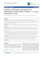
Electrocardiography showed regular sinus rhythm with-
out additional findings (Fig. 1)
Echocardiographic assessment revealed a severely calci-
fied aortic valve with a calculated valve area of 0,80 cm
2
and a mean pressure gradient of 55 mmHg.
Left ventricular ejection fraction was well preserved with
left ventricular wall thickness of 14 mm (septal and poste-
rolateral).
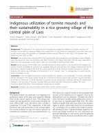
Preoperative coronary angiography revealed completely
normal coronary arteries (Fig 2).
The patient underwent aortic valve replacement surgery.
The procedure was performed using mini-sternotomy
with a single 2-stage venous cannula and normothermic
cardiopulmonary bypass. The use of normothermic tech-
niques has been reported to confer several advantages
over conventional hypothermia, such as reduced bleeding
and requirements for electrical defibrillation, shorter intu-
bation times, and improved hemodynamic parameters
postoperatively. Myocardial protection with cold ante-
grade and retrograde St. Thomas' cardioplegic (II) solu-
tion was obtained immediately after aortic cross
Preopoerative ECG: Regular sinus rhythmFigure 1
Preopoerative ECG: Regular sinus rhythm.
Preoperative coronary angiography (RAO view) showing the left coronary arteryFigure 2
Preoperative coronary angiography (RAO view)
showing the left coronary artery.
Journal of Cardiothoracic Surgery 2009, 4:29 />Page 3 of 4
(page number not for citation purposes)
clamping. The aortic valve is exposed through an oblique
aortotomy incision made well above the orifice of the
right coronary artery. The severely calcified stenotic aortic
valve was excised, replaced with a Sorin 23 mm mechani-
cal valve prosthesis, attached with subannular mattress
sutures of 2-0 Ethibond (Ethicon, Sommerville, New Jer-
sey, USA).
Early postoperative recovery was unremarkable.
12 weeks after surgery the patient was referred for evalua-
tion of recent onset of severe chest pain on mild exertion
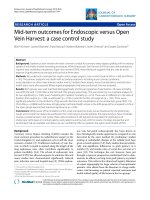
and at rest. ECG at admission showed inverted T-waves in
leads V2 to V5 and in lead aVL (Fig. 3).
During episodes of chest pain ECG revealed severe ST-seg-
ment depression in leads V2 to V5 (Fig 4).
Coronary angiography showed a 90% diameter reduction
involving the left coronary ostium and the left main stem
(Fig 5).
The patient was urgently referred for bypass surgery with a
left internal mammary artery graft to the LAD and a left
radial artery graft to the circumflex artery.
Postoperative recovery was unremarkable. Four months
later she is doing well without chest pain or signs of myo-
cardial ischemia.
Discussion
The present case underlines the importance of early diag-
nosis and treatment if angina pectoris occurs after aortic
valve replacement.
Coronary ostial stenoses can be detected in 0,1% of coro-
nary angiographies in unselected patients [10,11]. Apart
from atherosclerosis as the prime genesis, thromboses as
well as infections (lues) can provoke ostial stenoses. There
has also been a report about solitary ostial stenosis in a
patient with Takayasu's arteritis [12].
Iatrogenic coronary ostial stenosis is a well recognized,
but uncommon and potentially life threatening complica-
tion of aortic valve replacement. Symptoms include chest
pain during exercise or at rest, sudden onset of acute heart
failure and acute pulmonary edema. Usually, symptoms
ECG at admission three months after valve replacement with ST-T-abnormalities in the anterior leadsFigure 3
ECG at admission three months after valve replace-
ment with ST-T-abnormalities in the anterior leads.
ST segment depressions during severe angina at restFigure 4
ST segment depressions during severe angina at rest.
Postoperative coronary angiography (RAO view) showing the left coronary arteryFigure 5
Postoperative coronary angiography (RAO view)
showing the left coronary artery.
Journal of Cardiothoracic Surgery 2009, 4:29 />Page 4 of 4
(page number not for citation purposes)
occur within the first 6 months, though they may occur up
to 30 months after the operation [1].
Main stem stenosis after aortic valve replacement was first
recognised in 1969 by Trimble et al [2]. who described the
cases of three patients who underwent surgery for aortic
valve stenosis and/or insufficiency in 1965. Three to four
months after the operation they developed angina pec-
toris, in each case coronary angiography showed severe
stenosis of either one or both coronary ostia. According to
Lesage et al. the incidence of this severe complication may
be as high as 0.9% [3] – however, incidence is decreasing,
because of improved operative techniques [4].
Several pathogenic mechanisms have been suggested: aortic
root fibrosis secondary to turbulent flow around the prosthe-
sis [13]; the presence of perfusion catheters during valve sur-
gery that produce local pressure necrosis and subsequent
intimal proliferation leading to obstruction of the coronary
ostia [2]; balloon inflation in the proximal parts of the ves-
sels; turbulence that causes coronary artery intimal injury
that might explain the lesions often found distant to the
adherence of the cannulation devices; immunologic reaction
after valve replacement with heterograft [5].
There may also be a genetic predisposition for developing
this complication since 70% of the affected individuals as
compared to 10–15% in a control group had an epsilon 4
allele apolipoprotein E genotype [6].
Instrumentation with minimal trauma of the left main
stem is most likely the cause of early postoperative steno-
sis after aortic valve replacement surgery [7,8,14].
Avoiding cannulation of the coronary ostia for antegrade
cardioplegia, but instead using retrograde delivery as an
alternative method for myocardial perfusion during open
heart surgery may reduce the risk of postoperative coro-
nary ostial or left main stem stenosis [9,15].
Conclusion
If recurrent or newly onset angina pectoris occurs early
after aortic valve
Replacement, a high degree of suspicion os warranted to
recognize this rare but life threatening complication of
coronary ostial stenosis, even if preoperative coronary
angiography did not show coronary heart disease.
New operative techniques that reduce manipulation and
consequently avoid trauma of the coronary vessels may
prevent postoperative coronary ostial stenosis. Future
studies in patients undergoing this procedure using retro-
grade cardioplegia only will have to prove this hypothesis.
Consent
Written informed consent was obtained from the patient
for publication of this case report and accompanying
images. A copy of the written consent is available for
review by the Editor-in-Chief of this journal.
Competing interests
The authors declare that they have no competing interests.
Authors' contributions
SG was the main author and wrote the article. CKN was
the surgical consultant, was involved in data collection
and revised the manuscript. CS was the surgical consult-
ant was involved in data collection and interpretation. JA
was the cardiology consultant and gave final approval of
the manuscript. All authors have read and approved the
final manuscript.
References
1. Hadjimiltiades S, Harokopos N, Papadopoulos C, Gourassas I, Spanos
P, Louridas G: Left Main Coronary Artery Stenosis after Aor-
tic Valve Replacement. Hellenic J Cardiol 2005, 46(4):306-309.
2. Trimble AS, Bigelow WG, Wigle ED, Silver MD: Coronary ostial
stenosis. A late complication of coronary perfusion in open-
heart surgery. J Thorac Cardiovasc Surg 1969, 57(6):792-795.
3. Lesage CH, Vogel JH, Blount SG: Iatrogenic coronary occlusive
disease in patients with prosthetic heart valves. Am J Cardiol
1970, 26:123-129.
4. DePace NL, Lenwle GM, Wolf NW, Dowinsky S, Untereker W,
Spagna PM: Total Left Main Coronary Artery Occlusion after
Aortic Aneurysm Repair and Valve Replacement. Chest 1991,
99:515-517.
5. Tsukiji M, Akasaka T, Wada N, Okahashi N, Kume T, Yoshitani H,
Neishi Y, Watanabe N, Yoshida K: Bilateral coronary ostial sten-
osis after aortic valve replacement with freestyle stentless
bioprosthesis: a case report. J Cardiol 2004, 44:207-13.
6. Winkelmann BR, Ihnken K, Beyersdorf F: Left main coronary
artery stenosis after aortic valve replacement: genetic pre-
disposition for accelerated arteriosclerosis after injury of the
intact human coronary artery? Coron Artery Dis 1993, 4:659-667.
7. Roberts WC, Morrow AG: Late postoperative pathological
findings after cardiac valve replacement. Circulation. 1967,
35(4 Suppl ):I48-I62.
8. Roithinger FX, Berent R, Punzengruber C, Maurer E, Ng CK, Hartl P,
Pachinger O: Ostium stenosis of the left coronary artery after
aortic valve replacement-2 case reports. Wien Klin Wochenschr
1996, 108:552-4.
9. Menasché P, Subayi JB, Piwnica A: Retrograde coronary sinus car-
dioplegia for aortic valve operations: a clinical report on 500
patients. Ann Thorac Surg 1990, 49:556-63.
10. Yamanaka O, Hobbs RE: Solitary ostial coronary artery steno-
sis. Jpn Circ J 1993, 57:404-10.
11. Sasaguri S, Honda Y, Kanou T: Isolated coronary ostial stenosis
compared with left main trunk disease.
Jpn Circ J 1991,
55:1187-91.
12. Noma M, Sugihara M, Kikuchi Y: Isolated Coronary Ostial Sten-
osis in Takayasu's Arteritis: Case Report and Review of the
Literature. Angiology 1993, 44:839-844.
13. Reed GE, Spencer FC, Boyd AD, Engelman RM, Glassman E: Late
complications of intraoperative coronary artery perfusion.
Circulation 1973, 48(1 Suppl):III80-III84.
14. Pillai JB, Pillay TM, Ahmad J: Coronary Ostial Stenosis After Aor-
tic Valve Replacement, Revisited. Ann Thorac Surg 2004,
78:2169-2171.
15. Sethi G, Scott S, Takaro T: Iatrogenic coronary artery stenosis
following aortic valve replacement. J Thorac Cardiovasc Surg
1979, 77:760-767.