Báo cáo y học: " Perioperative infusion of low- dose of vasopressin for prevention and management of vasodilatory vasoplegic syndrome in patients undergoing coronary artery bypass grafting-A double-blind" potx
Bạn đang xem bản rút gọn của tài liệu. Xem và tải ngay bản đầy đủ của tài liệu tại đây (823.29 KB, 12 trang )
STUDY PROT O C O L Open Access
Perioperative infusion of low- dose of vasopressin
for prevention and management of vasodilatory
vasoplegic syndrome in patients undergoing
coronary artery bypass grafting-A double-blind
randomized study
Georgios Papadopoulos
1†
, Eleni Sintou
1†
, Stavros Siminelakis
2†
, Efstratios Koletsis
3*†
, Nikolaos G Baikoussis
3†
,
Efstratios Apostolakis
3†
Abstract
Preoperative medication by inhibitors of angiotensin-converting enzyme (ACE) in coronary artery patients predis-
poses to vasoplegic shock early after coronary artery bypass grafting. Although in the majority of the cases this
shock is mild, in some of them it appears as a situation, “intractable” to high-catecholamine dose medication. In
this study we examined the possible role of prophylactic infusion of low-dose vasopressin, during and for the four
hours post-bypass after cardiopulmonary bypass, in an effort to prevent this syndrome. In addition, we studied the
influence of infused vasopressin on the hemodynamics of the patients, as well as on the postoperative urine-out-
put and blood-loss. In our study 50 patients undergoing coronary artery bypass grafting were included in a blind-
randomized basis. Two main criteria were used for the eligibility of patients for coronary artery bypass graftin g:
ejection fraction between 30-40%, and patients receiving ACE inhibitors, at least for four weeks preoperatively. The
patients were randomly divided in two groups, the group A who were infused with 0.03 IU/min vasopressin and
the group B who were infused with normal saline intraoperativelly and for the 4 postoperative hours. Measure-
ments of mean artery pressure (MAP), central venous pressure (CVP), systemic vascular resistance (SVR), ejection
fracture (EF), heart rate (HR), mean pulmonary artery pressure (MPAP), cardiac index (CI) and pulmonary vascular
resistance (PVR) were performed before, during, and after the operation. The requirements of catecholamine sup-
port, the urine-output, the blood-loss, and the requirements in blood, plasma and platelets for the first 24 hours
were included in the data collected. The incidence of vasodilatory shock was significantly lower (8% vs 20%) in
group A and B respectively (p = 0,042). Generally, the mortality was 12%, exclusively deriving from group B. Post-
operatively, significant higher values of MAP, CVP, SVR and EF were recorded in the patients of group A, compared
to those of group B. In group A norepinephrine was necessary in fewer patients (p = 0.002) and with a lower
mean dose (p = 0.0001), additive infusion of epinephrine was needed in fewer patients (p = 0.001), while both
were infused for a significant shorter infusion-period (p = 0.0001). Vasopressin administration (for group A) was
associated with a higher 24 hour diuresis) (0.0001).
In conclu sion, low-dose of infused vasopressin during cardiopulmonary bypass and for the next 4 hours is benefi-
cial for its postoperative hemodynamic profile, reduces the doses of requirements of catecholamines and contri-
butes to prevention of the postcardiotomy vasoplegic shock in the patient with low ejection fraction who is
receiving ACE preoperatively.
* Correspondence:
† Contributed equally
3
Department of Cardiothoracic Surgery Department, Patras University
Hospital Patras, Greece
Papadopoulos et al. Journal of Cardiothoracic Surgery 2010, 5:17
/>© 2010 Papadopoulos et al; licensee BioMed Central Ltd. This is an Open Access article distributed under the terms of the Creative
Commons Attribution License ( which permits unrestricted use, distribution, and
reproduction in any me dium, pr ovided the original work is properly cited.
Background
Coronary artery bypass grafting by using cardiopulmon-
ary bypass (CPB) may be complicated by persistent
hypotension due to low systemic vascular resistance, in
5-22% of patients [1,2]. Different causes have been asso-
ciated with this situation, like hypothermia and duration
of CPB, total cardioplegic volume infused, reduced left
ventricular function, preoperative treatment with angio -
tensin-converting enzyme inhibitors, and systemic
inflammatory response syndrome (SIRS), or inappropri-
ate low arginine-vasopressin secretion. On the other
hand, different factors such as the reduced effect on the
pressor catecholamines, cellular acidosis, opening o f
ATP sensitive channels, efflux of K+ and hyperpolariza-
tion of the myocytes, which prevents Ca++ channels
from opening [3,4].
An advanced form of this post-cardiotomy hypoten-
sion is t he so-called vasodilatory or vasoplegic shock
which is a life-threatening condition, intractable in the
usual management with fluid administration, inotropes,
and even vasopressor catecholamines [4-7]. The inci-
dence of this syndrome is reported to range between 8.8
to 10% [8-10], but in patients with preoperative severe
left ventricular systolic dysfunction it may be observed
up to 42% of the cases [ 11]. In addition, the infusion of
catecholamines often complicates the cardiovascular sta-
bilization by producing arrhythmias and entering into a
circulus vicious [12,13].
Vasopressin has been introduced as adjunctive to cate-
cholamines in cardiac arrest and in advanced vasodila-
tory shock, and the results have shown that it is mor e
effective than vasopressor catecholamines [6,13,14].
We examined the effectiveness of intraoperative infu-
sion of arginine vasopressin in operated cardiac patients
to prevent the postoperative vasodilatory chock. The
aim of our study was to investigate the effects of pro-
phylactic administration of low-dose of vasopres sin (of
0.03 Units per minute for 4 hours), on the patients’
hemodynamic status, on the incidence of vasodilatory
shock, and on urine output and blood loss, for the 1
st
day after the operation.
Materials and methods
This study was conducted following approval from the
Ethics Committee and our hospital’ s Scientific Com-
mittee and after having obtained written informed con-
sent from all patients. A total of 50 patients, aged 32
to 81 years (61 ± 16 years), were operated between
January 2003 to Decembe r 2005 for coronary artery
disease. All the patients underwent selective coronary
artery bypass grafting by the same anesthetic and sur-
gical team. The inclusion criteria for the pat ients were
the following:
1. Patients were on ACE inhibitors therapy for at le ast
4 weeks prior to surgical procedure, and
2. Patients had impaired left ventricular ejection frac-
tion, expressed by a preoperatively estimated injection
fraction between 30-40% (by transthoracic or transeso-
phageal echo).
From the st udy patients were excluded, according t o
the following criteria:
1. injection fraction less than 30%,
2. in shock or critical hemodynamic state, confirmed
by the introduced TEE. In addition, patients with
appearance of shock or severe hemodynamic instability
“ intractable” in simple preload-manipulations (fluids
infusion) and in combination with simultaneous
(observed by TEE) impairment of left ventricular func-
tion during the operation and in the first 2 hours after
termination of cardiopulmonary bypass, were excluded,
3. confirmed hepatic, and/or renal, and/or t hyroid,
and/or adrenal disease,
4. significant carotid stenosis or any event of intrao-
perative brain ischemia documented by continuous tran-
scranial SvO
2
(INVUS),
5. significant peripheral obstructive arteriopathy,
6. documented pulmo nary hypertension, expressed by
systolic pulmonary pressure >30-35 mm Hg, and
7. chronic o bstructive pulmonary disease, confirmed
by preoperative spirometry, thorax X-rays and blood gas
analysis.
For all patients a double right internal jugular vein
catheterization was performed, with placement of a
three-way central catheter, as well as a Swan - Ganz
catheter for continuous measurement of pulmonary
artery pressure, cardiac outp ut and mixed venous blood
saturation. Next, a urinary catheter was introduced for
measurement of hourly diuresis. In addition, a tra nseso-
phageal ultrasound probe was introduced for intra- and
post-operative estimation of cardiac function. All three
catheters were retaine d for the first 24 h and removed
in ICU after this time.
Induction of anesthesia was performed using a contin-
uous remifentanyl infusion at a dosage o f 0.5 μg/Kg/
min, intravenous etomidate at a titrated dosage o f 0.2-
0.3 mg/Kg, and 0.6 mg/kg of rocuronium. For mainte-
nance of anesthesia, the following were used: re mifenta-
nyl, at a dose of 0.25-0.5 μg/Kg/min, sevoflurane, 1-2%,
and rocuronium in continuous infusion at a rate of 20
mg/h. The operation was performed using cardiopul-
monary bypass, systemic hypothermia at 30°C, and
intermittent (after each distal anastomosis) application
of cold blood cardioplegia in the same manner. Patients
were divided in a blind- manner in two groups. In
group A, continuous infusion of a solution of vasopres-
sin (Pitressin, Pfizer, Kalsruhe, Germany) 0.03 IU/min
Papadopoulos et al. Journal of Cardiothoracic Surgery 2010, 5:17
/>Page 2 of 12
was intravenously administered through a central line at
an infusion ra te of 22 ml/h. The infusion began 20 min-
utes before beginning cardiopulmonary bypass and was
continued throughout the operation for the next 4
hours after termination of the cardiopulmonary bypass.
In group B, a solution of normal saline was administered
in the same dose, way, and duration. Both solutions
were prepared by a nurse, and infused at an infusion
rate of 22 ml/h. Neither the surgeon nor the anesthetist
or any other in the operating room except from this
nursedidknowthekindofinfusedsolution,ineach
patient.
Ten minutes before termination of the cardiopulmon-
ary bypass, a solution of norepinephrine, at a dose of
0.03 μg/Kg/min was routinely administer ed (in continu-
ous iv infusion), and it was individually increased up to
0.05 μg/Kg/min during the next 24 hours until extuba-
tion, depending on the hemodynamic state of each
patient. An additional dose of epinephrine of 0.01-0.03
μg/Kg/min was selectively infused in patients to whom
the above dose of norepinephrine was insufficient in
order to restore a normal cardiac output, whereas in
every patient with vasodilatory shock.
After successful termination of the cardiopulmonary
bypass and the followed homeostasis, the patients were
transferred to the ICU, where the vasopressin or saline
solution was continued, until completion of the pre-speci-
fied infusion-time (4 hours after termination of cardiopul-
monary bypass). All the patients were sedated for the first
12-18 hours, and then they were extubated in the absence
of any hemodynamic instability. For maintenance of seda-
tion, a solution of Propofol in a dose of about 40 mg/h
was continuously administered until the time of extuba-
tion. Postoperative urine output and blood loss from
drains were hourly recorded, for the first 24 hours.
In all patients, the hemodynamic profile was routinely
recorded, at five phases. The first phase (phase-1) was
recorded at 20 minutes prior to initiation of extracor-
poreal circulation. The second (phase-2) was recorded
at 20 minutes after termination of the cardiopulmonary
bypass. The third ph ase (phase-3) was recorded at 40
minutes following termination of the cardiopulmonary
bypass. The fourth phase (phase-4) was recorded at 60
minutes after termination of the cardiopulmonary
bypass. Finally, the last phase (phase-5) was recorded at
2 hours following transfer of the patient in ICU. The
recorded parameters of hemodynamic profile were the
following: EF, HR, MAP, MPAP, CO, CVP, SVR, and
PVR. The rest of the data whi ch were recorded and
were considere d for the analysis of the results were the
following:
1. The preoperative medication,
2. Biometric data such as age and BSA,
3. Some intraoperative factors such as cardiopul-
monary bypass-time and ischemia-time,
4. The units of administered blood and/or blood
products,
5. The 24-hour patient dieresis,
6. The 24-hour blood-loss, and
7. Requirement for inotropes and their dosage, as
well as the mean dose and duration of norepinephr-
ine administration.
Statistical analysis
All data are expressed as mean value ± standard devia-
tion. Values in both groups passed the Kolmogorov-
Smirnof test for normality. Comparisons of co ntinuous
variables between groups were performed using the
unpaired student’s t-test. Comparison of categorical data
between the two groups of patients was performed b y
thechi-squaretestortheFischer’sexacttest,where
appropriate. p-values less than 0.05 were considered sta-
tistically significant. All analyses were performed using
the SPSS 16 statistical package.
Results
Three patients died (6%) in the postoperative period (48
hours, 88 hours and 4 days postoperatively), all of them
from the group B (12%) (0% versus 12%, p = 0.235). The
cause of death for all patients was the multiple organ-
system failure.
At first, the comparison between two groups was
made regarding the general characteristics (sex, mean
age, and BSA), clinical preoperative data (co-morbidity,
severity of CAD and intraoperative hemodynamic mea-
surements), preoperative medication and intraoperative
data (cardiopulmonary bypass-time, ischemia-time,
grafts number per patients, etc). All these data are pre-
sented in table 1 and 2. In table 3 the postoperative data
(mortality, hemodynamic profile, needed inotropic sup-
port, etc) for the two groups is shown.
According to all preoperative data, there were no sta-
tis tically significant differences between the two groups,
confirming the similarity of the groups at baseline (table
1). In the same way, from the comparison of postopera-
tive measurements (table 3), no statistical significant dif-
ferences were observed between two groups, concerning
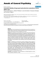
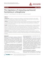
the factors HR, MPAP (fig. 1), CI (fig. 2) and PVR. On
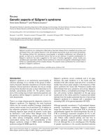
the contrary, comparison of values of MAP (fig. 3), CVP
(fig. 4), SVR (fig. 5), and EF (fig. 6) following extracor-
poreal circulation showed significantly higher values in
group A (table 2).
The mean vasopressin’s infusion-time was 404 ± 33
minutes and the mean total dose of infused vasopressin
in the patients of group A were 12.4 ± 1.3 Units (table 2).
Vasodilatory shock is considered the hemodynamic
state characterized by a systolic arterial pressure of less
Papadopoulos et al. Journal of Cardiothoracic Surgery 2010, 5:17
/>Page 3 of 12
than 80 mmHg (or mean arterial pressure < 70 mm Hg),
despite of a cardiac output more than 5 L/min (or a car-
diac index > 2.5 L/min/m
2
) (9, 10). According to this
definition, one (1) patient of the vasopressin group (4%),
and six (6) patients of the control group (24%) devel-
oped vasodilatory shock, during the first 24 hours of
postoperative observation (p = 0.042) (table 3).
It is of note that in none of the patients a hypertensive
crisis was observed. Inotropes infusion (norepinerhrine
and/or epinephrine) was individua lly decided, depending
on the postoperative hemodynamic status of the patients
for the first 24 hours. Norepinephrine was infused i n a
minimal dose of 0.03-0.05 μg/Kg/min in 6 patients
(24%) of group A and in 18 patients (72%) of group B
(p = 0.002). Epinephrine infusion was additionally neces-
sary in 5 patients (20%) of group A and in 17 (68%) of
group B (p = 0.001). Generally, the catecholamine infu-
sion-time was significantly lower in group A (10 ± 4
hours), in comparison to group B (18 ± 6 hours) (p =
0.0001) (table 3). Mean needed doses of norepinephrine
were significantly lower in group A (0.16 ± 0.04 μg/Kg/
min) than in group B (0.44 ± 0.07 μg/Kg/min) (p =
0.0001) (table 3).
Postoperative urine output during the first 24 hours
was significantly higher in group A (5603 ± 1450 ml), in
comparison to group B (3910 ± 1102 ml (p = 0.0001)
(table 3).
The needed transfusions for blood and platelet units
were statistically significantly lower for the patients of
Table 1 The comparative pre- operative data from the
patients both groups.
Group A Group B p
General characteristics
Number of patients 25 25 -
Male/female 20/5 21/4 1
Age (y/s) 66 ± 13 62 ± 15 0,319
Height (cm) 164 ± 9 168 ± 11 0,166
Weight (kg) 75 ± 11 72 ± 8 0,276
BSA 1.74 ± 7.4 1.82 ± 6.6 0,968
Clinical preoperative data
Hypertension 16 14 0.773
Diabetes mellitus 8 10 0.769
Euroscore 4.8 ± 2.2 4.5 ± 2.6 0,662
3-coronary vessel disease 19 17 0.754
2-coronary vessel disease 6 9 0.538
Significant Left main CAD 2 4 0.667
Ischemic mitral regurgitation 1+/4+ 7 4 0.496
Ischemic mitral regurgitation 2+/4+ 4 8 0.321
Ejection fraction 30-35% 9 12 0.567
Ejection fraction 35-40% 16 13 0.567
Cardiac Index (L/min/m
2
) 3.1 ± 0,6 3.2 ± 0.8 0,619
PCWP < 15 9 10 0.773
PCWP > 15 16 15 0.773
Preoperative medication
aMEA 25 25 -
b-blockers 14 17 0.377
Calcium channel blockers (pts) 11 8 0.561
Table 2 The comparative intra-operative data from the
patients both groups.
Group A Group B P
Total vasopressin infused (U) 12.4 ± 1.3 - -
Vasopressin’s infusion-time (min) 404 ± 33 - -
Operation’s-time (min) 238 ± 32 228 ± 26 0,231
Cardiopulmonary bypass-time (min) 169 ± 29 177 ± 20 0,262
Myocardial ischemia-time (min) 52 ± 14 47 ± 12 0.182
Mean hypothermia (°C) 31.4 ± 1.8 31.1 ± 1.5 0,525
LIMA-used (pts) 23 24 1.000
Radial artery used (pts) 9 6 0.538
3-grafts bypass 16 18 0.762
2-grafts bypass 9 6 0.538
1-graft bypass (LIMA) - 1 1.0
Table 3 The post-operative data from the patients both
groups
Characteristics Group A Group B p
Mortality
Surgical mortality 0(%) 3 (12%) 0.235
Hemodynamic profil
Cardiac Index (L/min/m
2
) 3.2 ± 0.7 3.0 ± 0.8 0,352
Heart rate (/min) 78 ± 11 83 ± 9 0,085
PVR 198 ± 40 214 ± 29 0,112
Mean PAP (mm Hg) 21 ± 4 19 ± 4 0,084
Mean AP (mm Hg) 84 ± 11 78 ± 7 0,026
SVR (dyn.cm/m
2
) 1210 ±
102
1103 ±
123
0.002
CVP (mm Hg) 8.5 ± 2.5 7 ± 1.8 0.019
EF 38.0 ± 3.9 35.5 ± 4.1 0.032
Vasodilatory shock (pts) 1 (8%) 6 (20%) 0.042
Inotropic needs
Needed norepinephrine (pts) 6180.002
Needed additional epinephrine (pts) 5170.001
Mean catecholamine infusion-time
(Hours)
10 ± 4 18 ± 6 0,000
Mean norepinephrine-dose μg/Kg/
min
0.16 ± 0.04 0.44 ± 0.07 0,000
Blood-loss and urine output
Mean blood loss (ml) 650 ± 125 975 ± 100 0,000
Mean urine volume (ml) 5603 ±
1450
3910 ±
1102
0.000
Transfusion needs
Mean erythrocytes’ units transfused 3.1 ± 1.7 4.2 ± 1.8 0.031
Mean plasma’s units transfused 6.1 ± 2.3 5.8 ± 3.1 0.699
Mean platelets’ units transfused 4.3 ± 1.8 5.7 ± 2.1 0,015
Papadopoulos et al. Journal of Cardiothoracic Surgery 2010, 5:17
/>Page 4 of 12
group A, in comparison to group B, in contrast to trans-
fused plasma units. Moreover the postoperative blood
loss for the first 24 hours was significantly lower in
group A (650 ± 125 ml), compared to group B (975 ±
100 ml) (p = 0.0001) (table 3).
Discussion
The vasodilatory shock is a state of abrupt hemody-
namic deterioration in the first hours following open
heart surgery. It is mainly characterized by a vasodila-
tory hypotension (systolic BP < 80 mmHg, while cardiac
output is restored >5 L/min) associated with lactic
acidosis, tachycardia, decreased systemic vascular
resistance and low filling pressures [11,15,16]. The hypo-
tension is characteristical ly unresponsive either to
catecholamine administration (or necessitating norepi-
nephrine administration more than 8 μg/min), or to
preload increase by excessive fluid infusion [17].
This situation is attributed to a loss of vascular tone, due
to either the inflammatory mediators produced by the car-
diopulmonary bypass or the administered vasodilators
suc h as phosphodiesterase inhibitors, nitrates, etc [5,16].
Some factors such as congestive heart failure (with EF <
35%), preoperative use of angiotensin-conver ting enzyme
inhibitors and/or b-blockers and/or amiodarone and phos-
phodiesterase inhibitors, seem to be related with increased
Figure 1 Mean Pulmonary Pressure during time-po ints T1 - T5. Distribution of values for mean pulmonary pressur e (MPAP) during time-
points T1 - T5 for group I (vasopressin, in blue boxplots) and group II (placebo, in green boxplots). (median = black line, boxplot = 50% of data
set, lines on both sides of the boxplot = dispersion for 99% of values, * = numbers outside of distribution range for 99% of values).
Papadopoulos et al. Journal of Cardiothoracic Surgery 2010, 5:17
/>Page 5 of 12
postoperative incidence of the vasodilatory shock
[11,15,18-20]. In our stu dy, the influence of low-dose of
vasopressin on postoperative vasodilatory shock was
examined in patients with two predisposing factors of this
syndrome: low ejection fraction and preoperative adminis-
tration of ACE inhibitors. In fact, according to Argengiano
et al [11], both low ejection fraction and use of ACE inhi-
bitors were independent risk factors for the development
of postoperative vasodilatory shock. In fact, while the inci-
dence of vasodilatory shock in patients with a normal ejec-
tion fraction was 3.3%, in patients with a low ejection
fraction or receiving ACE inhibitors, it was 26.9% and
26.7%, respectively [11]. In our study, the incidence of
vasodilatory shock was significantly lower in the group of
vasopressin, being 20% in the control group and 4% in the
vasopressin group (table 3), and much lower from those
values reported by Argengiano et al [21]. According to this
study, which included patients with end-stage heart failure
who were subjected to left ventricular assist device place-
ment, the incidence of postoperative vasodilatory shock
was 42% [21].
The mortality of post-cardiotomy vasodilatory syndrome
is high, dependent on its responsiveness in simultaneous
vasopressin and norepinephrine infusion [7,22]. According
to Gomes W, et al [8], the duration of norepinephrine
refractory vasoplegia -it may persist for longer than 36-48
hours- significantly influences outcomes, because the syn-
drome may complicate postoperative oozing that requires
Figure 2 Cardiac Index during time-points T1 - T5. Distribution of values for cardiac index (CI) during time-points T1 - T5 for group I
(vasopressin, in blue boxplots) and group II (placebo, in green boxplots). (median = black line, boxplot = 50% of data set, lines on both sides of
the boxplot = dispersion for 99% of values, * = numbers outside of distribution range for 99% of values).
Papadopoulos et al. Journal of Cardiothoracic Surgery 2010, 5:17
/>Page 6 of 12
blood and plasma transfusions. Generally, the mortality for
post-cardiotomy patients may be increased up to 25%
[8,9]. In our study, although the mortality for the patients
of group A was 0% and for group B 12% th is difference
wasn’t statistically significant. Of note, the mortality was
not obviously related to the syndrome, all deaths occurred
in patients with the syndrome, and at a later phase. There-
fore, the calculated mortality for the patients suffering
from the postcardiotom y vasoplegic shock syndrome was
50% (3 from the 6 pts) (table 3). The relative low mortality
in our study may be attributed to the design of our proto-
col: we used a very-low dose of infusion; we started it 20
minutes before cardiopulmonary bypass in combination
with norepinephrine infusion at the termination of cardio-
pulmonary bypass. Indeed, Patel B, et al [23] considers the
low dose of 0.03 IU/min, in combinat ion with its gradual
starting of infusion as a factor of its effectiveness. In addi-
tion, another study has shown that the combined infusion
of vasopressin with norepinephrine in post-cardiotomy
patients did not cause an increase in mortality as predicted
by Euroscore [24]. According to this study, the safety of
low dose of vasopressin (≤0.04 IU/min) combined with
norepinephrine was supported by the authors’ observation
that none of patients receiving vasopressin below 2 U/h
(0.033 IU/min), died [24].
Concerning t he appropriate dose of vasopres sin there
is not enough knowledge. It is mainly dependent on the
indication, namely the management of postoperative
vasodilatory shock or the prevention of the shock. For
management, it has been used by several investigators in
Figure 3 Mean arterial pressure values during time-points T1 - T5. Distribution of mean arterial pressure (MAP) values during time-points T1
- T5 for group I (vasopressin, in blue boxplots) and group II (placebo, in green boxplots). (median = black line, boxplot = 50% of data set, lines
on both sides of the boxplot = dispersion for 99% of values, * = numbers outside of distribution range for 99% of values).
Papadopoulos et al. Journal of Cardiothoracic Surgery 2010, 5:17
/>Page 7 of 12
different dosages, between 2-6, or even 15 U/h
[11,16,21]. Others have administered much lower
dosages as these of 0.03-1 U/h [16,25-29]. However,
infusion at a dose of about 6 U/hr seems to be effective,
because it obtains a plasma level of ≥150 pg/ml and
further increasing these levels does not offer additional
benefit [11,16,17,25]. In fact, Mutlu G and Factor P [29],
consider as appropriate the dose of <0.04 U/min and
showed that it is safe and effective, even for the treat-
ment of the septic vasodilatory shock. Higher dosages of
vasopressin may be associated with several complica-
tions such as decreased coronary blood flow and cardiac
output, ventricular arrhythmias and gut ischemia [28].
However, Torqersen C, et al [30] in their randomized
and controlled trial by comparing two doses of 0.033
and 0.067 IU/min of arginine vasopressin infusion in
patients with advanced vasodilatory shock, they showed
that the patients receiving dose of 0.067 IU/min
required significantly less norepinephrine, deve loped
lower metabolic acidosis, without significant differences
in MAP-levels, rate of adverse events and ICU-mortality,
even for the 48 hours after the operation.
Our study showed, that intraoperat ive total “ultra-low”
dose of 12.4 ± 1.3 Units of vasopressin may prevent the
Figure 4 Central Venous Pressure during time- points T1 - T5. Distribution of values for central venous pressure (CVP) during time- points T1
- T5 for group I (vasopressin, in blue boxplots) and group II (placebo, in green boxplots). (median = black line, boxplot = 50% of data set, lines
on both sides of the boxplot = dispersion for 99% of values, * = numbers outside of distribution range for 99% of values).
Papadopoulos et al. Journal of Cardiothoracic Surgery 2010, 5:17
/>Page 8 of 12
postoperative vasodilatory shock. Indeed, this “ultra-low”
dose of vasopressin according to our study, obtains a
significant increase of MAP (fig. 3), CVP (fig. 4), as well
as a significant increase of SVR (fig. 5). The increased
arterial pressure and systemic vascular resistance are
mainly due to the produced by vasopressin systemic
vasoconstrictive action, rather in p atients in shock than
in patients with a normal hemodynamic state [15,28].
Indeed, several studies in the past have shown that the
perioperative administration of vasopressin restores the
vascular tone in patients following cardiopu lmonary
bypass, especially in cases that are refractory to
norepinephrine [16,21,26]. This result could be war-
ranted by the known action of vasopressin: in low doses
it has little or no influence on blood pressure of the
normotensive patients, while the same doses in patients
in vasodilatory shock produce an effective constrictive
vessel action [15]. The increased cardiac index is attri b-
uted not only to th e preload and after load changes
[11,21,26,25,31], but also to the increased myocardial
contractility. In fact, vasopressin infusion in advanced
vasodilatory s hock tends to improve myocardial perfor-
mance by increasing of intramyocardia l calcium concen-
trations, and producing coronary artery vasodilatation,
Figure 5 Systemic Vascular Resistance during time-points T1 - T5. Distribution of values for peripheral resistance (SVR) during time-points T1
- T5 for group I (vasopressin, in blue boxplots) and group II (placebo, in green boxplots). (median = black line, boxplot = 50% of data set, lines
on both sides of the boxplot = dispersion for 99% of values, * = numbers outside of distribution range for 99% of values).
Papadopoulos et al. Journal of Cardiothoracic Surgery 2010, 5:17
/>Page 9 of 12
in combination with the increase of myocardial blood
flow due to increased systemic perfusion pressure
[12,14]. The observation of significant postoperative
increase of ejection fraction in our patients receiving
vasopressin (fig. 6), is confirmed only by our findings, as
to the best of our knowledge, no other study has
recorded and evaluated this hemodynamic parameter.
Our study also showed that pulmonar y vascular resis-
tanceandmeanpulmonaryarterypressurewerenot
affected by the vasopressin infusion (fig. 1). It may
attributed to the observed vasodilatory effect of
vasopressin in the pulmonary vasculature [21,31], influ-
ence (of a ction) which is already experimentally con-
firmed and is due to a release of NO by the endothelial
pulmonary capillaries [32]. Because of the above
described action, vasopressin has been successfully used
by Tayama E, et al [32], in cardiac surgical patients with
preoperative pulmonary hypertension.
Concerning the postoperative needs of norepinephr-
ine, our data showed that in the vasopressin group the
percentage of patients requiring a dministration was sig-
nificantly lower in comparison to the control group
Figure 6 Left ventricular Ejection Fraction during time-points T1 - T5. Distribution of values for left ventricular ejection fraction (E.F.) during
time-points T1 - T5 for group I (vasopressin, in blue boxplots) and group II (placebo, in green boxplots). (median = black line, boxplot = 50% of
data set, lines on both sides of the boxplot = dispersion for 99% of values, * = numbers outside of distribution range for 99% of values).
Papadopoulos et al. Journal of Cardiothoracic Surgery 2010, 5:17
/>Page 10 of 12
(6 pts or 24% versus 18 pts or 72%) (table 3). Similarly,
an impress ive difference was observed in the number of
patients requiring additive infusion of epinephrine.
While in the group of vasopressin o nly 5 pts (20%)
required additional infusion of epinephrine, in the group
of placebo it was 17 pts (68%) (table 3). An even more
impressive observation was the difference to the mean
administered dose of norepinephrine: this was signifi-
cantly lower (0.16 ± 0.04 versus 0.44 ± 0.07 μg/min) in
the vasopressin group. Similarly, the mean-time of cate-
cholamine’s infusion was significantly lower in the vaso-
pressin group (10 ± 4 versus 18 ± 6 hours) (table 3).
Several studies have demonstrated the augmented
vasoconstrictor action of vasopressin in patients with
hypotension not responding to high-dose of norepi-
nephrine, dopamine and fluid resuscitation [33], action
which persists for up to 2 hours [34] and with a serious
advantage: less pronounced vasoconstriction in the c or-
onary and cerebral circulation [35]. Finally, to this double
beneficial action of vasopressin for myocardium and
brain, the protective action for the kidneys can be added.
Experimental studies in protocols of short and prolonged
cardiac arrest have shown that vasopressin produced a
significantly higher vital organ blood flow and cerebral
oxygen delivery than epinephrine did [36,37].
The increased urine output represents a remarkable
result of infused vasopressin due -according to several
studies- to the increased mean arterial pressure of the
patient and therefore to the improvement of glomerular
filtration rate [37,3 8]. However, Bragadottir G, et al [35]
showed , that low to moderate doses of vasopressin (0.03-
0.08 IU/min) in post-cardiac surgery patients cause a sig-
nificant renal vasoconstricion and a decline in renal
blood flow, which was accompanied by an increased glo-
merular filtration rate, suggesting that vasopressin mainly
constricts efferent arterioles. It is of note that although
these patients were not in shock, vasopressin infusion
seems to produce an impairment of the r enal oxygen
demand/supply relationship [39]. Several studies have
shown that vasopressin receptors in the renal vasculature
are located in the efferent arterioles, in contrast to the
catecholamine receptors, which are located in the addu-
cing arterioles [33,35]. Therefore, although the vasocon-
strictive action of catecholamines leads to a decrease in
the filtration fraction, the action of vasopressin leads to
an increase in the filtration fraction and, hence, to an
increase of urine output [33,37,38].
We also observed this significantly increased 24-hour
diuresis in the patients of the vasopressin group (table
3). Morales D, et al [16], proposed the long-term admin-
istration (up to 12 hours) of vasopressin in patients with
postcardiotomy vasodilatory shock associated with renal
insufficiency (instead that of 2 to 3 ho urs in patients
with normal renal function), to maintain an improved
filtration rate and urine output.
Although vasopressin causes a decrea se of platelets in
a significant number of patients (up to 52%) [12,40], it
enhances blood coa gulation. This can be attributed to
an observed increase the plasma concentrations of fac-
tors VIII and von Willebrand [41,42]. The combination
of several factors as those just mentioned, like the pro-
duced vasoconstriction and probably the increased adhe-
sion of platelets (there are receptors V1 on them) [41],
may explain the observed statistically significant reduced
post-cardiotomy blood loss in the vasopressin gro up, in
our s tudy (table 3), this finding i s in accordance to the
less transfusions needs in the group A. Because desmo-
pressin -a known drug already used for the reduction of
postoperative bleeding in post-cardiotomy patients is an
analogue of vasopressin [36], could potentially offer an
additive “hemostatic role” in the vasopressin actions.
Conclusions
In summary, infusion of an “ultra-low” dose vasopressin
(0.03 U/min) during cardiopulmonary bypass and for
the first four hours after coronary artery bypass grafting
in patients with preoperative medication with ACE inhi-
bitors who are having low ejection fraction, is safe and
beneficial. It significantly reduces the required doses of
catecholamines, obtaining a better hemodynamic profile,
a higher urine output and lower blood loss for the first
24 hours. The use of an “ ultra-low ” dose vasopressin
seems to be preventive for the incidence of observed
post-cardiotomy vasodilatory shock. Finally, it may
decrease both catecholamine dose and duration of their
administration,itisconsideredasausefulagentfor
decreasing all their side-effects.
Author details
1
Department of Anesthesia and Intensive Care Unit, University Hospital of
Ioannina, Ioannina, Greece.
2
Cardiac Surgery Department, University Hospital
of Ioannina, Ioannina, Greece.
3
Department of Cardiothoracic Surgery
Department, Patras University Hospital Patras, Greece.
Authors’ contributions
All authors: 1) have made substantial contributions to conception and
design, or acquisition of data, or analysis and interpretation of data; 2) have
been involved in drafting the manuscript or revising it critically for important
intellectual content; and 3) have given final approval of the version to be
published.
Competing interests
The authors declare that they have no competing interests.
Received: 11 February 2010 Accepted: 28 March 2010
Published: 28 March 2010
References
1. Carrel T, Englberger L, Mohacsi P, Neidhart P, Schmidli J: Low systemic
vascular resistance after cardiopulmonary bypass: incidence, etiology,
and clinical importance. J Card Surg 2000, 15:347-353.
Papadopoulos et al. Journal of Cardiothoracic Surgery 2010, 5:17
/>Page 11 of 12
2. Sun X, Zhang L, Hill PC, Lowery R, Lee AT, Molyneaux RE, Corso PJ,
Boyce SW: Is incidence of postoperative vasoplegic syndrome different
between off-pump and on-pump coronary artery bypass grafting
surgery? Eur J Cardiothorac Surg 2008, 34:820-825.
3. Noto A, Lentini S, Versaci A, Giardina M, Risitano DC, Messina R, David A: A
retrospective analysis of terlipressin in bolus for the management of
refractory vasoplegic hypotension after cardiac surgery. Interact
CardioVascular and Thoracic Surgery 2009, 9:588-92.
4. Laffey JG, Boylan JF, Cheng DC: The systemic inflammatory response to
cardiac surgery: implications for the anesthesiologist. Anesthesiology 2002,
97:215-52.
5. Landry DW, Oliver JA: The pathogenesis of vasodilatory shock. N Engl J
Med 2001, 345:588-595.
6. Wenzel V, Krismer AC, Arntz HR, Sitter H, Stadlbauer KH, Lindner KH: A
comparison of vasopressin and epinephrine for out-of-hospital
cardiopulmonary resuscitation. N Engl J Med 2004, 350:105-113.
7. Gomes WJ, Carvalho AC, Palma JH, Goncalves I Jr, Buffolo E: Vasoplegic
syndrome: a new dilemma. J Thorac Cardiovasc Surg 1994, 107:942-3.
8. Gomes WJ, Carvalho AC, Palma JH, Teles CA, Branco JN, Silas MG, Buffolo E:
Vasoplegic syndrome after open heart surgery. J Cardiovasc Surg (Torino)
1998, 39:619-23.
9. Levin RL, Degrange MA, Bruno GF, Del Mazo CD, Taborda DJ, Griotti JJ,
Boullon FJ: Methylene blue reduces mortality and morbidity in
vasoplegic patients after cardiac surgery. Ann Thorac Surg 2004, 77:496-9.
10. Shanmugam G: Vasoplegic syndrome-the role of methylene blue. Eur J
Cardio-thoracic Surg 2005, 28:705-10.
11. Argenziano M, Chen J, Choundhri A, Cullinane S, Garfein E, Weinberg AD,
Smith CR Jr, Rose EA, Landry DW, Oz MC: Management of vasodilatory
shock after cardiac surgery: identification of predisposing factors and
use of a novel pressor agent. J Thorac Cardiovasc Surg 1998, 116:973-80.
12. Luckner G, Duenser M, Jochberger S, Mayr VD, Wenzel V, Ulmer H,
Schmid S, Knotzer H, Pajk W, Hasibeder W, Mayr AJ, Friesenecker B:
Arginine vasopressin in 316 patients with advanced vasodilatory shock.
Crit Care Med 2005, 33:2659-2666.
13. Duenser MW, Wenzel V, Mayr AJ, Hasibeder WR: Management of
vasodilatory shock: Defining the role of arginine vasopressin. Drugs 2003,
63:237-256.
14. Duenser MW, Mayr AJ, Ulmer H, Knotzer H, Sumann G, Pajk W,
Friesenecker B, Hasibeder WR: Arginine vasopressin in advanced
vasodilatory shock: A prospective, randomized, controlled study.
Circulation 2003, 107:2313-2319.
15. Morales D, Garrido M, Madigan J, Helman D, Faber J, Williams M, Landry D,
Oz M: A double-blind randomized trial: Prophylactic Vasopressin
Reduces Hypotension After Cardiopulmonary Bypass. Ann Thorac Surg
2003, 75:926-30.
16. Morales D, Gregg D, Helman D, Williams MR, Naka Y, Landry DW, Oz MC:
Arginine vasopressin in the treatment of fifty patients with
postcardiotomy vasodilatory shock. Ann Thorac Surg 2000, 69:102-6.
17. Raja S, Dreyfus G: Vasoplegic syndrome after Off-pump coronary artery
bypass surgery. Tex Heart Inst J 2004, 31:421-24.
18. Mekontso-Dessap A, Houel R, Soustelle C, Kirsch M, Thebert D, Loisance DY:
Risk factors for post-cardiopulmonary bypass vasoplegia in patients with
preserved left ventricular function. Ann Thorac Surg 2001, 71:1428-32.
19. Tuman KJ, McCarthy RJ, O’Connor CJ, Holm WE, Ivankovich AD:
Angiotensin-converting enzyme inhibitors increase vasoconstrictor
requirements after cardiopulmonary bypass. Anesth Analg 1995, 80:473-9.
20. Mets B, Michler RE, Delphin ED, Oz MC, Landry DW: Refractory vasodilation
after cardiopulmonary bypass for heart transplantation in recipients on
combined amiodarone and angiotensin-converting enzyme inhibitor
therapy: a role for vasopressin administration. J Cardiothorac Vasc Anesth
1998, 12:326-9.
21. Argengiano M, Choudhri A, Oz M, Rose E, Smith C, Landry D: A prospective
randomized trial of arginine vasopressin in the treatment of vasodilatory
shock after left ventricular assist device placement. Circulation 1997,
96:286-290.
22. Carrel T, Englberger L, Mohacsi P, Neidhart P, Schmidli J: Low systemic
vascular resistance after cardiopulmonary bypass: incidence, etiology,
and clinical importance. J Card Surg 2000, 15:347-53.
23. Patel B, Chittock D, Russell J, Walley K: Beneficial effects of short-term
vasopressin infusion during severe septic shock. Anesthesiology 2002,
96:576-82.
24. Suojaranta-Ylinen R, Vento R, Patila T, Kukkonen S: Vasopressin, when
added to norepinephrine, was not associated with increased predicted
mortality after cardiac surgery. Scand J Surg 2007, 96:314-18.
25. Morales DL, Landry DW, Oz MC: Therapy for vasodilatory shock: Arginine
vasopressin. Semin Anesth Periop Med 2000, 19:98-107.
26. Masetti P, Murphy SF, Kouchoukos NT: Vasopressin therapy for vasoplegic
syndrome following cardiopulmonary bypass. J Card Surg 2002, 17:485-9.
27. Malay MB, Ashton RC Jr, Landry DW, Townsend RN: Low-dose vasopressin
in the treatment of vasodilatory septic shock. J Trauma 1999, 47:699-705.
28. Albright T, Zimmerman M, Selzman C: Vasopressin in the cardiac surgery
intensive care unit. Am J Crit Care 2002, 11:326-332.
29. Mutlu G, Factor P: Role of vasopressin in the management of septic
shock. Intensive Care Med 2004, 30:1276-91.
30. Torqersen C, Duenser M, Wenzel V, Jochberger S, Mayr V, Schmittinger CA,
Lorenz I, Schmid S, Westphal M, Grander W, Luckner G: Comparing two
different arginine vasopressin doses in advanced vasodilatory shock: a
randomized, controlled, open-label trial. Intensive Care Med 2009, 36:57-65.
31. Argenziano M, Chen JM, Cullinane S, Choudhri AF, Rose EA, Smith CR,
Edwards NM, Landry DW, Oz MC: Arginine vasopressin in the
management of vasodilatory hypotension after cardiac transplantation. J
Heart Lung Transplant 1999, 18:814-817.
32. Tayama E, Ueda T, Shojima T, Akasu K, Oda T, Fukunaga S, Akashi H,
Aoyagi S: Arginine vasopressin is an ideal drug after cardiac surgery for
the management of low systemic vascular resistant hypotension
concomitant with pulmonary hypertension. Interact CardioVasc Thorac
Surg 2007, 6:715-719.
33. Leone M, Albanese J, Delmas A, Chaabane W, Garnier F, Martin C:
Terlipressin in catecholamine-resistant septic shock patients. SHOCK
2004, 22:314-319.
34. Novella S, Martínez C, Pagán R, Hernández R, García-Sacristán A, González-
Pinto A, González-Santos J, Benedito S: Plasma levels and vascular effects
of vasopressin in patients undergoing coronary artery bypass grafting.
Eur J Cardiothorac Surg 2007, 32:69-76.
35. Delmas A, Leone M, Rousseau S, Albanese J, Martin C: Clinical review:
Vasopressin and terlipressin in septic shock patients. Critical Care 2005,
9:212-222.
36. Wenzel V, Lindner K: Employing vasopressin during cardiopulmonary
resuscitation and vasodilatory shock as a lifesaving vasopressor.
Cardiovascular Research 2001, 51:529-541.
37. Wenzel V, Lindner K, Prengel A, Maier C, Voelckel W, Lurie KG,
Strohmenger HU: Vasopressin improves vital organ blood flow after
prolonged cardiac arrest with postcounter-shock pulseless activity in
pigs. Crit Care Med 1999, 27:486-92.
38. Holmes CL, Walley KR, Chittock DR, Lehman T, Russell JA: The effects of
vasopressin on hemodynamics and renal function in severe septic
shock: A case series. Intensive Care Med 2001, 27:1416-21.
39. Bragadottir G, Redfors B, Nygren A, Sellgren J, Ricksten SE: Low-dose
vasopressin increases glomerular filtration rate, but impairs renal
oxygenation in post-cardiac surgery patients. Acta Anesthesiol Scand 2009,
53:1052-59.
40. Duenser MW, Fries DR, Schobersberger W, Ulmer H, Wenzel V,
Friesenecker B, Hasibeder WR, Mayr AJ: Does arginine vasopressin
influence the coagulation system in advanced vasodilatory shock with
severe multiorgan dysfunction syndrome? Anesth Analg 2004, 99:201-206.
41. Treschan T, Peters J: The vasopressin system: Physiology and clinical
strategies. Anesthesiology 2006, 105:599-612.
42. Mannucci PM: Desmopressin (DDAVP) in the treatment of bleeding
disorders: The first twenty years. Haemophilia 2000, 6:60-7.
doi:10.1186/1749-8090-5-17
Cite this article as: Papadopoulos et al.: Perioperative infusion of low-
dose of vasopressin for prevention and management of vasodilatory
vasoplegic syndrome in patients undergoing coronary artery bypass
grafting-A double-blind randomized study. Journal of Cardiothoracic
Surgery 2010 5:17.
Papadopoulos et al. Journal of Cardiothoracic Surgery 2010, 5:17
/>Page 12 of 12