báo cáo khoa học: " A pilot histomorphology and hemodynamic of vasculogenic mimicry in gallbladder carcinomas in vivo and in vitro" pptx
Bạn đang xem bản rút gọn của tài liệu. Xem và tải ngay bản đầy đủ của tài liệu tại đây (12.7 MB, 12 trang )
RESEARC H Open Access
A pilot histomorphology and hemodynamic of
vasculogenic mimicry in gallbladder carcinomas
in vivo and in vitro
Wei Sun, Yue Z Fan
*
, Wen Z Zhang and Chun Y Ge
Abstract
Background: Vasculogenic mimicry (VM), as a new blood supply for tumor growth and hematogenous metastases,
has been recently described in highly aggressive human melanoma cells, etc. We previously reported VM in human
gallbladder carcinomas and its clinical significance. In this study, we further studied histomorphology and
hemodynamic of VM in gallbladder carcinomas in vivo and in vitro.
Methods: The invasive potential of human gallbladder carcinoma cell lines GBC-SD and SGC-996 were identified
by Transwell membrane. The vasculogenic-like network structures and the signal intensities i.e. hemodynamic in
gallbladder carcinomas stimulated via the three-dimensional matrix of GBC-SD or SGC-996 cells in vitro, the nude
mouse xenografts of GBC-SD or SGC-996 cells in vivo were observed by immunohistochemistry (H&E staining and
CD
31
-PAS double staining), electron microscopy and micro-MRA with HAS-Gd-DTPA, respectively.
Results: Highly aggressive GBC-SD or poorly aggressive SGC-996 cells preconditioned by highly aggressive GBC-SD
cells could form patterned networks containing hollow mat rix channels. 85.7% (6/7) of GBC-SD nude mouse
xenografts existed the evidence of VM, 5.7% (17/300) channels contained red blood cells among these tumor cell-
lined vasculatures. GBC-SD xenografts showed multiple high-intensity spots similar with the intensity observed at
tumor marginal, a result consistent with pathological VM.
Conclusions: VM existed in gallbladder carcinomas by both three-dimensional matrix of highly aggressive GBC-SD
or poorly aggressive SGC-996 cells preconditioned by highly aggressive GBC-SD cells in vitro and GBC-SD nude
mouse xenografts in vivo.
Keywords: Gallbladder neoplasm vasculogenic mimicry, 3-dimensional matrix, Xenograft model, Histomorphology,
Hemodynamic
Background
The formation of a microcirculation (blood supply)
occurs via the traditionally recognized mechanisms of
vasculogenesis (the differentiation of precursor cells to
endothelial cells that develop de novo vascular net-
works) and angiogenesis (the sprouting of new vessels
from preexisting vasculature in response to external
chemical stimulation). Tumors require a blood supply
for growth and hematogenous m etastasis, and much
attention has been focused on the role of angiogenesis
[1]. Recently, the concept of “vasculogenic mimicry
(VM)” was introduced to describe the unique ability of
highly aggressive tumor cells, but not to poorly aggressive
cells, to express endothelium and epithelium-associated
genes, mimic endothelial cells, and form vascular chan-
nel-like which could convey blood plasma and red blood
cells without the participation of endothelial cells (ECs)
[2]. VM consists of three formations: the plasticity of
malignant tumor cells, remodelling of the extracellular
matrix (ECM), and the connection of the VM channels
to the host microcirculation system [3-5 ]. Currently , two
distinctive types of VM have been described, including
tube (a PAS-positive patter n) and patterned matrix types
[6]. VM, a secondary circulation sys tem, has increasingly
been recognized as an important form of vasculogenic
* Correspondence:
Department of Surgery, Tongji Hospital, Tongji University School of
Medicine, Shanghai, China
Sun et al. Journal of Experimental & Clinical Cancer Research 2011, 30:46
/>© 2011 Sun et al; licensee BioMed Central Ltd. This is an Open Access article distributed under the terms of the Creative Commons
Attribu tion License ( which permits unrestricted use, distribution, and reproduction in
any medium, provided the original work is properly cited.
structure in solid tumors [2]. A lot of approaches have
suggested that these VM channels are thought to pro-
vide a mechanism of perfusio n and dissemination route
within the tumor that functions either independently of
or, simultaneously with angiogenesis [7-11]. VM chan-
nels and period ic acid-Schiff-positive (PAS) patterns are
also associated with a poor prognosis, worse survival
and the highest risk of cancer recurrence for the
patients with melanoma [2,12], cell renal cell carcinoma
[13], breast cancer [14], ovarian carcinoma [15], hepato-
cellular carcinoma [16-18], laryngeal squamous cell car-
cinoma [19], glioblastoma s [20], gastric adenocarcinoma
[21] colorectal cancer [22] and gastrointestinal stromal
carcinoma [23].
Gallbladder carcinoma (GBC) is the most common
malignancy of the biliary tract and the fifth common
malignant neoplasm of the digestive tract in western
countries [24,25]. It is also the most common malig-
nant lesion of the biliary tract, the sixth common
malignant tumor of the digestive tract and the leading
cause of cancer-related deaths in China and in Shang-
hai [26]. 5-year survival for the patients lies between
0% and 10% in most reported series [26,27]. The poor
prognosis of GBC patients is related to diagnostic
delay, low surgical excision rate, high local recurrence
and distant metastasis, and biological behavior of the
tumor. Therefore, it is an urgent task to reveal the
precise special biological behavior of GBC develop-
ment, and provide a novel perspective for anticancer
therapeutics. We previously reported the existence of
VM in human primary GBC specimens and its correc-
tion with the patient’s poor prognosis [28]. In addition,
the human primary gallbladder carcinoma cell lines
SGC-996, isolated from the primary mastoid adenocar-
cinoma of the gallbladder obtained from a 61-year-old
female patient i n Tongji Hospital w ere successfully
established by our groups in 2003, the doubling time
of cell proliferation was 48 h. Furthermore, we found
SGC-996 cells accorded with the general characteristic
of the cell line in vivo and in vitro. Based on these
results, we hypothesized that the two different tumor
cell lines, including GBC-SD and SGC-996, can exhibit
significant different invasive ability and possess discre-
pancy of VM channels formation.
In this study, we show evidence that VM exists in the
three-dimensional matrixes of human GBC cell lines
GBC-SD (highly aggressive) and SGC-996 (poorly
aggressive, but when placed on the aggressive cell-pre-
conditioned matrix) in vitro,andinthenudemouse
xenografts of GBC-SD cells in vivo. Taken together,
these results advance our present knowledge concerning
the biological characteristic of primary GBC and provide
the basis for new therapeutic intervention.
Methods
Cell culture
Two established human gallbl adder car cinoma cell lines
used in this study were GBC-SD (Shanghai Cell Biol ogy
Research Institute of Chinese Academy of Sciences,
CAS, China) and SGC-996 (a generous gift from
Dr. Yao-Qing Yang, Tumor Cell Biology Research Insti-
tute of Tongji University, China). These cells were
maintained and propagated in Dulbecco’smodified
Eagle’s media (DMEM, Gibco Company, USA) supple-
mented with 10% fetal bovine serum (FBS, Hangzhou
Sijiqing Bioproducts, China) and 0.1% gentamicin sulfate
(Gemini Bioproducts, Calabasas, Calif). Cells were main-
tained at log phase at 37°C with 5% carbon dioxide.
Invasion assay in vitro
The 35-mm, 6-well Transwell membranes (Coster
Company, USA) were used to measure the in vitro inva-
siveness of two tumor cells. Briefly, a p olyester (PET)
membrane with 8-μm pores was uniformity coated with
a defined basement membrane matrix consisting of 50
μl Matrigel mixture which diluted with serum-free
DMEM (2 volumes versus 1 volume) over night at 4°C
and used as the interv ening barrier to invasion. Upper
wells of chamber were respectively filled with 1 ml
serum-free DMEM containing 2 × 10
5
·ml
-1
tumor cells
(GBC-SD or SGC-996 cells, n = 3), lower wells of cham-
ber were filled with 3 ml ser um-free DMEM containing
1 × MITO+ (Collaborative Biomedical, Bedford, MA).
After 24 hr in a humidified incubator at 37°C with 5%
carbon dioxide, cells th at had invaded through the base-
ment membrane were stained with H&E, and counted
by light microscopy. Invasiveness was calcula ted as the
number of cells that had successfully invaded through
the matrix-coated membrane to the lower wells. Quanti-
fication was do ne by counting the number of cells in 5
independent microscopic fields at a 400- fold magnifica-
tion. Experiments were performed in duplicate and
repeated three times with consistent results.
Network formation assay in vitro
Thick gel of rat-tail collagen t ypeⅠwas made by mixing
together ice-cold gelation solution, seven volumes of
rat-tail collagen typeⅠ (2.0 mg·ml
-1
,SigmaCompany,
Germany) were mixed with two volumes of 10 × con-
centrated DMEM and one volume of NaHCO
3
(11.76
mg·ml
-1
). Then 50 μl cold thick gel of rat-tail collage-
nⅠand Matrigel (Becton Dickinson Company, USA) were
respectively dropped into a sterilized 35 mm culture
dish (one 18 × 18 mm
2
glass coverslips placed on the
bottom of dish) and allowed to polymerize for 30 min at
room temp erature, then 30 min at 37°C in a humidified
5% carbon dioxide incub ator. The 7.5 × 10
5
tumor cells
Sun et al. Journal of Experimental & Clinical Cancer Research 2011, 30:46
/>Page 2 of 12
were then seeded onto the gels and incuba ted at 37°C
with 5% carbon dioxide and humidity. The cultures
were maintained in DMEM supplemented with 10% FBS
and 0.1% gentamicin sulfate. The culture medium was
changed every 2 days. In addition, on the premise of dif-
ferent invasion of two kinds of tumor cells, for experi-
ments designed to analyze the ability of poorly
aggre ssive tumor cells to engage in VM when placed on
a matrix preconditioned by the highly aggressive tumor
cells, which were removed after three days with 20 mM
NH
4
OH followed by three quick washes with distilled
water, phosphate buffered saline (PBS), and then com-
plete medium. Followed by this experimental p rotocol,
the highly aggressive tumor cells were cultured on a
matrix preconditioned by the poorly aggressive tumor
cells to explore the changes of remodeling capabilities.
For experiments designed to analyze the ability of the
cells to engage in VM using phase contrast microscopy
(Olympus IX70, Japan). The images were taken digitall y
using a Zeiss Televal invertal microscopy (Carl Zeiss,
Inc., Thornwood, NY) and camera (Nickon, Japan) at
the time indicated.
Tumor Xenograft assay in vivo
All of procedures were performed on nude mice accord-
ing to the official recommendations of Chinese Commu-
nity Guidelines. BALB/C nu/nu mice, 4 weeks old and
about 20 gr ams, the ratio of male and female was 1:1 in
this study. All mice were provided by Shanghai Labora-
tory Animal Cen ter, Chinese Academy of Sc iences
(SLACCAS) and housed in specific pathogen free (SPF)
condition. A volume of 0.2 ml serum-free medium con-
taining single-cell suspensions of GBC-SD and SGC-996
(7.5 × 10
6
·ml
-1
) were respectively injected subcuta-
neously into the right axilback of nu/nu mice. In addi-
tion, the maximum diameter (a) and minimum diameter
(b) were measured with calipers two times each week.
The tumor volume was calculated by the following for-
mula: V (cm
3
)=∏ab
2
/6. The present study was carried
out with approval from Research Ethical Review Broad
in Tongji University (Shanghai, China).
Immunohistochemistry in vitro and in vivo
For H&E staining: 12 paraffin-embedded tissue speci-
mens of tumor xenografts were deparaffinized, hydrated,
and stained with H&E. Companion serial section were
stained with double staining of CD31 and PAS.
For CD
31
and PAS double staining: Briefly, 12 paraf-
fin-embedded tissue specimens (5 μm thickness) of the
tumor xenografts were mounted o n slides and deparaffi-
nized in three successive xylene ba ths for 5 min, then
each section was hydrated in ethanol baths with differ-
ent concentrations. They were air-dried; endogenous
peroxide activity was blocked with 3% hydrogen
peroxide for 10 min at room temperature. The slides
were washed in PBS (pH7.4), then pretreated with
citratc buffer (0.01 M citric acid, pH6.0) for twice 5 min
each time at 100°C in a microwave oven, then the slides
were allowed to cool at room temperature and washed
in PBS again, the sections were incubated with mouse
monoclonal anti-CD
31
protein IgG (Neomarkers, USA,
dilution: 1:50) at 4°C overnight. After being rinsed with
PBS again, the sections were incubated with goat anti-
mouse Envision Kit (Genetech, USA) for 40 min at 37°C
followed by incubation with 3, 3-diaminobenzidine
(DAB) chromogen f or 5 min at room temperature and
washing with distilled water, then the section were incu-
bated with 0.5% PAS for 10 min in a dark chamber and
washing with disti lled water for 3 min, final ly all of
these sections were counterstained with hematoxylin.
TheMicrovesselinmarginalareaoftumorxenografts
was determine d by light microscopy examination of
CD
31
-stained sections at the site with the greatest num-
ber of capillaries and small venules. The average vessel
count of five fields (×400) with the greatest neovascular-
ization was regarded as the microvessel density (MVD).
After glass coverslips with samples of three-dimen-
sional culture were taken out, the samples were fixed in
4% formalin for 2 hr followed by rinsing with 0.01 M
PBS for 5 min. The cultures were respectively stained
with H&E and PAS (without hematoxylin counters tain).
The outcome of immunohistochemistry was observed
under light microscope with ×10 and ×40 objectives
(Olympus CH-2, Japan).
Electron microscopy in vitro and in vivo
For transmission electron microscopy (TEM), fresh
tumor xenograft tissues (0.5 mm
3
) were fixed in cold
2.5% glutaraldehyde in 0.1 mol·L
-1
of sodium cacodylate
buffer and postfixed in a solution of 1% osmi um tetrox-
ide, dehydrated, and embedded in a standard fashion.
The specimens were then embedded, sectioned, and
stained by routine means for a JEOL-1230 TEM.
Dynamic MRA with intravascular contrast
agent for xenografts in vivo
On day 21, when all the tumors of xenografts had
reached at least 1.0 cm in diameter, they were examined
by dynamic micro-magnetic resonance angiography
(micro-MRA), MRI is a 1.5 T s uperconductive magnet
unit (Marconic Company, USA). Two kinds of tumor
xenograft nude mice (n = 2, for each, 7 weeks old, 35 ±
3 grams), anesthetized with 2% nembutal (45 mg·kg
-1
)
intraperitoneal injection and placed at the center of the
coils, were respectively injected I.V. in the tail vein with
human adult serum gadopentetic acid dimeglumine salt
injection (HAS-Gd-DTPA, 0.50 mmol (Gd)·l
-1
, Mr = 60-
100kD, 0.1 mmol (Gd)·kg
-1
,giftfromDepartmentof
Sun et al. Journal of Experimental & Clinical Cancer Research 2011, 30:46
/>Page 3 of 12
Radiology, Tongji Hospital of Tongji University, China)
before sacrifice. Micro-MRA was performed to analyze
hemodynamic in t he VM (central tumor) and angiogen-
esis (marginal tumor) regions. The images were acquired
before injection of the contrast agents and 2, 5, and 15
min after injection. Three regions of interest (ROI) in
the central area and the margina l area of the xeno-
grafted tumors and counted time-coursed pixel numbers
per mm
3
. Two experiments were performed on these
three gated ROI. All of the data (n = 6) were obtained
directly from the MRA analyzer and were expressed as
the mean ± SD.
Statistical analysis
All data were expresse d as mean ± SD and performed
using SAS version 9.0 software (SAS Institute Inc., Cary,
NC, USA). Statistical anal yses to determine significance
were tested with the c2 or Student-Newman-Keuls
t tests. P < 0.05 was considered statistically significant.
Results
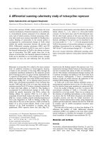
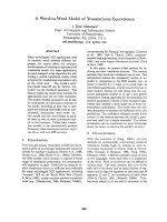
Invasive potential of GBC-SD and SGC-996 cells in vitro
The Transwell plates were used to measure the in vitro
ability of cells to invade a basement membrane matrix–
an important step in the metastatic cascade. We found
the GBC-SD cells were mainly composed of spindle-
shaped and polygonal cells. However, the SGC-996 cells
could mainly form multi-layered colonies. The invasion
results are summarized in Figure 1A. Both GBC-SD and
SGC-996 cells could successfully invade through the
matrix-coated membrane to the lower wells. However,
the number of GBC-SD cells were much more than that
of SGC-996 cells (137.81 ± 16.40 vs. 97.81 ± 37.66, t =
3.660, P = 0.0013). Hence, GBC-SD cells were defined
as highly invasive cell lines, whereas SGC-996 cells were
defined as poorly invasive cell lines (Figure 1B).
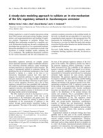
Vessel-like structure formation in three-dimensional
culture of GBC-SD and SGC-996 cells in vitro
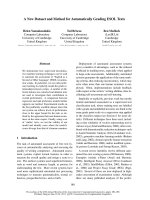
As shown in Figure 2, highly aggressive gallbladder
carcinoma GBC-SD cells wereabletoformnetworkof
hollow tubular structures when cultured on Matrigel
and rat-tail collagen typeⅠcomposed of the ECM gel in
the absence of endothelial cells and fibroblasts. The
tumor-formed networks initiated formation within 48 hr
after seeding the cells onto the matrix with optimal
structure formation achieved by two weeks. Microscopic
analysis demonstrated that the networks consisted of
tubular structures surrounding cluster of tumor cells.
During formation, the tubular networks became mature
channelized or holl owed vasculogenic-like structure
at two weeks after seeding the cells onto the gels. How-
ever, poorly aggressive SGC-996 cells were unable to
form the tubular-like structures with the same
conditions. After three days of incubation with the
aggressive GBC-SD cells, these cells were removed, and
poorly aggressive SGC-996 cells did assume a vasculo-
genic phenotype and initiated the formation of
patterned, vessel-like networks when seeded onto a
three-dimensional matrix preconditioned by aggressive
GBC-SD cells (Figure 2b5). GBC-SD cells could still form
hollowed vasculogenic-like structures when cultured on a
matrix preconditioned by SGC-996 cells (Figure 2a5).
The three-dimensional cultures of GBC-SD cells
stained with H&E showed the vasculogenic-like struc-
ture at two weeks (Figure 2a3). To address the role of
the PAS positive materials in tubular networks forma-
tion, the three-dimensional cultures of GBC-SD cells
were stained with PAS without hematoxylin counter-
stain. GBC-SD cells could secret PAS positive materials
and the PAS positive materials appeared around the sin-
gle cell or cell clusters. As an ingredient of the base-
membrane of V M, PAS positive materials were located
in granules and patches in the tumor cells cytoplasm
(Figure 2a4). In contrast, the similar phenomenon didn’t
occur in SGC-996 cells (Figure 2b3, 2b4).
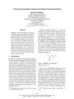
VM’s histomorphology of GBC-SD and SGC-996
xenografts in vivo
The tumor appeared gradually in subcutaneous area of
right axilback of nude mice from the 6th day after inocu-
lation. After 3 weeks, the tumor formation rates of nude
mouse xenografts were 100% (7/7) for GBC-SD and
71.4% (5/7) for SGC-996 respectively. In addition, the
medium tumor volume of GBC-SD xenografs was 2.95 ±
1.40 cm
3
(mean ± SD, range 1.73 to 4.86 cm
3
), while was
3.41 ± 0.56 cm
3
(mean ± SD, range 2.85 to 4.05 cm
3
)in
SGC-996 xenografts, there was no significant difference
between the two groups (Figure 3a1b1, P > 0.05).
H&E staining, dual -staining with CD
31
-PAS and TEM
were used for xenografts to observe the morphology
characteristic. Microscopically, in GBC-SD xenografts
(n = 7, 4 μm-thick serial tissue specimens per nude
mice model), the red blood cells were surrounded by
tumor cell-lined channel and tumor cells present ed var-
ious and obviously heteromorphism, necrosis was not
observed in the center of the tumor (Figure 3a3a4). The
channel consisted of tumor cells was negative of CD
31
and positive PAS. Abundant microvessels appeared
around the tumor, above all, in the marginal of the
tumor. VM positive rate was 85.7% (6/7). Among 24 tis-
sue sections, 10 high-power fields in each section were
counted to estimate the proportion of vessels that were
lined by tumor cells, 5.7% (17/300) channels were seen
to contain red blood cells among these tumor cell -lined
vasculatu res. However, in SGC-996 xenografts (n = 5, 4
μm-thick serial tissue specimens per nude mice model),
the phenomenon of tumor cell-lined channel containing
Sun et al. Journal of Experimental & Clinical Cancer Research 2011, 30:46
/>Page 4 of 12
Figure 1 Invasi ve potential of human gallbladder carcinoma cell lines GBC-SD and SGC-996 in vitro. (A) Representative phase contrast
microscopy pictures of GBC-SD cells (a
1-3
; original magnification, a
1
× 100, a
2
× 200, a
3
× 400) and SGC-996 cells (b
1-3
; original magnification, b
1
× 100, b
2
× 200, b
3
× 400) with HE staining. Both GBC-SD and SGC-996 cells could invade through the matrix-coated membrane to the lower
wells of Transwell plates. (B) The invaded number of GBC-SD cells were much more than that of SGC-996 cells (P = 0.0013).
Sun et al. Journal of Experimental & Clinical Cancer Research 2011, 30:46
/>Page 5 of 12
Figure 2 Phase contrast microscopy of human gallbladder carcinoma cell lines GBC-SD (a)andSGC-996(b) cultured three-
dimensionally on Matrigel (a
1
, b
1
; original magnification × 100) and rat-tail collagenⅠmatrix (a
2-5
, b
2-5
, original magnification × 200) in
vitro. Highly aggressive GBC-SD cells form patterned, vasculogenic-like networks when being cultured on Matrigel (a
1
) and rat-tail
collagenⅠmatrix (a
2
) for 14 days. Similarly, the three-dimensional cultures of GBC-SD cells stained with H&E showed the vasculogenic-like
structure at three weeks (a
3
); PAS positive, cherry-red materials found in granules and patches in the cytoplasm of GBC-SD cells appeared
around the signal cell or cell clusters when stained with PAS without hematoxylin counterstain (a
4
). However, poorly aggressive SGC-996 cells
did not form these networks when cultured under the same conditions (b
1-4
). GBC-SD cells cultured on a SGC-996 cells preconditioned matrix
were not inhibited in the formation of the patterned networks by the poorly aggressive cell preconditioned matrix (a
5
). Poorly aggressive SGC-
996 cells form pattern, vasculogenic-like networks when being cultured on a matrix preconditioned by the GBC-SD cells (b
5
).
Sun et al. Journal of Experimental & Clinical Cancer Research 2011, 30:46
/>Page 6 of 12
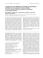
Figure 3 Characteristic appearance and the histomorphologic observation of GBC-SD and SGC-996 xenografts in vivo. (A) GBC-SD (a
1
)
and SGC-996 (b
1
) xenografts. Furthermore, SGC-996 xenografts exhibited different degree of tumor necrosis (red arrowhead).
Immunohistochemistry with CD
31
(original magnification × 200) revealed hypervascularity with a lining of ECs (red arrowheads), GBC-SD
xenografts showed more angiogenesis in marginal area of tumor (a
2
) than that of SGC-996 xenografts (b
2
)[P = 0.0115, (B)]. Using H&E (a
3
,b
3
)
and CD
31
-PAS double stain (a
4
,b
4
, original magnification × 200), sections of GBC-SD xenografts showed tumor cell-lined channels containing
red blood cells (a
3
, yellow circle) without any evidence of tumor necrosis. PAS-positive substances line the channel-like structures; Tumor cells
form vessel-like structure with single red blood cell inside (a
4
, yellow arrowhead). However, similar phenomenon failed to occur in SGC-996
xenografts (b
3
,b
4
) with tumor necrosis (b
3
, yellow arrowhead). TEM (original magnification × 8000) clearly visualized several red blood cells in
the central of tumor nests in GBC-SD xenografts (a
5
). Moreover, SGC-996 xenografts exhibited central tumor necrosis (b
5
, red arrowheads) which
consistent with morphology changes with H&E staining.
Sun et al. Journal of Experimental & Clinical Cancer Research 2011, 30:46
/>Page 7 of 12
the red blood cells were no t discovered; the central area
of tumor had the evidence of necrosis (Figure 3b3b4). In
addition, in the marginal area of GBC-SD xenografts,
hypervascularity with a lining of ECs was revealed, SGC-
996 xenografts (Figure 3b2) exhibited less angiogenesis
inthemarginalareaofthetumorthandidGBC-SD
(Figure 3a2). In the central area of tumor, GBC-SD
xenografts exhibited VM in the absence of ECs, central
necrosis, and fibrosis (Figure 3a3). Furthermore, t he
MVDofmarginalareaoftumorxenograftsbetween
GBC-SD and SGC-996 was compared. The MVD of
GBC-SD xenografts (n = 7) was higher than the GBC-
SD xenografts (n = 5, 13.514 ± 2.8328 vs. 11.68 ±
2.4617, t = 2.61, P = 0.0115) (Figure 3a2 b2).
For GBC-SD xenografts, TEM clearly showed single,
double, and several red blood cells existed in the central
of tumor nests. There was no vascular structure between
the surrounding tumor cells and erythrocytes. Neither
necrosis nor fibrosis was observed in the tumor nests
(Figure 3a5). In contrast, the necrosis in GBC-SD xeno-
grafts specimens could be clearly found (Figure 3b5).
These finding demonstrated that VM existed in GBC-
SD xenografts and assumed the same morphology and
structure characteristic as VM existed in human primary
gallbladder carcinomas reported by us [28].
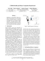
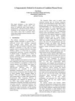
Hemodynamic of VM and angiogenesis in GBC-SD and
SGC-996 xenografts in vivo
Two-mm-interval horizontal scanning of two different
gallbladder carcinoma xenografts (GBC-SD and SGC-
996) were conducted to compare tumor signal intensi-
ties between mice by dynamic Micro-MRA with an
intravascular macromolecular MRI contrast agent
named HAS-Gd-DTPA. As shown in Figure 4, the
tumor marginal area of GBC-SD and SGC-996 xeno-
grafts exhibited gradually a high-intensity signal that
completely surrounded the xenografted tumor, a finding
consist ent with angiogenesis. In th e tumor center, GBC-
SD xenografts exhibited multiple high-intensity spots
(which is consistent with the intensity observed at
tumor marginal), a result consistent with pathological
VM. However, SGC-996 xenografts exhibited a low
intensity signal or a lack of signal, a result consistent
with central necrosis and disappearance of nuclei. Exam-
ination of the hemodynamic of VM revealed blood flow
with two peaks of intensity and a statistically significant
time lag relative to the hemodynamic of angiogenesis.
Discussion
In the present study, we examined the capacity of GBC-
SD and SGC-996 cell phenotypes and their invasive
potential to participate in vessel-like structures forma-
tion in vitro, and succeeded in establishing GBC-SD and
SGC-996 nude mouse xenograft models. In addition,
highly invasive GBC-SD cells when grown in three-
dimensional cultures c ontaining Matrigel or typeⅠcolla-
genintheabsenceofendothelial cells and fibroblasts,
and poorly aggressive SGC-996 cells when placed on the
aggressive cell-preconditioned matrix could all form pat-
terned networks containing hollow matrix channels.
Furthermore, we identified the existence of VM in
GBC-SD nude mouse xenografts by immunohistochem-
istry (H&E and CD31-PAS double-staining), electron
microscopy and micro-MRA technique with HAS-Gd-
DTPA. To our knowledge, this is the first study to
report that VM not only exists in the three-dimensional
matrixes of human gallbladder carcinoma cell lines
GBC-SD in vi tro, but also in the nude mouse xenografts
of GBC-SD cells in vivo, which is consistent with our
previous finding [28].
PAS-positive patterns are also associated with poor
clinical outcome for the patients with melanoma [12]
and cRCC [13]. In this study, we confirmed that VM, an
intratumoral, tumor cell-lined, PAS-positive and
patterned vasculogenic-like network, not only exists in
the three-dimensional matrixes of human gallbladder
carcinoma cell lines GBC-SD in vitro,butalsointhe
nude mouse xenografts of GBC-SD cells in vivo.Itis
suggested that the PAS positive materials, secreted by
GBC-SD cells, maybe be an important ingredients of
base membrane of VM.
Tumor cell plasticity, which has also been demon-
strated in prostatic carcinoma [29-31], bladder carci-
noma [32], astrocytoma [33], breast cancer [34-38] and
ovarian carcinoma [39-41], underlies VM. Consistent
with a recent report, which show that poorly aggr essive
melanoma cells (MUM-2C) could form patterned,
vasculogenic-like networks when cultured on a matrix
preconditioned by the aggressive melanoma cells
(MUM-2B). Furthermore, MUM-2B cells cultured on a
MUM-2C preconditioned matrix were not inhibited in
the formation of the patterned networks [42]. Our
results showed that highly aggressive GBC-SD cells
could form channelized or hollowed vasculogenic-like
structure in three-dimensional matrix, whereas poorly
aggressive SGC-996 cells failed to form these structures.
Interestingly, the poorly aggressive SGC-996 cells
acquired a vasculogenic phenotype and formed tubular
vasculogenic-like networks in response to a metastatic
microenvironment (preconditioned by highly aggressive
GBC-SD cells). GBC-SD cells could still form hollowed
vasculogenic-like structures when cultured on a matrix
preconditioned by SGC-996 tumor cells. These data
indicate that tumor matrix microenvironment plays a
critical role in cancer progression. To date, several
genes in tumor matrix micro environment were revealed
to participate in the process of VM and tumor cell
plasticity. For example, over-expression of migration-
Sun et al. Journal of Experimental & Clinical Cancer Research 2011, 30:46
/>Page 8 of 12
Figure 4 Dynamic micro-MRA of the xenografts (a
1-6
) and hemodynamic of VM and angiogenesis in GBC-SD and SGC-996 xenografts
(b
1-6
) in vivo. (A) The images were acquired before the injection of the contrast agents (HAS-Gd-DTPA, pre), 1, 3, 5, 10, and 15 min after
injection. The tumor marginal area (red circle) of both GBC-SD and SGC-996 exhibited a signal that gradually increased in intensity. In the tumor
center (yellow circle), GBC-SD exhibited spots in which the signal gradually increased in intensity (consistent with the intensity recorded for the
tumor margin). However, the central region of SGC-996 maintained a lack of signal. (B) Hemodynamic of VM and angiogenesis in GBC-SD and
SGC-996 nude mouse xenografts. All data are expressed as means ± SD. The time course of intensity of the tumor center (corresponding to the
hemodynamic of VM) was consistent with the time course of intensity of tumor margin (corresponding to the hemodynamic of angiogenesis).
Sun et al. Journal of Experimental & Clinical Cancer Research 2011, 30:46
/>Page 9 of 12
inducing protein 7 (Mig-7) was found in aggressive
invasive melanoma cells capable of VM but no t in
poorly invasive that do not form the tumor-lined struc-
ture. Over-expression of Mig-7 increased g2chain
domain Ⅲ fragments known to contain epidermal
growth factor (EGF)-like repeats that can activate EGF
receptor. Laminin 5 is the only laminin that contains
the g2 chain, which following cleavage into promigratory
fragments , the domain Ⅲ region, causes increased levels
of matrix metalloproteinase-2 (MMP-2), and matrix
metalloproteinase-14 (MMP-14) cooperate to cleave g2
chain into fragments that promote melanoma cell inva-
sion and VM [43,44]. However, in this study, we did not
determine the molecular epigenetic effects induced by
the matrix microenvironmentpreconditionedbyhighly
aggressive GBC-SD cells. Molecular signal regulations of
VM formation in GBC are supposed to be further stu-
died. On the other hand, Sood et al [41] revealed the
detailed scanning and transmission electron micrographs
of ovarian cancer cell cultures grown on three-dimen-
sional collagenⅠmatrices. The evident hollow tubular
structures lined by flattened ovarian cancer cells could
be observed by electron microscopy. In addition, they
also found the tumor-formed networks initiated forma-
tion within 3 days after seeding the aggressive ovarian
cancer cells onto the matrix. Furthermore, the tubular
networks became channelized or hollowed during for-
mation, and were stable through 6 weeks after seeding
the cells onto a matrix, which is similar to our data,
suggesting that hollow tubular structures might be the
mature structures of VM when aggressive tumor cells
were cultured on Matrigel or rat-tail collagen type Ⅰ.
VM, referred to as the “fluid-conducting-meshwork”,
may have significant implications for tumor perfusion
and dissemination. Several papers evidenced the VM
channel functional role in tumor circulation by microin-
jection method [3,7] and MRA technique [ 8,9,11]. We
observed that VM only exists in GBC-SD xenografts by
using H&E staining, CD
31
-PAS double staining and
TEM, 5.7% channels were seen to contain red blood
cells among these tumor cell-lined vasculatures, which
is consistent with the ratio of human GBC samples
(4.25%) [28]. We also found that GBC-SD xenografts
exhibited much more microvessel in the marginal area
of the tumor than did SGC-996 xenografts. In the cen-
tral area of tumor, GBC-SD xenografts exhibited VM in
the absence of ECs, central necrosis, and fibrosis. In
contrast, SGC-996 xenografts exhibited central tumor
necrosis as tumor grows in the absence of VM. This
might suggest that the endothelial sprouting of new ves-
sels from preexisting vessels as a result of over-expres-
sion of angiogenic factors. On the premise of
successfully establishing GBC-SD and SGC-996 nude
mouse xenografts, we furthermore performed dynamic
micro-MR A analysis, using HAS-Gd-DTPA (60-100kD),
which was much larger than Gd-DTPA (725D, generally
MRI contrast agent) in molecule weight and volume.
Thus the HAS-Gd-DTPA assumed much less leakage
through the vascular wall than Gd-DTPA. Our results
indicated that the hemodynamic of VM revealed blood
flow with two peaks of intensity and a statistically signif-
icant time lag, relative to the hemodynamic of angiogen-
esis, whi ch is consis tent with the reported findings
[9,11], suggesting that VM might play role in perfusion
and dissemination of GBC-SD xenografted tumors as
the fluid-conducting-meshwork. Taken together, these
data also provided strong evidence the connection
between angiogenesis and VM in GBC-SD xenografts.
Conclusions
In conclusion, the present study reveals that VM exists
in GBC by both three-dimensional matrix of highly
aggressive GBC-SD or poorly aggressive SGC-996 cells
preconditioned by highly aggressive GBC-SD cells in
vitro and GBC-SD nude m ouse xenografts in vivo.This
study has a limitation that only two different established
GBC cell lines in China were enrolled in present study.
Hence, we couldn’t draw a comprehensive conclusion
about biological characteristic of GBC. However, our
study provides the b ackground for continuing study for
VM as a potential target for anticancer therapy i n
human GBC. Therefore, furthermore studies are needed
to clarify the molecular mechan ism of VM in the devel-
opment and progression of GBC.
Abbreviations
VM: vasculogenic mimicry; ECs: endothelial cells; ECM: extracellular matrix;
PAS: periodic acid-Schiff-positive; GBC: Gallbladder carcinoma; SPF: specific
pathogen free; DMEM: Dulbecco’s modified Eagle’s media; FBS: fetal bovine
serum; MVD: microvessel density; TEM: transmission electron microscopy;
HAS-Gd-DTPA: human adult serum gadopentetic acid dimeglumine salt
injection; ROI: regions of interest; Mig-7: migration-inducing protein 7; EGF:
epidermal growth factor; MMP: matrix metalloproteinase.
Acknowledgements
This work was supported by a grant from the National Nature Science
Foundation of China (No.30672073). We are grateful to Prof. An-Feng Fu and
Mei-Zheng Xi (Department of Pathology, Shanghai Jiaotong University,
China) for their technical assistance. We also grateful to Prof. Lian-Hua Ying,
Feng-Di Zhao, Chao Lu, Yan-Xia Ning and Ting-Ting Zhou (Department of
Pathophysiology, Fudan University, China) for their advice and technical
assistance. In addition, we also gratefully acknowledge access to SGC-996
cell lines provided by Prof. Yao-Qing Yang (Tumor Cell Biology Research
Institute, Medical College of Tongji University, China). In particular we thank
Prof. Xiang-Yao Yu, Hao Xi and Han-Bao Tong (Department of Pathology,
Shanghai Tenth People’s Hospital, Tongji University, China) for reviewing the
tissue specimens.
Authors’ contributions
W Sun and YZ Fan were responsible for data collection and analysis,
experiment job, interpretation of the results, and writing the manuscript. W
Sun carried out the Invasion assay and three-dimensional culture of GBC-SD
and SGC-996 cells in vitro. WZ Zhang and CY Ge carried out the nude
mouse xenografts of GBC-SD and SGC-996 cells. W Sun and WZ Zhang were
Sun et al. Journal of Experimental & Clinical Cancer Research 2011, 30:46
/>Page 10 of 12
responsible for the existence of VM in GBC by using immunohistochemistry
staining, TEM and micro-MRA technology in vitro and in vivo, respectively. All
authors have read and approved the final manuscript.
Competing interests
The authors declare that they have no competing interests.
Received: 18 January 2011 Accepted: 29 April 2011
Published: 29 April 2011
References
1. Folkman J, Klagsbrun M: ANGIOGENIC FACTORS. Science 1987, 235:442-447.
2. Maniotis AJ, Folberg R, Hess A, Seftor EA, Gardner LM, Pe’er J, Trent JM,
Meltzer PS, Hendrix MJ: Vascular channel formation by human melanoma
cells in vivo and in vitro: vasculogenic mimicry. Am J Pathol 1999,
155:739-752.
3. Frenkel S, Barzel I, Levy J, Lin AY, Bartsch DU, Majumdar D, Folberg R,
Pe’er J: Demonstrating circulation in vasculogenic mimicry patterns of
uveal melanoma by confocal indocyanine green angiography. Eye (Lond)
2008, 22:948-952.
4. Zhang S, Guo H, Zhang D, Zhang W, Zhao X, Ren Z, Sun B:
Microcirculation patterns in different stages of melanoma growth. Oncol
Rep 2006, 15:15-20.
5. Folberg R, Hendrix MJ, Maniotis AJ: Vasculogenic mimicry and tumor
angiogenesis. Am J Pathol 2000, 156:361-381.
6. Folberg R, Maniotis AJ: Vasculogenic mimicry. APMIS 2004, 112:508-525.
7. Clarijs R, Otte-Holler I, Ruiter DJ, de Waal RM: Presence of a fluid-
conducting meshwork in xenografted cutaneous and primary human
uveal melanoma. Invest Ophthalmol Vis Sci 2002, 43:912-918.
8. Kobayashi H, Shirakawa K, Kawamoto S, Saga T, Sato N, Hiraga A,
Watanabe I, Heike Y, Togashi K, Konishi J, et al: Rapid accumulation and
internalization of radiolabeled herceptin in an inflammatory breast
cancer xenograft with vasculogenic mimicry predicted by the contrast-
enhanced dynamic MRI with the macromolecular contrast agent G6-
(1B4M-Gd)(256). Cancer Res 2002, 62:860-866.
9. Shirakawa K, Kobayashi H, Heike Y, Kawamoto S, Brechbiel MW, Kasumi F,
Iwanaga T, Konishi F, Terada M, Wakasugi H: Hemodynamics in
Vasculogenic mimicry and angiogenesis of inflammatory breast cancer
xenograft. Cancer Research 2002, 62:560-566.
10. Ruf W, Seftor EA, Petrovan RJ, Weiss RM, Gruman LM, Margaryan NV,
Seftor RE, Miyagi Y, Hendrix MJ: Differential role of tissue factor pathway
inhibitors 1 and 2 in melanoma vasculogenic mimicry. Cancer Res 2003,
63:5381-5389.
11. Shirakawa K, Kobayashi H, Sobajima J, Hashimoto D, Shimizu A, Wakasugi H:
Inflammatory breast cancer: vasculogenic mimicry and its
hemodynamics of an inflammatory breast cancer xenograft model.
Breast Cancer Res 2003, 5:136-139.
12. Warso MA, Maniotis AJ, Chen X, Majumdar D, Patel MK, Shilkaitis A,
Gupta TK, Folberg R: Prognostic significance of periodic acid-Schiff-
positive patterns in primary cutaneous melanoma. Clin Cancer Res 2001,
7:473-477.
13. Vartanian AA, Stepanova EV, Gutorov SL, Solomko E, Grigorieva IN,
Sokolova IN, Baryshnikov AY, Lichinitser MR: Prognostic significance of
periodic acid-Schiff-positive patterns in clear cell renal cell carcinoma.
Can J Urol 2009, 16
:4726-4732.
14.
Shirakawa K, Wakasugi H, Heike Y, Watanabe I, Yamada S, Saito K, Konishi F:
Vasculogenic mimicry and pseudo-comedo formation in breast cancer.
Int J Cancer 2002, 99:821-828.
15. Sood AK, Fletcher MS, Zahn CM, Gruman LM, Coffin JE, Seftor EA,
Hendrix MJ: The clinical significance of tumor cell-lined vasculature in
ovarian carcinoma: implications for anti-vasculogenic therapy. Cancer Biol
Ther 2002, 1:661-664.
16. Sun B, Zhang S, Zhang D, Du J, Guo H, Zhao X, Zhang W, Hao X:
Vasculogenic mimicry is associated with high tumor grade, invasion and
metastasis, and short survival in patients with hepatocellular carcinoma.
Oncol Rep 2006, 16:693-698.
17. Sun BC, Zhang SW, Zhao XL, Hao XS: Vasculogenic mimicry is associated
with shorter survival in hepatocellular carcinomas. Laboratory
Investigation 2006, 86:1302.
18. Guzman G, Cotler SJ, Lin AY, Maniotis AJ, Folberg R: A pilot study of
vasculogenic mimicry immunohistochemical expression in hepatocellular
carcinoma. Archives of Pathology & Laboratory Medicine 2007,
131:1776-1781.
19. Wang W, Lin P, Han C, Cai W, Zhao X, Sun B: Vasculogenic mimicry
contributes to lymph node metastasis of laryngeal squamous cell
carcinoma. J Exp Clin Cancer Res 2010, 29:60.
20. El Hallani S, Boisselier B, Peglion F, Rousseau A, Colin C, Idbaih A, Marie Y,
Mokhtari K, Thomas JL, Eichmann A, et al: A new alternative mechanism in
glioblastoma vascularization: tubular vasculogenic mimicry. Brain 2010,
133:973-982.
21. Li M, Gu Y, Zhang Z, Zhang S, Zhang D, Saleem AF, Zhao X, Sun B:
Vasculogenic mimicry: a new prognostic sign of gastric adenocarcinoma.
Pathol Oncol Res 2010, 16:259-266.
22. Baeten CI, Hillen F, Pauwels P, de Bruine AP, Baeten CG: Prognostic role of
vasculogenic mimicry in colorectal cancer. Dis Colon Rectum 2009,
52:2028-2035.
23. Sun B, Qie S, Zhang S, Sun T, Zhao X, Gao S, Ni C, Wang X, Liu Y, Zhang L:
Role and mechanism of vasculogenic mimicry in gastrointestinal stromal
tumors. Hum Pathol 2008, 39:444-451.
24. Gourgiotis S, Kocher HM, Solaini L, Yarollahi A, Tsiambas E, Salemis NS:
Gallbladder cancer. Am J Surg 2008, 196:252-264.
25. Reddy SK, Clary BM: Surgical management of gallbladder cancer. Surg
Oncol Clin N Am 2009, 18:307-324.
26. Hsing AW, Gao YT, Devesa SS, Jin F, Fraumeni JF Jr: Rising incidence of
biliary tract cancers in Shanghai, China. Int J Cancer 1998, 75:368-370.
27. Shukla PJ, Barreto SG: Gallbladder cancer: we need to do better! Ann Surg
Oncol 2009, 16:2084-2085.
28. Fan YZ, Sun W, Zhang WZ, Ge CY:
Vasculogenic mimicry in human
primary
gallbladder carcinoma and clinical significance thereof.
Zhonghua Yi Xue Za Zhi 2007, 87:145-149.
29. Liu C, Huang H, Donate F, Dickinson C, Santucci R, El-Sheikh A, Vessella R,
Edgington TS: Prostate-specific membrane antigen directed selective
thrombotic infarction of tumors. Cancer Res 2002, 62:5470-5475.
30. Sharma N, Seftor REB, Seftor EA, Gruman LM, Heidger PM, Cohen MB,
Lubaroff DM, Hendrix MJC: Prostatic tumor cell plasticity involves
cooperative interactions of distinct phenotypic subpopulations: Role in
vasculogenic mimicry. Prostate 2002, 50:189-201.
31. Chung LW, Huang WC, Sung SY, Wu D, Odero-Marah V, Nomura T,
Shigemura K, Miyagi T, Seo S, Shi C, et al: Stromal-epithelial interaction in
prostate cancer progression. Clin Genitourin Cancer 2006, 5:162-170.
32. Fujimoto A, Onodera H, Mori A, Nagayama S, Yonenaga Y, Tachibana T:
Tumour plasticity and extravascular circulation in ECV304 human
bladder carcinoma cells. Anticancer Res 2006, 26:59-69.
33. Yue WY, Chen ZP: Does vasculogenic mimicry exist in astrocytoma? J
Histochem Cytochem 2005, 53:997-1002.
34. Shevde LA, Metge BJ, Mitra A, Xi Y, Ju J, King JA, Samant RS: Spheroid-
forming subpopulation of breast cancer cells demonstrates vasculogenic
mimicry via hsa-miR-299-5p regulated de novo expression of
osteopontin. J Cell Mol Med 2010, 14:1693-1706.
35. Robertson FM, Simeone AM, Lucci A, McMurray JS, Ghosh S, Cristofanilli M:
Differential regulation of the aggressive phenotype of inflammatory
breast cancer cells by prostanoid receptors EP3 and EP4. Cancer 2010,
116:2806-2814.
36. Basu GD, Liang WS, Stephan DA, Wegener LT, Conley CR, Pockaj BA,
Mukherjee P: A novel role for cyclooxygenase-2 in regulating vascular
channel formation by human breast cancer cells. Breast Cancer Res 2006,
8:R69.
37. Hoffmeyer MR, Wall KM, Dharmawardhane SF: In vitro analysis of the
invasive phenotype of SUM 149, an inflammatory breast cancer cell line.
Cancer Cell Int 2005, 5:11.
38. Shirakawa K, Furuhata S, Watanabe I, Hayase H, Shimizu A, Ikarashi Y,
Yoshida T, Terada M, Hashimoto D, Wakasugi H: Induction of
vasculogenesis in breast cancer models. Br J Cancer 2002, 87:1454-1461.
39. Hess AR, Seftor EA, Seftor RE, Hendrix MJ: Phosphoinositide 3-kinase
regulates membrane Type 1-matrix metalloproteinase (MMP) and MMP-
2 activity during melanoma cell vasculogenic mimicry. Cancer Res 2003,
63:4757-4762.
40. Sood AK, Fletcher MS, Hendrix MJ: The embryonic-like properties of
aggressive human tumor cells. J Soc Gynecol Investig 2002, 9:2-9.
41. Sood AK, Seftor EA, Fletcher MS, Gardner LM, Heidger PM, Buller RE,
Seftor RE, Hendrix MJ: Molecular determinants of ovarian cancer
plasticity. Am J Pathol 2001,
158:1279-1288.
Sun et al. Journal of Experimental & Clinical Cancer Research 2011, 30:46
/>Page 11 of 12
42. Seftor EA, Meltzer PS, Kirschmann DA, Margaryan NV, Seftor RE, Hendrix MJ:
The epigenetic reprogramming of poorly aggressive melanoma cells by
a metastatic microenvironment. J Cell Mol Med 2006, 10 :174-196.
43. Robertson GP: Mig-7 linked to vasculogenic mimicry. American Journal of
Pathology 2007, 170:1454-1456.
44. Petty AP, Garman KL, Winn VD, Spidel CM, Lindsey JS: Overexpression of
carcinoma and embryonic cytotrophoblast cell-specific Mig-7 induces
invasion and vessel-like structure formation. Am J Pathol 2007,
170:1763-1780.
doi:10.1186/1756-9966-30-46
Cite this article as: Sun et al.: A pilot histomorphology and
hemodynamic of vasculogenic mimicry in gallbladder carcinomas in
vivo and in vitro. Journal of Experimental & Clinical Cancer Research 2011
30:46.
Submit your next manuscript to BioMed Central
and take full advantage of:
• Convenient online submission
• Thorough peer review
• No space constraints or color figure charges
• Immediate publication on acceptance
• Inclusion in PubMed, CAS, Scopus and Google Scholar
• Research which is freely available for redistribution
Submit your manuscript at
www.biomedcentral.com/submit
Sun et al. Journal of Experimental & Clinical Cancer Research 2011, 30:46
/>Page 12 of 12