Advanced Therapy in Gastroenterology and Liver Disease - part 5 ppsx
Bạn đang xem bản rút gọn của tài liệu. Xem và tải ngay bản đầy đủ của tài liệu tại đây (1.03 MB, 74 trang )
Metabolic Bone Disease in Gastrointestinal and Liver Patients / 337
M
onegal A, Navasa M, Guanabens N, et al. Osteoporosis and bone
mineral metabolism disorders in cirrhotic patients referred
for orthotopic liver transplantation. Calcif Tissue Int
1997;60:148–54.
M
ora S, Barera G, Ricotti A, et al. Reversal of low bone density
w
ith a gluten-free diet in children and adolescents with celiac
disease. Am J Clin Nutr 1998;67:477–81.
Mora S, Weber G, Barera G, et al. Effect of gluten-free diet on
bone mineral content in growing patients with celiac disease.
A
m J Clin Nutr 1993;57:224–8.
Moran CE, Sosa EG, Martinez SM, et al. Bone mineral density
in patients with pancreatic insufficiency and steatorrhea. Am
J Gastroenterol 1997;92:867–71.
Motley RJ, Clements D, Evans WD, et al. A four-year longitudi-
nal study of bone loss in patients with inflammatory bowel
disease. Bone Miner 1993;23:95–104.
Neer RM, Arnaud CD, Zanchetta JR, et al. Effect of parathyroid
hormone (1-34) on fractures and bone mineral density in
postmenopausal women with osteoporosis. N Engl J Med.
2001;344:1434–41.
Nemetz A, Toth M, Garcia-Gonzalez MA, et al. Allelic variation
at the interleukin 1beta gene is associated with decreased bone
mass in patients with inflammatory bowel diseases. Gut
2001;49:644–9.
Newton J,Francis R, Prince M, et al. Osteoporosis in primary bil-
iary cirrhosis revisited. Gut 2001;49:282–7.
Ninkovic M, Love S, Tom BD, et al. Lack of effect of intravenous
pamidronate on fracture incidence and bone mineral density
after orthotopic liver transplantation. J Hepatol 2002;37:93–100.
Nuti R, Martini G, Valenti R, et al. Prevalence of undiagnosed
coeliac syndrome in osteoporotic women. J Intern Med
2001;250:361–6.
Ormarsdottir S, Ljunggren O, Mallmin H, et al. Circulating lev-
els of insulin-like growth factors and their binding proteins
in patients with chronic liver disease: lack of correlation with
bone mineral density. Liver 2001;21:123–8.
Pollak RD, Karmeli F, Eliakim R, et al. Femoral neck osteopenia in
patients with inflammatory bowel disease. Am J Gastroenterol
1998;93:1483–90.
Reeves HL, Francis RM, Manas DM, et al. Intravenous bisphos-
phonate prevents symptomatic osteoporotic vertebral collapse
in patients after liver transplantation. Liver Transpl Surg
1998;4:404–9.
Reid D,Cohen S, Pack S, et al. Risedronate reduces the incidence
o
f
v
e
r
t
ebral fractures in patients on chronic corticosteroid
therapy. Arthritis Rheum 1998;41:S136.
Reid D, Cohen S, Pack S. Risedronate is an effective and well-
t
olerated therapy in both the treatment and prevention of
corticosteroid-induced osteoporosis. Bone 1998;23:S402.
Reid D,Devogelaer JP, Hughes R, et al. Risedronate is effective and
well tolerated in treating corticosterd-induced osteoporosis.
American College of Rheumatology Annual Meeting; 1998.
Reid IR, Brown JP, Burckhardt P, et al. Intravenous zoledronic
acid in postmenopausal women with low bone mineral den-
sity. N Engl J Med 2002;346:653–61.
Ringe JD,Welzel D. Salmon calcitonin in the therapy of corticoid-
induced osteoporosis. Eur J Clin Pharmacol 1987;33:35.
Robinson RJ, Carr I, Iqbal SJ, et al. Screening for osteoporosis in
Crohn’s disease.A detailed evaluation of calcaneal ultrasound.
Eur J Gastroenterol Hepatol 1998;10:137–40.
R
obinson RJ, Iqbal SJ,Wolfe R, et al. The effect of rectally admin-
istered steroids on bone turnover: a comparative study.
Aliment Pharmacol Ther 1998;12:213–7.
Romaldini CC, Barbieri D, Okay TS, et al. Serum soluble
i
nterleukin-2 receptor,interleukin-6, and tumor necrosis fac-
t
or-alpha levels in children with celiac disease: response to
treatment. J Pediatr Gastroenterol Nutr 2002;35:513–7.
Rosen CJ, Brown S. Severe hypocalcemia after intravenous bis-
phosphonate therapy in occult vitamin D deficiency. N Engl
J
Med 2003;348:1503–4.
Ruegsegger P, Medici TC, Anliker M. Corticosteroid-induced bone
loss. A longitudinal study of alternate day therapy in patients
with bronchial asthma using quantitative computed tomo-
graphy. Eur J Clin Pharmacol 1983;25:615–20.
Saag K, Emkey R, Cividino A. Effects of alendronate for two years
on BMD and fractures in patients receiving glucocorticoids.
Bone 1998;23:S182.
Saag KG, Emkey R, Schnitzer TJ, et al. Alendronate for the pre-
vention and treatment of glucocorticoid-induced osteoporo-
sis.
Glucocorticoid-Induced Osteoporosis Intervention Study
Group. N Engl J Med 1998;339:292–9.
Saito JK, Davis JW, Wasnich RD, Ross PD. Users of low-dose glu-
cocorticoids have increased bone loss rates: a longitudinal
study. Calcif Tissue Int 1995;57:115–9.
Sambrook P, Birmingham J, Kelly P, et al. Prevention of cortico-
steroid osteoporosis. A comparison of calcium, calcitriol, and
calcitonin. N Engl J Med 1993;328:1747–52.
Schoon EJ, Bollani S, Mills PR, et al. Budesonide versus pred-
nisolone: effect on bone mineral density in patients with ileo-
cecal Crohn’s disease. Am J Gastroenterol 2002;97:A827.
Schulte C, Dignass AU, Mann K, Goebell H. Reduced bone min-
eral density and unbalanced bone metabolism in patients with
inflammatory bowel disease. Inflammatory Bowel Diseases
1998;4:268–75.
Schulte C, Goebell H, Roher HD, Schulte KM. Genetic determi-
nants of IL-6 expression levels do not influence bone loss in
inflammatory bowel disease. Dig Dis Sci 2001;46:2521–8.
Schulte CM, Dignass AU, Goebell H, et al. Genetic factors deter-
mine extent of bone loss in inflammatory bowel disease.
Gastroenterol 2000;119:909–20.
Scott EM, Gaywood I, Scott BB. Guidelines for osteoporosis in
coeliac disease and inflammatory bowel disease. British Society
of Gastroenterology. Gut 2000;46:1–8.
Sher KS, Jayanthi V, Probert CS, et al. Infertility, obstetric and gynae-
c
olog
ical pr
o
blems in coeliac sprue. Dig Dis 1994;12:186–90.
Solerio E, Isaia G, Innarella R, et al. Osteoporosis: still a typical com-
plication of primary biliary cirrhosis? Dig Liver Dis 2003;35:339–46.
St
ockbrugger RW, Schoon EJ, Bollani S, et al. Discordance
between the degree of osteopenia and the prevalence of spon-
taneous vertebral fractures in Crohn’s disease. Aliment
Pharmacol Ther 2002;16:1519–27.
Suda T, Nakamura I, Jimi E, Takahashi N. Regulation of osteo-
clast function. J Bone Miner Res 1997;12:869–79.
Szathmar
i M,
T
ulassa
y T
, Arato A, et al. Bone mineral content and
d
ensity in asymptomatic children with coeliac disease on a
gluten-free diet. Eur J Gastroenterol Hepatol 2001;13:419–24.
Takahashi N, Udagawa N, Suda T. A new member of tumor necro-
sis factor ligand family,ODF/OPGL/TRANCE/RANKL, regu-
lat
es osteoclast differentiation and function. Biochem Biophys
Res Commun 1999;256:449–55.
338 / Advanced Therapy in Gastroenterology and Liver Disease
T
homason K, West J, Logan RF, et al. Fracture experience of
patients with coeliac disease: a population based survey. Gut
2003;52:518–22.
Thomson BM, Mundy GR, Chambers TJ. Tumor necrosis factors
a
lpha and beta induce osteoblastic cells to stimulate osteo-
c
lastic bone resorption. J Immunol 1987;138:775–9.
Thomson BM, Saklatvala J, Chambers TJ. Osteoblasts mediate
interleukin 1 stimulation of bone resorption by rat osteoclasts.
J Exp Med 1986;164:104–12.
T
idermark J, Zethraeus N, Svensson O, et al. Femoral neck frac-
tures in the elderly: functional outcome and quality of life
according to EuroQol. Qual Life Res 2002;11:473–81.
Tovey FI, Godfrey JE, Lewin MR. A gastrectomy population: 25-
30 years on. Postgrad Med J 1990;66:450–6.
Trautwein C, Possienke M, Schlitt HJ, et al. Bone density and
metabolism in patients with viral hepatitis and cholestatic liver
diseases before and after liver transplantation. Am J
Gastroenterol 2000;95:2343–51.
Trombetti A, Herrmann F, Hoffmeyer P, et al. Survival and poten-
t
ial years of life lost after hip fracture in men and age-matched
women. Osteoporos Int 2002;13:731–7.
Tsuboi M, Kawakami A, Nakashima T, et al. Tumor necrosis fac-
tor-alpha and interleukin-1beta increase the Fas-mediated apop-
tosis of human osteoblasts. J Lab Clin Med 1999;134:222–31.
van Staa TP,Leufkens HGM,Abenhaim L, et al. Use of oral corticos-
teroids and risk of fractures.J Bone Miner Res 2000;15:993–1000.
Vasquez H, Mazure R, Gonzalez D, et al. Risk of fractures in celiac
disease patients: a cross-sectional, case-control study. Am J
Gastroenterol 2000;95:183–9.
V
estergaard P, Mosekilde L. Fracture risk in patients with celiac
disease, Crohn’s disease, and ulcerative colitis: a nationwide
follow-up study of 16,416 patients in Denmark. Am J
Epidemiol 2002;156:1–10.
V
estergaard P. Bone loss associated with gastrointestinal disease:
p
revalence and pathogenesis. Eur J Gastroenterol Hepatol
2003;15:851–6.
Vogelsang H, Klamert M, Resch H, Ferenci P. Dietary vitamin D
intake in patients with Crohn’s disease. Wiener Klinische
W
ochenschrift 1995;107:578–81.
Votta BJ,Bertolini DR. Cytokine suppressive anti-inflammatory com-
pounds inhibit bone resorption in vitro. Bone 1994;15:533–8.
Walters J, Weiser M. Calcium transport by rat duodenal vil-
lus and crypt basolateral membranes. Am J Physiol
1987;252:G170.
Weinstein RS, Jilka RL, Parfitt AM, Manolagas SC. Inhibition
of osteoblastogenesis and promotion of apoptosis of
osteoblasts and osteocytes by glucocorticoids. Potential
mechanisms of their deleterious effects on bone. J Clin
I
nvest 1998;102:274–82.
West J, Logan RF, Card TR, et al. Fracture risk in people with celiac
disease: a population-based cohort study. Gastroenterol
2003;125:429–36.
Westerholm-Ormio M, Garioch J, Ketola I, Savilahti E. Inflammatory
cytokines in small intestinal mucosa of patients with potential
coeliac disease. Clin Exp Immunol 2002;128:94–101.
Wiener H, Turnheim K. Calcium-activated potassium channels in
basolateral membranes of colon epithelial cells; reconstitution and
functional properties. Wien Klin Wochenschr 1990;102:622–8.
339
CHAPTER 56
DIETARY-INDUCED SYMPTOMS
LAWRENCE R. SCHILLER,MD
ESOPHAGUS
In the esophagus, the repeated swallows associated with eat-
ing and the postprandial rise in serum gastrin levels decrease
lower esophageal sphincter (LES) tone. In addition, gas-
tric distention due to ingested food and intragastric gas pro-
duction as acid is neutralized by food increase the number
of transient LES relaxations (the “belching reflex”) and per-
mit gastroesophageal reflux to occur. Patients with gas-
troesophageal reflux disease often note a distinct increase
in symptoms postprandially. Fatty foods and hypertonic
beverages may be particular problems (see later in chapter).
S
TOMACH
Eating stimulates gastric acid secretion, increasing the vol-
ume of material in the stomach. The ability of the stom-
ach to hold the additional fluid and the meal is due to
gastric accommodation, which allows the gastric wall to
relax. This vagally mediated reflex is disturbed in some
patients with FD and in patients after vagotomy, who can-
not accommodate large volumes in the stomach. This may
aggravate gastroesophageal reflux, speed gastric empty-
ing of liquids, and trigger sensations of bloating or early
satiety. Antral motility also is stimulated by eating.
S
MALL BOWEL
In the small bowel, ingestion of food rapidly converts the
fasting pattern of motility, which features cyclical migrat-
ing motor complexes, into the more chaotic postprandial
pattern. Chyme emptied from the stomach is joined by
pancreatic and biliary secretions, which distend the small
bowel and stimulate peristalsis.
The bowel wall is sensitive to distention and eating acti-
vates afferent nerves that may produce painful sensations
in some individuals. The entry of chyme into the duode-
num also results in release of many peptides and other sig-
naling s
ubstances
that p
roduce effects elsewhere in the gut
and even outside the GI tract.
COL
ON
F
ood residues enter the
c
olon
hour
s after ingestion.
Carbohydrate that is not absorbed in the small intestine
Most patients with gastrointestinal (GI) symptoms
attribute their symptoms to “something”they ate and want
advice from the doctor about what to eat to minimize their
symptoms. Symptoms after food ingestion most often are
due to normal food-induced physiological changes, such
as the gastrocolic reflex, or to the effects of food digestion,
such as the generation of gas. They rarely are due to food
allergy or to immunologic reactions to food breakdown
products, such as in celiac disease. Specific problems will
not be discussed further in this chapter. There are separate
chapters on food allergies (Chapter 57, “Gastrointestinal
Food Allergy”), celiac disease (Chapter 61, “Celiac Sprue
and Related Problems”), and lactose intolerance (Chapter
62, “Lactose Intolerance”).
Food-related symptoms often occur when organic prob-
lems are present, but probably occur most often in patients
with common functional bowel disorders, such as func-
tional dyspepsia (FD) or irritable bowel syndrome (IBS).
Patients with organic problems, such as short bowel syn-
drome, will have exacerbation of symptoms like diarrhea
when eating, with some foods producing more problems
than others. Patients with functional problems tend to be
unusually sensitive to distention and other digestive events,
and, therefore, may have aggravation of their basic symp-
toms when ingesting any foods. However some foods may
be more problematic than others. It is not that these foods
cause the fundamental functional problem, only that the
offending foods aggravate the symptoms of those condi-
tions (O’Sullivan and O’Morain, 2003).
Meal-Related Physiological Changes
Response to a Meal
Intestinal fluid and electrolyte transport and motility con-
tinue during fasting, but ingestion of a meal results in a
prompt alteration of activity.
This is not different conceptually than what happens to
the cardiovascular system with exercise, but it typically
involves changes that are an order of magnitude greater.
Thus salivary, gastric, biliary, and pancreatic secretion
increase 10-fold or more over basal levels, and motility pat-
terns abruptly change from fasting to fed patterns.
340 / Advanced Therapy in Gastroenterology and Liver Disease
(poorly absorbed carbohydrate and fiber) enters the right
colon and is fermented by the colonic bacterial flora. The
products of fermentation are short chain fatty acids—up
to 80 g of which can be produced by the colonic flora—
a
nd voluminous amounts of gas (carbon dioxide and
hydrogen gas) (Hammer et al, 1989; Hammer et al,1990).
Every 10 g of carbohydrate can yield about 1 L of gas. Gas can
distend the colon, stimulating motility and causing bloat-
ing, cramps and pain
in some people.
Effects of Specific Foods and Food
Additives
People ingest a variety of substances that consist of mixtures
of chemicals that can have specific effects on the body.These
chemicals include primary macronutrients, such as carbo-
hydrates, fats, and proteins; micronutrients, such as vitamins
and minerals; and incidental chemicals that have no nutri-
tive value,but are part of the animals and plants that we eat,
such as
caffeine in coffee or theobromine in chocolate. These
incidental chemicals may be biologically active in the gut
and elsewhere in the body and may produce symptoms.
Carbohydrates
Carbohydrates are responsible for a variety of food-induced
symptoms (Table 56-1). These symptoms can be due to
hypertonicity or to malabsorption of carbohydrate.
Ingestion of hypertonic carbohydrate solutions results
in entry of water into the gut lumen to produce osmotic
equilibration. This is mostly a problem for individuals with
unregulated gastric emptying, such as those who have had
g
astric surgery, and can produce a dumping syndrome with
bloating, nausea, diarrhea, flushing, and hypotension.
Hypertonic carbohydrate solutions, such as fruit juices, also
can produce dyspepsia, probably by stimulation of recep-
tors in the esophagus and stomach.
Malabsorption of carbohydrate in the small intestine
results in delivery of excess fermentable substrate to the
colon. This can be due to generalized malabsorption (eg,
celiac disease or short bowel syndrome) or to malabsorp-
tion of specific carbohydrate moieties. In addition, excess
dietary fiber ingestion will load the colon with additional
carbohydrate. When smaller amounts of carbohydrate are
delivered to the colon, excess gas, bloating and cramps
develop as gas is produced as a byproduct of fermentation.
Diarrhea is produced when larger amounts are ingested or
insufficient time is allowed for absorption due to accelerated
transit. Symptoms can develop with as little as 5 to 10 g of
excess carbohydrate entering the colon. Symptoms relate
more to the total amount of fermentable carbohydrate enter-
ing the colon than to the specific type of carbohydrate that
is malabsorbed. In some cases, “tolerance” to gradually
increasing amounts of carbohydrate develops, which is prob-
ably related to changes in bacterial metabolism.
Specific carbohydrates may be incompletely absorbed
by the small intestine in certain individuals. These include
fructose, sucrose, lactose, and the sugar alcohols: mannitol
and sorbitol.
MANNITOL AND SORBITOL
A
bso
rption of
mannit
o
l
and s
or
bitol
is int
r
insically limited
by the absence of carriers or pores in the intestine that per-
mit their transport. Thus, everyone malabsorbs these sub-
stances. These agents are used as non-nutritive sweeteners
in a variety of dietetic foods and as sweeteners in “sugar-
free” chewing gum (Carefree, Dentyne, Extra) and medi-
cines. Sorbitol is also a natural component of some fruits
and fruit juices, such as apple juice and pear juice
(Rumessen and Gudmand-Hoyer, 1988; Perman, 1996).
FRUCTOSE
Fructose absorption is mediated by facilitated diffusion
across the brush border, but the capacity for transport is
limited. Thus, symptoms of fructose malabsorption will
develop when the amount ingested is greater than a thresh-
old amount.
Fructose is found naturally in many fruits and
vegetables, and high fructose corn syrup is a popular sweet-
ener in soft drinks and processed foods (Rumessen and
G
udmand-Hoyer, 1988; Perman, 1996; Ravich et al, 1983).
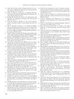
TABLE 56-1. Carbohydrate-Induced Symptoms in
Specific Situations
Symptoms Situations
Due to hypertonicity
Dumping syndrome Gastric surgery
Bloating Vagotomy
Nausea
Pylor
oplasty
Diarrhea Antrectomy
Flushing Gastrojejunostomy
Hypotension
Bariatric pr
ocedur
es
Dyspepsia after fr
uit juice
Gastr
oesophageal
r
eflux disease
Due to malabsorption or ingestion of poorly absorbed carbohydrates
Gas, bloating, diar
r
hea, pain
Generalized
malabsorption
Celiac disease
Short bowel syndrome
Specifi
c malabsorption
Fructose
Sucrose
Lactose
Poorly absorbed substances
Mannitol, sorbitol
Dietar
y fi
ber
Dietary-Induced Symptoms / 341
Sucrose is rarely malabsorbed, but some individuals have
an inherited defect in sucrase-isomaltase, the brush border
enzyme necessary for its absorption. Individuals with vil-
lous atrophy may have an acquired enzyme deficiency.
LACTOSE
There is a separate chapter on lactose intolerance (see
Chapter 62,“Lactose Intolerance”). The most common car-
bohydrate that is malabsorbed is lactose or milk sugar (Vesa
et al, 2000). Lactose is the primary carbohydrate in milk,
and all mammals depend on lactase activity in the intes-
tine to digest and absorb this substrate in infancy. Most
mammals retain lactase activity until weaning and then
turn off production of this enzyme, because milk is no
longer a part of the diet. Most human populations retain
lactase expression through adolescence and then become
lactase insufficient.
In some populations (particularly the individuals in the
Northern European gene pool), lactase activity is main-
tained into adulthood, but is typically lost gradually, pro-
ducing some degree of lactose intolerance with aging.
The degree of lactose intolerance is highly variable and
the development of symptoms depends not only on the
amount consumed (eg, 12.5 g per glass of milk), but also
on such factors as the amount of other fermentable sub-
strates ingested with the meal, coexisting mucosal disease,
and the rate of transit through the intestine. Lactose toler-
ance can be tested by assessing symptoms after ingestion
of 1 to 2 cups of milk (240 to 480 mL) or, more formally,
by breath hydrogen testing after a lactose load (typically
25 g). Use of milk that has been treated to hydrolyze the
lactose can reduce symptoms. In sensitive individuals, care
must also be taken with the ingestion of processed foods
that ha
v
e been fortified with nonfat dry milk to improve
their nutritional characteristics (Paige et al, 1975).
Fats
Fats also can produce a number of symptoms (Table 56-2).
Fat digestion is a complex process and the GI tract is orga-
nized to slow the movement of fats through the intestine to
allow sufficient time for fat digestion to occur. Thus fatty
meals slow gastric emptying and intestinal transit and, ulti-
mately, induce satiety and stop food intake. This is medi-
ated by duodenal receptors that recognize the presence of
fat within the lumen, cause the release of cholecystokinin
(CCK) into the blood, and set off neural reflexes. The con-
sequences of reduced gastric emptying may include exac-
erbation of gastroesophageal reflux, bloating, and early
s
atiety. However, as shown by Lin and colleagues (1999), fat
intolerance is associated with rapid gastric emptying.
Most fat is absorbed in the jejunum and fat entering the
lower ileum triggers the “ileal brake”by release of peptide YY,
which inhibits gastric emptying and proximal small bowel
transit. Patients who have had substantial ileal resections lack
this mechanism and may flood the colon with unabsorbed
nutrients after meals, producing diarrhea, gas, and cramps.
Dietary fat also has an effect on colonic motility. One of
the key activators of the gastrocolic reflex is fat entering the
duodenum. Because of this many patients with diarrhea have
bowel movements after meals and soon learn to restrict their
food intake to avoid diarrhea. This promotes development
of food aversions and weight loss in patients with chronic
diarrhea. An exaggerated gastrocolic reflex may also play a
role in postprandial urgency in patients with IBS.*
Proteins
Proteins are less likely than other macronutrients to cause
the kind of GI symptoms that we are discussing (nonim-
munologically mediated symptoms) because they are
ingested as large polymers that do not exert much osmotic
activity, and they are efficiently digested and absorbed by
the intestine. Some foods, however, contain
bioactive
amines
and peptides that may influence gut activity. An
example is coffee, which, in addition to caffeine, contains
dozens of
peptides that may influence gastric acid secretion
and other GI events. Amino acids stimulate CCK secretion
and may induce abdominal pain by stimulation of pan-
cr
eat
ic exocrine secretion if the pancreatic duct is struc-
turally or functionally obstructed.
Capsaisin, Caffeine, and Others
Other dietary components that may induce GI symptoms
include capsaisin, caffeine, and various minerals. Capsaisin
is the “active”ingredient in hot peppers and reacts with spe-
cific receptors in the mucosa that activate enteric sensory
nerves. The physiological “purpose”of these receptors is not
clear at the present time. Caffeine and other bioactive amines
have pharmacological effects when ingested in milligram
amounts. In addition to central nervous system effects, the
co-editor of this text and his colleagues have shown that caf-
feine can increase intestinal chloride secretion by inhibiting
phosphodiesterase, which may exaggerate diarrhea in
pat
ients with ileostomies and in those with IBS (Wald et al,
1976). Minerals such as calcium, aluminum, and iron tend
TABLE 56-2. Fat-Associated Symptoms and Situations
Symptoms Situations
Dyspepsia Gastroesophageal reflux disease
Bloating, early satiety Gastric surgery
Postprandial urgency of defecation Irritable bowel syndrome
Distension
Postprandial upper abdominal pain
*Editor’s Note: Patients learn to eat grilled chicken breast rather
than hamb
urg
e
r at fast food restaurants. (TMB)
342 / Advanced Therapy in Gastroenterology and Liver Disease
to be constipating, whereas magnesium may cause diarrhea.
Many patients ingest dietary supplements containing these
elements and may not be aware of their effects on bowel
function. Finally, many patients ingest “health foods”which
o
ften contain herbal products, including senna and aloe,
which can have profound effects on gut function. Every
patient needs to be asked about ingestion of these products.
There is a separate chapter on alternative medicines (Chapter
58, “Complementary and Alternative Medicines in
Gastrointestinal Disease”).*
Impact of Food Intolerances in Specific
Conditions
Symptoms in many GI conditions may be aggravated by
food intolerance. Several disorders in which dietary factors
should be explored by the physician are discussed below
and summarized in Table 56-3.
Functional Syndromes
Functional syndromes, such as IBS, FD, chronic diarrhea, and
chronic constipation are common disorders, affecting up to
20% of the US population. Although their pathophysiology
is gradually being unraveled, management is still based on
symptom control. Food intolerance may play an important
role in aggravating symptoms and should be probed.
Motility Disorders
M
otility disorders
,
such as gastroparesis and chronic intesti-
nal pseudo-obstruction, can also be affected by diet.
Although controlled clinical studies to prove efficacy have
not been conducted, dietary management is key to the long
term treatment of these conditions.
Post-Surgery Syndromes
Post-surgery syndromes are another fruitful area for dietary
therapy. No surgical intervention on the gut is without the
potential for disturbing function, and when the distur-
bance is severe enough to produce symptoms, careful
dietary management can improve matters substantially.
Conditions in which food intolerance may aggravate symp-
toms include postvagotomy or postgastrectomy dumping
syndrome, short bowel syndrome, ileostomy diarrhea,
postresection diarrhea, and ileoanal pouch dysfunction.
There are separate chapters on some of those situations.
Evaluation of Symptoms That May Be
Related to Food Ingestion
Many different symptoms may be due to food ingestion
and its consequences. These include abdominal pain in any
area, heartburn, bloating, nausea, abdominal distention,
*Editor’s Note: Garlic is widely touted as a health food. However,
there are numerous biologically active amines in garlic that are
used as vermifuges in animals and children. Some individuals,
including the aforementioned editor are, as adults, highly sensitive
to an alcohol soluble fraction of garlic with a cramping laxative
effect. (TMB)
TABLE 56-3. Potential Food Intolerance in Clinical Conditions
Condition Potential Food Intolerance Mechanism
Gastroesophageal reflux disease Hypertonic, carbohydrate-rich liquids Osmoreceptors
Fatty foods Delayed gastric emptying, reduced LES pressure
Gastroparesis Hypertonic beverages Delayed gastric emptying
Fatty foods Delayed gastric emptying
Raw fr
uits, vegetables
Impaired trituration
Dumping syndr
ome
Hypertonic carbohydrate Intestinal hormone release, osmotic fluid shifts
Functional dyspepsia
Many Gastric distention, delayed gastric emptying, abnormal gastric motility,
hypersensitivity to distention
Chronic intestinal pseudo-obstruction Fiber Potential substrate for bacterial overgrowth
Short bowel syndrome Caffeine Increased secretion, motility
Carbohydrate Fermentation
Fatty foods Accentuated gastrocolic reflex
Irritable bowel syndrome Carbohydrate (lactose, sorbitol, fructose) Osmotic diarrhea, fermentation
Fatty foods
Accentuated gastr
ocolic r
efl
ex
Chr
onic diarrhea (including functional,
postresection, and ileostomy diarrhea) Fatty foods Accentuated gastrocolic reflex
Chronic constipation Fiber Fermentation
Ileal pouch–anal anastomosis Carbohydrate (lactose, sorbitol, fructose) Osmotic diarrhea, fermentation
LES = lower esophageal sphincter
.
Dietary-Induced Symptoms / 343
gas, and diarrhea. The physician needs to establish the tim-
ing of the symptoms in relation to a meal and should try
to establish a link between specific foods and the present-
ing symptoms. This can be done best by a diet and symp-
t
om diary in which the temporal relation between ingestion
of certain foods and the onset of symptoms can be deter-
mined. However, bacterial fermentation of unabsorbed car-
bohydrates may occur many hours after ingestion.
Reproducibility of symptom induction by specific foods
should be the basis for trial of an elimination diet.
Registered dietitians can be a valuable help in sorting
through the patient’s history and recommending alterna-
tive diets.*
*Editor’s Note: The careful reader who employs the concepts nicely
demonstrated in this chapter will help a lot of patients obtain sig-
nificant relief. As stated in the first sentence of this chapter, many
patients do attribute some of their symptoms to “something they
ate!” (TMB)
Supplemental Reading
Burden S. Dietary treatment of irritable bowel syndrome: cur-
rent evidence and guidelines for future practice. J Hum Nutr
Diet 2001;14:231–41.
H
ammer HF, Fine KD, Santa Ana CA, et al. Carbohydrate mal-
absorption. Its measurement and its contribution to diarrhea.
J Clin Invest 1990;86:1936–44.
Hammer HF, Santa Ana CA, Schiller LR, Fordtrans JS. Studies
o
f osmotic diarrhea induced in normal subjects by ingestion
o
f polyethylene glycol and lactulose. J Clin Invest
1989;84:1056–62.
Lin HC, Van Citters GW, Zhao XT, Waxman A. Fat intolerance
depends on rapid gastric emptying. Dig Dis Sci 1999;44:330–5.
O
’Sullivan M, O’Morain C. Food intolerance: dietary treatments
in functional bowel disorders. Curr Treat Options
Gastroenterology 2003;6:339–45.
Paige DM, Bayless TM, Huang SS,Wextner R. Lactose hydrolyzed
milk. Am J Clin Nutr 1975;28:898–22.
Perman JA. Digestion and absorption of fruit juice carbohydrate.
J Am Coll Nutr 1996;15Suppl 5:12–17S.
Ravich WJ, Bayless TM, Thomas M. Fructose: incomplete intesti-
nal absorption in humans. Gastroenterology 1983;84:26–9.
Rumessen JJ, Gudmand-Hoyer E. Functional bowel disease: mal-
absorption and abdominal distress after ingestion of fructose,
sorbitol, and fructose–sorbitol mixtures. Gastroenterology
1988;95:694–700.
Vesa TH, Marteau P, Korpela R. Lactose intolerance. J Am Coll
Nutr 2000;19Suppl 2:165–75S.
Wald A, Back C, Bayless TM. Effect of caffeine on the human
small intestine. Gastroenterology 1976;71:738–42.
Gastrointestinal Food Allergy / 345
is the major cause of anaphylactic reactions in industrial-
ized societies including the United States, Australia, and
Europe. The prevalence of peanut allergy (0.5 to 7% of
adults in the United States and the United Kingdom) and
i
ts potentially fatal consequences has had significant effect
on the operational policies of groups ranging from school
districts to the airline industry. Fatal anaphylaxis can result
from exposure to minute amounts of antigen such as that
imparted by a kiss.
Food-associated exercise-induced ana-
phylaxis
is a rare type of anaphylaxis in which the food only
elicits an anaphylactic reaction when the subject exercises
within several hours of ingesting that food. Acetylsalicylic
acid can also augment type I allergic symptoms when com-
bined with food and exercise in such individuals.
Pollen-Food Allergy Syndrome
The oral allergy syndrome or pollen-food allergy syndrome
results from various plant proteins that cross-react with cer-
tain inhalant antigens, particularly birch, ragweed, and mug-
wort (Sloane and Sheffer, 2001). Exposure to the
cross-reacting foods may lead to pruritis, tingling and/or
swelling of the tongue, lips, palate, or oropharynx, and,
occasionally, to bronchospasm or more systemic reactions.
Foods that cross-react with birch include raw potatoes, car-
rots, celery, apples, pears, hazelnuts, and kiwi. Those indi-
viduals that are allergic to ragweed may react to fresh melons
and to bananas. It is important to educate patients with
inhalant allergies about potential cross-reacting foods.
Latex-Food Allergy Syndrome
Latex-food allergy syndrome, also referred to as the latex-fruit
syndrome,is a specific form of food allergy in which food anti-
gens cross-react with various latex antigens (Blanco, 2003).
Natural rubber latex contains over 200 proteins, 10 of which
bind IgE
Hevea brasiliensis latex protein allergens (HEV b 1 to
10) and cross-react with a variety of food antigens including
kiwi (HEV b 5), potato and tomato (HEV b 7), and avocado,
chestnut, and banana (HEV b 6). In latex-sensitive individu-
als exposure to these foods can result in the same symptoms
as if exposed to latex ranging from pruritis, eczema,oral-facial
swelling, asthma, GI complaints, and anaphylaxis. A large
number of studies from around the world indicate that the
natural rubber latex allergy is increasing in prevalence and
that the frequency of associated food allergy varies from 21 to
58% (Blanco, 2003). Worldwide, banana, avocado, chestnut
and kiwi are the most common causes of food-induced symp-
toms associated with latex allergy.
Other Immune-Mediated GI Adverse
Reactions to Food
Immunologic reactions to foods involving mechanisms
other than immediate hypersensitivity,such as cell-mediated
immunity (see Table 57-2), play a role in food protein-
induced enterocolitis syndromes (FPIES),such as cow’s milk
protein enteropathy, and also celiac disease. FPIES also known
as food protein-induced enteropathies, present in infancy or
e
arly childhood and are most commonly due to cow’s milk
protein followed by soy protein and less commonly, egg, fish,
and other food antigens (Nowak-Wegrzyn et al, 2003).
Clinical manifestations include diarrhea, vomiting, anemia,
bleeding, and failure to thrive. As with many other food aller-
gies, such cases are managed by elimination of the specific
food antigen until the disease resolves with age. It is com-
mon practice to switch infants with enterocolitis from a
cow’s milk-based formula to a
soy-protein derived formula,
but because over half will react to soy protein, continued
problems may result from the development of soy–protein-
induced enterocolitis.
Hypoallergenic or elemental feeds are
often necessary in such cases.
Celiac Disease
Celiac disease is one of the best-recognized diseases result-
ing from an immunologic reaction to food. Dietary inges-
tion of gliadin found in wheat, hordelein in rye, and secalin
on barley, induces an enteropathy in genetically suscepti-
ble individuals. Removal of the offending grains from the
diet restores normal small bowel function and appearance,
with improvement in symptoms that can range from diar-
rhea, weight loss, and failure to thrive, to the more com-
mon but less often recognized complaints of fatigue,
dyspepsia, neurological dysfunction, and musculoskeletal
problems.As with other immune-mediated ARF, elimina-
tion of the offending food substance (gluten) is the pri-
mary method of management in celiac disease. However,
unlike most other food protein-induced enteropathies,
g
l
uten must be eliminated from the diet on a lifelong basis
in celiac disease. See Chapter 61,“Celiac Sprue and Related
Problems” for a more complete discussion of celiac-sprue.
Eosinophilic Gastroenteritis
Food allergy is thought to play a role in some cases of
eosinophilic gastroenteritis, a relatively rare condition char-
acterized by eosinophilic infiltration of the gut and, often,
peripheral eosinophilia. Approximately half the patients
with eosinophilic gastroenteritis have atopic features,
including food allergy. Strategies to identify and eliminate
food antigens should be followed as in other food allergic
conditions, but often other measures, particularly corti-
costeroids, are necessary to manage patients with
eosinophilic gastroenteritis. Even after thorough evalua-
t
ion for parasites, an
e
mpiric course
o
f
ant
ihelminthic ther-
apy may be given before embarking on a course of
corticosteroids. Allergic eosinophilic esophagitis presents
in infancy thr
ough adolescence and manifests with symp-
toms of gastroesophageal reflux that are often refractory
346 / Advanced Therapy in Gastroenterology and Liver Disease
to typical antisecretory therapy (Hill et al, 2000). As with
lower GI presentations of allergic eosinophilic conditions,
this disorder is characterized by eosinophilic infiltration of
the mucosa, but also the histologic hallmarks of gastro-
e
sophageal reflux disease (GERD) and abnormal 24-hour
pH monitoring. Young children with this diagnosis usually
have a clinical and histologic benefit from eliminating spe-
cific foods.
Nonimmune Adverse Reactions to Food
The vast majority of ARF are not immunologic in origin
(see Table 57-1) and by virtue of their prevalence, are
important considerations in the examination of patients
complaining of ARF. Food toxicity or food poisoning
results from microbial contamination of food causing pri-
marily GI manifestations due to preformed toxins (eg,
staphylococcal enterotoxin) or replication of enteric
pathogens (
Campylobacter, Salmonella, Shigella, Escherichia
coli). These reactions can be distinguished from other ARF
because they usually do not recur and have fairly charac-
teristic presentations. Occasionally, a self-limited infection
may result in a postinfectious irritable bowel syndrome
(IBS). This is discussed in the chapter on IBS (see Chapter
39, “Irritable Bowel Syndrome”) and the chapter on trav-
eler’s diarrhea (see Chapter 50,“Traveler’s Diarrhea”).
Anaphylactoid or Pseudoallergic
Anaphylactoid or pseudoallergic reactions to food result
from foods that mimic the effects of mast cell degranula-
tion but do not involve IgE antibodies.
Strawberries and
shellfish may cause this type of ARF. Certain food ingredi-
ents, including additives such as
salicylates, benzoates, and
tar
t
razine
,
ind
uce pseudoallergic reactions. As with true
food allergy, patients exhibiting such reactions should be
instructed to avoid the offending food substance if iden-
tifiable. Pharmacological reactions to food or food addi-
tives represent a relatively common type of ARF, although
most of these reactions cause symptoms outside of the GI
tract. Histamine found in certain cheeses or in scrombroid
fish, such as tuna, can cause headaches and diffuse ery-
thema of the skin. Certain individuals develop migraine
headaches to various foodstuffs, including those rich in
amines. Sulfites, tartrazine and monosodium glutamate
(MSG) have all been associated with asthma, and MSG can
cause a characteristic syndrome consisting of a burning or
warm sensation, chest tightness, headache, and gastric dis-
c
omfort shortly after its ingestion.
Lactose Intolerance
Globally, lactose intolerance is the most common adverse
reaction to a specific food, with most cases the result of
declining levels of intestinal lactase activity in later childhood
and adult life, although rare congenital deficiencies can occur.
Symptoms of lactase insufficiency are usually dose related
and include bloating, flatulence, and diarrhea. Secondary lac-
tase deficiency can result from viral gastroenteritis, radiation
e
nteritis, Crohn’s disease (CD), and celiac sprue. It is impor-
tant from a management standpoint to understand that indi-
viduals with constitutive lactose intolerance (1) do not suffer
severe and potentially life-threatening complications of
ingesting lactose and (2) are able to consume naturally lac-
tose free diary products including most cheeses and yogurts.
This contrasts with cow’s milk allergic individuals who may
suffer anaphylactic or asthmatic reactions to dairy products
and must avoid all foods containing the culprit cow’s milk
protein allergen, usually casein or
β-lactoglobulin. There is a
chapter on carbohydrate intolerance (see Chapter 62,
“Lactose Intolerance”).
Psychological Reactions
In certain individuals, reactions to food may be psycho-
logical (Kelsay, 2003). This is a difficult type of ARF to diag-
nose because the mechanisms giving rise to such reactions
are poorly understood. Individuals who are not confirmed
to have ARF have higher rates of hypochondria, hysteria,
somatization, and anxiety than those with ARF confirmed
by food challenge. An individual who experienced a severe
ARF may avoid the culprit food for fear of further reac-
tions, and there is also some evidence that hypersensitiv-
ity reactions to food may be triggered through central
neural mechanisms so that, eventually, just the thought
of ingesting the food can trigger allergic symptoms in the
absence of antigen. Food allergy itself may lead to psycho-
logical distress, and studies of food allergic subjects report
an altered quality of life for the individual and their fam-
il
y
, with severe manifestations such as anaphylaxis result-
ing in a
post-traumatic stress situation.
Physiologic Reactions
Perhaps the most common form of ARF results from phys-
iologic reactions to food components or additives. It is well
known that starches found in legumes serve as substrate
for gas production by colonic flora and many other foods
are associated with “gas,” including onions, cabbage, bran
fiber, and other vegetables and grains. Certain foods and
food additives affect the lower esophageal sphincter,
whereas foods high in fat delay gastric emptying, resulting
in symptoms of heartburn and dyspepsia. These physio-
logic reactions to foods are typically noted by patients with
functional bowel disease, many of whom exhibit height-
e
ned endocrine, motor and sensory responses to normal
digestive events. Because elimination of the offending
food(s) may provide some benefit in select patients, it is
imp
ortant to determine whether specific food intolerances
exist in this group of patients. The reader is referred to
evaluation of food allergies have recently been published as
a medical position statement by the American Gastro-
enterological Association (Sampson et al, 2001). It is essen-
tial to obtain a careful history correlating symptoms with
s
pecific foods. Most immediate hypersensitivity reactions
to food include a set of symptoms that consistently occur
minutes to hours after ingesting certain foods. In some
individuals, other factors, such as medications or exercise,
may modulate the reaction to a specific food. Specificity
of the reaction does not always imply a food allergy because
patients with anaphylactoid reactions or lactose intolerance
report defined reactions to specific foods. However, the
nature of the reaction will help differentiate lactose intol-
erance (gas, bloating, diarrhea) from an allergy to cow’s milk
protein (often urticaria, swelling of the lips and oral mucosa,
and/or asthmatic symptoms occur in addition to GI symp-
toms).
Dairy, Elimination, Challenge
If a specific food or group of foods cannot be identified by
the initial history,the patient should keep a diet diary for sev-
eral weeks in an attempt to correlate foods with GI and other
symptoms. After certain foods are identified as possible cul-
prits by history or a diet diary, these items should be elimi-
nated from the diet for several weeks to determine the effect
on symptoms. If a benefit is seen, the patient may reintro-
duce the putative allergen(s) in an attempt to prove the asso-
ciation. Such open food challenges are subject to bias and
should be corroborated by another more objective method
before permanent elimination from the diet, particularly if
the patient is young and the food(s) in question represent a
major component of the diet (eg, eggs, milk, wheat). Skin
testing, in vitro testing and blinded oral challenges may be
he
lp
ful in this regard and are briefly discussed below.
T
ABLE 57-3.
Approach to Patient With Suspected F
ood
Allergies as a Cause of Gastrointestinal Symptoms
1.
Establish foods and food additives that r
epr
oducibly cause symptoms
•
Careful history
• Diet diary
•
Elimination diet
• Skin testing and/or RAST
• Food antigen challenge
2. Exclude and manage other disorders that may mimic GI food allergy
• CBC, peripheral blood eosinophil count
• Celiac serology
•
Lactose hydr
ogen br
eath test
• Stool studies
• Endoscopy and biopsy
3.
Initiate tr
eatment for food aller
gy
• Avoidance of specific foods
• Medications for after accidental exposure (antihistamines, epinephrine,
corticosteroids)
• Preventive measures (Oral cromoglycate, avoid co-precipitating factors,
eg, medications)
•
Education about hidden sour
ces of antigens and cross-reacting foods
CBC = complete blood count; GI = gastrointestinal; RAST = radioallergosorbent test.
Gastrointestinal Food Allergy / 347
Chapter 56,“Dietary-Induced Symptoms”for a further dis-
cussion of dietary-induced GI symptoms.
GI Disorders and ARF
Functional Disorders
It is human nature for patients with GI disorders to believe
that something in their diet has caused their condition even
in the absence of a history of food intolerance. A signifi-
cant number of GI conditions are associated with ARF but
food plays a causal role in only some of these disorders. For
patients with GERD, nonulcer dyspepsia, IBS, and other
functional conditions, nonspecific physiological reactions
to food can provoke symptoms. It is generally advisable
to instruct these patients to avoid foods that cause symp-
toms, but nondietary measures are usually also necessary
to manage their complaints. However, food protein intol-
erance or allergy may play a role in infants with GERD
symptoms. There is no generalized role for hypoallergenic
diets in IBS, although a few studies report benefit from such
diets (reviewed by Spanier et al, 2003) and, in some
instances, instituting a rigorous diet is helpful in convinc-
ing patients that specific dietary factors are not the sole
cause of their illness.
I
NFLAMMATORY BOWEL DISEASE
There are many studies that have examined the role of diet
in inflammatory bowel disease (IBD) but there is no evi-
dence that specific immune-mediated reactions to food
play a role in the majority of patients with either CD or
ulcerative colitis. Elemental enteral feeding and parenteral
nutrition can assist in the management of IBD patients
with benefits that appear related to improved nutrition and
bowel rest (and decreased fecal flow) rather than removal
of specific allergens from the diet. Patients in remission
should be encouraged to eat a nutritionally balanced diet
without restrictions unless they experience intolerance to
specific foods. It is typical for IBD patients to be instructed
to avoid dairy products but this is unnecessary in most
cases. Apart from those with symptomatic lactose intoler-
ance (in which case they should still be able to eat most
cheeses and yogurts) or rare instances of cow’s milk pro-
tein allergy, IBD patients should be encouraged to consume
dairy products because they are excellent sources of bio-
logically available calcium in a population at increased risk
of osteoporosis.
Approach to Patients
Complaining of ARF
A significant component of the difficulty in managing food
allergy is determining whether the patient has food allergy
or another form of ARF (Table 57-3). Guidelines for the
348 / Advanced Therapy in Gastroenterology and Liver Disease
Hypoallergenic or Elimination Diet
I
f specific foods are not identified by the clinical history or
a diet diary, a hypoallergenic or elimination diet, such as that
shown in Table 57-4, may be tried for 2 to 3 weeks. In most
cases of suspected GI adverse reactions to foods or food
additives, this approach is without benefit because the
majority of patients will have functional bowel disease with
nonspecific reactions to foods. In cases where a benefit is
seen, new foods are gradually introduced in an attempt to
identify specific foods that may contribute to the illness. It
should be recognized that the hypoallergenic diet can be
falsely interpreted as a negative test because a minority of
subjects can react to antigens contained in a typically
hypoallergenic diet.
Differential Diagnosis
It is important to consider the differential diagnosis of
patients who complain of food-associated GI complaints
because the majority will not have food allergy. The major
syndrome in which patients complain of adverse reactions
to foods is IBS, and other functional bowel presentations.
Lactose intolerance is the most common form of food intol-
erance worldwide and may coexist with other GI condi-
tions as well as food allergy. A complete medical history is
often helpful because most patients with a history of food
a
llergy have a
f
amily history of atopy
,
and may have a per-
sonal history of other allergic conditions, such as asthma
and dermatitis. A history of latex allergy should alert the
practitioner to the large number of fruits that can cross-
react with latex. Similarly, the
oral allergy syndrome occurs
in response to inhalant plant allergens, but cross-reactivity
with fruit, nut, and certain vegetable antigens is common.
Finally, it is well recognized that
exercise and medications
such as aspirin may act as cofactors in allergic reactions to
various types of antigens.
Tests for the Diagnosis and Management of
Food Allergy
Methods to detect food-specific IgE including prick skin
testing and measurements in blood are helpful in clinical
practice but standardized tests to detect non-IgE mediated
food allergy are not as well developed. Skin prick testing
provides a readily available and relatively inexpensive
means to assess a panel of food allergens in both children
and adults. The major limitation of skin testing is its poor
positive predictive value (many asymptomatic patients
exhibit reactions to food allergens) but a negative test in
the absence of antihistamine drugs strongly suggests that
immediate hypersensitivity is an unlikely mechanism for
the patient’s food-induced complaints. Skin testing is not
helpful in predicting who might outgrow their food aller-
gies, and, in fact, skin reactivity to foods can persist with-
out clinical manifestations while the individual goes on
to develop inhalant allergies. Although quite widely used
b
y v
arious practitioners, sublingual challenge or neuro-
muscular testing for food antigens are
not considered to be
scientifically acceptable methods to diagnose food allergy.
Blood Tests
A radioallergosorbent test (RAST) can be used as an alter-
native to skin testing in very young children, those with
severe atopic dermatitis, those who cannot discontinue
antihistamines, and those reporting anaphylactic reactions
to foods or food additives. The limitations of RAST are the
expense, lower sensitivity, and relatively limited number of
antigens that can be tested when compared with skin test-
ing. A modification of the traditional RAST test, the CAP
System FEIA (Pharmacia), is reported to be more sensitive
than a standard RAST. Levels of food-specific IgE above
w
hich a patient has a
> 95% lik
elihood of experiencing an
allergic reaction after the ingestion of specific food have
been established (Sampson, 2002).An oral food challenge
is r
ecommended at lower levels of food-specific IgE because
the clinical significance of such levels cannot be predicted.
TABLE 57-4. Elimination Diet
Food Category Allowed Avoid
Meat and meat Lamb Pork
alternatives Chicken Beef
Turkey Fish
Eggs
Milk and milk products
Seafood
Grains Rice Wheat
(Barley) Oats
Tapioca Corn
Arrowroot Rye
Legumes and nuts Avoid all dried peas,
beans, nuts
Vegetables All except corn and peas
Fruits All except citrus fruits,
strawber
ries, and tomatoes
Sweeteners
Sugar (cane or beet)
Maple syr
up
Honey
Fats Olive oil Soy, corn, peanut oils
Safflower oil Butter
Crisco
Mar
garine
Miscellaneous White vinegar Coffee, tea
Water Alcohol
(Ginger ale)
Chocolate
Salt
Colas
(Pepper) Spices
Fruit juices Chewing gum
Note: Also known as an exclusion or hypoaller
genic diet. Foods in brackets ( ) may cause adverse
reactions in some individuals and these may be omitted from the trial elimination diet. If an
allowed food is one that has caused a reaction in the past, it should also be omitted. While on
the trial elimination diet a record of symptoms is kept, and it is also noted if there are changes
from symptoms on the previous regular diet. If there are symptoms, the patient or family should
note if there is any relationship to specific foods.
In North America the Food Allergy and Anaphylaxis
Network (1-800-929-4040, <www.foodallergy.org>) is a
source of valuable information for those with various types
of food allergy. Similarly, it is important for celiac patients
t
o join local celiac disease foundations and support groups
that can provide valuable information used to determine
sources of gluten free foods and medications.
Infants with cow’s milk protein allergy present a unique
situation because avoidance of their major source of nutri-
tion poses difficulty in this age group. Formulas with
reduced antigenicity have been developed and include
those in which milk proteins are partially hydrolyzed by
heat or enzymes, as well as more extensively hydrolyzed
preparations. It is recommended that extensively rather
than partially hydrolyzed preparations are used for those
who are truly allergic to cow’s milk protein because only
the latter are truly hypoallergenic. For the 10% of infants
that still react to even the more hydrolyzed formulas, amino
acid based preparations should be used. For infants with
IgE-mediated cow’s milk allergy there is only a small chance
they will also be allergic to soy protein, whereas infants with
cow’s milk protein-induced enteropathy involving other
immune mechanisms have a
> 50% likelihood of devel-
oping soy protein-induced enterocolitis.
Anaphylactic Reactions
Because it is often difficult to prevent accidental exposure
to food antigens, patients with a history of an
anaphylac-
tic reaction
should be instructed to carry an epinephrine-
containing syringe for emergency administration. As
reactions may be biphasic in nature, patients must be
instructed to go to a local emergency facility even after con-
trol of the initial symptoms. Individuals who are at
incr
ease
d risk of anaphylaxis include those with a past his-
tory of anaphylaxis, those with reactions with respiratory
symptoms, those with episodes due the ingestion of
peanuts, tree nuts, fish or seafood, and those taking
β-blockers or angiotensin converting enzyme inhibitor
therapy. Antihistamines, ketotifen, oral cromolyn, leukotriene
antagonists and corticosteroids may modify symptoms to
food allergens but apart from first generation histamine
receptor antagonists, their efficacy in food allergic condi-
tions is largely unproven. Realizing that there are limited
studies available from which to make evidence-based deci-
sions, a trial of an orally administered mast cell stabiliz-
ing drug, sodium cromoglycate, 100 to 200 mg up to 4 times
daily, may be helpful in preventing episodes of allergy par-
t
icularly for patients who have to eat outside of their own
home and/or have multiple food alllergies.
Diet
ary Restrictions
D
ietary restrictions for food allergy associated with ana-
phylaxis and celiac disease should be maintained on a long
Gastrointestinal Food Allergy / 349
It is important to inform patients that unless there is clini-
cal evidence of adverse reactions to foods identified by skin
testing or in vitro methods, these foods do not need to be
eliminated from the diet in most instances.
Patch Testing
Diagnostic tests for non-IgE-mediated food allergies
include food allergy patch testing, T-cell cytokine assays,
and measurements of markers of eosinophil activation.
Conventional patch testing is used to diagnose contact
hypersensitivity reactions involving T cells and has been
applied to the evaluation of food allergy in the setting of
atopic dermatitis and allergic eosinophilic esophagitis, pri-
marily to cow’s milk proteins (De Boissieu et al, 2003).
Other tests may be useful in specific conditions, such as 24-
hour pH monitoring in eosinophilic esophagitis. Occult
parasitic infections should be excluded in order to diagnose
idiopathic or allergic eosinophilic syndromes and, occa-
sionally, a course of empiric antihelminthic therapy may
be indicated. Histological analysis is important in many
presentations of food allergy including eosinophilic
esophagitis, food protein-induced enterocolitis and proc-
tocolitis, and celiac disease.
Placebo Controlled Food Challenge
Because reactions to food antigens by RAST or skin test-
ing are neither specific nor sensitive, a double-blinded
placebo-controlled food challenge (DBPCFC), in which
food antigens are administered by
nasogastric tube or
gelatin capsules, should be performed if possible. This tech-
nique is considered the gold standard for diagnosing food
allergy but is not widely available. The DBPCFC is also less
reliable when assessing for delayed reactions to foods and
food additives. Clinical history and the results of skin test-
ing help guide the choice of foods to include in the oral
challenge. A number of investigators have performed the
GI equivalent of skin testing by
injecting the GI mucosa with
a panel of antigens and observing for a wheal-and-flare
response by endoscopy but this form of testing has not
been incorporated into routine clinical practice.
Treatment of Food Allergy
The cornerstone of the management of food allergy is
avoidance of the offending allergen. This is particularly
important in cases of peanut allergy where trace amounts
of allergen can cause significant reactions. Most fatalities
due to food allergy have been due to peanut allergy. Patients
with food allergies should learn to read and understand
labels for hidden food allergens and to recognize the poten-
tial for foods to cross-react with other antigens (eg, banana
and kiwi with latex, and b
irch pollen with apple, carrot, and
hazel nut
).
350 / Advanced Therapy in Gastroenterology and Liver Disease
term basis, whereas such measures can be lessened in other
types of food allergy that resolve with time, particularly those
presenting in early childhood. At one time it was thought
that unlike other food allergies, peanut allergy was not out-
grown. However, there are recent studies that indicate that
there may be as high as a 50% chance of outgrowing a peanut
allergy. As noted above, skin testing cannot be used to pre-
dict loss of clinical reactivity because skin tests may remain
positive in a child who no longer has clinical manifestations
of food allergy. Instead a decline in specific IgE levels fol-
lowed by a negative oral challenge provides a better index of
clinical loss of reactivity to a specific food antigen.
To date, there is no definite evidence that oral desensi-
tization, injection immunotherapy, or similar techniques
used for allergies to inhalant allergens, insect venoms, and
medications, are beneficial in the prevention or modula-
tion of food allergy. One exception to this is the
oral allergy
syndrome
in which desensitization to the pollen benefits not
only the symptoms of rhinitis but also food-induced oral
manifestations. Immunomodulation via oral, subcutaneous
and sublingual desensitization remain an area of contro-
versy and these techniques are not routinely recommended
in the management of food allergy.
Prevention of Food Allergy
The optimum means to prevent the development of aller-
gies in high risk individuals remains an area of controversy.
Recommendations have been made in the United States and
in Europe for infants with a strong family history of atopy
at risk of developing food and other allergies and include
the exclusive use of breastfeeding for at least 4 to 6 months,
delayed introduction of solid foods until after 4 to 6 months
of age, particularly allergenic foods such as egg, wheat, nuts,
and fish, avoidance of all CMP, and if formula is needed,
to use only extensively hydrolyzed or amino-acid based for-
mulas. Partially hydrolyzed cow’s milk, soy, and goat or
sheep milk products are not recommended. Hypoallergenic
diets have been recommended during pregnancy and with
breastfeeding for atopic mothers to reduce the incidence of
f
ood allergy in their offspring.
Probiotics offer another means to prevent the develop-
ment of food allergy. The rationale for using probiotics in
allergic diseases is that normal enteric flora established
shortly after birth provides counter regulatory signals
against a sustained T-helper type 2 cell (Th2)-skewed
immune response (Isolauri, 2002). A number of random-
iz
ed placebo controlled studies show that
L
actobacillus GG
(also called Lactobacillus rhamnosus [ATCC 53103]) given
to
women before and during subsequent breastfeeding reduced
the occurrence of allergic eczema in their offspring
. Other stud-
ies suggest that
probiotics s
uc
h as
L
ac
tobacillus GG
ma
y also
be beneficial in
ame
lior
at
ing the s
e
v
e
rity of allergic responses
in established food al
le
r
gy
par
t
icular
l
y in y
oung
e
r s
ubjects.
Newer Therapies for Food Allergy
B
iologic Therapy
Perhaps the most exciting developments in the field of food
a
llergy are new therapeutic approaches that modulate
immune responses to foods (Nowak-Wegrzyn, 2003).
These include tolerogenic peptides, recombinant epitopes,
anti-IgE and DNA vaccination, as well as administration of
Th1 type cytokines, such as interleukin (IL)-12 and inter-
feron-
γ, or strategies to antagonize the actions of Th2
cytokines, such as IL-4 and Il-5. The benefit of such
approaches in food allergy was recently documented in a
double blind randomized, placebo controlled, dose-ranging
trial, in which a humanized monoclonal IgG1 antibody
against IgE that recognizes and masks an epitope in the
CH3 region of IgE responsible for binding to the FcR
εI on
mast cells and basophils was administered subcutaneously
in peanut allergic subjects (Leung et al, 2003).A statistically
significant improvement (subjects increased their toler-
ance for peanuts from an average of 1.5 peanuts to 9
peanuts at one time) was seen between the highest dose
and placebo. The long term benefit and practical applica-
tion of this treatment is unknown but these initial results
are promising for the population who are at risk of poten-
tially fatal reactions from peanut allergy.
Modify Antigenic Structure
Methods to genetically or chemically modify the antigenic
structures of foods to reduce their allergic potential are also
being developed. For example, it is known that single amino
acid substitutions in the IgE binding site of a peanut aller-
gen can lead to the loss of binding to these epitopes.
Mutated
protein
or peptide immunotherapies are promising but
unp
r
oven strategies to induce desensitisation to food anti-
gens.
Traditional Chinese medicine (herbal) used for allergic
disorders has been shown to modulate the immune response
and to block anaphylaxis in a murine model of peanut
allergy suggesting that such treatments may be beneficial
in human food allergy. Other experimental therapies are
being directed to modifying the intestinal barrier so it is less
permeable to food and other types of antigens. Although all
these developments hold some promise for food allergy suf-
ferers, none are at a stage of development so as to signifi-
cantly impact the current way food allergy is treated.
Summary
ARF r
esulting in GI symptoms are common in the gen-
eral population and although only a minority of individ-
uals will have symptoms due to food allergy, GI food
al
lergies do exist in both children and adults. It is impor-
tant to recognize potential cases of food allergy in order
to correctly diagnose and manage the small subset of
pat
ients with immunologically mediated ARF. Potentially
fatal reactions to food necessitate careful instruction and
monitoring on the part of health care workers involved in
the care of individuals at risk of anaphylaxis. This is par-
ticularly true in westernized countries where the preva-
l
ence of allergy is increasing and food allergy is now the
major cause of anaphylaxis.
Supplemental Reading
American Gastroenterological Association Position Statement:
Guidelines for the Evaluation of Food Allergies. Gastroenterol
2001;120:1023–5.
Blanco, C. Latex-Fruit Syndrome. Curr Allergy Asthma Rep
2003;3:47–53.
Crowe SE, Perdue MH. Gastrointestinal food hypersensitivity:
Basic mechanisms of pathophysiology. Gastroenterol
1992;103:1075–v95.
De Boissieu D, Waguet JC, Dupont C. The atopy patch tests for
detection of cow’s milk allergy with digestive symptoms.
J Pediatr 2003;142:203–5.
Hill DJ, Heine RG, Cameron DJ, et al. Role of food protein intol-
erance in infants with persistent distress attributed to reflux
esophagitis. J Pediatr 2000;136:641–7.
Ingelfinger FJ, Lowell FC, Franklin W. Gastrointestinal allergy.
N Engl J Med 1949;241:303–40.
Isolauri E, Rautava S, Kalliomaki M, et al. Role of probiotics in food
hypersensitivity.Curr Opin Allergy Clin Immunol 2002;2:263–71.
Kelsay K. Psychological aspects of food allergy. Curr Allergy
A
sthma Rep 2003;3:41–6.
Leung DY, Sampson HA, Yunginger JW, et al. Effect of anti-IgE
therapy in patients with peanut allergy. N Engl J Med
2003;348:986–93.
N
owak-Wegrzyn A. Future approaches to food allergy. Pediatrics
2
003;111:1672–80.
Nowak-Wegrzyn A, Sampson HA, Wood RA, Sicherer SH. Food
protein-induced enterocolitis syndrome caused by solid food
proteins. Pediatrics 2003;111:829–35.
Sampson HA. Food allergy. J Allergy Clin Immunol 2003;111
(2 Suppl):S540–7.
Sampson HA. Improving in-vitro tests for the diagnosis of food
hypersensitivity.Curr Opin Allergy Clin Immunol 2002;2:257–61.
Sampson HA, Sicherer SH, Birnbaum AH.AGA technical review
on the evaluation of food allergy in gastrointestinal disorders.
Gastroenterol 2001;120:1026–40.
Sicherer SH. Food Allergy. Lancet 2002;360:701–10.
Sloane D, Sheffer A. Oral allergy syndrome. Allergy Asthma Proc
2001;22:321–5.
Spanier JA, Howden CW, Jones MP. A systematic review of alter-
native therapies in the irritable bowel syndrome. Arch Intern
Med 2003;163:265–74.
Gastrointestinal Food Allergy / 351
Complementary and Alternative Medicine in Gastrointestinal Disease / 353
early during the course of their disease (Astin, 1998).
Others may be comfortable with conventional medicine
and only seek out CAM when they believe they have seen
the limitations of conventional medicine.
I
n general, patients do
n
ot
a
bandon conventional medi-
cine in favor of complementary therapies. Instead they tend
to use both, often hoping that there will be a synergistic effect
or that the complementary therapy will ameliorate or pre-
vent side effects from the conventional medicine.
Patients report obtaining a number of benefits through
their use of CAM. One of the most common benefits is a
greater sense of being in control of their disease. They also
frequently feel that by using a complementary therapy they
have taken a more active role in the management of their
disease. These benefits may overshadow any improvement
in their symptoms resulting from their use of CAM.
Therefore, a patient may not appreciate any improvement
in their disease but still be satisfied and report higher qual-
ity of life with their use of CAM. Therefore, CAM use can
be considered a
coping strategy used by those with chronic
diseases. It is important for the gastroenterologist to under-
stand this because it helps explain why rational patients
demonstrate, what is from the gastroenterologist’s per-
spective, an irrational health behavior—the use of an
unproven therapy. Understanding patients’ use of CAM
requires looking beyond symptoms to the impact the dis-
ease and its treatment has on every aspect of patients’ lives.
We have also found that the degree of satisfaction with
CAM is partly associated with the patient’s
health beliefs
(Hilsden et al, 1999). Patients whose health beliefs were
more congruent with CAM are more likely to report a high
degree of satisfaction with the CAM they used than
patients whose health beliefs were more congruent with
conventional medicine.
Patients frequently
exclude their gastroenterologist when
deciding to use a CAM and then often do not inform them
about using it (Hilsden and Verhoef, 1998). Patients often
indicate that they withhold this information because they
are afraid of their physician rejecting their use of CAM or
because they do not see their physician as being know-
ledgeable about these therapies.
In summary, patients commonly use CAM as part of
the treatment of their disease in combination with their
conventional medicines, they often do so because of prob-
lems they have had with their conventional treatments, and
they frequently do not include their physician in the deci-
sion making process.
Efficacy and Safety of CAM
One of the main problems facing both patients and
physicians is the lack of information on the safety and
efficacy of CAM. In fact, we found that GI patients rated
CAM as one of their most important information needs.
Gastroenterologists are well aware of the potential for
severe hepatotoxicity from some herbal products. However,
in general, there are no major safety concerns with the
common forms of therapy (herbs and nutritional supple-
ments) used by GI patients. Potential risks include allergic
r
eactions, contamination or mislabeling of herbal prod-
ucts, nutritional deficiencies resulting from restrictive diets,
and neck and spine injury resulting from spinal manipu-
lation. However, physicians and patients should be aware
that some therapies are associated with the risk of serious
side effects due to the therapy’s chemical constituents (eg,
hepatic veno-occlusive disease from herbs such as comfrey
that contain pyrrolizidine alkaloids), contamination with
heavy metals (reported with some medicines prepared in
Asia), and the potential risk for toxicity to the fetus.
The potential for interactions between complementary
and conventional medicines exists, but is poorly docu-
mented for most therapies (Crone and Wise, 1998). Many
herbs can affect the absorption or metabolism of conven-
tional medicines. Patients on immunosuppressants or
other medications with a narrow therapeutic window
should be especially careful.
Many complementary therapies based on traditional
healing practices have a rich folk history supporting their
use, however there is little, if any, direct scientific evidence
supporting the benefits of most forms of CAM. Much of
the evidence that patients and physicians have access to is
anecdotal. Some controlled trials of specific therapies have
been conducted but these are often reported in journals
unfamiliar to practicing physicians, are flawed, and exam-
ine treatments not widely used or available. Conducting
randomized controlled trials of some forms of CAM are
methodologically difficult due to the highly individualized
nature of the therapies, lack of placebos, patient and
provider preferences, and different beliefs about health and
disease (Hilsden and Verhoef, 1998). There is a body of
ethnopharmacology and basic science research on some
herbal products that support a possible role in the treat-
ment of IBD. Systematic reviews of some therapies used
for common GI conditions are available (Spainer et al,
2003; Jacobs et al, 2002).
Approach to the Patient Using or Wishing to Use a
Complementary Therapy
Counseling patients about CAM use is important and can
help the patient make a more informed choice. To be effec-
tive, it must be done in a sensitive and nonjudgmental fash-
ion. As with any attempt to modify health behavior,
g
astroenterologists should avoid an authoritative “advice-
giving” or direct persuasion approach because this can
push the patient into a more resistant and defensive posi-
t
ion. This is not to say that physicians must agree with their
patients use of CAM. In our experience, patients often rec-
ognize that they are obtaining only one side of the story
fr
om those promoting a complementary therapy. However,
354 / Advanced Therapy in Gastroenterology and Liver Disease
they want more than just a “No, don’t use it”from their gas-
troenterologist. They value their gastroenterologist as an
information source and want an open discussion of the
potential value or risks associated with a therapy, even if ulti-
m
ately the gastroenterologist disagrees with their use of it.
Eisenberg (1997) has written a valuable article on advis-
ing patients who seek alternative medical therapies. It is
directed towards the patient who is seeking care from a
complementary practitioner. Even though we find that
most patients self-treat with complementary therapies
rather than see a complementary practitioner, Eisenberg’s
guidelines are still appropriate. Below are the steps that one
of us (RJH) uses when counseling a patient.
1. Document CAM use
Determining current and past use of CAM should
be part of the routine medical history for all
patients. There are several reasons for doing so.
First, use of a CAM may be an indication that the
patient is dissatisfied with their current treatment
either because they are not achieving the benefits
they desire or because they are suffering side effects.
Second, the use of potentially dangerous therapies
can be discovered. Third, potential drug–CAM
interactions can be anticipated. Finally, the effects
of the CAM, either good or bad, will not be mis-
construed as resulting from a conventional treat-
ment.
Patients, however, are reluctant to reveal their use
of CAM especially if they view their gastroenterol-
ogist as being intolerant or uninformed. Therefore,
this is not the correct situation to use terms such as
quakery, fraudulent, or unconventional therapies. I
routinely ask the patient whether they (1) use herbal
or natural therapies, (2) have made any dietary
changes, and (3) use any other therapies for their
condition or general health.
2. Determine reasons for seeking CAM
A patient who is using or considering using CAM
should be asked about their reasons for doing so.
This is important because determining specific areas
of dissatisfaction with their conventional treatment
could allow modifications to be made. Again care-
ful questioning is required, as the patient may be ret-
icent to reveal issues that they feel may be perceived
as criticism by their gastroenterologist. Open-ended
questions such as “What do you see as the potential
benefits of using this therapy” and “Do you have any
concerns about your current treatment that is lead-
ing you to consider this new therapy” allow the
patient to openly discuss their perspective on their
treatment. It is also valuable to obtain some sense of
the patient’s health beliefs. If the patient is a firm
believer in the principles of complementary medi-
cine, then it is unlikely that they will be convinced
not to use one. If on the other hand the patient is
more comfortable with conventional medicine but
is seeking alternatives because they are experiencing
problems, then they may be willing to first try a
m
odification in their conventional medical treat-
ment.
3. Explore the patient’s knowledge and source of infor-
mation about CAM
Many sources of information available to the
patient, for example books and Internet sites, pro-
vide an overly optimistic and one-sided account of
the effectiveness of a therapy and are often based
only on testimonials. Often information about
safety is not provided. The patients understanding
of how the therapy works and its potential bene-
fits and harms should be determined. Some patients
have very realistic expectations. They may under-
stand that their chance of obtaining some benefit is
low but they are willing to try it on the off chance
that they do benefit. However, many patients have
unrealistic expectations and expect a quick cure. In
the short time available for counseling a patient, it
is impossible and impractical to teach them the
principles of scientific medicine and the random-
ized controlled trial. However, the patient should
be encouraged to define realistic treatment goals
and to reevaluate their use of a therapy after a set
period of time.
Many patients often believe that CAM is without
risk, often because they are “natural”therapies. This
belief is often promoted by advertisements for the
therapy. Therefore, the patient may not have con-
sidered the possibility of side effects. Physicians
should ask patients whether they know the possible
side effects of a therapy and should warn patients
about the possibility of interactions with alcohol or
other drugs. The patient and the physician may have
difficulty finding any specific information about the
risks of these with a given product. Patients should
be encouraged to think of any therapy in terms of
a trade-off between potential benefits and potential
risks. Often patients think more about the poten-
tial benefits and neglect to consider whether they
are willing to incur the risks of a complementary
therapy, including its cost.
4. Determine how the patient will obtain and use the
therapy
If the patient is seeing or will be seeing an alterna-
tive practitioner, the gastroenterologist can provide
the pat
ient with questions they should ask the prac-
titioner. These would include (1) is the practitioner
licensed and what was their training, (2) how expe-
r
ienced are they in treating patients with IBD, (3)
what can the patient expect from the treatment in
apies used in complementary medicine, and (3) diagnostic
t
echniques used in complementary medicine. Within the
symptoms and disorders section, therapies used for a variety
of medical conditions are discussed. There is a section on GI
conditions, although neither ulcerative colitis nor Crohn’s dis-
e
ase is specifically included. Each therapy is given a rating as
to the likelihood of achieving a therapeutic benefit and poten-
tial risks are listed. The level of evidence supporting any ben-
efits (ie, case reports, clinical trials) is given, but unfortunately
no references are provided.
P
rofessionals Handbook of Complementary and Alternative Medicines
(C. W. Feltrow and J. R. Avila, Springhouse Publishers). This
book describes the chemical components, actions, reported
uses, and suggested doses of many herbs and alternative med-
icines. The authors also list potential adverse events and drug
interactions. The book is less critical of claims made about the
efficacy of the treatments than the above two books.
Side Effects/Drug Interventions
Herb Contraindications and Drug Interactions (F. Brinker, Eclectic
Medical Publications). This book describes known and spec-
ulated side effects, contraindications, and drug interactions
of herbs. The book is well referenced and indexed.
Web Sites
National Center for Complementary and Alternative Medicine
< of National Institutes
of Health that has the goals of supporting rigorous research on
CAM, training researchers in CAM, and disseminating infor-
mation to the public, and professionals on which CAM modal-
ities work, which do not, and why. The Web site includes several
systematic reviews of various therapies for GI conditions.
The Research Council for Complementary Medicine <www.rccm.
org.uk>. Includes a centralized information service on com-
plementary medicine with links to clinical trials and Cochrane
reviews.
Quackwatch <www.quackwatch.com> Described as “Your guide
to health fraud, quackery and intelligent decisions.”
Supplemental Reading
Astin JA. Why patients use alternative medicine: results of a
natio
nal st
ud
y
. JAMA 1998;279:1548–53.
Crone CC, Wise TN. Use of herbal medicines among consulta-
tion-liason populations. Psychosomatics 1998;39:313.
E
isenberg DM. Advising patients who seek alternative medical
therapies. Ann Intern Med 1997;127:61–9.
Er
nst E. Prevalence of use of complementary/alternative medicine:
a systematic review. Bull World Health Organ 2000;78:252–7.
Giese LA. A study of alternative health care use for gastrointesti-
nal disorders. Gastroenterol Nurs 2000;23:19–27.
H
a
y
d
e
n CW, Bernstein CN, Hall RA, et al. Usage of supplemental
alt
ernative medicine by community-based patients with gas-
troesophageal reflux disease (GERD). Dig Dis Sci 2002;47:1–8.
Hilsden RJ, Scott CM,Verhoef MJ. Complementary medicine use
by patients with inflammatory bowel disease. Am J
Gast
roenterol 1998;93:697–701.
Complementary and Alternative Medicine in Gastrointestinal Disease / 355
terms of benefits and side effects, (4) what is the
basis for these expectations, and (5) what will be the
cost of the treatment.
Patients should not start a number of different ther-
a
pies, especially a combination of conventional and
complementary therapies, at the same time. If this
is done, it will be impossible to determine which
therapy resulted in any benefits or side effects. Some
method for monitoring for side effects should be
agreed upon. This will depend upon the potential
risks associated with a therapy.
Information Sources About CAM
There are a variety of valuable information sources avail-
able for physicians, although none of them are specifically
focused on GI disease. Patients should not rely too heav-
ily on the advice or recommendations provided by employ-
ees of health food stores or other stores selling herbal and
nutritional supplements (Verhoef et al, 2002). Often only
the owner or the manager of the store has much experi-
ence and knowledge about the therapies. Other employees
may have relatively little training and may not be able to
distinguish between treatments that are safe and appro-
priate for a given condition and those more commonly
used for other GI complaints. General intestinal remedies
sold at health food stores often contain laxatives.
Below are several sources of information on CAM that
we find useful. We have chosen these because they critically
review therapies and provide supporting evidence for any
claims made.All of them are relatively inexpensive (at least
compared to medical textbooks).
In conclusion, CAM is not likely to disappear in the near
future. Patients will continue to incorporate it into their
health car
e,
and certain therapies such as probiotics, will
be incorporated into conventional medicine if their effi-
cacy is demonstrated. Physicians will need to address the
questions and concerns of patients using CAM and will
need to be able to safely manage patients using conven-
tional and complementary therapies concomitantly.
General Sources
The American Pharmaceutical Association Practical Guide to
N
atural Medicines
(A.
Pierce). Reference book directed at
potential users of natural products, includes sections on
assessing saf
e
ty and efficacy.
The Honest Herbal (V. E. Taylor PhD, Pharmaceutical Products
Press). This comprehensive book provides a wealth of critical
and referenced information on many herbs. The author
describes the putat
ive active ingredients, recommended uses
and supporting evidence of efficacy for each herb, and also
debunks unwarranted claims.
The Complete Book of Symptoms and Treatments (E. Ernst, Editor,
Element Books Limited). This book is divided into the fol-
lowing three sections: (1) symptoms and disorders, (2) ther-
356 / Advanced Therapy in Gastroenterology and Liver Disease
H
ilsden RJ, Meddings JB, Verhoef MJ. Complementary and alter-
native medicine use by patients with inflammatory bowel dis-
ease: an internet survey. Can J Gastroenterol 1999;13:327–32.
Hilsden RJ,Verhoef MJ. Complementary and alternative medi-
c
ine: evaluating its effectiveness in inflammatory bowel dis-
e
ase. Inflamm Bowel Dis 1998;4:318–23.
Hilsden RJ,Verhoef MJ, Best A, Pocobelli G. Complementary and
alternative medicine use by Canadian patients with inflam-
matory bowel disease: results from a national survey. Am J
G
astroenterol 2003;98:1563–8.
J
acobs BP, Dennehy C, Ramirez G, et al. Milk thistle for the treat-
ment of liver disease: a systematic review and meta-analysis.
Am J Med 2002;113:506–15.
Spanier JA, Howden CW, Jones MP. A systematic review of alter-
n
ative therapies in the irritable bowel syndrome [review]. Arch
I
ntern Med 2003;163:265–74.
Verhoef MJ, Rapchuk I, Liew T, et al. Complementary practi-
tioners’ views of treatment for inflammatory bowel disease.
Can J Gastroenterol 2002;16:95–100.
357
CHAPTER 59
OBSCURE GASTROINTESTINAL BLEEDINGS
ANDREW I. SABLE,MD,AND JAMIE S. BARKIN, MD, FACP, MACG
Obscure gastrointestinal bleeding (OGIB) is defined as
bleeding of unknown origin that persists or recurs after
negative initial or primary endoscopy, including
colonoscopy and upper endoscopy. It poses a profound
diagnostic and therapeutic challenge for gastroenterolo-
gists and surgeons alike because these patients often have
recurrent bleeding and use a plethora of health care
resources. Bleeding may arise from virtually any location
within the gastrointestinal (GI) tract and patients present
with great variability, from chronic occult bleeding to
acute bleeding with visible blood loss. Either of these pre-
sentations may result in iron deficiency anemia (IDA)
and necessitate blood transfusion.
OGIB is categorized into the following two clinically
distinct entities: (1)
obscure-occult, which is characterized
by IDA and/or recurrent positive fecal occult blood test
(FOBT), and (2)
obscure-overt, in which recurrent bleed-
ing is clinically evident by the presence of melena,
maroon stools, or hematochezia (Zuckerman et al, 2000).
There is little data regarding the frequency and natural
history of OGIB. Despite timely upper endoscopy and
colonoscopy, bleeding remains unexplained in approxi-
mately 5% of patients (Hayat et al, 2000). Failure to iden-
tify a bleeding source at the time of endoscopy may be the
result of the following:
1. Overlooked lesions, such as nonbleeding lesions or
those obscured by the presence of blood or thickened
gastric folds
2. Lesions beyond the reach of traditional endoscopes
(ie, third portion of duodenum)
3. Difficult to diagnose lesions (ie, Dieulafoy’s malfor-
mation or gastric antral vascular ectasias (GAVE)
4. The discovery of an equivocal finding at the time of
endoscopy that may or may not be the bleeding
source
Colonoscopy with ileal intubation and upper
endoscopy are requisite in the initial evaluation of
patients with OGIB. Repeated bidirectional endoscopy
may be both indicated and necessary before a diagnosis is
made. A second or third endoscopic look may identify
lesions that are commonly missed (ie, Cameron’s erosions
and arteriovenous malformations) and/or difficult to
identify (ie, Dieulafoy’s disease and celiac sprue). If bleed-
ing continues and the source remains unidentified, fur-
ther evaluation should be directed to the small bowel, a
rare but important source of blood loss and the over-
whelming location of bleeding of obscure origin (Lahoti
and Fukami, 1999).
Examination of the small bowel was previously limit-
ed by the poor application of conventional studies. There
is little use for radioisotope bleeding scans and angiogra-
phy in OGIB of occult origin. Small bowel series and
enteroclysis generally have a low diagnostic yield for
lesions that commonly cause OGIB (ie, vascular ectasia
and ulcerations). With the advent of wireless capsule
endoscopy (WCE) direct, noninvasive examination of the
entire small bowel is possible thus enabling the identifica-
tion of clinically relevant lesions as well as determining
their approximate location. This chapter will discuss our
approach to OGIB with attention to the small bowel.
Approach to Diagnosis of Patients with
OGIB
The identification of a bleeding source begins with a thor-
ough history and physical examination. Pharmacologic
agents, including aspirin and nonsteroidal anti-
iflammatory drugs (NSAIDs), which are cyclooxygenase-
1-selective inhibitor sparing and nonselective, are toxic to
the intestinal mucosa and predispose to ulceration and
mucosal bleeding. The pathogenesis, although complex, is
well established. In addition to the suppression of
prostaglandins, there is likely local, topical mucosal injury.
These toxic effects, although well described in the
stomach and duodenum, are now known to also occur in
the colon (ie, NSAID-induced colitis) and small intestine
(ie, erosions, ulcerations, and webs) and at a higher fre-
quency compared with controls than previously known
(58% small bowel lesions in NSAID users versus 17% in
nonusers) (Graham et al, 2003). Small bowel and colonic
injury may occur independently of symptoms of gastro-
d
uodenal irritation. The identification of NSAID intake is
fundamental in the management of patients with bleed-
ing of obscure origin. There is a separate chapter
(C
hapter 60, “Nonsteroidal Antiinflammatory Drug-
Induced Small and large Intestinaal Injury”) on NSAID-
induced injury to the small and large intestine.
A family history may reveal hereditary disorders
resulting in OGIB, including hereditary hemorrhagic
telangectasias (HHT), Osler-Weber-Rendu disease
(OWR) and polyposis syndromes. Patients with previous
a
ortic aneurysm repair
s
hould have mandatory examina-
tion of the third portion of the duodenum to evaluate for
the presence of aortoenteric fistula. Patients with easy
bruisibility or other clinical manifestations suggesting a
coagulation disorder should be examined with a coagula-
tion profile. In addition, patients with aortic stenosis
acquire defects in von Willebrand’s factor.
The clinical pattern of blood loss may help localize
bleeding. Hematemasis, although a rare presentation of
OGIB, may help to localize bleeding proximal to the liga-
ment of Treitz. Stool color is less helpful in predicting the
site of blood loss because it is primarily a function of the
transit time of the blood bolus (Hilsman, 1950). Patients
with slow oozing from the distal small bowel and proxi-
mal colon can have melena, whereas those with brisk
blood loss from the proximal intestine often present with
hematochezia and hypotension, but without
hematemasis or bloody nasogastric aspirate. The details
of the history and physical examination may be helpful in
providing clues to a source of bleeding as depicted in
Table 59-1
.
The frequency of upper intestinal lesions in patients
with positive FOBT is reportedly as high as 75% (Geller
et al, 1993). Furthermore, of patients with obscure-overt
bleeding approximately 50% will have identifiable lesions
within the reach of a standard gastroscope (Jensen, 2003).
Repeat upper endoscopy may therefore, in many cases,
identify missed lesions not seen on initial evaluation.
Specific lesions presenting with OGIB, occult or overt,
that may be missed initially if they are not actively bleed-
ing are in Table 59-2.
Coagulation Studies
The determination of coagulation parameters in patients
with OGIB is necessary. Abnormalities in the bleeding time
and partial thromboplastin time may reflect von
Willebrand’s disease (vWD). Whereas prolonged interna-
tional normalized ratio (INR) may reflect advanced liver
disease, disseminated intravascular coagulation, or surrepti-
tious anticoagulation intake. Abnormal platelet function
test (prolonged bleeding time) may indicate acetylsalicyclic
acid (ASA) and/or NSAID use. Hemorrhagic tendency in
vWD is variable and dependent on the type and severity of
disease.
Patients with Types 1 and 2 vWD may have mild,
occult bleeding associated with IDA. Type 3 vWD disease is
associated with telangiectasis of the small and large bowel
and may present with severe obscure-overt bleeding. vWD
may be acquired in individuals who were previously normal
and is associated with mitral valve prolapse and aortic steno-
sis (Vincentelli et al, 2003; Heyde, 1958; Mant et al, 1968;
Mannucci et al, 1973). Uremia in patients with acute renal
failure or chronic renal insufficiency who have OGIB may
indicate a need for vasopressin to correct platelet dysfunc-
tion, whereas plasma infusion and/or vitamin K supplemen-
tation may be required in patients with acute or chronic liver
failure. Additionally, hospitalized patients on broad spec-
trum antibiotics may develop vitamin K deficiency and sup-
plementation may be helpful in the event of bleeding.
Diagnostic Modalities
Radiologic Procedures
SMALL BOWEL SERIES/ENTEROCLYSIS
The overall yield of barium examination of the small bowel
is extremely low. These techniques are employed after neg-
at
ive enteroscopy or when enteroscopy is not immediately
3
58
/
Advanced Therapy in Gastroenterology and Liver Disease
TABLE 59-1. Clues to Diagnosis
History or Physical Finding Cause of Bleeding
GERD Cameron’s erosion/ulcers
Abdominal pain Tumor/ischemia
Hx of pancreatic injury/pancreatitis Hemosuccus pancreaticus
RUQ surgery/injury Hemobilia
Arthritis/NSAIDs SB/colon ulceration/colitis
Hx of radiation Neovascularization
Surgery Anastomotic
Chronic renal insufficiency Vascular ectasia
Abdominal aortic aneurysm repair Aorto-enteric fistula
Diarrhea CD
Liver disease/spider angiomata Gastro/duodenal/rectal varices
Connective tissue disorder GAVE
Age < 40 years Meckel’s diverticulum
Aortic stenosis/MVP Acquired von Willebrand’s factor defect
CD = Crohn’s disease; GAVE = gastric antral vascular ectasias; GERD = gastroesophageal reflux
disease; Hx = medical histor
y; MVP = mitral valve prolapse; NSAID = nonsteroidal anti-inflam-
matory drug; SB = small bowel.
TABLE 59-2. Proximal Intestinal Lesions Causing
Obscure Gastrointestinal Bleeding
Esophagus Esophagitis/ulcers
Mallory-Weiss tear
Cameron’s erosions/ulcer
Stomach AVM
Dieulafoy’s malformation
GAVE
PHG
Duodenum Dieulafoy’s malformation
AVM
CD
Hemobilia
Hemosuccus Pancreaticus
Aorto-enteric fistula
Adapted from Mujica and Barkin, 1996.
AVM = arteriovenous malformations; CD = Crohn’s disease, GAVE = gastric antral vascular
ectasias; PHG = portal hypertensive gastropathy.
endoscopy should only undergo small bowel examination
with enteroclysis or SBFT to effectively rule out signifi-
cant lesions, particularly if a response to oral iron replace-
ment therapy was observed (Rockey and Cello, 1993.).
H
owever, in our view, this was similar to treating an auto-
mobile oil leak with oil replacement only, rather than fix-
ing its source. In clinical practice, an occult malignancy
may be missed and/or a bleeding lesion progress causing
increased morbidity and mortality. Patients with OGIB
and comorbid disease, and/or those who require blood
transfusions, should certainly be subject to a more exten-
sive evaluation. Our current algorithmic approach to
patients with OGIB is outlined in Figure 59-1.
After they have undergone negative upper endoscopy
and colonoscopy with ileoscopy (repeated if initially nega-
tive), enteroscopy, preferably performed with use of an
overtube, is our standard approach (O’Loughlin and
Barkin, 2004). Enteroscopes vary in length from 220 to
250 cm and with the use of fluoroscopy and an overtube,
they can generally reach to a depth of 100 to 110 cm beyond
the ligament of Treitz. However a physician’s choice of
instrument and technique (pediatric colonoscopes/use of
available. In the absence of obstructive symptoms, small
bowel follow-through (SBFT) leads to a diagnosis in about
5% of patients with OGIB. The yield with enteroclysis,
although better (about 10%), is still minimal, because
t
hese methods are inadequate for detecting mucosal
lesions like
vascular ectasia, which are overwhelmingly the
most frequent cause for small bowel bleeding. Comparison
of WCE with small bowel enteroclysis in patients with
OGIB has shown the superiority of WCE for the detection
of lesions (Liangpunsakul et al, 2003).
R
ADIONUCLEOTIDE STUDIES
Scintigraphy with technetium-labeled red blood cells may
help to confirm hemorrhage that may be originating in
the small bowel; however, it does not accurately locate the
site of bleeding. Despite high sensitivity for detection of
bleeding (positive if bleeding is less than or equal to
0.1 mL/min) (Alavi, 1982), its low specificity limits its use-
fulness. Its role, therefore, continues to be controversial.
Delayed scans, performed at 12 to 24 hours postinjection,
may be misleading by identifying luminal blood that is
pooled at sites other than the bleeding source. The
Meckel’s scan is based on an isotope labeled compound
that localizes in the ectopic gastric mucosa found in
Meckel’s diverticulum. As this isotope normally accumu-
lates in the stomach and bladder, both should be empty at
the time of examination to increase its diagnostic yield.
Increased sensitivity is achieved by using histamine-2
receptor antagonists, which causes increased activity in
the ectopic parietal cells. This test has a more specific role
in the examination of patients with OGIB who are below
the age of 40 years.
A
NGIO
GR
APHY
Selective mesenteric angiography is expensive and inva-
sive, yet offers both diagnostic and therapeutic modalities.
Although it is not as sensitive to low rate or intermittent
bleeding as bleeding scans (rate 0.5 to 1 mL/min), angiog-
raphy has the potential ability to localize and treat bleed-
ing lesions. Provocative maneuvers performed at the time
of exam include the use of anticoagulants and/or vasodila-
tors, both of which may precipitate bleeding and improve
the diagnostic yield of angiography. The large arcade of
mesenteric vasculature makes identification of smaller
vascular ectasia difficult. The use of nuclear scans to select
those actively bleeding patients who will undergo angiog-
raphy is controversial but may improve diagnostic yield
and lower overall cost by avoiding unnecessary exams.
Enteroscopy
In the past it was believed that asymptomatic patients
with obscure-occult bleeding over 60 years of age who
have undergone negative colonoscopy and upper
Obscure Gastrointestinal Bleedings / 359
History/physical (exclude NSAIDS)
Repeat EGD/Colonoscopy with Ileoscopy
Treat
Push Enteroscopy with overtube
Treat
No alarm symptoms or
contraindications
Alarm symptoms: abd
pain, symptoms of
subacute obstruction
Small bowel
follow-through
Wireless Capsule
Endoscopy
CD/Ulcerations:
Medical treatment, biopsy
AVMs:
Endoscopic and/or
pharmacologic tx
(hormones)
Other lesions:
Tumors, strictures
Intaoperative
Enteroscopy (IOE)
+
+
_
_
Continued bleeding
FIGURE 59-1. Schematic approach to obscure gastrointestinal
bleeding. abd = abdominal; AVM = arteriovenous malformations;
CD
=
Cr
ohn’
s disease; EGD = esophagogastr
oduodenoscopy;
NSAIDs = nonsteroidal anti-inflammatory drugs.
overtube), as well as experience, affect the depth of inser-
tion. Regardless of technique the overall yield of diagnosis
using enteroscopy in patients with OGIB is 30 to 50%.
Enteroscopy has higher diagnostic yield compared with
s
mall bowel radiography and, thus, we feel that enteroscopy
should be performed earlier in the evaluation of OGIB.
Although this order has not been compared in randomized
trials, it is generally accepted as standard of care
(Zuckerman et al, 2000.)
Sonde enteroscopy permits more distal examination of
the small bowel. Its insertion depth and amount of mucosa
examined depends on intestinal motility. Lengthy examina-
tion, patient discomfort, and lack of therapeutic capabilities
make this technique impractical and largely abandoned.
Until recently,
intraoperative enteroscopy has been the
most complete, direct diagnostic modality available for the
evaluation of OGIB. It is also the most invasive and is gen-
erally reserved for patients with severe, recurrent GI bleed-
ing in which the source remains obscure after complete,
exhaustive examination. It has the considerable advantage
of allowing complete small bowel examination while also
directing therapy. Invariably, mucosal trauma causes arti-
fact bleeding, which may itself obscure potential bleeding
sites. Its yield varies from 77 to 87% (Bashir and Al-Kawas,
1996) and is dependent on technical expertise of the sur-
geon and endoscopist. To minimize artifact, the lumen
should be examined in anterograde fashion as the bowel is
manipulated over the scope. Potential bleeding sites identi-
fied by the endoscopist in the intestinal mucosa or on the
intestinal serosa by the surgeon are marked with sutures for
subsequent resection.
WCE
W
CE using the M2A (Gi
ven Imaging, Yoqneam, Israel)
capsule endoscope is a monumental development that
allows noninvasive visualization of the entire small bowel.
First approved for use in 2001, WCE has recently been rec-
ognized by the US Food and Drug Administration as a
standard first line diagnostic tool for patients with sus-
pected small bowel disorders. It is performed easily in the
a
mbulatory setting, has few complications and/or con-
traindications (Table 59-3) and has opened novel diag-
nostic vistas in the study of small bowel disorders, includ-
ing GI bleeding (see Table 59-3).
U
TILITY OF WCE IN OGIB
With the introduction of capsule endoscopy, the
approach to OGIB has changed. The identification and
approximate location of clinically relevant lesions previ-
ously inaccessible to the “umbilicated” endoscope is pos-
sible. The capsule functions essentially as an extension of
the enteroscope.
C
OMPARISON OF WCE TO RADIOLOGIC TECHNIQUES
Capsule endoscopy is superior to SBFT in the examination
of patients with OGIB of suspected small bowel origin.
The overall low diagnostic yield of SBFT and enteroclysis
limit their usefulness. Costamagna and colleagues (2002)
prospectively compared the yield of SBFT to WCE in
patients with suspected small bowel diseases, two-thirds of
whom had OGIB. They found that WCE was superior to
SBFT in yielding a diagnosis (45% versus 27%).
Interestingly, approximately 10% of the patients were
excluded from this study secondary to suspected small
bowel strictures discovered with barium SBFT radiogra-
phy. This finding and the subsequent clinical investigations
involving the capsule has raised awareness of the possibili-
ty of so called nonnatural excretion of the capsule (ie,
hang-up of the capsule in the areas of luminal narrowing).
Thus, there may be a potentially definable role for small
bowel radiography prior to capsule endoscopy in certain
subgroups of patients with suspected luminal narrowing.
Clinically significant capsule “hang-up” (or nonnatural
excretion requiring surgical intervention/retrieval) occurs
in approximately 0.75% of performed studies. Barkin and
Friedman (2002) reported that in each patient an intestin-
al structural abnormality accounted for the capsule “hang-
up,” including narrowing as a result of Crohn’s disease,
radiation, tumor, or NSAIDs.
3
60
/
Advanced Therapy in Gastroenterology and Liver Disease
TABLE 59-3. Indications for Capsule Endoscopy
Absolute Obscure gastrointestinal bleeding
Relative
Diar
r
hea
Suspected Crohn’s disease
Not indicated
Chr
onic abdominal pain
T
ABLE 59-3A.
Contraindication to
Wireless Capsule
Endoscopy
Known or suspected obstruction/stricture/fistula; extensive CD disease
Swallowing disor
ders
Pseudo-obstruction
Motility disorders
Car
diac pacemakers (r
elative)
Implanted defibrillators and electr
omechanical devices
Relative Contraindications
Pregnancy
Long-standing NSAID use
Large and numerous diverticuli
Zenker’s diverticulum
Gastroparesis
Prior pelvic or abdominal surgery
CD = Crohn’s disease; NSAID = nonsteroidal anti-inflammatory drug.
small bowel tumors. Other less common sources include
drug-induced ulcerations, Crohn’s disease (CD), Dieulafoy’s
malformation, and metastatic tumors to the small bowel.
The likely bleeding source varies depending on the age of
t
he affected individual (Table 59-4).
OGIB in patients 40 years of age and younger is more
likely caused by small bowel tumors (primary and metasta-
tic), CD, Meckel’s diverticulum, and vascular malforma-
tions. Whereas patients older than 40 years of age with
comorbidities, tend to have more AVMs, Dieulafoy’s dis-
ease, and small bowel ulcerations secondary to NSAID use.
Specific management strategies, depending on etiology, are
discussed below.
I
NTESTINAL ANGIOECTASIAS
The etiology of AVMs is not known but there is clear asso-
ciation with specific clinical conditions such as valvular
heart disease, chronic liver/renal disease, collagen vascular
disorders, intestinal radiation, vWD, and hereditary disor-
ders like OWR. Most AVMs remain clinically asympto-
matic; therefore, their prevalence is difficult to estimate.
Unless specifically identified as the cause of bleeding they
should not be treated. Bleeding is typically painless, and
may be chronic, subacute, or, in approximately 15% of
patients, acute and massive. Treatment is directed at both
the underlying condition and the AVM itself. When diag-
nosed as the likely cause of bleeding, endoscopic therapy
seems like a reasonable approach. However, the visualized
AVMs may be indicative of others located distally, which
can be the source of ongoing bleeding. Endoscopic thera-
py with electrocoagulation may be only temporizing and
repeat endoscopic sessions at regular intervals may be nec-
essary.
Thermal contact, which can be applied effectively
w
ith
heat
e
r probe, bipolar electrocoagulation
(BICAP) and
argon plasma coagulation (APC), are generally available
and easy to use. Conversely, injection sclerotherapy is
Comparison of WCE to Enteroscopy
W
CE detects more distal small bowel lesions in patients
with OGIB than does push enteroscopy. Lewis and Swain
(2002) reported a yield of 55% (11/20) for capsule
endoscopy. Ell and colleagues (2002) reported on a het-
erogeneous group of patients with OGIB and found a
diagnostic yield of 66% for capsule endoscopy and 28%
for push enteroscopy. Mylonaki and colleagues (2003)
reported on 50 patients with OGIB. Using WCE, a bleed-
ing source was discovered in the small bowel in 34 of the
50 patients (68%), whereas push enteroscopy found a
source in 32% (16/50). This is not unexpected as WCE
visualizes small bowel mucosa far beyond that seen with
push enteroscopy. However, WCE inadequately visualizes
the esophagus and stomach and lacks therapeutic capa-
bility. Therefore, these are not competitive but rather
complimentary procedures. Ciorba and colleagues (2003)
reported that push enteroscopy was recommended
after
capsule endoscopy in 16% of patients, primarily for
examination and treatment.We recommend that push
enteroscopy be performed before WCE, as bleeding sites
in the esophagus, stomach and duodenum can be both
diagnosed and treated. WCE should not replace
enteroscopy, but rather it should be viewed as its extension.
Comparison of WCE to Intraoperative Enteroscopy
Three abstracts at Digestive Disease Week (DDW) 2003
reported the findings of intraoperative enteroscopy (IOE)
in patients having previously undergone WCE that
revealed lesions. IOE was negative in up to approximately
10% of cases (Katz et al, 2003; Hartmann et al, 2003; Wolff
et al, 2003). Whether these are false positive WCE or false
negative IOE remains to be determined. Obviously IOE is
performed in a nonphysiologic state and lesions can be
overlooked. A 10% false positive rate of WCE may be rea-
sonable. Conversely, to our knowledge, there is no study in
which patients with negative WCE and continued bleed-
ing undergo IOE. This study may allow us to determine
the false negative rate of WCE and refine its utility. Our
current approach to patients with negative WCE, especial-
ly those younger than 40 years of age with severe OGIB, is
early laparotomy and IOE as the frequency of small intes-
tinal tumors and Meckel’s diverticulum may be higher
than those older than 40 years of age (Geller et al, 1993).
Specific Etiologies and Treatments
The small bowel is an unusual but important source of GI
blood loss. In patients with negative repeat endoscopy
and colonoscopy, the small bowel should become the
focus of further investigation. The overwhelming major-
ity of small bowel bleeding originates from v
ascular
lesions
(ie, arteriovenous malformations [AVM]) and
Obscure Gastrointestinal Bleedings / 361
T
ABLE 59-4.
Obscure Gastrointestinal Bleeding
Etiolog
y Depending On Age
Age 40 Years or Younger Older Than Age 40 Years
Small bowel tumors
A
VM
Crohn’s disease GAVE
Meckel’s diverticulum Cameron’s ulcer
Small bowel tumors
—
Polyposis syndr
omes
Dr
ug-induced small bowel injury
AVM Dieulafoy’s malformation
Dieulafoy’s malformation Amyloidosis
von Willebrand’s disease von Willebrand’s disease
Drug-induced small bowel injury Portal hypertensive intestinal “opathy”
Por
tal hypertensive intestinal “opathy” Pancreatic hemosuccus
Pancreatic hemosuccus Osler-Weber-Rendu
Adapted from Mujica and Barkin, 1996. AVM = arteriovenous malformations; GAVE = gastric
antral vascular ectasias.
rather ineffective. Hemostasis can be achieved in 50 to
85% of the lesions regardless of which contact endoscop-
ic technique is used (Van Cutsem and Piessevaux, 1996).
In patients with large and multiple AVMs, such as those
w
ith OWR, coaptation in a centripetal pattern with
BICAP or APC is preferred to obliterate the AVM. Control
of bleeding may be difficult. Massive or recurrent, severe
bleeding may warrant angiographic and/or surgical inter-
vention with enterotomy and resection.
S
MALL BOWEL TUMORS
Neoplasms of the small intestine are uncommon and
often remain clinically unrecognized. Bleeding occurs in
25 to 50% of patients with small bowel tumors (Bashir
and Al-Kawas, 1996) and comprises approximately 5 to
10% of cases of bleeding of obscure origin. Benign
tumors are more likely to bleed than malignant lesions.
When recognized, most will warrant endoscopic resec-
tion or, when not amenable to endoscopic resection, sur-
gical evaluation and resection. Benign small bowel lesions
include adenomas, leiomyomas, lipomas, hamartomas,
and rarely neural tumors. Occasionally, pain or obstruc-
tive-type symptoms may lead to their diagnosis.
Although a pattern of obscure-occult bleeding is more
characteristic of benign small bowel tumors, lesions in
the duodenum may present with frank hematemesis and
those in the ileum with hematochezia. Adenomas are
usually found proximal to the ligament of Treitz and
account for 25% of benign lesions. All adenomas in the
small bowel should be viewed as premalignant lesions
and removed regardless of bleeding. Duodenoscopy with
a side viewing endoscope may be necessary for diagnosis
and treatment of periampullary adenomas such as those
seen in familial adenomatous polyposis (FAP).
L
eiomyomas are the second most common tumor of the
small intestine and are also the most likely small bowel
tumors to bleed. They are composed primarily of smooth
muscle cells and as they enlarge, tumor necrosis results in
a central umbilication and ulceration that predisposes to
bleeding. If they become large, small bowel series may
reveal an intraluminal mass. These are very vascular
tumors and 86% will demonstrate a tumor blush on
angiography (Cho and Reuter, 1980). Surgical resection
is mandatory for large lesions as they are grossly indistin-
guishable from leiomyosarcomas. Lipomas rarely bleed
and, in general, require no specific treatment. When larg-
er than 4 cm, superficial ulceration may occur that can be
treated locally with injection therapy or thermal coagula-
tion.
Small bowel malignancies are rare, accounting for
<
2% of all GI cancers. Primary small bowel lesions include
adenocarcinomas, carcinoid tumors, lymphoma, and
leiomyosarcomas. Metastatic lesions may arise from
melanoma, Kaposi’s sarcoma, lung, breast, and renal cell
carcinoma. They are more commonly seen in patients in
their fifth to seventh decade of life. Adenocarcinomas are
the most common small bowel malignant tumors to
cause intestinal bleeding with an incidence approaching
60% (Bashir and Al-Kawas, 1996). In the setting of CD,
adenocarcinomas tend to occur distally and are more
common in the small bowel than the colon. Endoscopic
treatment of small bowel malignancies is highly unsuc-
cessful and associated with the occurrence of a high rate
of complications; therefore, treatment with surgical
resection, if possible, is preferred.
M
ISCELLANEOUS
Ulcers distal to the ligament of Treitz are rare causes of GI
bleeding and are difficult to diagnose. Their multiple caus-
es and associated conditions are reflected in Table 59-6.
Recently, capsule endoscopy has shown that 58% of
NSAID users develop small bowel lesions as compared to
17% of nonusers(Graham et al, 2003). Risk factors are like-
ly similar to those for more proximal ulceration and
include duration of use, age over 60 years, associated
comorbidities, concurrent steroid use, and use of multiple
NSAIDs, alcohol, and tobacco. Interestingly, Goldstein and
c
olleagues confirmed that approximately
14% o
f healthy
volunteers also have lesions (petechiae, erosions, and
mucosal breaks) on capsule endoscopy (Goldstein et al,
2003) r
eminding us that visualized pathology may not nec-
essarily constitute a definitive diagnosis.
3
62
/
Advanced Therapy in Gastroenterology and Liver Disease
TABLE 59-5. Causes of Small Bowel Ulceration
Drugs
NSAIDs
Potassium
Salicylates
Neoplasms
Adenocar
cinoma
L
ymphoma
Melanoma
Infections
CMV
Tuberculosis
Syphilis
Yersinia
Campylobacter
Inflammator
y Diseases
Cr
ohn’s disease
Celiac disease
Behçet’s syndrome
Ulcerative jejunoileitis
Vasculitis
Radiation enteritis
Adapted from Bashir and Al-Kawas, 1996.
CMV = cytomegalovirus; NSAID = nonsteroidal anti-inflammatory drug.
G
oldstein J, Eisen G, Lewis B, et al. Abnormal small bowel find-
ings are common in healthy subjects for a multi-center, dou-
ble blind, randomized, placebo-controlled trial using cap-
sule endoscopy [abstract 284]. DDW 2003.
G
raham DY, Qureshi WA, Willingham F, et al. A controlled
s
tudy of NSAID-induced small bowel injury using video
capsule endoscopy [abstract 147]. DDW 2003.
Hartmann D, Schmidt H, Schilling D, et al. Proscpective controlled
multicentric trial comparing wireless capsule endoscopy with
i
ntraoperative enteroscopy in patients with chronic gastrointesti-
nal bleeding: Preliminary results [abstract M1870]. DDW 2003.
Hayat M, Axon AT, O’Mahoney S. Diagnostic yield and effect on
clinical outcomes of push enteroscopy in suspected small-
bowel bleeding. Endoscopy 2000;32:369–72.
Heyde EC. Gastrointestinal bleeding in aortic stenosis. N Engl J
Med 1958;259:196.
Hilsman JH. The color of blood containing feces following the
instillation of citrated blood at various levels of small intes-
tine. Gastroenterol 1950;15:131–4.
J
ensen, DJ. Current diagnosis and treatment of severe obscure
GI hemorrhage. Gastrointest Endosc 2003;58:256–66.
Katz D, Lewis B, Katz LB. Surgical experience following capsule
endoscopy [abstract M1882]. DDW 2003.
Lahoti S, Fukami N. The small bowel as a source of gastrointestinal
blood loss [review]. Curr Gastroenterol Rep 1999;1:424–30.
Lewis B, Swain P. Capsule endoscopy in the evaluation of
patients with suspected small intestinal bleeding: results of a
pilot study. Gastrointest Endosc 2002;56:349–53.
Liangpunsakul S, Chadalawada V, Rex DK, et al. Wireless cap-
sule endoscopy detects small bowel ulcers in patients with
normal results from state of the art enteroclysis. Am J
Gastroenterol 2003;98:1295–8.
Mannucci PM, Lombardi R, Bader R, et al. von Willebrand’s
syndrome presenting as an acquired bleeding disorder in
association with a monoclonal gammopathy. Blood
1973;42:429.
Mant MH, Hirsh J, Gauldie J, et al. Acquired von Willebrand’s
syndrome in systemic lupus erythematosis. Blood 1968;
31:806.
Mylonaki M, Fritscher-Ravens A, Swain P. Wireless capsule
endoscopy: a comparison with push enteroscopy in patients
with gastroscopy and colonoscopy negative gastrointestinal
bleeding. Gut 2003;52:1122–6.
O’Loughlin C, Barkin JS. Wireless capsule endoscopy.
Gast
r
o
intest End
osc C
lin N Am.[In press]
Rockey D,Cello JP.The evaluation of the gastrointestinal tract in patients
with iron deficiency anemia. N Engl J Med 1993;329:1691–5.
V
an Cutsem E, Piessevaux H. Pharmacologic therapy of arteriove-
nous malformations. Gastrointest Clin N Am 1996;6:819–32.
Vincentelli A, Susen S, Le Tourneau T, et al. Acquired von
Willebrand syndrome in aortic stenosis
. N Engl J Med
2003;349:343–9.
Wolff RS, Cave D, Doherty S, et al. Surgical experience after
v
id
e
o caps
ule e
ndoscopy: the fantastic voyage to the operat-
ing r
oom [abstract M1932]. DDW 2003.
Zuckerman GR, Prakash C, Askin MP, Lewis BS. AGA technical
review on the evaluation and management of occult and obscure
gastrointestinal bleeding. Gastroenterol 2000;118:201–21.
Hormonal Therapy
I
n addition to iron supplementation, hormonal therapy
may be beneficial in patients with disseminated AVMs who
have recurrent, transfusion requiring blood loss.
Combination hormone therapy (estradiol 0.035 to 0.05 mg,
norethisterone 1 mg) has been found to be highly effective
in the prevention of rebleeding in patients with both sus-
pected and verified AVMs and OGIB (Barkin and Ross,
1998). Treatment courses are recommended in six-month
intervals to minimize side effects, including breast tender-
ness and vaginal bleeding in woman and gynecomastia and
decreased libido in men.
Summary
Until recently the approach to diagnosis of OGIB has
been fairly standardized, and for many physicians, often
frustrating. Patients are subjected to meticulous exami-
nation that at times may seem inefficient and ineffectual.
With the development of WCE, the algorithm has
changed with the promise of fewer patients with obscure
bleeding going undiagnosed.
Supplemental Reading
Alavi A. Radionucleotide localization of GI hemorrhage.
Radiology 1982;142:801–3.
American Gastroenterological Association Medical Position
Statement. Evaluation and management of occult and obscure
gastrointestinal bleeding. Gastroenterology 2000;118:197–200.
Barkin JS, Friedman S. Wireless capsule endoscopy requiring sur-
gical intervention: the world’s experience. Am J Gastroenterol
2002;97:S298.
Barkin JS, Ross BS. Medical therapy for chronic gastrointestinal
bleeding of obscure origin. Am J Gastroenterol
1998;93:1250–4.
Bashir RM, Al-Kawas FH. Rare causes of occult small intestinal
b
leeding, including aortoenteric fistula, small bowel tumors,
and small bowel ulcers. Gastrointest Endosc Clin N Am
1996;6:709–38.
Cho KJ, Reuter SR. Angiography of duodenal leiomyomas and
leiomyosarcomas. AJR 1980;135:31.
Ciorba M, Jonnalagadda S, Zuckerman G, et al. Capsule
e
nd
osc
o
p
y: varied outcomes over short term follow-up
[abstract M1876]. DDW 2003.
Coastamagna G, Shah SK, Riccioni ME et al. A prospective trial
c
omparing small bowel radiographs and wireless capsule
endoscopy for suspected small bowel disease.
Gast
r
oenterology 2002;123:999–1005.
Ell C, Remke S, May A, et al. The first prospective controlled trial
comparing wireless capsule endoscopy with push enteroscopy in
chronic gastrointestinal bleeding. Endoscopy 2002;34:685–9.
Geller AJ, Kolts BE, Achem SR, Wears R. The high frequency of
upper intestinal pathology in patients with fecal occult
blood and colon polyps. Am J Gastroenterol 1993;88:1184.
Obscure Gastrointestinal Bleedings / 363