Fatty Liver Disease : Nash and Related Disorders - part 5 pdf
Bạn đang xem bản rút gọn của tài liệu. Xem và tải ngay bản đầy đủ của tài liệu tại đây (422.75 KB, 34 trang )
CYTOKINES AND INFLAMMATORY RECRUITMENT IN NASH
127
and certain types of liver lymphocytes, including NKT
cells, express adrenergic receptors and respond to
norepinephrine (NE) by producing various cytokines
[32–35]. Neurotransmitters may also regulate the hep-
atic accumulation of certain lymphocyte subpopula-
tions. Minagawa et al. [36] reported that pretreatment
with adrenergic receptor antagonists virtually abolished
the accumulation of NKT cell populations in the livers
of mice that were subjected to partial hepatectomy.
The latter finding intrigued us because ob/ob mice
are known to have both reduced NE levels and decreased
hepatic NKT cells. Therefore, we decided to evaluate
the hypothesis that ob/ob mice are sensitized to LPS
hepatotoxicity because reduced NE inhibits the hep-
atic accumulation of NKT cells and results in Th-1
polarization of hepatic cytokine production in leptin-
deficient mice. If NE proves to be a major proximal
regulator of hepatic NKT cell populations, then
changes in NE activity may alter hepatic NKT cell
numbers and influence hepatic cytokine production
independently of leptin. This, in turn, suggests a
mechanism for sensitization to LPS hepatotoxicity
that may have general relevance to the pathogenesis
of steatohepatitis.
Norepinephrine increases hepatic NKT cells in
leptin-deficient mice
Because leptin deficiency induces multiple neuronal,
hormonal, metabolic and immunological abnormalit-
ies, including relative deficiency of NE, it is difficult to
predict which factors are predominately responsible
for decreasing NKT cells in the livers of leptin-deficient
mice. To assess the significance of NE deficiency to the
hepatic depletion of NKT cells that occurs in leptin-
deficient mice, we implanted minipumps containing
NE or saline vehicle subcutaneously into ob/ob mice.
Three weeks later, hepatic mononuclear cells were
isolated and fluorescent antibody cell sorting (FACS)
analysis was performed to determine if NE altered
hepatic mononuclear cell populations. NE significantly
increased hepatic NKT cells in the leptin-deficient mice,
demonstrating that reduced NE has an important role
in decreasing hepatic NKT cells during leptin deficiency.
Moreover, evidence that supplemental NE restores
hepatic NKT cell populations despite persistent leptin
deficiency demonstrates that this sympathetic neuro-
transmitter does not require leptin to increase hepatic
NKT cells.
Norepinephrine reduces hepatic NKT cell apoptosis
in leptin-deficient mice
To gain insight into the mechanisms by which NE
increases hepatic NKT cells, we assessed the effects of
NE on NKT cell apoptosis. Similar to obese humans
with NASH, ob/ob mice overexpress TNF-α, a factor
that causes NKT cell apoptosis. Therefore, we sus-
pected that NKT cell apoptosis might be increased
in ob/ob livers. To assess this possibility, we isolated
hepatic mononuclear cells from NE-treated ob/ob
mice and vehicle-treated ob/ob and lean mice and meas-
ured the levels of apoptotic cells using Annexin V. We
found that hepatic NKT cell apoptosis is increased
significantly in ob/ob mice. Moreover, 3 weeks of
NE treatment decreased hepatic NKT cell apoptotic
activity to normal levels. To determine if interleukin
15 (IL-15), another factor that increases NKT cells,
also reduces hepatic NKT cell apoptosis, these studies
were repeated in ob/ob mice that were treated with IL-
15. Compared to NE, IL-15 is a much less effective
inhibitor of hepatic NKT cell apoptosis. This finding
suggests that IL-15 and NE may act by different mech-
anisms to promote hepatic accumulation of NKT cells.
Norepinephrine reverses Th-1 polarization of hepatic
cytokine production during leptin deficiency
The livers of leptin-deficient mice are unusually sensit-
ive to LPS-induced injury, a process that is mediated
by Th-1 cytokines, such as TNF-α and interferon-γ
(IFN-γ). Studies with TNF-α neutralizing antibodies
demonstrate that TNF-α is required for LPS liver
injury. However, IFN-γ sensitization to TNF-α is also
critically important, because mice that are genetically
deficient in IFN-γ are completely protected from LPS
hepatotoxicity despite persistent TNF-α expression
[37]. NKT cell populations produce both IFN-γ and
IL-4. While the former exacerbates TNF-α toxicity,
the latter is a key inducer of anti-inflammatory (Th-2)
cytokines, which generally attenuate the toxic effects
of TNF-α. Therefore, it is difficult to predict the ulti-
mate effects of hepatic NKT cell depletion on hepatic
cytokine production and LPS sensitivity.
ELLISPOT assays of mononuclear cells harvested
from ob/ob livers demonstrate significantly reduced
production of IL-4 [27]. This suggested that in liver, as
in other epithelial tissues, reducing NKT cell popula-
tions promotes unbalanced overproduction of Th-1
CHAPTER 10
128
cytokines. To evaluate this possibility, we treated ob/ob
mice with NE or vehicle, isolated hepatic mononuclear
cells and measured intracellular cytokines. Results
from both ob/ob groups were also compared to those
of lean control mice. Production of IFN-γ and TNF-α
are increased significantly in total liver mononuclear
cells from ob/ob mice compared to controls. These dif-
ferences reflect increases in Th-1 cytokine production
by several different cell populations, as demonstrated
by increased IFN-γ and/or TNF-α expression in hep-
atic T cells and NK cells. Treatment with doses of NE
that restore hepatic NKT cell numbers reduced Th-1
cytokine production by all of the hepatic mononuclear
cell populations evaluated.
TNF-a, hepatic insulin resistance and NASH in
ob/ob mice
The aforementioned studies clearly demonstrate that
cytokine-producing cells in ob/ob livers are Th-1
polarized. This microenvironment favours the perpetu-
ation of inflammatory signals. Sustained activation
of inflammatory kinases, including Jun N-terminal
kinase (JNK) [38] and inhibitor of κ kinase-β (IKK-β)
[39,40], was recently found to cause cellular insulin
resistance. The latter information identifies a potential
mechanism for hepatic insulin resistance in leptin-
deficient mice, because both kinases are targets for
TNF-α-initiated activation [41]. Others have already
reported that breeding ob/ob mice with mice that are
genetically deficient in TNF function generates off-
spring with improved systemic sensitivity to insulin
[42]. However, the role of TNF-α in NAFLD patho-
genesis has remained controversial. To evaluate the
role of TNF-α in hepatic insulin resistance, we treated
obese adult ob/ob mice with vehicle or neutralizing
anti-TNF antibodies for 1 month and compared hep-
atic activities of JNK and IKK-β in the two groups
[43]. Inhibiting TNF-α significantly reduced the hep-
atic activities of both kinases, thereby supporting the
concept that excessive TNF-α activity contributes to
hepatic insulin resistance in leptin-deficient mice.
A strong positive correlation has been noted between
hepatic insulin resistance and NAFLD in many experi-
mental animals and humans. Also, increased TNF-α
activity has a major role in the pathogenesis of ASH.
To determine if antibodies that inhibit TNF-α activity
and improve hepatic insulin resistance in ob/ob mice
also reduce NASH, we compared histological and bio-
chemical parameters of liver injury in ob/ob mice that
had been treated with vehicle or neutralizing TNF-α
antibodies for 4 weeks. Inhibition of TNF-α activity
with anti-TNF antibodies significantly improved liver
histology and serum aminotransferases in ob/ob mice
[43]. Hence, as in humans with NASH, TNF-α activity
and the severity of NASH are well-correlated in leptin-
deficient mice.
Intestinal bacterial products and hepatic
Th-1 cytokine activity
In experimental animal models of ASH, products of
intestinal bacteria induce TNF-α and enhance alcohol-
related liver damage. To determine if products of the
intestinal flora might also be one of the endogenous
signals that trigger hepatic cytokine production, insulin
resistance and NASH, we fed probiotics (a mixture of
live lactobacillus and bifidobacteria) to another group
of ob/ob mice. Although probiotics did not inhibit hep-
atic expression of TNF-α mRNA, they did significantly
downregulate JNK and IKK-β activities. Similar to
anti-TNF antibodies, probiotics also improved histo-
logical and biochemical evidence of steatohepatitis
[43]. These findings suggest that intestinal bacterial
products might regulate TNF-α activity by post-
transcriptional mechanisms in ob/ob mice. One such
mechanism might involve altered production of other
cytokines that are known to enhance (e.g. IFN-γ)
or inhibit (e.g. IL-10, IL-15, transforming growth
factor-β) TNF-α activity. Further study is needed to
evaluate this possibility directly.
Hepatic innate immune system
abnormalities in leptin-sufficient
models for NAFLD
There has been considerable controversy about the rel-
evance of findings in leptin-deficient mice to NAFLD
pathogenesis in mice and humans with normal leptin
genes. To address this issue, we evaluated another
widely studied mouse model of NAFLD to determine
if hepatic NKT cell depletion also occurs in mice that
develop NAFLD despite having normal genes for
leptin and leptin receptors. Normal adult mice of the
same genetic background (C57BL-6) as ob/ob mice
were fed MCD diets to increase TNF-α production
and induce steatohepatitis [44]. Age- and gender-
CYTOKINES AND INFLAMMATORY RECRUITMENT IN NASH
129
matched control C57BL-6 mice were fed a nutrition-
ally replete diet from the same manufacturer. Liver
mononuclear cells were isolated from both groups
and analysed by FACS. Compared to normal controls,
mice fed MCD diets have reduced numbers of NKT
cells, including CD4
+
NKT cells. Indeed, the degree
of NKT-cell depletion in the MCD diet model of
NAFLD is similar to that noted in ob/ob mice with
NAFLD. These findings suggest that defects in the hep-
atic innate immune system are likely to be conserved
among different NAFLD models. This supports the
concept that the early stages of NALFD (steatosis and
steatohepatitis) may be a common end-point of diverse
insults that promote excessive hepatic sensitivity to
Th-1 cytokines.
Conclusions
Immunological mechanisms mediate most kinds of
chronic liver disease, including NAFLD/NASH. Studies
in genetically obese leptin-deficient ob/ob mice, a
murine model for NAFLD, demonstrate some of these
immunological alterations and also suggest mechan-
isms that might be driving them. In this model, the
cytokine milieu of the liver is pro-inflammatory (Th-1
polarized). Thus, when cytokine production is induced
by secondary stimuli (e.g. LPS), pro-inflammatory
cytokines (e.g. TNF-α, IFN-γ) accumulate and their
activities become sustained because anti-inflammatory
(Th-2) cytokines (which normally inhibit Th-1
cytokines) are relatively deficient. Th-1/Th-2 cytokine
imbalance develops because hepatic NKT cell popula-
tions are reduced significantly during leptin deficiency.
Liver NKT cell depletion results from excessive
apoptosis in this cell population. Apoptosis increases
because leptin deficiency inhibits the production of
factors, such as norepinephrine, that are required for
hepatic NKT cell viability. When ob/ob mice are
treated with supplemental norepinephrine, NKT cell
populations are restored in the liver and production of
Th-1 cytokines is downregulated to normal levels.
Treatments (e.g. anti-TNF-α antibodies) that inhibit
Th-1 cytokine activity directly also improve hepatic
insulin resistance and NASH in ob/ob mice.
It remains to be seen if similar immunological
abnormalities occur in other animal models of fatty
liver disease, and in humans with NASH. The following
evidence supports the concept that common immune
mechanisms mediate the pathogenesis of NASH:
• The livers of mice that have been fed MCD diets to
induce NASH are also depleted in NKT cells and over-
express TNF-α.
• Excessive hepatic activity of Th-1 cytokines, such as
TNF-α, mediates steatohepatitis that is induced by
alcohol in mice and rats.
• Gene polymorphisms that enhance TNF-α activity
have been associated with NASH (and ASH) in patients.
• In humans with ASH, certain anti-inflammatory
agents (e.g. corticosteroids, pentoxifyline) are beneficial.
• Inhibition of TNF-α activity is also a common
property of diverse drugs (e.g. metformin, thiazoliden-
diones, betaine, vitamin E) that have been reported to
improve NASH in obese insulin-resistant patients.
Thus, a growing body of evidence supports a role
for hepatic Th-1 cytokine polarization in the patho-
genesis of hepatic insulin resistance and NASH.
These results suggest various therapeutic strategies
that might be utilized to improve these conditions in
obese individuals.
References
1 Ludwig J, Viggiano RT, McGill DB. Non-alcoholic
steatohepatitis: Mayo Clinic experiences with a hitherto
unnamed disease. Mayo Clin Proc 1980; 55: 342–8.
2 Kamimura S, Tsukamoto H. Cytokine gene expression by
Kupffer cells isolated from ethanol-fed rats. Hepatology
1995; 22: 1304–9.
3 Bird GLA, Sheron N, Goka AKJ, Alexander GL, Williams
RS. Increased plasma tumor necrosis factor in severe
alcoholic hepatitis. Ann Intern Med 1990; 112: 917–
20.
4 McClain CJ, Cohen DA. Increased tumor necrosis
factor production by monocytes in alcoholic hepatitis.
Hepatology 1989; 9: 349–51.
5 Hill D, Marsano L, Cohen D et al. Increased plasma inter-
leukin-6 activity in alcoholic hepatitis. J Lab Clin Med
1992; 119: 507–12.
6 Iimuro Y, Gallucci RM, Luster MI, Kono H, Thurman RG.
Antibodies to tumor necrosis factor-α attenuates hepatic
necrosis and inflammation caused by chronic exposure to
ethanol in the rat. Hepatology 1997; 26: 1530–7.
7 Adachi Y, Moore LE, Bradford BU, Gao W, Thurman
RG. Antibiotics prevent liver injury in rats following
long-term exposure to ethanol. Gastroenterology 1995;
108: 218–24.
8 Nanji AA, Khettry U, Sadrzadeh SM. Lactobacillus feed-
ing reduces endotoxemia and severity of experimental
CHAPTER 10
130
alcoholic liver disease. Proc Soc Exp Biol Med 1994; 205:
243–7.
9 Wanless IR, Lentz JS. Fatty liver hepatitis (steatohepatitis)
and obesity: an autopsy study with analysis of risk factors.
Hepatology 1990; 12: 1106–10.
10 Koteish A, Diehl AM. Animal models of steatosis. Semin
Liver Dis 2001; 21: 89–104.
11 Kern PA, Saghizaheh M, Ong JM et al. The expression of
tumor necrosis factor in human adipose tissue: regulation
by obesity, weight loss, and relationship to lipoprotein
lipase. J Clin Invest 1995; 95: 2111–9.
12 Li Z, Lin HZ, Yang SQ, Diehl AM. Murine leptin
deficiency alters Kupffer cell production of cytokines that
regulate the innate immune system. Gastroenterology
2002; 123: 1304–10.
13 Ridker PM, Rifai N, Rose L, Buring JE, Cook NR. Com-
parison of C-reactive protein and low-density lipoprotein
cholesterol levels in the prediction of first cardiovascular
events. N Engl J Med 2002; 347: 1557–65.
14 Crespo J, Cayon A, Fernandez-Gil P et al. Gene expres-
sion of tumor necrosis factor α and TNF-receptors,
p55 and p75, in non-alcoholic steatohepatitis patients.
Hepatology 2001; 34: 1158–63.
15 Valenti L, Fracanzani AL, Dongiovanni P et al. Tumor
necrosis factor α promoter polymorphisms and insulin
resistance in non-alcoholic fatty liver disease. Gastro-
enterology 2002; 122: 274–80.
16 Wigg AJ, Roberts-Thomson IC, Dymock RB et al. The
role of small intestinal bacterial overgrowth, intestinal
permeability, endotoxaemia, and tumour necrosis factor
α in the pathogenesis of non-alcoholic steatohepatitis.
Gut 2001; 48: 206–11.
17 Loguercio C, DeSimone T, Federico A et al. Gut-liver
axis: a new point of attack to treat chronic liver damage?
Am J Gastroenterol 2002; 97: 2144–6.
18 Jarvelainen HA, Orpana A, Perola M et al. Promoter
polymorphism of the CD14 endotoxin receptor gene as a
risk factor for alcoholic liver disease. Hepatology 2001;
33: 1148–53.
19 Zhang Y, Proenca R, Maffei M et al. Positional cloning of
the mouse obese gene and its human homologue. Nature
1994; 372: 425–32.
20 Yang SQ, Lin HZ, Lane MD, Clemens M, Diehl AM.
Obesity increases sensitivity to endotoxin liver injury:
implications for pathogenesis of steatohepatitis. Proc
Natl Acad Sci USA 1997; 94: 2557–62.
21 Faggioni R, Feingold KR, Grunfeld C. Leptin regulation
of the immune response and the immunodeficiency of
malnutrition. FASEB J
2001; 15: 2565–71.
22 Zarkesh-Esfahani H, Pockley G, Metcalfe RA et al.
High-dose leptin activates human leukocytes via
receptor expression on monocytes. J Immunol 2001; 167:
4593–9.
23 Lord GM, Matarese G, Howard JK et al. Leptin
modulates the T-cell immune response and reverses
starvation-induced immunosuppression. Nature 1998;
394: 897–901.
24 Sanna V, Di Giacomo A, La Cava A et al. Leptin surge
precedes onset of autoimmune encephalomyelitis and cor-
relates with development of pathogenic T cell responses.
J Clin Invest 2003; 111: 241–50.
25 Loffreda S, Yang SQ, Lin HZ et al. Leptin regulates
proinflammatory immune responses. FASEB J 1998; 12:
57–65.
26 Howard JK, Lord GM, Matarese G et al. Leptin protects
mice from starvation-induced lymphoid atrophy and
increases thymic cellularity in ob/ob mice. J Clin Invest
1999; 104: 1051–9.
27 Guebre-Xabier M, Yang SQ, Lin HZ et al. Altered hep-
atic lymphocyte subpopulations in obesity-related fatty
livers. Hepatology 1999; 31: 633–40.
28 Matsui K, Yoshimoto T, Tsutsui H et al. Propionibac-
terium acnes treatment diminishes CD4
+
NK1.1
+
T cells
but induces type 1 T cells in the liver by induction of IL-12
and IL-18 production from Kupffer cells. J Immunol
1997; 159: 97–106.
29 Makimura H, Mizuno TM, Roberts J et al. Adrena-
lectomy reverses obese phenotype and restores hypotha-
lamic melanocortin tone in leptin-deficient ob/ob mice.
Diabetes 2000; 49: 1917–23.
30 Knehans AW, Romsos DR. Reduced norepinephrine
turnover in brown adipose tissue of ob/ob mice. Am J
Physiol 1982; 242: E253–61.
31 Cohen P, Zhao C, Cai X et al. Selective deletion of leptin
receptor in neurons leads to obesity. J Clin Invest 2001;
108: 1113–21.
32 Spengler RN, Chensue SW, Giacherio DA, Blenk N,
Kunkel SL. Endogenous norepinephrine regulates tumor
necrosis factor-α production from macrophages in vitro.
J Immunol 1994; 152: 3024–31.
33 Kalinichenko VV, Mokyr MB, Graf LH Jr, Cohen RL,
Chambers DA. Norepinephrine-mediated inhibition of
antitumor cytotoxic T lymphocyte generation involves a
β-adrenergic receptor mechanism and decreased TNF-α
gene expression. J Immunol 1999; 163: 2492–9.
34 Elenkov IJ, Chrousos GP, Wilder RL. Neuroendocrine
regulation of IL-12 and TNF-α/IL-10 balance: clinical
implications. Ann NY Acad Sci 2000; 917: 94–105.
35 Zhou M, Yang S, Koo DJ et al. The role of Kupffer cell 2-
adrenoceptors in norepinephrine-induced TNF-α pro-
duction. Biochim Biophys Acta 2001;1537: 49–57.
36 Minagawa M, Oya H, Yamamoto S et al. Intensive
expansion of natural killer T cells in the early phase of
hepatocyte regeneration after partial hepatectomy in
mice and its association with sympathetic nerve activa-
tion. Hepatology 2000; 31: 907–15.
CYTOKINES AND INFLAMMATORY RECRUITMENT IN NASH
131
37 Shimizu Y, Margenthaler JA, Landeros K et al. The resist-
ance of P. acnes-primed interferon-γ-deficient mice to
low-dose lipopolysaccharide-induced acute liver injury.
Hepatology 2002; 35: 805–14.
38 Hirosumi J, Tuncman G, Chang L et al. A central role for
JNK in obesity and insulin resistance. Nature 2002; 420:
333–6.
39 Kim JK, Kim YJ, Fillmore JJ et al. Prevention of fat-
induced insulin resistance by salicylate. J Clin Invest
2001; 108: 437–46.
40 Yuan M, Konstantopoulos N, Lee J et al. Reversal of
obesity- and diet-induced insulin resistance with salicy-
lates or targeted disruption of IKK-β. Science 2001; 293:
1673–7.
41 Aggarwal BB. Tumour necrosis factors receptor associ-
ated signalling molecules and their role in activation of
apoptosis, JNK and NK-κB. Ann Rheum Dis 2000; 59
(Suppl. 1): I6–I16.
42 Uysal KT, Wiesbrock SM, Marino MW, Hotamisligil GS.
Protection from obesity-induced insulin-resistance in
mice lacking TNF-α function. Nature 1997; 389: 610–
4.
43 Li Z, Yang S, Lin H et al. Probiotics and antibodies
to TNF inhibit inflammatory activity and improve
non-alcoholic fatty liver disease. Hepatology 2003; 37:
343–50.
44 Chitturi S, Farrell GC. Etiopathogenesis of non-alcoholic
steatohepatitis. Semin Liver Dis 2001; 21: 27–41.
132
Abstract
Rich diet and lack of exercise can result in obesity,
insulin resistance and steatosis, which may evolve into
non-alcoholic steatohepatitis (NASH). Patients with
this ‘primary’ (metabolic) form of NASH have high
levels of hepatic free fatty acids (FFA), and sometimes
high blood glucose levels. Enhanced mitochondrial fatty
acid β-oxidation increases the delivery of electrons
to the mitochondrial respiratory chain. The resultant
reduction of oxygen forms reactive oxygen species
(ROS), which oxidize fatty acids to release lipid perox-
idation products. In turn, these react with mitochon-
drial DNA (mtDNA) and proteins to partially block
the flow of electrons in the respiratory chain. The
imbalance between high electron input and restricted
electron flow may cause over-reduction of respiratory
chain components, which react with oxygen to gener-
ate ROS. Increased mitochondrial ROS formation
further damages mtDNA, proteins and lipids, depletes
antioxidants, and stimulates the formation of tumour
necrosis factor-α (TNF-α). In patients with NASH,
mitochondria exhibit ultrastructural lesions, mtDNA
depletion and decreased activity of respiratory chain
complexes. The in vivo ability to resynthesize adeno-
sine triphosphate (ATP) after a fructose challenge is
decreased. Hepatic lipid peroxidation products are
increased. Blood vitamin E can be decreased, and liver
tests can improve after vitamin E supplementation.
In steatohepatitis resulting from other causes such
as drugs and alcohol, mitochondrial ROS formation
increases to a greater extent because of the direct
toxic effects of the aetiological agent. This exaggerated
ROS formation promotes more lipid peroxidation
Mitochondrial injury and NASH
Bernard Fromenty & Dominique Pessayre
11
Key learning points
1 High mitochondrial fatty acid β-oxidation increases the delivery of electrons to the mitochondrial respir-
atory chain.
2 Reactive oxygen species (ROS) oxidize fat deposits to release lipid peroxidation products that react
with mitochondrial DNA (mtDNA) and proteins to partially block the flow of electrons in the respiratory
chain.
3 The imbalance between high electron input and restricted electron flow may cause over-reduction of
respiratory chain components, which react with oxygen to generate ROS.
4 Increased mitochondrial ROS formation further damages mtDNA, proteins and lipids, increases tumour
necrosis factor formation and can deplete antioxidants.
5 In steatohepatitis, mitochondria exhibit ultrastructural lesions, mtDNA depletion and decreased activity
of respiratory chain complexes.
Fatty Liver Disease: NASH and Related Disorders
Edited by Geoffrey C. Farrell, Jacob George, Pauline de la M. Hall, Arthur J. McCullough
Copyright © 2005 Blackwell Publishing Ltd
MITOCHONDRIAL INJURY AND NASH
133
and cytokine induction, triggering more pronounced
apoptosis, inflammation and fibrogenesis than in steato-
hepatitis resulting from metabolic causes (NASH).
Introduction
As a result of a lipid-rich diet and lack of exercise,
the populations of affluent countries are becoming
increasingly obese. This thrifty trend in energy storage
as fat is associated with a parallel surge in prevalence
of hepatic steatosis characterized by an accumulation
of fat droplets within the cytoplasm of hepatocytes
[1–3]. In some patients, this hepatic steatosis remains
isolated (without other liver injury; see Chapter 2),
while in others it triggers mild hepatocyte injury (bal-
looning degeneration, apoptosis and necrosis) and a
mild inflammatory cell infiltrate, termed steatohepat-
itis; there may be a slow development of hepatic fibrosis
which can progressively evolve over a period of years
or decades into cirrhosis [2].
In addition to steatohepatitis associated with
obesity, type 2 diabetes and insulin resistance (NASH),
there are also several ‘secondary’ forms of steatosis
and steatohepatitis, including jejuno-ileal bypass, total
parenteral nutrition, alcohol abuse, Wilson’s disease
and administration of some drugs [2]. Steatohepatitis
tends to be more severe in these cases with a known
cause (see Chapters 20 and 21).
Accumulating evidence suggests a major role for
lipid peroxidation, mitochondrial dysfunction, ROS
formation, cytokine induction and apoptosis in steato-
hepatitis (see Chapters 8 and 10). To understand these
mechanisms, it may be useful to first recall the normal
role of mitochondria in fat metabolism, energy produc-
tion and formation of ROS.
Normal role of mitochondria in hepatic
fat metabolism, energy production and
reactive oxygen species formation
Hepatic fat metabolism
Hepatic FFA are taken up by the liver from the plasma
FFA that are released by adipose tissue, or generated
in the liver from the hydrolysis of chylomicrons com-
ing from the intestine, or are directly synthesized de
novo within hepatocytes [2]. These hepatic FFA either
enter the mitochondria to undergo mitochondrial β-
oxidation, or are esterified into triglycerides (a storage
form of lipid in which FFA molecules are esterified to
glycerol).
Hepatic triglycerides, surrounded by a single
monolayer of phospholipids, either accumulate as
fat droplets within the cytoplasm of hepatocytes, or
are secreted as very-low-density lipoproteins (VLDL).
Plasma VLDL particles comprise lipid (triglycerides
and cholesterol esters) surrounded by phospholipids
and a large protein termed apolipoprotein B (apo B; see
Chapter 9). Apo B is co-translationally lipidated in the
endoplasmic reticulum lumen by microsomal triglyc-
eride transfer protein (MTP) and is further lipidated in
the Golgi apparatus [4]. The extent of lipidation
directs the fate of apo B molecules. Fully lipidated apo
B quickly follows vesicular flow, to be secreted into
the plasma. In contrast, incompletely lipidated apo
B molecules fail to completely translocate into the
endoplasmic reticulum lumen and/or undergo retrotrans-
location to the cytosol where they are ubiquitinated,
and finally digested by the proteasome [5]. For a
detailed discussion of fat metabolism (see Chapter 9).
Mitochondrial fatty acid oxidation
The entry of long-chain FFA into the mitochondria is
critically dependent on carnitine palmitoyltransferase
1 (CPT-1), an outer membrane enzyme whose activ-
ity is inhibited by malonyl-CoA [6]. Malonyl-CoA is
formed by acetyl-CoA carboxylase and is the first step
in the synthesis of fatty acids from acetyl-CoA [6].
After a carbohydrate meal, high blood glucose and
insulin levels stimulate brisk hepatic synthesis of fatty
acids [6]. This produces abundant malonyl-CoA, which
inhibits CPT-1, thereby blocking FFA entry into mito-
chondria and β-oxidation [6]. The undegraded FFA
are directed towards the formation of triglycerides,
which are secreted as VLDL [6].
In contrast, in the fasting state, FFA are released by
adipose tissue and taken up by the liver. During fasting,
hepatic FFA synthesis and thus malonyl-CoA levels
are low, permitting extensive mitochondrial import of
FFA and extensive β-oxidation. Successive β-oxidation
cycles split FFA into acetyl-CoA subunits. Acetyl-CoA
can then be completely degraded to CO
2
by the
tricarboxylic acid cycle. However, during fasting con-
ditions, acetyl-CoA is mostly condensed into ketone
bodies, which are secreted by the liver to be oxidized in
muscles and other peripheral tissues [6].
CHAPTER 11
134
Mitochondrial energy production
The oxidation of FFA in mitochondria, and the oxida-
tion of other fuels both elsewhere and in mitochondria,
are associated with the conversion of oxidized cofac-
tors (NAD
+
and FAD) into reduced cofactors (NADH
and FADH
2
) (Fig. 11.1) [7]. These reduced cofactors
are then re-oxidized by the mitochondrial respiratory
chain, which is attached to the mitochondrial inner
membrane. This re-oxidation regenerates the NAD
+
and FAD necessary for other cycles of fuel oxidation
[7].
During their re-oxidation, NADH and FADH
2
transfer their electrons to the first complexes of the
respiratory chain. Electrons then migrate along the
repiratory chain and this flow of electrons is coupled
with the extrusion of protons from the mitochondrial
matrix into the mitochondrial intermembranous space.
Proton extrusion creates a large electrochemical poten-
tial across the inner membrane, thus creating a reservoir
of latent potential energy.
When energy is needed, protons re-enter the matrix
through the F
0
portion of ATP synthase, causing the
rotation of a molecular rotor in the F
1
portion of
ATP synthase and the conversion of adenosine diphos-
phate (ADP) into ATP. The adenine nucleotide trans-
locator then extrudes the formed mitochondrial ATP,
in exchange for cytosolic ADP [2]. Cytoplasmic ATP is
then used to power all the cell processes that require
energy.
Mitochondrial reactive oxygen species formation
Most of the electrons, which are donated to the respir-
atory chain, migrate all the way along the respiratory
chain, to finally reach cytochrome c oxidase (the ter-
minal oxidase), where they safely combine with oxygen
and protons to form water [2]. However, at several
upstream sites of the respiratory chain, a fraction of
these electrons can react directly with oxygen, to form
the superoxide anion radical. This radical is then
dismutated by mitochondrial manganese superoxide
dismutase (MnSOD) into hydrogen peroxide, which is
detoxified into water by mitochondrial glutathione
peroxidase [2].
Due to the intermediate formation of the superoxide
anion radical and hydrogen peroxide, which can form
the hydroxyl radical, mitochondria are the main site of
ROS formation in the cell [7]. This high basal rate of
mitochondrial ROS formation is further increased
whenever the electron flow in the respiratory chain is
partially hampered [2]. This may occur when hepatic
Insulin
resistance
in adipocytes
and muscles
Sustained
adipocyte
lipolysis
Increased
plasma
FFA
Increased glucose/insulin levels
Increased
FFA
synthesis
Increased
hepatic
FFA pool
Increased
ß-oxidation
Increased
fat deposits
Increased
triglyceride pool
Triglyceride secretion (increased
in obesity, but decreased in NASH?)
FATTY LIVER
(1+2) = (3+4)
2
3
4
1
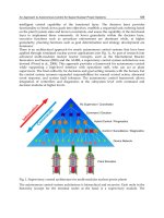
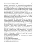
Fig. 11.1 Insulin resistance and
hepatic steatosis in obese subjects.
Insulin resistance in adipocytes
increases adipocyte lipolysis, which
increases plasma free fatty acids (FFA)
and hepatic FFA uptake. Insulin
resistance in myocytes increases
glucose and/or insulin levels, which
may increase hepatic FFA synthesis.
Hepatic FFAs are increased because
of increased uptake and increased
synthesis, in equilibrium with an
expanded pool of triglycerides, with
triglyceride deposits in the cytoplasm.
A new steady state is achieved whereby
these increased input pathways are
compensated by an increased
oxidation of fatty acids. The hepatic
secretion of triglycerides is also
increased in obese patients without
NASH, but might be decreased in
patients with NASH. (Modified from
Pessayre [3].)
MITOCHONDRIAL INJURY AND NASH
135
steatosis develops as a result of diverse causes, includ-
ing obesity.
Obesity, insulin resistance and steatosis
Obesity
In the past, prolonged overeating was self-regulating,
as excess weight soon impaired the physical fitness
required to gather food and handle predators or foes
[1]. For the first time in history, a large fraction of the
population in affluent countries can concomitantly
indulge in rich food and physical idleness, causing a
surge in obesity. About 22.5% of US citizens are obese,
and this prevalence could reach 40% by the year 2025
[8] unless drastic lifestyle changes can curb present
trends.
Obesity involves the accumulation of fat not only in
adipocytes, but also in muscle cells, and this accumula-
tion can cause insulin resistance in adipocytes and
muscles.
Insulin resistance in adipocytes and muscles
After a meal in lean persons, a mild increase in blood
glucose causes a minor increase in insulin. Insulin acts
on its receptor on the surface of adipocytes and myocytes
to trigger the phosphorylation of insulin receptor
substrates (IRS), which activate phosphatidyl inositol
3-kinase and Akt/protein kinase B, to eventually cause
the translocation of GLUT-4 glucose transporters from
intracellular storage vesicles to the plasma membrane
[9]. Abundant expression of GLUT-4 transporter on
the membrane causes efficient glucose uptake, which
limits the increase in blood glucose and insulin levels.
In obese people, however, adipocytes may produce
less GLUT-4 transporter [9]. More importantly, both
fat-engorged adipocytes and fat-laden myocytes are
resistant to the signalling effects of the insulin receptor
[9]. It is suggested that acyl-CoA or other derivatives
of FFA may limit the activation of IRS and phos-
phatidyl inositol 3-kinase [9]. The mechanism could
involve the activation of Jun N-terminal kinase and,
hence, the serine phosphorylation and thus inactiva-
tion of IRS [10]. Whatever the mechanism, insufficient
translocation of GLUT-4 to the plasma membrane lim-
its glucose uptake by adipocytes and myocytes [9].
This insufficient uptake results in an increase of blood
glucose and a compensatory increase in the release of
insulin by pancreatic β cells (the insulin-secreting cells
of the pancreas) [11]. In some subjects, however, this
compensatory insulin increase is not enough, or secon-
darily fails, and frank diabetes develops. Therefore,
insulin resistance in adipocytes and muscles tends to
result in increased C-peptide, insulin and blood glucose
levels (after eating).
Another normal effect of the activation of Akt/
protein kinase B by the insulin receptor in adipocytes,
is to activate a phosphodiesterase, which degrades
cyclic adenosine monophosphate (AMP) [12]. This
degradation prevents the cyclic AMP-mediated activa-
tion of protein kinase A and then hormone-sensitive
lipase, which otherwise would hydrolyse triglycerides
into fatty acids. As a final consequence, a normal effect
of insulin is to block adipose tissue lipolysis. How-
ever, this normal effect of insulin is hampered during
insulin resistance. Indeed, whereas the adipocytes of
lean insulin-sensitive persons release FFA during fast-
ing but then store fat after meals, in contrast, the fat-
engorged insulin-resistant adipocytes of obese people
keep releasing FFA after meals, causing a sustained
increase in plasma FFA [13].
Thus, in obese persons, insulin resistance causes
not only high blood insulin and glucose levels, but
also high plasma FFA. Both effects may be involved in
the development of hepatic steatosis in obese persons
[3].
Hepatic steatosis
High plasma FFA levels increase hepatic FFA uptake,
while high glucose and insulin levels may increase hep-
atic FFA synthesis in some obese patients (Fig. 11.1)
[3]. Indeed, insulin increases the transcription of sterol
regulatory element-binding protein-1 (SREBP-1), and
genetically obese ob/ob mice have increased levels of
SREBP-1 mRNA and protein [14]. SREBP-1 upregu-
lates the expression of acetyl-CoA carboxylase and
fatty acid synthase, to increase hepatic fatty acid syn-
thesis [14]. Interestingly, stearoyl-CoA desaturase is
also increased in ob/ob mice, resulting in a consider-
able increase in oleic acid [14], an unsaturated fatty
acid that is a substrate for lipid peroxidation.
In obese persons, the increased uptake and synthesis
of FFA expand the hepatic FFA pool [3]. These increased
input pathways are compensated by an increased rate
of hepatic mitochondrial FFA β-oxidation (Fig. 11.1)
[15]. By contrast, the hepatic secretion of triglyceride
CHAPTER 11
136
This large basal ROS formation is further enhanced
in steatotic livers. First, mitochondrial ROS formation
is increased (see below). Secondly, CYP2E1 is also
increased [22], which further increases ROS formation
in hepatocytes. Finally, endotoxin receptors on Kupffer
cells are upregulated in animals with either obesity- or
alcohol-mediated hepatic steatosis [23,24]. Increased
sensitivity of Kupffer cells to bacterial endotoxin may
increase ROS formation by these cells (Fig. 11.2). This
abundant formation of ROS may start to oxidize the
unsaturated lipids of fat deposits to cause lipid peroxi-
dation (Fig. 11.2) [1,2].
Indeed, 11 different treatments causing acute or
chronic steatosis always increased hepatic thiobar-
bituric acid reactants and ethane exhalation, an in
vivo index of lipid peroxidation, in mice [25]. After a
single dose of tetracycline or ethanol, there was a
parallel time course in the rise and fall of hepatic
triglycerides, and the rise and fall of lipid peroxidation
products. This is consistent with a cause-and-effect
relationship between the presence of oxidizable fat
in the liver and lipid peroxidation [25]. Extensive
lipid peroxidation also occurs in animals with hepatic
steatosis resulting from a methionine- and choline-
deficient diet [26], genetically obese leptin-deficient
ob/ob mice (personal unpublished results) and patients
might be differently affected in obese persons without
NASH and obese patients with NASH (Fig. 11.1).
Thus, in obese persons without NASH, the secretion
of apo B tended to be slightly increased [16], which
may explain why these patients tend to have hyper-
triglyceridaemia. Likewise, in obese ob/ob mice, MTP
expression and hepatic lipoprotein secretion were
both increased [17]. However, in obese persons with
NASH, hepatic apo B secretion was decreased [16],
which infers decreased secretion of VLDL.
The reasons for the differences in apo B secretion in
patients with and without NASH are unknown. There
are two possible mechanisms. First, NASH may be
associated with even higher insulin and TNF levels,
which both downregulate MTP production [18,19].
Although insulin resistance in the liver could perhaps
hamper insulin effects, increased TNF may decrease
MTP-mediated apo B lipidation and thus the secre-
tion of triglyceride-rich VLDL particles in patients
with NASH. Secondly, subjects with an inborn par-
tial deficiency in MTP expression could excrete less
hepatic VLDL and could therefore store more fat in
the liver, to be at increased risk of developing NASH
[20].
Although a new equilibrium is achieved between
input and output pathways in insulin-resistant per-
sons (with or without NASH), this new equilibrium
is achieved at the expense of expanded pools of
hepatic FFA and triglycerides, thus causing steatosis
(Fig. 11.1) [3].
Harmful effects of fat in the liver
Although the reasons for the deleterious effects of
steatosis are still incompletely understood, there is
growing evidence that the presence of oxidizable fat
in the liver can trigger lipid peroxidation, mitochon-
drial dysfunction and increased mitochondrial ROS
formation.
Lipid peroxidation
Even in the basal (fat-free) state, hepatocytes produce
large amounts of ROS. These ROS are formed mainly
in mitochondria, but also at other sites, including
microsomal cytochrome P450 (CYP). Yet another
potential source of ROS is the NADPH oxidase of
Kupffer cells (Fig. 11.2) [21].
Kupffer
cell
Hepatocyte
Endotoxin
MITO
ROS
Lipid
peroxidation
-CH=CH-
Fat deposits
CYP2E1
Increased endotoxin receptor
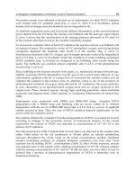
Fig. 11.2 The presence of fat in the liver triggers lipid
peroxidation. Mitochondria (MITO) and cytochrome P450
2E1 (CYP2E1) generate reactive oxygen species (ROS).
In several models of steatosis, the endotoxin receptors
of Kupffer cells are increased, which might trigger ROS
formation by these cells. When fat accumulates in the liver,
ROS oxidize the unsaturated lipids of fat deposits to cause
lipid peroxidation. (Modified from Pessayre [3].)
MITOCHONDRIAL INJURY AND NASH
137
complexes is decreased [30], as is the in vivo resynthesis
of ATP after a fructose challenge [31].
Increased input of electrons
Contrasting with this partial block in the flow of elec-
trons in the respiratory chain, the input of electrons
into this chain may be enhanced by the increased
mitochondrial β-oxidation of FFA [15], which forms
NADH and FADH
2
, that transfer their electrons to
the respiratory chain (Fig. 11.3). In diabetic patients,
increased blood glucose levels and enhanced glucose
oxidation could further increase this influx of electrons.
Increased mitochondrial reactive oxygen
species formation
The imbalance between an increased electron input
into the respiratory chain and a partially hampered
flow of electrons within this chain may cause over-
reduction of respiratory chain components. These can
then react with oxygen to form the superoxide anion
radical, thus increasing mitochondrial ROS formation
(Fig. 11.3) [3]. An increased mitochondrial ROS forma-
tion has indeed been demonstrated in genetically obese
ob/ob mice [32] and in mice fed a choline-deficient
diet [33]. This increased mitochondrial ROS formation
will in turn cause several vicious cycles (see below).
with NASH [15]. This extensive lipid peroxidation
releases several reactive substances that can damage
mitochondria.
Mitochondrial dysfunction
Restricted electron flow
The peroxidation of hepatic triglycerides releases react-
ive aldehydes, such as 4-hydroxynonenal and malon-
dialdehyde, that damage mtDNA (Fig. 11.3) [27]. This
may secondarily impair the flow of electrons in the res-
piratory chain, because mtDNA encodes some of the
respiratory chain polypeptides [1]. Lipid peroxidation
products also directly attack and inactivate respiratory
chain polypeptides, including cytochrome c oxidase, the
terminal oxidase of the respiratory chain [28]. These
two effects of lipid peroxidation products could thus
partially hamper the flow of electrons in the respiratory
chain (Fig. 11.3) [1].
The livers of patients with NASH have been shown
to exhibit ultrastructural mitochondrial lesions, with
the presence of crystalline inclusions in megamito-
chondria [15] (see Chapte 2, Fig. 2.1). These patients
have mtDNA depletion and decreased expression of a
mtDNA-encoded cytochrome oxidase subunit II [29].
The ex vivo activity of mitochondrial respiratory chain
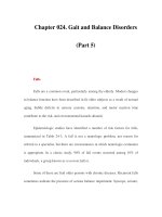
Fig. 11.3 Electron overflow and
reactive oxygen species (ROS). In
the normal liver, the electrons that
are given to the respiratory chain
mostly flow along this chain, up to
cytochrome c oxidase (the terminal
oxidase), where they safely combine
with oxygen and protons to form
water. In the fatty liver of insulin-
resistant patients, high β-oxidation
rates increase the delivery of electrons
to the respiratory chain, while ROS
and lipid peroxidation products, such
as 4-hydroxynoneal (HNE), may
partially block the flow of electrons
within this chain. The imbalance
between a high input and a restricted
flow of electrons may cause over-
reduction of respiratory chain
components, which directly transfer
their electrons to oxygen to form the
superoxide anion radical and other
ROS. (Adapted from Pessayre [3].)
CHAPTER 11
138
ance. In other causes of steatohepatitis, the situation
is exacerbated because the causative disease process
itself directly increases ROS formation [1]. This sub-
ject has been reviewed elsewhere [7,37].
From reactive oxygen species formation
to the development of NASH
ROS cause lipid peroxidation, which releases reactive
aldehydes, such as malondialdehyde, and 4-hydrox-
ynonenal [1]. ROS also increase the expression of sev-
eral cytokines, including transforming growth factor-β
(TGF-β), interleukin-8 (IL-8), TNF and Fas ligand [1].
Both lipid peroxidation products and cytokines seem
to be involved in steatohepatitis liver injury [1].
Inflammation and Mallory bodies
TGF-β, IL-8 and 4-hydroxynonenal are chemo-
attractants for human neutrophils, which may account
for the neutrophilic infiltrate in steatohepatitis [1].
TGF-β also induces tissue transglutaminase [1]. This
enzyme is associated with the cytoskeleton, includ-
ing intermediary filaments. Transglutaminase catalyses
the formation of ε-lysine–gamma-glutamyl cross-links
between a lysine on one polypeptide chain and a
Fig. 11.4 Vicious cycles involving
reactive oxygen species (ROS) and
mitochondria. The increased
formation of mitochondrial ROS
(mtROS) can further damage
mitochondria and further increase
mtROS formation through several
vicious cycles. 1. ROS directly damage
mitochondrial DNA, proteins and
cardiolipin to impair the flow of
electrons in the respiratory chain and
increase mitochondrial ROS
formation. 2. ROS activate NF-κB,
which increases TNF-α and further
damages mitochondria. 3. ROS
deplete antioxidants such as vitamin E.
4. ROS trigger further lipid
peroxidation, whose products impair
the flow of electrons in the respiratory
chain to further increase
mitochondrial ROS formation.
(Adapted from Pessayre [3].)
Reactive oxygen species-dependent vicious cycles
Because ROS themselves damage mitochondria to fur-
ther increase mitochondrial ROS formation (Fig. 11.4),
an increase in mitochondrial ROS production can
trigger the following processes that amplify injury:
1 ROS directly damage mtDNA, respiratory chain
polypeptides and mitochondrial cardiolipin [1].
2 ROS cause NF-κB activation, which induces the
synthesis of TNF [34]. TNF is also synthesized by
fat-engorged adipocytes and is released by Kupffer
cells stimulated by endotoxin. TNF damages mito-
chondria and increases mitochondrial ROS formation
(see below).
3 ROS may deplete some tissue antioxidants, thereby
further aggravating ROS-induced damages. Thus, low
serum vitamin E levels are found in some obese chil-
dren with steatohepatitis [35] and supplementation
with vitamin E can decrease serum aminotransaminase
(AT) levels in obese children [36].
4 Increased mitochondrial ROS formation further
increases lipid peroxidation, thereby releasing more
reactive aldehydes that further damage mtDNA and
respiratory chain polypeptides [1].
These vicious cycles can damage mitochondria and
enhance ROS formation in patients with NASH sec-
ondary to obesity, type 2 diabetes and insulin resist-
Mitochondrial
dysfunction
mt
ROS
Lipid
peroxidation
products
Depletion
of antioxidants
Fat
MITOCHONDRIAL INJURY AND NASH
139
hepatocyte to cause fratricidal apoptosis (Fig. 11.5)
[38]. ROS also increase the synthesis of TNF, and
patients with steatohepatitis have high hepatic TNF
mRNA levels [39]. TNF is also synthesized by fat-
engorged adipocytes in obese people, and may be
released in excess by Kupffer cells stimulated by bac-
terial endotoxin, because of the overexpression of
endotoxin receptors on these cells (Fig. 11.5).
The interaction of Fas ligand with Fas, or the
interaction of TNF with TNF receptor 1 (TNFR1),
activates procaspase 8 into caspase 8, which cuts BH3
interacting domain death agonist (Bid) [38]. Truncated
Bid enters the outer mitochondrial membrane to per-
meabilize this membrane. Bid also induces a confor-
mational change in Bcl-2-associated x protein (Bax)
glutamine on another polypeptide chain. The induc-
tion of tissue transglutaminase by TGF-β could poly-
merize cytokeratins to form Mallory bodies, which are
formed of cross-linked cytoskeletal proteins, in particu-
lar cytokeratins [1].
Death receptors, mitochondria and apoptosis
Normally, hepatocytes express Fas (a membrane
receptor), but not Fas ligand, preventing them from
killing their neighbours [38]. However, several con-
ditions leading to increased ROS formation, such as
drugs, alcohol abuse or Wilson’s disease, cause Fas
ligand expression by hepatocytes, so that Fas ligand on
one hepatocyte can now interact with Fas on another
Fig. 11.5 Death receptors, mitochondria and apoptosis. In
hepatocytes, ROS trigger the expression of Fas ligand (Fas L)
and TNF-α. The latter is also formed by the adipocytes of
obese subjects and by endotoxin-stimulated Kupffer cells.
The interaction of Fas L with Fas, or TNF-α with TNF-α
receptor 1 (TNFR1), activates procaspase 8 into caspase 8,
which cuts BH3 interacting domain death agonist (Bid).
Truncated Bid (tBid) enters the outer mitochondrial
membrane to permeabilize this membrane. Bid also induces
a conformational change in Bcl-2-associated x protein
(Bax) and its analogue Bak, which translocate to the outer
mitochondrial membrane to permeabilize this membrane.
This increased permeability causes the release of cytochrome
c from the intermembranous space, thus blocking the flow
of electrons into the respiratory chain and increasing
mitochondrial ROS formation. ROS could then act on the
same or other mitochondria to open an inner membrane
pore called the mitochondrial permeability transition pore
(MPTP). Pore opening causes matrix expansion and
outer membrane rupture. As a result of both increased
permeability and rupture of the outer membrane,
cytochrome c, procaspases and other pro-apoptotic factors
leave the mitochondrial intermembrane space to activate
caspase 9 and effector caspases in the cytosol and trigger
apoptosis. (Adapted from Pessayre [3].)
Fas L
TNF-α
Endotoxin-
stimulated
Kupffer
cells
Adipocytes
ROS
in
hepatocytes
Fas
Caspase 8 TNFR1
ROS
MPTP
Cytochrome c
Bax/Bak
tBid
Hepatocyte
Caspases
APOPTOSIS
CHAPTER 11
140
Implications in clinical management
Although overweight patients should lose weight, severe
dieting or total fasting increases peripheral lipolysis
and the release of FFA, which uncouple and inhibit
mitochondrial respiration [1]. Fasting may also cause
glutathione depletion [45], which enhances lipid per-
oxidation and cytokine-mediated cell death [46]. It is
therefore not surprising that rapid weight loss result-
ing from starvation, severe dieting, jejuno-ileal bypass
or gastroplasty paradoxically increases liver inflamma-
tion and fibrosis in obese patients (for a detailed dis-
cussion see Chapter 20) [1].
Instead, the combination of physical exercise and
a moderately hypocaloric diet (high in vegetables but
low in sugar, starch and fat) can progressively decrease
adipocyte fat stores, improve liver tests and stop fibro-
genesis [1]. The role of hypolipidaemic peroxisome
proliferator receptor-α agonists, metformin, vitamin E
and betaine (to improve VLDL secretion) are discussed
in Chapter 16.
Conclusions
In affluent countries, new lifestyle habits combining
rich diet and lack of physical activity have resulted
in an ever-increasing prevalence of obesity. Excess
weight can trigger insulin resistance in adipocytes and
muscle. Insulin resistance increases blood glucose and
insulin levels and causes persistent adipocyte lipolysis,
which can cause a fatty liver. As insulin resistance
causes hepatic steatosis, and steatosis can develop into
NASH, there is an almost universal association of prim-
ary NASH with insulin resistance. Insulin resistance
can also be present in patients with hepatic steatosis
but without NASH [15] and, conversely, steatohepatitis
can occur when hepatic steatosis is triggered by mech-
anisms other than insulin resistance. During chronic
hepatic steatosis, several vicious cycles involving lipid
peroxidation, mitochondrial damage, ROS formation,
depletion of antioxidants and cytokine release may
cause necroinflammation and fibrogenesis in genetic-
ally susceptible patients. Further studies are required
to understand better how these diverse effects interact
with each other, which genetic or environmental factors
are involved in individual susceptibility, and which treat-
ments or combinations of treatments are best used in
patients who fail to lose weight, despite medical advice.
and its analogue Bak, which translocate to mitochon-
dria to form channels in the outer mitochondrial
membrane (Fig. 11.5).
Increased permeability of the outer mitochondrial
membrane may release cytochrome c from the inter-
membranous space of some mitochondria, thus block-
ing the flow of electrons into the respiratory chain
and increasing mitochondrial ROS formation. ROS
could then act on the same or other mitochondria
to open an inner membrane pore, whose opening
causes matrix expansion and outer membrane rupture
(Fig. 11.5) [40].
Because of increased permeability and rupture of the
outer membrane, cytochrome c and other pro-apoptotic
factors leave the mitochondrial intermembranous space
to activate caspase 9 in the cytosol. Caspase 9 activates
effector caspases, which trigger apoptosis (Fig. 11.5)
[40]. It is therefore noteworthy that apoptosis seems to
have an important role in both NASH and alcoholic
steatohepatitis [41,42].
Implications for genetic susceptibility
In hepatic steatosis, the tendency of different subjects
to develop steatohepatitis varies considerably [2]. For
the same amount of excess weight, or the same alcohol
consumption, some subjects only have steatosis while
others develop cirrhosis.
Genetic polymorphisms could also be in involved
(see Chapter 6). For example, in obesity-related NASH,
a genetic polymorphism, which decreases MTP activ-
ity, may cause less hepatic VLDL secretion and thus
more fat accumulation, more lipid peroxidation, more
ROS formation and more liver lesions [20]. Also, a
genetic dimorphism affects the mitochondrial target-
ing sequence of MnSOD. The alanine-containing
sequence confers an α-helical structure to the import
peptide, causing better mitochondrial import than the
valine sequence, which confers a β-sheet structure to
the peptide [43]. The genetic polymorphism therefore
modulates the mitochondrial import of MnSOD, which
may affect the mitochondrial detoxication of ROS
[43]. Although this MnSOD dimorphism has been
implicated in susceptibility to severe alcoholic liver
disease in a French population, this finding was not
confirmed in a larger English study [44]. Other studies
are required to evaluate further the role of this dimor-
phism in both NASH and alcoholic liver disease.
MITOCHONDRIAL INJURY AND NASH
141
and mitochondrial abnormalities. Gastroenterology 2001;
120: 1183–92.
16 Charlton M, Sreekumar R, Rasmussen D, Lindor K,
Nair S. Apolipoprotein synthesis in non-alcoholic steato-
hepatitis. Hepatology 2002; 35: 898–904.
17 Bartels ED, Lauritsen M, Nielsen LB. Hepatic expression
of microsomal triglyceride transfer protein and in vivo
secretion of triglyceride-rich lipoproteins are increased in
obese diabetic mice. Diabetes 2002; 51: 1233–9.
18 Lin MCM, Gordon DC, Wettereau JR. Microsomal
triglyceride transfer protein (MTP) regulation in HepG2
cells: insulin negatively regulates MTP gene expression.
J Lipid Res 1995; 36: 1073–81.
19 Navasa M, Gordon DA, Hariharan N et al. Regulation of
microsomal triglyceride transfer protein mRNA expres-
sion by endotoxin and cytokines. J Lipid Res 1998; 39:
1220–30.
20 Day CP, Saksena S, Leathart J et al. Genetic evidence
supporting the two-hit model of NASH pathogenesis.
Hepatology 2002; 36: 82A.
21 Kono H, Rusyn I, Yin M et al. NADPH oxidase-derived
free radicals are key oxidants in alcohol-induced liver dis-
ease. J Clin Invest 2000; 106: 867–72.
22 Weltman MD, Farrell GC, Hall P, Ingelman-Sundberg M,
Liddle C. Hepatic cytochrome P450 2E1 is increased in
patients with non-alcoholic steatohepatitis. Hepatology
1998; 27: 128–33.
23 Fiorini RN, Shafizadeh SF, Chavin KD. Primary non-
function in steatotic livers is due to differential Toll-like
expression and endotoxin sensitivity. Hepatology 2002;
36: 198A.
24 Enomoto N, Takei Y, Hirose M et al. Thalidomide pre-
vents alcoholic liver injury in rats through suppression
of Kupffer cell sensitization and TNF-α production.
Gastroenterology 2002; 123: 291–300.
25 Lettéron P, Fromenty B, Terris B, Degott C, Pessayre D.
Acute and chronic steatosis lead to in vivo lipid peroxida-
tion in mice. J Hepatol 1996; 24: 200–8.
26 Leclercq IA, Farrell GC, Field J et al. CYP2E1 and CYP4A
as microsomal catalysts of lipid peroxides in murine
non-alcoholic steatohepatitis. J Clin Invest 2001; 105:
1067–75.
27 Hruszkewycz AM. Evidence for mitochondrial DNA
damage by lipid peroxidation. Biochem Biophys Res
Commun 1988; 153: 191–7.
28 Chen J, Schenker S, Frosto TA, Hensderson GI. Inhibition
of cytochrome c oxidase activity by 4-hydroxynonenal
(HNE): role of HNE adduct formation with the enzyme
catalytic site. Biochem Biophys Acta
1998; 1380: 336–44.
29 Haque M, Mirshahi F, Campbell-Sargent C et al. Non-
alcoholic steatohepatitis (NASH) is associated with hepa-
tocyte mitochondrial DNA depletion. Hepatology 2002;
36: 403A.
References
1 Pessayre D, Berson A, Fromenty B, Mansouri A.
Mitochondria in steatohepatitis. Semin Liver Dis 2001;
21: 57–69.
2 Pessayre D, Mansouri A, Fromenty B. Mitochondrial dys-
function in steatohepatitis. Am J Physiol Gastrointest
Liver Physiol 2002; 282: G193–9.
3 Pessayre D. Mitochondrial injury in steatohepatitis. In:
Suchy FJ, Gregory FJ, Maher JJ. American Association
for the Study of Liver Diseases Postgraduate Course
2002. Mechanisms of Acute and Chronic Liver Diseases:
Implications for Diagnosis, Pathogenesis and Treatment.
AASLD Postgraduate Course, 2002: 97–103.
4 Tran K, Thorne-Tjomsland G, DeLong CJ et al.
Intracellular assembly of very-low-density lipoproteins
containing apolipoprotein B100 in rat hepatoma McA-
RH777 cells. J Biol Chem 2002; 277: 31187–200.
5 Liao W, Yeung SCJ, Chan L. Proteasome-mediated
degradation of apolipoprotein B targets both nascent
peptides cotranslationally before translocation and full-
length apolipoprotein B after translocation into the
endoplamic reticulum. J Biol Chem 1998; 273: 27225–
30.
6 McGarry JD, Foster DW. Regulation of hepatic fatty acid
oxidation and ketone body production. Ann Rev Biochem
1980; 49: 395–420.
7 Fromenty B, Pessayre D. Inhibition of mitochondrial β-
oxidation as a mechanism of hepatotoxicity. Pharmacol
Ther 1995; 67: 101–54.
8 Kopelman PG. Obesity as a medical problem. Nature
2000; 404: 635–43.
9 Shepherd PR, Kahn BB. Glucose transporters and insulin
action: implications for insulin resistance and diabetes
mellitus. N Engl J Med 1999; 341: 248–57.
10 Hirosumi J, Tuncman G, Chang L et al. A central role for
JNK in obesity and insulin resistance. Nature 2002; 420:
353–6.
11 Chitturi S, Abeygunasekera S, Farrell GC et al. NASH
and insulin resistance: insulin hypersecretion and specific
association with the insulin resistance. Hepatology 2002;
35: 373–9.
12 Valet P, Tavernier G, Castan-Laurell I et al. Understand-
ing adipose tissue development from transgenic animal
models. J Lipid Res 2002; 43: 835–60.
13 Gorden ES. Non-esterified fatty acids in blood of obese
and lean subjects. Am J Clin Nutr 1960; 8: 740–7.
14 Shimomura I, Bashmakov Y, Horton JD. Increased levels
of nuclear SREBP-1c associated with fatty livers in two
mouse models of diabetes mellitus. J Biol Chem 1999;
274: 30028–32.
15 Sanyal AJ, Campbell-Sargent C, Mirshahi F et al. Non-
alcoholic steatohepatitis: association of insulin resistance
CHAPTER 11
142
Cameron RG, Feuer G, eds. Apoptosis and its Modulation
by Drugs: Handbook of Experimental Pharmacology.
Heidelberg: Springer Verlag, 2000, 142: 59–108.
39 Crespo J, Cayon A, Fernadez-Gil P et al. Gene expression
of tumor necrosis factor-α and TNF-receptors p55 and
p75 in non-alcoholic steatohepatitis patients. Hepatology
2001; 34: 1158–63.
40 Feldmann G, Haouzi D, Moreau A et al. Opening of the
mitochondrial permeability transition pore causes matrix
expansion and outer membrane rupture in Fas-mediated
hepatic apoptosis in mice. Hepatology 2000; 31: 674–83.
41 Natori S, Rust C, Stadheim LM et al. Hepatocyte apop-
tosis is a pathologic feature of human alcoholic hepatitis.
J Hepatol 2001; 34: 248–53.
42 Rodriguez CM, Cortez-Pinto H, Sola S et al. Apoptosis is
a prominent feature of human alcoholic and non-alcoholic
steatohepatitis. Hepatology 2001; 34: 672A.
43 Sutton A, Khoury H, Prip-Buus C et al. The Ala-9Val
dimorphism modulates the import of human manganese
superoxide dismutase into rat liver mitochondria. Phar-
macogenetics 2003; 13: 145–57.
44 Stewart SF, Leathart JB, Chen Y et al. Valine-alanine
manganese superoxide dismutase polymorphism is not
associated with alcohol-induced oxidative stress or liver
fibrosis. Hepatology 2002; 36: 1355–60.
45 Pessayre D, Dolder A, Artigou JY et al. Effect of fasting
on metabolite-mediated hepatotoxicity in the rat. Gastro-
enterology 1979; 77: 264–71.
46 Haouzi D, Lekehal M, Tinel M et al. Prolonged, but
not acute, glutathione depletion promotes Fas-mediated
mitochondrial permeability transition and apoptosis in
mice. Hepatology 2001; 33: 1181–8.
30 Perez-Carrera M, Del Hoyo P, Martin M et al. Activity of
the mitochondrial respiratory chain enzymes is decreased
in the liver of patients with non-alcoholic steatohepatitis.
Hepatology 1999; 30: 379A.
31 Cortez-Pinto H, Chatham J, Chacko VP et al. Alterations
in liver ATP homeostasis in human non-alcoholic steato-
hepatitis: a pilot study. J Am Med Assoc 1999; 282:
1659–64.
32 Yang SQ, Zhu H, Li Y et al. Mitochondrial adaptations to
obesity-related oxidant stress. Arch Biochem Biophys
2000; 378: 259–68.
33 Hensley K, Kotake Y, Sang H et al. Dietary choline
restriction causes complex I dysfunction and increased
H
2
O
2
generation in liver mitochondria. Carcinogenesis
2000; 21: 983–9.
34 Yin M, Gäbele E, Wheeler MD et al. Alcohol-induced free
radicals in mice: direct toxicants or signaling molecules?
Hepatology 2001; 34: 935–42.
35 Strauss RS. Comparison of serum concentrations of α-
tocopherol and β-carotene in a cross-sectional sample of
obese and non-obese children (NHANES III). J Pediatr
1999; 134: 160–5.
36 Lavine JE. Vitamin E treatment of non-alcoholic steato-
hepatitis in children: a pilot study. J Pediatr 2000; 136:
739–43.
37 Berson A, De Beco V, Leteron P et al. Steatohepatitis-
inducing drugs cause mitochondrial dysfunction and lipid
peroxidation in rat hepatocytes. Gastroenterology 1998;
114: 764–74.
38 Pessayre D, Feldmann G, Haouzi D et al. Hepatocyte
apoptosis triggered by natural substances (cytokines,
other endogenous substances and foreign toxins). In:
143
Cell biology of NASH: fibrosis and
cell proliferation
Isabelle A. Leclercq & Yves Horsmans
12
Key learning points
1 In the liver, injury triggers a physiological wound healing response that contains the injurious agent,
isolates damaged cells and effects wound closure, leading to restoration of normal hepatic structure and
function. The process requires coordination of the inflammatory reaction, cell proliferation, differentiation
and death (apoptosis), fibrogenesis and matrix remodelling.
2 Activation of the fibrotic cascade is part of the response to liver injury. The organized sequence of
responses include activation of hepatic stellate cells (HSC) and other cell types (Kupffer cells and endothelial
cells), migration and proliferation of HSC, synthesis and deposition of extracellular matrix, remodelling and
degradation of scar tissue, and deactivation or apoptosis (cell deletion) of the effector cells.
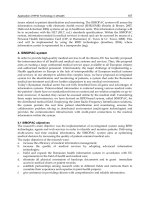
3 Pathological examination of liver tissue from non-alcoholic steatohepatitis (NASH) patients reveals
increased numbers of α-smooth muscle actin-reactive HSC, mainly located in the perivenular area,
confirming the presence of activated HSC in this setting.
4 Fibrosis associated with NASH can be understood as the physiological consequence of chronic hepatic
injury, necrosis and inflammation (steatohepatitis). However, profibrotic mechanisms specifically related to
the context of NASH are emerging: steatosis and insulin resistance, oxidative stress generated by CYP2E1 and
4A or from other sources, dysregulation of leptin expression and signalling, peroxisome proliferator-activated
receptor-α and -γ expression and signalling, inflammation and release of cytokine and fibrogenic mediators.
5 Clinical observations suggest an impairment of hepatocyte proliferation in non-alcoholic fatty liver
disease (NAFLD)/NASH. However, it remains to be confirmed that such altered adaptative response to liver
injury participate in the pathogenesis of the disease.
6 From animal studies, it has been shown that liver regeneration and hepatocyte proliferation are normal in
several models of fatty liver as well as in fibrosing steatohepatitis. In contrast, liver regeneration is markedly
impaired in fatty liver because of disrupted leptin signalling. To date, the parts played by altered lipid metabol-
ism, insulin resistance and/or leptin deficiency in the control of liver regeneration remain to be established.
Abstract
Fibrosis is the most significant pathological con-
sequence associated with non-alcoholic steatohepatitis
(NASH). Activation of hepatic stellate cells (HSC) into
extracellular matrix (ECM) producing myofibroblasts,
the central event in hepatic fibrosis, is recognized in
NASH. Hepatic fibrogenesis could represent the healing
and tissue repair response to chronic necroinflammat-
ory injury associated with NASH. However, there is
Fatty Liver Disease: NASH and Related Disorders
Edited by Geoffrey C. Farrell, Jacob George, Pauline de la M. Hall, Arthur J. McCullough
Copyright © 2005 Blackwell Publishing Ltd
CHAPTER 12
144
acute injury, the effectiveness of the wound healing
process is mostly dependent on the ability of the liver
to isolate the damage and reconstitute functional liver
mass by hepatocyte proliferation, the process known
as liver regeneration. When the injury is chronic or
repeated, as in NASH, there are multiple cycles of
tissue repair and deposition of ECM or scarring
(fibrosis). Chronic activation of the scarring response
leads to hepatic fibrosis, which can be considered as
the highly integrated response to any type of chronic
liver injury, irrespective of aetiology. Hepatic com-
plications develop when fibrosis has progressed to
cirrhosis. They result from portal hypertension and
loss of hepatic cell mass, which leads to hepatocellular
failure, or from imbalance between hepatocellular pro-
liferation and apoptosis, which can result in tumour
formation. This review discusses these tissue and cell
biological responses to chronic liver injury, and con-
siders mechanisms and outcomes relevant to NAFLD/
NASH.
Hepatic fibrosis
Fibrosis is the most significant pathological consequence
of liver disease associated with hepatic steatosis. It is
therefore important to identify profibrotic stimuli asso-
ciated with fatty liver disease.
The mechanism of the fibrotic process in chronic
metabolic steatohepatitis is likely to be similar in
nature to that in response to other forms of chronic
liver injury. Considerable insight into these mechan-
isms has been gained in the past decade, as reviewed
elsewhere [1,2]. Fibrosis associated with NASH can
be understood as the physiological consequence of
chronic hepatic injury, necrosis and inflammation
(steatohepatitis). However, profibrotic mechanisms spe-
cifically related to the context of NASH have emerged
recently. The activation of these pathways might deter-
mine or at least partly explain the propensity for fibrosis
progression between individuals with NASH. This
section briefly recalls the general features of the fibro-
genic process, and discusses special aspects pertaining
to fibrogenesis in NASH.
Role of hepatic stellate cells
The hepatic scar consists of a broad accumulation of
ECM. In turn, this comprises macromolecules from
no strict link between the intensity of the necroin-
flammation and the intensity of fibrosis.
Recently, profibrotic mechanisms specifically related
to the context of NASH have been identified and
might explain the propensity for fibrosis progression
in this metabolic disorder. These include oxidative
stress and lipid peroxidation, imbalanced intrahepatic
lipid metabolism, and insulin resistance. Hormones
and transcription factors involved in the control of
glucose and/or lipid metabolism have been implicated
in fibrogenesis. They operate directly by stimulating
HSC activation and collagen synthesis, or indirectly
by modulating the inflammatory response and the
release of profibrotic cytokines and mediators. Funda-
mental insights into leptin biology and its dysregula-
tion associated with the insulin resistance (metabolic)
syndrome (IRS), peroxisome proliferator-activated
receptor (PPAR) transcription factors and their dual
effects on the control of insulin sensitivity and biology
of HSCs, and cytokine signalling are all likely to bene-
fit our understanding of NASH-associated fibrosis.
NASH is characterized by chronic hepatocellular
injury. Clinical observations suggest an impairment
of hepatocyte proliferation in non-alcoholic fatty liver
disease (NAFLD)/NASH. Animal studies provide evid-
ence that intrahepatic lipid overload per se does not
appear to compromise the proliferative response of
the liver. However, liver regeneration is impaired in
animals with disrupted leptin signalling, resistance to
insulin and immune perturbations, a phenotype that
closely resembles that of patients with NASH. Con-
ceptually, these factors, alone or together, could poten-
tially dampen the adaptative response of the liver to
injury and could contribute to NASH pathogenesis,
but this remains to be confirmed.
Introduction
The wound healing process is integral to any organ’s
response to injury. In the liver, injury triggers a physio-
logical wound healing response that operates contain-
ment of the injurious agent, isolation of damaged cells
and wound closure, leading to restoration of normal
hepatic structure and function. The entire process
requires precise coordination of several important
cellular actions: the inflammatory reaction, cell pro-
liferation and differentiation, fibrogenesis and matrix
remodelling, as well as apoptosis. In response to an
CELL BIOLOGY OF NASH
145
three main families: collagens, glycoproteins and pro-
teoglycans. As the liver becomes fibrotic, production
and accumulation of collagen and non-collagen com-
pounds increases and there are qualitative changes in
the composition of the ECM [1–3]. More interstitial
type matrix molecules are produced: fibril-forming
collagens (types I and III) become prominent, proteo-
glycans and structural glycoproteins such as laminin
and fibronectin are deposited and accumulate in the
subendothelial space (space of Disse).
An additional factor is that excessive matrix deposi-
tion is no longer compensated by enhanced degradation
because of an imbalance between expression of matrix
metalloproteinases (MMP) and their inhibitors (tissue
inhibitors of metalloproteinases, TIMP) [1,3,4]. The
accumulation of matrix and the formation of fibrous
septa disrupts the normal architecture of the vascular
and cellular compartments in the liver, impairing the
ready exchange of oxygen and nutrients and progress-
ively leading to cirrhosis.
As part of the acute response to liver injury, the
fibrotic cascade is activated. The short duration of the
stimulus allows recognition of an organized sequence
of responses. These include the activation of HSC and
other cell types (Kupffer cells and endothelial cells),
migration and proliferation of HSC, synthesis and
deposition of ECM, remodelling and degradation of
the scar tissue and deactivation or apoptosis (cell
deletion) of the effector cells. It is yet to be determined
whether these events represent a continuum or are
independent but interconnected phenomena triggered
by specific or multiple factors. During chronic liver
injury, injurious and healing processes evolve simul-
taneously; thus, organization of the fibrotic cascade is
disrupted, leading to scar formation and distortion of
liver architecture [1,2].
ECM deposition in perivascular and pericellular areas
is the hallmark of hepatic fibrosis. Hepatic matrix pro-
ducing cells are diverse populations that exist in both
the normal and injured liver [5–7]. They differ by their
content in vitamin A, the expression of intermedi-
ate filaments and other characteristics of myogenic
or neural crest cells, and their location (perisinusoidal
HSC or perivascular and periportal fibroblasts). The
embryological origin of these cells and the way they par-
ticipate in the fibrogenic process is still debated [4–7].
HSC and myofibroblasts have been identified as the
principal cell types in the liver responsible for ECM
production during fibrogenesis [4,5,8]. In normal liver,
HSC are found in the space of Disse and beneath the
endothelial cells. They emit multiple star-like projec-
tions and establish direct contacts via gap junctions
with other stellate cells, hepatocytes and probably
endothelial cells [5,8]. Regardless of the nature of
the insult, liver injury provides stimuli that transform
HSC from a quiescent retinoid-storing phenotype into
a proliferative migrating ECM-producing and con-
tractile myofibroblast-like phenotype [1,2,5,8]. This
process, characterized by morphological and func-
tional changes of the HSC, is known as ‘activation’; it
is a programmed response occurring in a reproducible
sequence at the onset of injury. Stellate cell activation
is initiated by paracrine stimuli from injured and
neighbouring parenchymal and non-parenchymal cells,
recruited inflammatory cells as well as subtle changes
in ECM composition. Activation is then perpetuated
through enhanced cytokine expression and respons-
iveness to cytokines mediated by autocrine and
paracrine stimuli and by accelerated ECM remodelling
[1,4,5,9,10]. It follows that, if the injurious factor is
not removed, the fibrotic process is perpetuated.
Alternatively, if the injurious process is interrupted or
if the injury is self-limited, loss of activated stellate
cells through phenotypical reversion or apoptosis may
occur with resultant resolution of fibrosis [10,11].
Amongst the best-studied paracrine stimuli for
HSC activation are fibronectin, lipid peroxides and
cytokines. The latter include platelet-derived growth
factor (PDGF), transforming growth factor-β1 (TGF-
β1), endothelin-1 (ET-1) and epidermal growth factor
(EGF) [1,4,5,10,12,13]. Transcription factors involved
in HSC activation include c-myb, nuclear factor-κB
(NF-κB), activator protein-1 (AP-1), signal transducer
and activator of transcription-1 (STAT-1) and mem-
bers of the Krüppel-like transcription factors family,
such as KLF6 or SP-1 [9,13]. Once activated, stellate
cells undergo phenotypical changes that concur with
the accumulation of ECM, as summarized in Fig. 12.1.
1 HSC proliferate under the action of paracrine
and autocrine mediators, such as PDGF, ET-1, and
TGF-β1.
2 They gain contractile capability; this can increase
resistance to portal blood flow resulting in portal
hypertension. The key contractile mediator is ET-1,
but vasopressin and/or eicosanoids also participate in
this action.
3 HSC are stimulated to produce large amounts of
fibril-forming collagen. The most potent stimulus for
CHAPTER 12
146
is uncommon (but see Chapter 24), although reversion of
fibrosis has been described in other types of liver disease
following treatment that removes the cause of injury
[11]. Interestingly, the recurrence of NASH following
liver transplantation for cryptogenetic cirrhosis impli-
cates the operation of an underlying extrahepatic or
metabolic abnormality that may account for the disease
in both native and transplanted livers (see Chapter 17).
Fibrosis in a characteristic chicken-wire pericellular
distribution has long been recognized as a hallmark
of NASH and a criterion for severity of the disease (see
Chapter 2). Pathological examination of liver tissue
from NASH patients, compared to normal controls,
reveals increased numbers of α-smooth muscle actin
(α-SMA)-reactive HSC, mainly located in the peri-
venular area, confirming the presence of activated
HSC in this setting [14,15]. To date, there are no true
indicators for fibrosis progression in NAFLD. How-
ever, increasing obesity and type 2 diabetes correlate
with severity of fibrosis and risk of cirrhosis (see
Chapter 3). Other risk factors for fibrosis in NASH
include necroinflammatory activity, increased alanine
fibrogenesis is TGF-β1, but other factors such as inter-
leukin 1β (IL-1β), tumour necrosis factor (TNF) and
lipid peroxides can induce synthesis of ECM.
4 PDGF and monocyte chemoattractant peptide-1
(MCP-1) direct migration of HSC. This process, together
with HSC proliferation, participates in recruitment of
active matrix-depositing cells at the site of injury.
5 The production of MMP and their inhibitors (TIMP)
modifies the fibrolytic activity and the remodelling of
the matrix.
6 Cytokines acting at the site of injury are critical for
the perpetuation of HSC activation.
Fibrosis in NASH
Ten to 20% of patients with NAFLD will progress to
NASH (see Chapters 1 and 3). Patients with steatosis
and minimal or no inflammation appear to exhibit a
benign course. In contrast, in patients with NASH,
fibrotic evolution of the disease seems to be the rule
and can lead to cirrhosis (see Chapter 2). Once this pro-
cess is initiated, spontaneous histological improvement
INJURY
Oxidative stress,
Lipoperoxides,
Cytokines,
Fibronectin,
Quiescent HSC
Increased cytokine
release.
Perpetuation of
pro-fibrotic signals
KC activation
WBC recruitment
and activation
scar
If injury ceases, HSC
undergo apoptosis,
fibrogenesis is
interrupted and
collagenolysis occurs
HSC phenotypic change:
– loss of vitamin A
– decreased PPARγ
– expression of α-SMA
– expression of leptin
Proliferation
Migration and HSC
chemotaxis
Contractility
Fibrogenesis
– collagen & ECM
production
– TIMPS>>MMPs
Leucocyte chemoattraction
and activation
Fig. 12.1 Schematic representation of hepatic fibrogenesis
and hepatic stellate cells (HSC) activation during liver injury
and resolution. In response to injury to the liver, HSC
undergo activation, characterized by phenotypical changes
from quiescent, vitamin A storing cells to proliferative,
migrating, contractile and extracellular matrix (ECM)
producing cells. α-SMA, α-smooth muscle actin; KC,
Kupffer cells; MMP, metalloproteinase; PPARγ, peroxisome
proliferator-activated receptor γ; TIMP, tissue inhibitors
of metalloproteinases; WBC, white blood cells (mainly
lymphocytes, polymorphonuclear neutrophils and recruited
macrophages).
CELL BIOLOGY OF NASH
147
aminotransferase (ALT) above twice the normal value
or inversion of the aspartate aminotransferase : alanine
aminotransferase (AST : ALT) ratio, age [16] and sys-
temic hypertension [17], possibly reflecting the IRS
(see Chapter 5).
The grading and staging systems used for evaluation
of NASH on liver biopsy allow the assessment of fib-
rosis, but have no value for predicting the risk for fibrosis
development or fibrosis progression (see Chapter 2).
One study performed on 15 NASH biopsies and five
controls showed a significant association between the
labelling index for activated HSC and portal and
lobular inflammation, but not with severity of fibrosis
[14]. This suggests that a worse prognosis in NASH is
determined by the presence of inflammation. By con-
trast, another study of a larger number of patients found
that the degree of HSC activation correlated with
severity of fibrosis but not with inflammatory activity,
severity of steatosis or stainable iron [15]. Further, in
one-third of these patients (25 out of 76) the degree of
HSC activation was greater than expected on the basis
on fibrosis intensity, raising the question as to whether
these patients are at higher risk of disease progression.
In a study of 551 morbidly obese patients undergo-
ing antiobesity surgery, steatosis was found in 86%,
fibrosis in 74%, with cirrhosis in 2%; mild inflamma-
tion was present in only 24% of cases. There was a
strong correlation between fibrosis and extent of
steatosis. Further, the risk of fibrosis increased seven-
fold among patients with insulin resistance or type 2
diabetes [18]. It is therefore possible that the IRS, via
impaired glucose tolerance or insulin resistance, could
be linked directly to fibrosis, and necroinflammation
might not always be essential for HSC activation and
fibrosis progression.
This hypothesis is supported by immunohistochem-
ical studies. Washington et al. [15] demonstrated that
HSC activation and fibrosis occur not only in NASH
but also in fatty liver in the absence of significant
necroinflammation. Likewise, in alcoholic liver disease,
Reeves et al. [19] have shown a correlation between
severity of steatosis and degree of fibrosis in the
absence of hepatic inflammation.
Pathogenesis of fibrosis associated with
NAFLD/NASH
No clear picture has yet emerged from studies invest-
igating the source and nature of fibrotic stimuli in
NAFLD/NASH. It is unclear whether fibrogenesis is
sustained by steatosis, metabolic factors (especially
insulin resistance and altered lipid metabolism), hepa-
tocyte injury, the inflammatory response or a com-
bination of several of these factors. It is essential to
address these issues in order to identify potential thera-
peutic targets for the prevention and the treatment
of NAFLD-related fibrosis. Several pathways could
contribute to fibrogenesis in NAFLD/NASH:
1 Steatosis and insulin resistance
2 Oxidative stress generated by CYP2E1 and 4A, or
other sources (see Chapters 7, 8 and 10)
3 Dysregulation of leptin expression and signalling
4 PPARα and γ expression and signalling
5 Inflammation and release of cytokine and other
fibrogenic mediators (summarized in Table 12.1).
These factors are inter-related but, for clarity, are
described separately.
Steatosis and insulin resistance
Obesity, and in particular truncal (central) obesity, is
associated with hepatic steatosis and insulin resis-
tance. It is increasingly recognized as an important risk
factor for fibrosis progression in many chronic liver
diseases [20–22]. A growing body of evidence suggests
that steatosis and insulin resistance have a causal role
in the progression from steatosis to NASH. In NASH,
as well as in alcohol-induced liver disease, activation
of HSC and early fibrosis can been evidenced in the
absence of demonstrable inflammation or hepatocyte
necrosis [15,18,19]. Together with the recognition of
obesity as a risk factor for fibrosis, this observation
suggests that the presence of fat might be directly fibro-
genic. In hepatitis C, the amelioration of fibrosis and
resolution of hepatic steatosis achieved by weight
reduction [23] is further support for a causal link
between steatosis and fibrosis.
Using animal models, the importance of dietary
fat on liver disease induced by alcohol intake has been
well documented [24, and references therein]. Together
with intragastric administration of alcohol, a high
dietary fat content (25% of energy intake) is necessary
for hepatic fibrosis to develop. There is also a cor-
relation between the dietary content of unsaturated
fatty acids and the severity of fibrosis. Thus, rats fed
unsaturated fat (corn oil) develop severe liver disease
and hepatic fibrosis, while those fed pork or bovine
fat develop substantially less or no injury, respect-
ively. This has been shown to be directly related to the
CHAPTER 12
148
PPARα agonist prevented steatosis and also oxidative
stress, necroinflammation and fibrosis [26]. Further,
this treatment reversed steatohepatitis and fibrosis in
well-established steatohepatitis [27]. It is therefore
likely that accumulation of triglyceride and free fatty
acids (FFA) in the liver beyond the oxidative and trans-
port capabilities of the liver (see Chapter 9) provides
substrate for reserve pathways of lipid metabolism. In
turn, these can result in the formation of profibrotic
lipoperoxides, ceramides and other lipid mediators of
inflammation. These observations reveal a relationship
between the quantity and the quality of fatty acids
accumulating in the liver and fibrogenesis. The balance
between the amount and nature of intrahepatic lipids,
and the capacity for their oxidative and non-oxidative
intrahepatic metabolism, appears to be a determinant
for NASH progression and fibrogenesis.
amount of linoleic acid in the diet (18 : 2, n-6). Rats fed
ethanol with fish oil develop even more severe fibrosis
than those fed ethanol with corn oil. Fish oil contains
the highest percentage of polyunsaturated fatty acids
(more than two double bounds) and hence is highly
susceptible to lipid peroxidation. It therefore appears
that an increased amount of peroxidable fatty acids
is associated with greater severity of liver injury and
fibrosis in alcohol-induced liver injury.
Steatosis has been conceptualized as the background
upon which steatohepatitis develops [25]. In vivo
animal experiments have demonstrated that stimula-
tion of fatty acid β-oxidation by PPARα agonists
increases fat combustion and thereby reduces intra-
hepatic storage of fat. In an experimental model of
steatohepatitis, induced by a lipogenic diet deficient in
methionine and choline (MCD), administration of a
Table 12.1 Possible pathways for NASH-related fibrogenesis.
Steatosis ↑ Polyunsaturated FA Prone to lipid peroxidation
↑ FA beyond metabolic oxidative ↑ Non-oxidative metabolism
capabilities ↑ Production of lipoperoxides, ceramides
Mitochondrial dysfunction and lipid mediators of inflammation
Generate ROS
Insulin resistance ↑ Hepatic availability of free FA ↑ Substrate for lipid peroxidation and ROS
High insulin and high glucose Stimulate HSC proliferation and collagen production
Stimulate CTGF expression
Oxidant stress Generated by altered lipid metabolism, Direct fibrogenic stimulation of HSC
microsomal enzymes (CYP2E1, Stimulate inflammatory reaction
CYP4A), mitochondrial dysfunction,
inflammatory reaction
Leptin Altered leptin signalling Associated with insulin resistance, altered
↑ Circulating and intrahepatic leptin intrahepatic lipid metabolism and steatosis
and altered leptin signalling Increase TGF-β1 activity
Modulate paracrine/autocrine regulation of the
cytokine microenvironment conducive to fibrotic
response
PPARγ PPARγ signalling Modulate the response to insulin
Dysregulation of PPARγ pathway Could lead to loss of the maintenance of the quiescent
phenotype of HSC
Inflammatory reaction Paracrine and autocrine release of Stimulate profibrotic response
inflammatory and fibrogenic mediators Perpetuation of pro-inflammatory and profibrotic
such as TGF-β1, leptin, CTGF, signals
chemokines and TNF
FA, fatty acids; CTGF, connective tissue growth factor; HSC, hepatic stellate cells; PPARγ, peroxisome proliferator-activated
receptor-γ; ROS, reactive oxygen species; TGF-β, transforming growth factor β; TNF, tumour necrosis factor.
CELL BIOLOGY OF NASH
149
Insulin resistance is a risk factor for fibrosis and
fibrosis progression in NASH (see Chapters 3, 5, 14 and
24). The consequences of insulin resistance on intra-
hepatic lipid metabolism are numerous (see Chapter
9). In particular, availability of FFA for intrahepatic
metabolism is increased in the insulin resistance state.
This potentially leads to increased production of reactive
oxygen species (ROS) and non-oxidative metabolites
that have consequences for fibrogenesis (see below).
Insulin may also have direct effects on HSC biology.
Both insulin and insulin-like growth factors stimulate
HSC mitogenesis and collagen synthesis [28]. Increased
glucose levels slightly induced collagen expression
in cultured HSC [29]. Moreover, in cultured HSC,
high glucose concentrations and insulin stimulate the
expression of connective tissue growth factor (CTGF),
a profibrogenic molecule [29].
CTGF expression has also been detected in liver
biopsies from patients with NASH, but not in normal
liver. The induction of profibrogenic CTGF provides one
possible direct link between metabolic features associ-
ated with NASH and fibrosis. However, the observation
that CTGF is also upregulated in hyperinsulinaemic
and hyperglycaemic Zucker rats [29], in which fatty
liver has no propensity for fibrosis, indicates that an
increase of CTGF is not sufficient to induce fibrosis.
Rather, it might act as a surrogate factor to enhance the
fibrotic process in the context of type 2 diabetes.
Oxidative stress
Oxidative stress is associated with important patho-
biological processes in a variety of liver diseases, and in
particular with hepatic fibrosis. It has been proposed
that free radicals and lipid peroxidation products may
modulate injury and fibrosis by their ability to damage
cellular components, such as membranes, proteins and
DNA, to recruit inflammatory cells, and/or to directly
modulate collagen gene expression in HSC [30, and
references therein].
It is now established that oxidative stress is present
in several animal models of NAFLD and steatohepatitis
(see Chapter 8), including rodents fed a MCD diet [31],
and acyl-CoA deficient mice [32]. Oxidative stress is
also prominent in the liver of humans with NASH [33–
35]. Adducts between proteins and 4-hydroxynonenal
(HNE), an end-product of lipid peroxidation, could be
are detected by immunohistochemistry in 80% of cases
of uncomplicated NAFLD and 100% of NASH biopises.
Also, 8-hydroxydeoxyguanosine, a DNA base modified
by oxygen-derived free radicals, was detected in 20–
30% of NASH biopsies, but not in NAFLD [33]. These
indices appear to correlate with the grade of necroin-
flammation and the stage of fibrosis. Hence, oxidative
stress has been proposed as an important pathogenic
factor for disease progression and fibrosis in NASH
[25,31,36]. The possible sources for oxidative stress in
NAFLD and NASH are discussed in Chapters 8–11.
In the rodent MCD diet model of steatohepatitis,
intrahepatic oxidative stress and lipid peroxidation occur
as early events, preceding necroinflammatory changes,
HSC activation and collagen deposition [31,37]. The
causal link between oxidative stress and fibrosis is
further supported by the observation that vitamin E,
a potent antioxidant, decreases oxidative stress and
also ameliorates the severity of fibrosis in experimental
steatohepatitis [38]. A reduction of fibrosis scores has
also been obtained in humans with NASH with such
antioxidant therapies as α-tocopherol, combination of
vitamins E and C, or betaine (see Chapter 16) [39].
Although it has been questioned whether HSC are
directly subjected to oxidative stress in experimental
fibrosis, exposure of cultured human or rat HSC to
pro-oxidant systems or to medium containing prod-
ucts released from hepatocytes undergoing oxidative
stress is followed by increased gene expression and
synthesis of collagen type I [30]. Antioxidants and nitric
oxide (NO) donors prevented this effect. Collagen gene
expression in HSC is also strongly elicited by very low
levels of end-products of lipid peroxidation (HNE,
malondialdehydes and related hydroxyalkenals) in the
culture medium, as well as by H
2
O
2
[30].
Microsomal metabolism has been proposed as a
possible source for oxidative stress and lipoperoxida-
tion in NASH [31]. In particular, CYP2E1 and 4A
exhibit high NADPH oxidase activities and extensively
release O
2
, H
2
O
2
and hydroxyethyl radicals. These
enzymes also possess an endogenous lipoperoxidase
activity. A role for either intracellular and extracellular
reactive intermediates (such as ROS) as profibrogenic
mediators has been confirmed in HSC transfected
with human CYP2E1 cDNA, as well as in co-culture
experiments of HSC and CYP2E1-transfected hepato-
cytes [40,41]. In these in vitro systems, collagen type I
expression was found to be proportional to CYP2E1
levels. Morevover, CYP2E1-transfected HSC overex-
pressed collagen type I after exposure to ethanol or
arachidonic acid, an effect prevented by antioxidants
or by specific inhibition of CYP2E1 activity [42].
CHAPTER 12
150
acetamide [43,44]. More pertinent to this discussion,
leptin-deficient mice also fail to develop fibrosis during
the evolution of MCD diet-induced steatohepatitis,
despite equivalent levels of oxidative stress and inflam-
matory infiltration as in leptin-competent controls [43].
Importantly, these experiments clearly demonstrate
that fibrogenic capability could be restored by adminis-
tration of recombinant leptin in physiological amounts
[43]. This confirms that leptin is directly involved in
the control and modulation of the profibrogenic pro-
cess in steatohepatitis. Conversely, a pharmacological
increase in leptin levels enhanced fibrosis severity dur-
ing the wound healing response to toxic liver injury
[46].
Several mechanisms could underlie the role of leptin
in steatosis-related hepatic fibrosis (Fig. 12.2). First,
in leptin-deficient animals, the absence of fibrotic
response to CCl
4
administration is associated with a
defect in the production of TGF-β1 [43], the most
potent profibrotic cytokine [12]. Ikejima et al. [44]
have further shown that leptin upregulates TGF-β1
production by endothelial cells and Kupffer cells.
There are few data available on the molecular mech-
anisms involved in oxidative stress-mediated activa-
tion of HSC and upregulation of collagen. Nieto et al.
[42] have shown that CYP2E1-generated oxidative
stress acts at the post-transcriptional level to increase
the efficiency of translation of type I collagen mRNA.
HNE and ROS can also activate transcription factors
involved in the control of HSC activation, such as
NF-κB and AP-1 [8,13,30]. In addition, H
2
O
2
may act
as an intracellular mediator of the profibrotic action of
TGF-β1 [30]. Some experimental results also suggest
that oxidative stress may modulate collagen synthesis
through activation of the Na
+
/H
+
exchanger and the
resultant increase in intracellular pH [30].
Dysregulation of leptin expression and signalling
Leptin, an adipocyte-derived cytokine, has a wide
range of biological actions. Recently, the essential role
of leptin in fibrogenesis has been elucidated [43–
45]. Leptin-deficient ob/ob obese mice are protected
from experimental hepatic fibrosis caused by repeated
administration of carbon tetrachloride (CCl
4
) or thio-
Adipocytes
Endothelial cells
Kupffer cells
Inflammatory cells
Directly stimulate
collagen expression?
Modulate
response
to cytokines?
Bio-active
TGF-β1
Change in cytokine
microenvironment
Fibrogenesis
Activated HSC
Leptin
Fig. 12.2 Profibrogenic effect of leptin: possible mechanisms
of action. Leptin, derived from adipocytes or produced
locally during fibrogenesis by activated HSC, participates
in the paracrine–autocrine regulation of the cytokine
microenvironment of the liver. Leptin increases the release
and the bioactivation of the most potent fibrogenic cytokine,
TGF-β1. Leptin is involved in the maturation and activation
of immune cells, and thereby modulates cytokine balance.
Also, leptin regulates the expression of several cytokine
receptors and responsiveness to cytokines. In addition, a
direct effect of leptin on collagen I expression has been
proposed.
CELL BIOLOGY OF NASH
151
Secondly, HSC express and produce leptin during their
in vitro transformation to the activated phenotype
[47]. Leptin expression has also been demonstrated
in HSC isolated from liver of rats with MCD diet-
induced steatohepatitis (Leclercq et al., unpublished
data), and in rat fibrotic liver [44]. Thus, leptin pro-
duced locally at the site of injury is likely to participate
in the paracrine–autocrine regulation of the fibrotic
response. In addition, it has been proposed that leptin
might act directly on HSC to increase collagen gene
expression via activation of STAT-3 by ligand-binding
activation to the long form of the leptin receptor [45].
However, others have not reproduced these results
[44; Leclercq et al., unpublished data]. Finally, in
experimental animals, expression of CYP2E1 appears
to be dependent on leptin [48], so the profibrogenic
activity of leptin could also be partly mediated through
its ability to induce oxidant stress via CYP2E1 activity.
Hyperleptinaemia is usually encountered in the
IRS and is observed in NASH patients (see Chapter 5).
The increased risk of fibrosis and fibrosis progression
observed in these patients could therefore be attribut-
able to leptin. However, hyperleptinaemia is thought
to be the consequence of a resistance to leptin action.
Central resistance to leptin has been demonstrated
but little is known about leptin signalling in peripheral
tissues in this setting [49]. To reconcile this apparent
paradox, there is still much to be learnt from studies
of leptin receptor expression, function and leptin sig-
nalling in peripheral tissues. In particular, the thresh-
old of stimulation for central leptin receptors might
be different from that of peripheral receptors; the level
of leptin-receptor expression in specific cell types could
then allow signalling events despite central leptin
resistance. Alternatively, local production of leptin
could be sufficient to increase local concentrations,
thereby effecting receptor signalling at the specific site
of fibrogenesis by autocrine and paracrine pathways
of humoral regulation.
On the contrary, because leptin acts to prevent
excessive fat accumulation and its consequences in
non-adipose tissues [50,51], it is possible that intra-
hepatic resistance to leptin protects the liver against
the deleterious effects of steatosis and steatohepatitis.
With normal leptin signalling, the prevalence of
fibrosis and the rate of fibrosis progression in NAFLD
and NASH could be significantly increased. Detailed
studies of hepatic leptin-receptor expression and func-
tion in humans with obesity and hyperleptinaemia are
therefore mandatory to address the leptin paradox,
and to exploit it therapeutically.
PPAR
α
and
γ
expression and signalling
PPARs are a family of ligand-activated transcription
factors that regulate cell differentiation and prolifera-
tion, control the metabolism of glucose and lipids
and modulate the response to insulin. They are of
particular interest here because NASH is clearly part
of the IRS (see Chapter 5). PPARα in rodents is known
to stimulate fatty acid β-oxidation (see Chapter 11),
thereby preventing accumulation of lipid substrates for
non-oxidative metabolism as well as steatosis, inflam-
mation and fibrosis [26].
PPARγ is expressed at high levels in adipose tissue
where it controls adipocyte differentiation and metabol-
ism. By stimulating PPARγ, thiazolidinedione drugs
induce triglyceride storage in adipocytes. As a conse-
quence, there is a decrease in FFA available for hepatic
uptake, and the response to insulin is improved (see
Chapter 4). Quiescent HSC express PPARγ, and this
transcription factor is thought to participate in the
maintenance of the quiescent phenotype [52]. Thus,
PPARγ stimulation by specific agonists prevents activa-
tion of HSC in vitro and prevents fibrosis in several
experimental models [52].
Thiazolidinedione drugs (PPARγ ligandsarosiglita-
zone and pioglitazone), which are currently used as
insulin-sensitizers in the treatment of type 2 diabetes,
could have additional effects in preventing fibrosis in
NASH [39,53]. However, transformation of the HSC
phenotype is associated with decreased or loss of
PPARγ expression [54]. Therefore, HSC already activ-
ated and involved in fibrogenesis become insensitive to
PPARγ agonists. Current animal studies and clinical
trials will determine whether these drugs, by inhibiting
de novo recruitment of HSC for fibrogenesis, are effec-
tive in the treatment of fibrosing NASH or whether
they are only effective in preventing fibrosis (see
Chapters 16 and 24).
Inflammation and release of cytokines and other
fibrogenic mediators
Infiltration of inflammatory cells into the hepatic
parenchyma is a key feature of fibrosing steatohepat-
itis, although to date it has not been resolved whether
inflammation is the cause or a consequence of hepato-
cellular injury. Any chronic inflammatory process in the
liver is associated with fibrosis, largely attributable to