Primary Care of Musculoskeletal Problems in the Outpatient Setting - part 9 potx
Bạn đang xem bản rút gọn của tài liệu. Xem và tải ngay bản đầy đủ của tài liệu tại đây (836.23 KB, 35 trang )
severe injury. It may take up to 3 months before the patient can return to full
involvement in strenuous physical activity. Return to full activity should be
accompanied by preparticipation conditioning and stretching exercises.
10. Popliteus Tendonitis
This is not a common problem but one that needs to be considered in patients
with pain in the popliteal area (the back of the knee). The popliteus is the pri-
mary internal rotator of the tibia. Its origin is the posterior, medial border of
the tibia. It inserts on the lateral femoral condyle anterior and inferior to the
origin of the fibular collateral ligament. Popliteus tendonitis can be confused
with lateral meniscus and lateral collateral ligament injury as well as gastroc-
nemius injury.
The patients usually complain of posterolateral knee pain that extends
into the popliteal fossae. The onset of symptoms is gradual and it increases
with activity. Examination reveals tenderness in the popliteal fossae and the
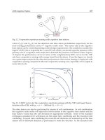
posterior lateral area of the knee. Resisted external rotation (Figure 13.8)
while palpating the popliteus produces pain. This test is performed with
patients lying on their back with the painful leg placed in 90° of hip flexion
282 E.J. Shahady
FIGURE 13.8. Resisted external rotation.
and 90° of knee flexion. The clinician stands on the lateral side of the knee
with one hand supporting the knee and the other placed on the foot resist-
ing external rotation.
10.1. Imaging
An MRI may be needed to make a definitive diagnosis.
10.2. Treatment
Excessive quadriceps fatigue strains the popliteus so a rehabilitation program
emphasizing strengthening of the quadriceps muscle should be instituted.
A 2-week course of NSAIDs should also be included in the treatment.
Recalcitrant cases may require a local injection of a steroid. Maintaining
good quadriceps strength is the key to preventing a recurrence.
11. Retrocalcaneal Bursitis
The retrocalcaneal bursa is located behind the calcaneus and in front of the
Achilles tendon at its insertion site onto the calcaneus. The history is generally
that of slow onset of dull aching pain in the retrocalcaneal area aggravated by
activity and certain shoe wear. A common complaint is start-up pain after sit-
ting or when arising in the morning. Examination reveals swelling in between
the Achilles tendon and the calcaneus. There is generally a prominence in the
area of the superior portion of the heel. Palpation may reveal the presence of
fluid within the bursa. Dorsiflexion of the foot usually increases the pain in
the area. Retrocalcaneal bursitis may be a manifestation of systemic arthritis
or gout. Treatment is similar to that used for Achilles tenonitis.
12. Achilles Tendon Disorders
Commonly called “Achilles tendinitis” by many clinicians, posterior heel pain
in the setting of exercise and overuse represents spectrum of problems caused
by both inflammation and degeneration. Entities include tendonitis with and
without partial rupture, retrocalcaneal bursitis, and complete tear caused by
an acute injury. Achilles tendon disorders occur most often in patients
involved in activities where running is an important part of the activity. Like
other overuse injuries, training errors, improper footwear, and foot pronation
predispose to Achilles injury. Long standing tendon degeneration may occur
without symptoms or pain. But if a change in exercise intensity occurs the
patient will develop symptoms.
A classic history is postexercise pain usually relieved by rest. The pain is
located about 4 to 6 cm proximal to where the tendon inserts on the heel.
13. Lower Leg Problems 283
A change in activity levels or training techniques usually precedes the
onset of symptoms. Patients usually take some NSAIDs, rest a little, and
return to activity. If no change in training or correction of other predis-
posing factors occurs, the pain will return quickly. As the tendonitis con-
tinues, pain may occur during exercise and interfere with activities of daily
living. Familial hypercholesterolemia, which is present in one of 500
patients, is associated with recurrent Achilles tendonitis. So inquiring
about a family history of premature cardiovascular disease or lipid disor-
ders is appropriate if recurrent Achilles tendonitis is present. A complete
rupture of the tendon is usually an acute event accompanied by pain and
inability to plantar-flex the foot. The patient usually complains of a sud-
den severe calf pain as if someone hit them with a rock. They will have dif-
ficulty bearing weight.
Clinical examination of the foot should be performed with the patient
first standing and then prone. Inspection for pronation and palpation of
the tendon for swelling, asymmetry, thickening, erythema, tenderness,
crepitation and nodules should start the examination. Pain anterior to the
tendon at its insertion is a sign of retrocalcaneal bursitis. If the tendon has
ruptured acutely, the patient may have a defect in the tendon about 2 to 3
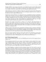
in. from its insertion. The Thompson test (Figure 13.9) should be per-
formed to assess the integrity of the Achilles tendon. With the patient
kneeling on a chair grasp the calf and note the ability of the foot to plan-
tar-flex. Plantar flexion will not occur with a torn tendon. The test is best
performed within 48 h of the rupture.
284 E.J. Shahady
FIGURE 13.9. Thompson test.
12.1. Diagnostic Tests
Ultrasound and MRI are sometimes used if it is difficult to make the diag-
nosis. Although ultrasound is less expensive, both are costly and should be
used with discretion. History and examination are usually sufficient to make
the diagnosis and start treatment unless a complete tear is likely.
12.2. Treatment
Initial management should focus on symptom relief and correcting the train-
ing errors and mechanical problems. Cessation of running and cross training
with a stationary bike or swimming plus the use of NSAIDs will help decrease
the symptoms. There is no place for injection of steroids into the tendon but
steroid injection may be considered for retrocalcaneal bursitis. Ice massage as
described in Chapter 1 can also decrease symptoms and help with inflamma-
tion. Exercises to stretch and strengthen the tendon as described at the end of
this chapter are important. Orthotics for pronation and a heel lift also help. The
heel lift should be used for a short time to decrease the discomfort. Operative
treatment may be needed in a small number of patients for excision of adhe-
sions and degenerated nodules, or decompression of the tendon by longitudi-
nal tenotomies. If the tendon is completely ruptured, surgery may be indicated
depending on the age, level of activity, and medical status of the patient.
13. Fractures of the Tibia and Fibula
Fracture of the tibia secondary to trauma are not usually a diagnostic prob-
lem. Type 1 growth plate fractures in children may be a little more difficult
to diagnose because the X-ray is usually negative. Any child or adolescent
less than age 16 may have open growth plates. Any child in this age group
with significant lower leg pain, inability to bear weight, and a negative X-ray
should be considered to have a growth plate fracture until proven wrong.
The key symptom is inability to bear weight. Because of the potential
impact on bone growth, a consultation with an orthopedic surgeon is
recommended.
Isolated fibula fractures, especially of the distal fibula, are not usually
problematic because the fibula is not a weight-bearing bone. Proximally the
fibula anchors the lateral supports of the knee and distally it is the lateral
buttress for the talus and ankle joint. In patients with tibial fractures, stabil-
ity of the fibula assumes more importance. Fixation of the fibula may be
indicated in order to restore stability and alignment for the tibia. An intact
fibula in association with a tibial shaft fracture is actually a marker for a less
severe injury and an improved prognosis.
Most fibular fractures are distal and associated with an ankle inversion
injury. If there is a fracture in the proximal fibula be alert for a Maisonneuve’s
13. Lower Leg Problems 285
fracture. This is a proximal fibula fracture with an associated ankle fracture or
ankle deltoid ligament tear. This fracture is also associated with partial or com-
plete disruption of the syndesmotic membrane between the tibia and fibula.
An orthopedic surgeon should manage Maisonneuve’s fracture.
Treatment of truly isolated fibular shaft fractures is symptomatic. A well-
padded splint or cast may be useful briefly for comfort, but is not required.
A lightly wrapped elastic bandage is applied over the padding. Elevation,
ice, crutches (with weight bearing as tolerated), and NSAIDs as needed are
helpful. Once the pain and swelling have largely resolved (usually in 1 to 2
weeks), progressive weight bearing is encouraged, and activities are encour-
aged. This fracture is treated as inversion ankle injury, which is discussed in
Chapter 14.
14. Medical Problems
14.1. Baker’s Cyst
A Baker’s, or popliteal, cyst should be considered in a patient with a bulge or
pain in the back of the knee, also known as the popliteal region. The cyst rep-
resents a herniation of the synovial membrane through the posterior aspect
of the capsule of the knee. Fluid may escape through the normal communi-
cation of the bursa with the knee joint producing a budge. The herniation can
sometimes also occur laterally. The underlying problem is always internal
derangement of the knee (loose body, meniscal tear, and degenerative arthri-
tis) that produces synovitis and fluid accumulation. As the severity of the
synovitis increases more fluid is produced and the size of the cyst (bulge) will
increase. This is an important piece of information in the history, as these
patients may not have a prior history of posterior pain but one of a posterior
knee mass that fluctuates in size.
The clinical challenge comes when there is rupture of the cyst and escape
of fluid into the calf. This produces significant pain and a clinical picture sim-
ilar to thrombophlebitis and gastrocnemius strain or tear. Baker’s cyst are
usually present in older not-too-active patients who have a history of
osteoarthritis and a fluctuating posterior knee mass. Nevertheless, there are
active patients in their middle ages that can have a baker’s cyst and/or tear
their gastrocnemius muscle. A meticulous history and physical using the sug-
gestions listed in other parts of this chapter will usually help establish the
diagnosis. If knee pathology is present, a focused knee history and examina-
tion, as discussed in Chapter 12, should help establish the diagnosis of the
knee problem. The diagnosis of thrombophlebitis will be by exclusion of the
other entities and presence of circumstances that predispose the patient to
thrombophlebitis. If you suspect thrombophlebitis, please consult another
source of information.
286 E.J. Shahady
Treatment for Baker’s cyst is primarily for the underlying cause of the cyst
(usually osteoarthristis). Spontaneous disappearance is common but occasion-
ally aspiration and or surgical excision may be required. Differentiation from
other clinical entities may require aspiration, ultrasound, or an MRI scan.
Once the underlying intra-articular pathology is understood, appropriate treat-
ment and prevention measures can be instituted.
15. Spinal Stenosis
Spinal stenosis is mentioned in this chapter because it can cause exertional
lower leg pain (neurogenic claudication). The patients are usually over age
60 and have had a 5- to 10-year history of back pain and other signs of
osteoarthritis of large joints like the knees or the hips. The classical symp-
toms are back pain radiating into the calf and foot brought on by exercise.
It is also classical that the symptoms are relieved by bending over and rest.
Bending backward as demonstrated in Figure 10.3 (page 183) increases all
the symptoms. Chapter 10 has a more extensive discussion of spinal steno-
sis. Vascular claudication must also be ruled out by accessing for loss or
diminishing of dorsalis pedis and posterior tibial pulses with exercise in
these patients. Both entities may be present in some patients.
16. Lower Leg Exercises
Figures for these exercises can be found in Chapter 14.
1. Towel stretch (see Figure 14.10): Sit with your injured leg stretched out in
front of you. Loop a towel around the ball of your foot and pull the towel
toward your body keeping your knee straight. Hold this position for 10 s
then relax. Repeat five times.
2. Standing calf stretch (see Figure 14.12): Facing a wall, put your hands
against the wall at about eye level. Keep the injured leg back, the uninjured
leg forward, and the heel of your injured leg on the floor. Slowly lean into
the wall until you feel a stretch in the back of your calf. Hold for 15 to 30
s. Repeat three times. Do this exercise several times each day.
3. Anterior leg muscle stretch (see Figure 14.13): Stand next to a chair or the
kitchen counter and grasp one of them with your hand to maintain bal-
ance. Bend your knee and grab the front of your foot on your injured leg.
Bend the front of the foot toward your heel. You should feel a stretch in
the front of your shin. Hold for 10 to 15 s. Repeat five times.
4. Heel raises A (see Figure 14.14): Stand behind a chair or counter to balance
yourself. With your feet internally rotated, raise your heels by standing on
the tips of the toes for 5 s. Do this 20 times and repeat two times a day.
13. Lower Leg Problems 287
5. Heel raises B (see Figure 14.15): Stand behind a chair or counter to balance
yourself. With your feet straight, raise your heels by standing on the tips of
the toes for 5 s. Do this 20 times and repeat two times a day.
6. Heel raises C (see Figure 14.16): Stand behind a chair or counter to balance
yourself. With your feet externally rotated, raise your heels by standing on
the tips of the toes for 5 s. Do this 20 times and repeat two times a day.
7. Heel raises on the stairs (see Figure 14.17): Stand on a stairs (grab a banis-
ter for support) and support your body weight on the tips of your toes.
Rise up on your toes for 5 s and then lower the heel down below the toes
to increase dorsiflexion for 5 s. Work up to achieving 10 repetitions three
times a day. The ankle will be stiff and hard to dorsiflex (see Fig. 14.3 on
page 293) initially but will become more flexible with increased repetitions.
Once the degree of dorsiflexion in the injured ankle is the same as the unin-
jured ankle, activity-specific training can begin.
8. Standing toe raises (see Figure 14.18): Stand with your feet flat on the floor,
rock back onto your heels, and lift your toes off the floor. Hold this for 5
s. Repeat the exercise 10 times and do it two times a day.
9. Activity-specific training: If you will be involved in a recreational activity
or competitive sport, gradually acclimatize your ankle to the routines and
stress of this activity. Start with a combined walk–jog–run that is charac-
teristic of this activity/sport. The running/jogging component should grad-
ually increase and replace the walking. Gradually increase the distance and
add figures of eight and backward walking/jogging to the routine. The last
routine attempted should be sharp cutting movement after coming to a
stop.
A trainer, physical therapist, or coach may be able to help you with all of the
above exercises.
Suggested Readings
Hootman JM, Macera CA, Ainsworth BE, et al. Predictors of lower extremity injury
among recreationally active adults. Clin J Sport Med. 2002;12(2):99–106.
Glorioso J, Wilckens J. Exertional leg pain. In: O’Connor F, Wilder R, eds. The
Textbook of Running Medicine. New York: McGraw-Hill; 2001:181–198.
288 E.J. Shahady
14
Ankle Problems
EDWARD J. SHAHADY
This chapter covers primary care problems that occur with the ankle. The most
common problem seen by primary care clinicians is the common ankle sprain.
Unfortunately, ankle sprains are not always treated appropriately. Often, a
patient is evaluated in the emergency department where an X-ray is performed
without much of a history and physical examination and the recommended
treatment is “take it easy or use a set of crutches until you see your doctor”and
no rehabilitation exercises are prescribed. Nonindicated X-rays raise the cost of
initial care and lack of appropriate rehabilitation delays return to activity and
increases the risk of recurrent ankle injury. Other ankle problems like fractures
and osteoarthritis (OA), although less frequent, are discussed.
A focused history that includes the mechanism of injury will help catego-
rize the problem so that a focused examination can be performed. Common
ankle problems seen in primary care are listed in Table 14.1. The decision to
obtain X-rays with acute trauma is facilitated by following the Ottawa ankle
rules (Table 14.2). Following these rules helps decrease unneeded X-rays. An
effective treatment plan should include some form of rehabilitation exercises.
As with all musculoskeletal problems, a good working knowledge of the epi-
demiology, anatomy, associated symptoms, and examination reduce confu-
sion and enhance the diagnostic and therapeutic process.
1. Anatomy
The talus articulates with the tibia and fibula to form the ankle joint. The
talar dome is wider at its anterior margin than the posterior margin by an
average of 2 to 3 mm. This difference in width imparts relative ankle insta-
bility in plantar flexion and increased stability during ankle dorsiflexion. This
partially explains the reason why ankle injury is most common in the plan-
tar-flexed position. Lateral ankle stability is enhanced by the lateral ankle lig-
aments. The lateral ankle ligaments include the anterior talofibular ligament
(ATFL), the calcaneofibular ligament (CFL), and the posterior talofibular
ligament (PTFL) (see Fig. 14.1). The ATFL and CFL are the most important
clinically because they are the most commonly injured ankle ligaments.
289
The ATFL originates from the anterior aspect of the distal fibula and
inserts on the lateral aspect of the talar neck. The CFL originates from the
distal tip of the fibula and inserts at the lateral wall of the calcaneus (Figure
14.1). When the ankle is in dorsiflexion, the ATFL is perpendicular to the
axis of the tibia and the CFL is oriented parallel to the tibia. In neutral dor-
siflexion, the CFL provides resistance to inversion stress or varus tilt of the
talus. In plantar flexion, the most common position for lateral ankle inver-
sion injuries, the ATFL is parallel and the CFL is perpendicular to the
axis of the tibia. This position places the ATFL in the precarious situation of
providing resistance to inversion stress.
Isolated testing of the individual ankle ligaments demonstrates that the
ATFL is the first to fail and the ATFL is considered the weakest lateral ankle
ligament. Sixty-five percent of ankle sprains are secondary to partial or com-
plete rupture of the ATFL. (Figure 14.2). Another 30% are caused by a
sprain or rupture of both the ATFL and the CFL. As previously mentioned,
the PTFL is seldom, if ever, involved in ankle sprains seen in the primary care
setting.
Medial ankle stability is provided by the strong deltoid ligament, the ante-
rior tibiofibular ligament, and the bony mortise. The anterior tibiofibular lig-
ament is located between the distal portions of the tibia and fibula. The
deltoid ligament is composed of four strong ligaments: posterior and anterior
tibiotalar ligament, tibiocalcaneal ligament, and the tibionavicular ligament.
They are named for the bones where they originate and insert.
290 E.J. Shahady
TABLE 14.1. Common ankle problems.
Ankle sprains
●
Lateral ankle sprains
●
Medical ankle sprains
●
High ankle sprains
Fractures
●
Lateral and medial malleolus
●
Talar dome
●
Maisonneuve fracture
Arthritis
●
Osteoarthritis
●
Rheumatoid arthritis
TABLE 14.2. Ottawa ankle and foot rules.
An ankle radiographic series is indicated if a patient has
1. Inability to bear weight immediately in the emergency department or physician’s office or
2. Pinpoint bone tenderness at the posterior portions of the lateral and medial malleolus
A foot radiographic series is indicated if a patient has pinpoint pain over the base of the fifth
metatarsal or the navicular bones
Adapted from Stiell IG, McKnight RD, Greenberg GH, McDowell I, Nair RC, Wells GA,
et al. Implementation of the Ottawa ankle rules. JAMA. 1994;271:827–832.
Because of the support of the bony articulation between the medial malleo-
lus and the talus, medial ankle sprains are less common than lateral sprains. In
medial ankle sprains, the mechanism of injury is excessive eversion and dorsi-
flexion. Medial ankle sprains are more problematic and take more time to heal.
14. Ankle Problems 291
Posterior
Talofibular
Calcanofibular
Anterior
Talofibular
Ligament
FIGURE 14.1. Lateral ankle ligaments. (Reproduced from Shahady E, Petrizzi M, eds.
Sports Medicine for Coaches and Trainers. Chapel Hill, NC: University of North
Carolina Press; 1991:119, with permission.)
Anterior
Talofibular
Ligament
FIGURE 14.2. Anterior talofibular ligament tear. (Reproduced from Shahady E,
Petrizzi M, eds. Sports Medicine for Coaches and Trainers. Chapel Hill, NC: University
of North Carolina Press; 1991:120, with permission.)
2. Focused History
Establish whether the problem is acute or chronic or if other chronic diseases
that have musculoskeletal components are present. This will get you started
down the right path. The mechanism of injury will many times pinpoint the
anatomy involved in the injury. Questions like the following help put the
pieces of the puzzle together. If the problem is chronic and getting worse ask
how it is related to exercise. Is it only present with exercise? Does it stop or
continue when exercise is over? Certain characteristics like intensifying one’s
exercise routine, changing the terrain like hills or the beach, or a change of
shoes are all areas that may be causative. Chronic problems like OA usually
wax and wane with time. Patients with OA usually have evidence of other
joint involvement like the hands (Heberden’s nodes) and large joints like the
knees and hips. Rheumatoid arthritis (RA) may involves the ankle and foot
and the first signs of rheumatoid may be in the foot and ankle.
Ask about prior ankle injury. Old ankle sprains that were not properly
rehabilitated lead to increased risk of new ankle sprains. Ability to bear
weight after acute trauma is a critical piece of information. Third-degree
ankle sprains and fractures are likely when the patient is unable to bear
weight. How the injury occurred also helps. Inversion injury leads to tears
of the lateral ligaments and eversion injury tears the deltoid ligaments.
A “pop” followed by immediate swelling usually indicates a torn ligament.
When the swelling occurred is important to note. Swelling that occurred the
day after an injury or after using heat rather than ice is less significant than
swelling that occurs immediately and is disabling. A mechanism of injury
that leads to twisting or rotation of the lower leg with eversion and inver-
sion should lead the clinician to consider a syndesmosis or high ankle
sprain injury. High ankle sprains may have significant pain with minimal
swelling.
Patients with chronic or recurrent ankle sprain may complain of weakness,
apprehension, loss of coordination, periodic swelling, and episodes of the
ankle “giving away.” Running on uneven or loose surfaces brings out many
of the symptoms of chronic ankle sprains.
3. Examination
Even if the patient’s history suggests an inversion injury, the examination
should not be limited to the lateral ankle ligaments. The examination should
rule out ATFL sprain, CFL sprain, syndesmosis sprain, deltoid sprain, per-
oneal tendon tear, lateral malleolus fracture, and talar dome osteochondral
injury. First, observe for swelling and deformities. Remember RA may first
manifest itself in the ankle and foot. Palpation for tenderness over
the ATFL and the CFL as well as the bones of the navicular, malleoli,
and the fifth metatarsal should be performed. Feel for nodules of the
292 E.J. Shahady
Achilles tendon and other extensor surfaces. These nodules may be associ-
ated with RA or familial hypercholesterolemia. Range of motion of the
ankle is assessed with the patient seated and relaxed. Maximal dorsiflexion
and plantar flexion are observed both passively and actively. Dorsiflexion
(Figure 14.3) can normally be accomplished to 15° to 20° and plantar flex-
ion up to 40° to 55° (Figure 14.4). Compare the injured to the uninjured
side to access for differences. Initial measurements can be used as a baseline
to evaluate progress.
Next, a series of tests to evaluate for ligamentous injury and stability are
performed. The squeeze test identifies tibiofibular syndesmosis disruption
(the interosseus membrane between the tibia and fibula). The test is per-
formed by compression of the midleg from posterior lateral to anterior
medial area, as noted in Figure 14.5. This test is positive when the compres-
sion produces pain secondary to separation of the fibula from the tibia in the
lower ankle.
The talar tilt test is performed with the lower leg secured with one hand
and the heel grasped from behind with the opposite hand. An inversion force
is placed in an effort to produce a talar tilt. Perform the test against resistance
in both ankle neutral and plantar-flexed positions (Figure 14.6). Inversion
14. Ankle Problems 293
FIGURE 14.3. Dorsiflexion.
294 E.J. Shahady
FIGURE 14.4. Plantar flexion.
FIGURE 14.5. Squeeze test.
stress in the neutral position tests the stability of the CFL and inversion stress
in the plantar-flexed position tests the stability of the ATFL.
4. Test
The anterior drawer test tests the integrity of the ATFL. While the patient is
seated the lower leg is grasped with one hand and the foot with the other
(Figure 14.7). An anterior force (see arrow in Figure 14.7) is used in an effort
to produce forward translation. Perform the test in both ankle neutral and
plantar flexion positions. A few millimeters of translation is normal.
Compare one side with the other. The test is more reliable in chronic insta-
bility than in acute because of the negative inhibition of pain during an acute
sprain. Thus, a negative test is not always reliable with acute ankle sprain and
should be repeated when the pain subsides.
14. Ankle Problems 295
FIGURE 14.6. Talar tilt (inversion stress).
5. Case
5.1. History and Exam
A healthy 23-year-old female student who regularly runs 3 miles three times
per week comes to your office with ankle pain and swelling. She tripped on a
tree root in the woods yesterday while running and twisted her right ankle.
She did not hear or feel a pop, but noticed immediate pain on the lateral side
of the ankle. She was able to bear weight and walk out of the woods. As
she walked, the ankle became more painful and began to swell. She has no
history of past ankle injuries and she has no known medical problems. Based
on the advice of a friend she used a heating pad to help decrease the swelling
and pain. The next morning she noted increased swelling and moderate dis-
comfort while walking.
The examination revealed swelling, ecchymosis, and diffuse tenderness over
the anterior portions of right lateral malleolus. Dorsiflexion is limited to 10°
on the right foot compared with 20° on the left. Plantar flexion is equal bilat-
erally to 45°. The anterior and medial portions of the malleolus are tender
but not the posterior portion of the lateral malleolus. The squeeze and ante-
rior drawer tests are negative. The talar tilt test produces pain in the plantar
flexed position but no instability. Palpation of the base of the fifth metatarsal
reveals no tenderness.
296 E.J. Shahady
FIGURE 14.7. Anterior drawer.
The patient was prescribed a nonsteroidal anti-inflammatory drug
(NSAID), and taught some initial exercises to stretch and strengthen her ankle
ligaments. After 2 weeks, the patient returned to running but was unable to
complete her runs without lateral ankle pain. She then was treated by a physi-
cal therapist and after 2 months has returned to her full routine of running.
5.2. Thinking Process
The history suggests that the patient has probably stretched or torn one of
her lateral ankle ligaments. The anterior talar fibular and CFLs are the most
common lateral ankle ligaments injured. The immediate swelling indicates
bleeding and the use of a heating pad suggests increased bleeding secondary
to the vasodilatation caused by the heat. The ability to walk and bear weight
is a significant piece of history. Fractures and complete ligamentous tears are
associated with an inability to bear weight. Therefore, the diagnosis is most
likely a first- or second-degree sprain. Her lack of a history of ankle injury
goes against this, being a chronic ankle sprain. Rheumatoid arthritis can
present with ankle pain but it is not usually associated with an acute event.
There is no pinpoint tenderness over the posterior portions of the lateral and
medial malleolus tests, indicating fracture is unlikely. A negative squeeze test
makes a syndesmosis tear unlikely and the negative anterior drawer suggests
that the ATFL is intact. The talar tilt test produces pain in the plantar-flexed
position but no instability, indicating complete stability of the ATFL and
CFL. An avulsion fracture of the fifth metatarsal at the insertion of the per-
oneus brevis is not likely because no tenderness was demonstrated at the base
of the fifth metatarsal. After reviewing the Ottawa Ankle Rules (Table 14.2)
a decision was made not to perform an X-Ray of the ankle or foot. The
patient was able to bear weight and had no pinpoint tenderness on the pos-
terior portions of the lateral malleolus or the base of the fifth metatarsal.
6. Ankle Sprains
Ankle sprains are classified according to signs and symptoms. Grade I is char-
acterized by a stretching of the ATFL and the CFL, producing mild tender-
ness and swelling. Usually no ecchymosis is present and no loss of function or
motion. The patient is able to bear weight and walk with minimal pain.
Examination reveals no instability with the talar tilt and anterior drawer test.
The patient usually recovers with minimal treatment in 6 to 8 days.
Grade II is an incomplete tear of the ATFL and stretching of the CFL with
moderate pain and swelling. There is ecchymosis, more swelling and tender-
ness, and some loss of function and motion. The patient has pain with weight
bearing but is able to walk usually with a limp. Examination reveals mild to
moderate instability with the tilt and anterior drawer tests. These patients may
take 3 to 6 weeks to recover and chronic instability is more likely.
14. Ankle Problems 297
Grade III is a complete tear of the ATFL and CFL and partial tears of the
posterior talofibular and the tibiofibular ligaments. There is immediate and sig-
nificant swelling. The ecchymosis is more significant. The patient loses function
and motion and is unable to bear weight or ambulate. The talar tilt test and
anterior drawer tests are positive indicating significant instability. Many times
the tests cannot be performed because of the marked discomfort with move-
ment. These patients are probably best referred to an orthopedic surgeon. It is
debatable whether they do better with surgery or casting.
7. Mechanism of Injury
Up to 90% of ankle sprains are caused by inversion of the plantar-flexed
ankle. The ATFL and CFL ligaments are the most commonly injured when
the ankle is inverted and the ATFL is the most easily injured. Significant
instability can occur when both ligaments are injured. Both grade II and III
sprains can lead to significant chronic instability if effective rehabilitation is
not accomplished. Excessive eversion and dorsiflexion produces sprains of
the strong deltoid ligament medially. Medial injury is not that common
because of the stability provided by the bony articulation. Injury to the (syn-
desmosis) tibiofibular ligament and the interosseus membrane between the
tibia and fibula (Figure 14.8) usually occurs with a combination of twisting
and plantar flexion.
8. Evaluation
The evaluation includes an assessment of the grade of sprain and an appli-
cation of the Ottawa ankle rules (Table 14.2). After this evalution the clini-
cian can more readily make decisions about imaging, prognosis and
treatment. The history should include a description of the mechanism of
injury, past history of ankle problems, ability to bear weight after the injury,
and what treatment the patient used prior to your evaluation. Examination
should include inspection, palpation of the malleoli, weight-bearing status,
and all the tests listed in the focused examination. If you suspect a syn-
desmosis injury, palpate the entire length of the tibia and fibula to detect a
fracture of the proximal fibula (Maisonneuve fracture). Palpate the base of
the fifth metatarsal to rule out an avulsion fracture.
Two unusual but significant scenarios should be kept in mind. One is a
possible talar dome fracture. These patients will not be able to bear weight but
the X-ray may not be positive for 2 to 4 weeks. The fracture may be between
the talus and the fibula or the talus and the tibia. Persistent pain and limita-
tion of dorsiflexion and plantar flexion should raise suspicion of this fracture.
The other scenario is significant pain and disability, minimal swelling, and ten-
derness over the distal tibiofibular joint. This usually indicates a syndesmosis
298 E.J. Shahady
sprain or high ankle sprain. In addition to the “squeeze test” (Figure 14.5) for
disruption of the syndesmosis, an additional test called the “external rotation
test” will also help identify syndesmosis disruption. This test (Figure 14.9) is
performed by externally rotating the foot with the ankle in dorsiflexion. Pain
in the distal tibiofibular junction indicates syndesmosis injury.
9. Imaging
The Ottawa ankle rules (Table 14.2) can be used to determine when radi-
ographic studies are needed. Location of the pain and inability to bear weight
are the key indicators. Use of these rules has decreased costs, waiting time,
and unneeded tests. The rules are highly sensitive for malleolar and midfoot
fractures. If X-rays are obtained, they should include anteroposterior, lateral,
and mortise views to rule out the most common types of fractures. In addi-
tion to looking for fractures, observe for an increase in the space between the
tibia and fibula. This so-called “clear space” is increased in size with disrup-
tion of the syndesmosis. Talar dome fractures are not always seen initially on
the X-ray. If after 6 weeks, the patient has persistent symptoms and the X-ray
14. Ankle Problems 299
Interosseus
Tibiofibular
FIGURE 14.8. Injury to the tibiofibular ligament and the interosseus membrane
between the tibia and fibula. (Reproduced from Shahady E, Petrizzi M, eds. Sports
Medicine for Coaches and Trainers. Chapel Hill, NC: University of North Carolina
Press; 1991:120, with permission.)
remains negative, a magnetic resonance imaging (MRI) or computerized
tomography (CT) scan may be helpful.
10. Treatment
Most ankle sprains can be managed conservatively. Disability from sprains
is related to the loss of motion especially dorsiflexion. Initial goals are to
prevent or decrease swelling and maintain motion. Rest, ice, compression,
and elevation (RICE) are the keys to early management. Initially use
crutches to decrease weight bearing and provide relative rest for 48 to 72
hours and ice to decrease pain and swelling. The ice can be placed in a plas-
tic bag applied to the ankle over a thin cloth. Ice decreases metabolism and
limits hypoxic injury. Immersion in a cold-water bath can also be used. Ice
massage can be applied by filling paper cups with water and placing them in
a freezer. The patient then removes the cup and peels a portion of the rim to
expose the ice and use it to massage the ice into the area affected. The ankle
should be cooled for 20 min every 3 to 4 h during the first 48 h. Heat should
not be used for an acute ankle sprain because it increases swelling and
inflammation. Compression with an ace wrap on the ankle helps limit the
swelling. Elevation is the most poorly followed of all the components of
RICE. The elevation should be above the level of the heart to not only
300 E.J. Shahady
FIGURE 14.9. External rotation test.
decrease the swelling but also displace it. Effective elevation will cause
the blood to pool away from the area of injury and go to a higher spot on the
leg or into the lower foot. Blood is a proinflammatory agent. If the blood
stays in the area of the torn ligaments, it aids in the promotion of the inflam-
mation. Elevation causes the blood to go away from the area of the original
ligamentous tear. Following these few simple steps will decrease recovery
time.
Prolonged immobilization of ankle sprains is a common treatment error.
Movement of the ankle stimulates the incorporation of stronger replacement
collagen. Sprained ankles tend to stiffen in a plantar-flexed, slightly inverted
position and preventing this stiffening is critical to more rapid recover.
Rehabilitation begins on the day of injury and continues until pain-free gait
and activity can be attained. Range-of-motion and muscle strengthening
exercises and proprioceptive and activity-specific training are the key com-
ponents of rehabilitation. Grade I and II rehabilitation can start immediately
but grade III sprain rehabilitation will not begin until the ankle is judged sta-
ble. Rehabilitation begins with ROM and progresses to muscle strengthening
followed by proprioceptive and activity-specific training.
Painless ROM exercises should begin within hours of the injury. Patients
are asked to write the alphabet with their toes on the floor without bearing
weight and pull a book on a towel toward them. After 48 h, instruct patients
to pull their toes with a towel to stretch the Achilles tendon and prevent loss
of dorsiflexion (Figure 14.10). Once they are able to bear weight without pain
and a limp they can use an air-filled or gel-filled ankle brace. These braces
restrict inversion–eversion and allow plantar and dorsiflexion. Casting is
14. Ankle Problems 301
FIGURE 14.10. Towel stretch.
discouraged as it limits plantar flexion and dorsiflexion. Other exercises are
now started to regain strength and balance. These exercises are described at
the end of the chapter. A program for activity-specific training should also be
recommended. A physical therapist can also be a big help if the patient needs
more encouragement or is not responding as well as anticipated.
Occasionally it may be necessary for the clinician to recommend no weight
bearing and use crutches or a cast for 10 to 14 days and then reevaluate for
recovery or the presence of a talar dome or other fracture.
11. Fractures
Ankle fractures can occur at the lateral malleolus (distal fibula), medial
malleolus, posterior portion of the medial malleolus, and the talar dome.
Stable ankle fractures involve only one side of the ankle joint like the distal
fibula fracture with no involvement of the medial bony or ligamentous struc-
tures. Unstable ones involve two or three sides of the ankle joint. Bimalleolar
fractures include fractures of the lateral and medial malleoli or the lateral
malleolus and a disrupted deltoid ligament. Trimalleolar fractures have the
additional involvement of the posterior portion of the medial malleolus.
Stable fractures are treated nonoperatively but the unstable ones require
orthopedic evaluation and possible surgery. The history is usually one of
acute twisting trauma and immediate onset of pain. The examination will
reveal an immediate effusion and significant pinpoint tenderness. Palpation
should always include the proximal fibula to rule out the Maisonneuve frac-
ture and tenderness of the tibiofibular ligament and the syndesmosis.
X-rays should include anteroposterior, lateral, and mortise views to rule
out the most common types of fractures. Talar dome fractures are not always
seen initially on the X-ray. If after 6 weeks, the patient has persistent symp-
toms and the X-ray remains negative an MRI or CT scan may be helpful.
Initial treatment should include protection of the ankle, ice, and relief of
pain with a narcotic if needed. As the fibula is a non-weight-bearing bone a
cast may not be needed. Small tip avulsion fractures of the distal fibula can
be treated like an ankle sprain. Braces and weight-bearing casts can be used
for other distal fibular fractures. The unstable fractures will require non-
weight-bearing casts and more prolonged immobilization. Surgery may be
needed for some unstable fractures.
12. Arthritis
Rheumatoid arthritis may affect the ankle. Ankle swelling without trauma in a
patient less than 50 should make the clinician suspicious of RA. Ten to fifteen
percent of patients with RA have symptoms in the ankle and foot. In a small
percentage, the first symptoms may be in the ankle and foot. The talonavicular
joint is the most common one involved in RA. Swelling and decreased ROM
302 E.J. Shahady
(mostly dorsiflexion) are the most common presenting symptoms. Foot symp-
toms may also be present. The gait may be abnormal because of the pain with
walking. Examination may reveal a warm tender joint that has decreased plan-
tar flexion and dorsiflexion. Rheumatoid nodules (firm nontender movable
masses) on the extensor surfaces may also be present. X-rays may show joint
space changes early in the disease. If RA is suspected, be sure to obtain appro-
priate laboratory tests and consider referral for disease-modifying drug ther-
apy. Many of these patient eventually become candidates for surgery.
Osteoarthritis is the most common nontraumatic cause of ankle pain. These
patients are usually over 50 and may have had some type of trauma to the ankle
in the past. The tibiotalar, talonavicular, and talocalcaneal joints are most com-
monly affected by OA. Other joints like the first metatarsal phalangeal, hip,
knee, and distal interphalangeal are commonly involved at the same time as the
ankle. Symptoms are pain, swelling, and difficulty walking. Examination may
reveal decreased ROM and walking with the leg externally rotated. Treatment
consists of using Tylenol and/or NSAIDs, orthotics, and firm but comfortable
shoes. Ankle injection with lidocaine and a steroid may also help. The injection
should be into the tibiotalar joint. Palpate for a soft area just medial to the
anterior tibial tendon to identify the tibiotalar joint (Figure 14.11).
13. Ankle Exercises
Tell the patient to repeat each of the following exercises two times a day.
Rotate from one exercise to the other. Do one set of one exercises and then
rotate to another exercise and do a set. Do not exercise past the point of pain.
Pain means stop.
14. Ankle Problems 303
Anterior Tibial Tendon
F
IGURE 14.11. Area for injection of the tibiotalar joint.
304 E.J. Shahady
FIGURE 14.12. Standing calf stretch.
1. Towel stretch (Figure 14.10): Sit with your injured leg stretched out in front
of you. Loop a towel around the ball of your foot and pull the towel
toward your body keeping your knee straight. Hold this position for 10 s,
then relax. Repeat five times.
2. Standing calf stretch (Figures 14.12): Face a wall, and put your hands
against the wall at about eye level. Keep the injured leg back, the uninjured
leg forward, and the heel of your injured leg on the floor. Slowly lean into
the wall until you feel a stretch in the back of your calf. Hold for 15 to 30 s.
Repeat three times. Do this exercise several times each day.
3. Anterior leg muscle stretch (Figure 14.13): Stand next to a chair or the
kitchen counter and grasp one of them with your hand to maintain
balance. Bend your knee and grab the front of your foot on your injured
leg. Bend the front of the foot toward your heel. You should feel a
stretch in the front of your shin. Hold for 10 to 15 s. Repeat five times.
4. Heel raises A (Figure 14.14): Stand behind a chair or counter to balance
yourself. With your feet internally rotated, raise your heels by standing on
the tips of the toes for 5 s. Do this 20 times and repeat two times a day.
5. Heel raises B (Figure 14.15): Stand behind a chair or counter to balance
yourself. With your feet straight, raise your heels by standing on the tips of
the toes for 5 s. Do this 20 times and repeat two times a day.
14. Ankle Problems 305
FIGURE 14.13. Anterior leg muscle stretch.
FIGURE 14.14. Heel raises A.
306 E.J. Shahady
FIGURE 14.15. Heel raises B.
6. Heel raises C (Figure 14.16): Stand behind a chair or counter to balance
yourself. With your feet externally rotated, raise your heels by standing on
the tips of the toes for 5 s. Do this 20 times and repeat two times a day.
7. Heel raises on the stairs (Figure 14.17): Stand on a stair (grab a banister
for support) and support your body weight on the tips of your toes. Raise
up on your toes for 5 s and then lower the heel down below the toes to
increase dorsiflexion for 5 s. Work up to achieving 10 repetitions three
times a day. The ankle will be stiff and hard to dorsiflex initially but will
become more flexible with increased repetitions. Once the degree of dor-
siflexion in the injured ankle is the same as the uninjured ankle, activity-
specific training can begin.
8. Standing toe raises (Figure 14.18): Stand with your feet flat on the floor,
rock back onto your heels, and lift your toes off the floor. Hold this for
5 s. Repeat the exercise 10 times and do it two times a day.