Blood Disorders in the Elderly - part 2 docx
Bạn đang xem bản rút gọn của tài liệu. Xem và tải ngay bản đầy đủ của tài liệu tại đây (880.22 KB, 50 trang )
38 Oscar A. Cepeda, Julie K. Gammack, John E. Morley
53. Tinetti ME, Baker DI, McAvay G, et al. A multifactorial
intervention to reduce the risk of falling among elderly
people living in the community. N Engl J Med 1994;
331: 821–7.
54. Mathias S, Nayak US, Isaacs B. Balance in elderly
patients: the “get-up and go” test. Arch Phys Med
Rehabil 1986; 67: 387–9.
55. Foley K, Palmer RM. Offi ce evaluation of the frail older:
practical tips. Home Med 1996; 32: 21.
56. Marwick C. NHANES III health data relevant for aging
nation. JAMA 1997; 227: 100–2.
57. Potter J, Langhorne P, Roberts M. Routine protein
energy supplementation in adults: systematic review.
BMJ 1998; 317: 495–501.
58. Guigoz Y, Vellas B, Garry PJ. Mini nutritional
assessment: a practical assessment tool for grading the
nutritional state of elderly patients In Facts, Research,
Interventions in Geriatrics (New York: Serdi, 1997),
15–60.
59. Balducci L, Wallace C, Khansur T, Vance RB, Thigpen JT,
Hardy C. Nutrition, cancer and aging: an annotated
review. J Am Geriatr Soc 1986; 34: 127–36.
60. Aslani A, Smith RC, Allen BJ, Pavlakis N, Levi JA. The
predictive value of body protein for chemotherapy-
induced toxicity. Cancer 2000; 88: 796–803.
61. Fantl JA, Newman DK, Colling J. Urinary Incontinence
in Adults: Acute and Chronic Management. AHCPR
publication No 96-0682 (Rockville, MD: US Department
of Health and Human Services, 1996).
62. Salive ME, Guralnik J, Christen W, Glynn RJ, Colsher P,
Ostfeld AM. Functional blindness and visual impair-
ment in older adults from three communities.
Ophthalmology 1992; 99: 1840–7.
63. Sommer A, Tieslch JM, Katz J, et al. Racial differences
in the cause-specifi c prevalence of blindness in east
Baltimore. N Engl J Med 1991; 325: 1412–17.
64 Tieslch JM, Sommer A, Witt K, Katz J, Royall RM.
Blindness and visual impairment in an American urban
population: the Baltimore Eye Survey. Arch Ophthalmol
1990; 108: 286–90.
65. Macphee GJA, Crowther JA, McAlpine CH. A sim-
ple screening test for hearing impairment in elderly
patients. Age Ageing 1988; 17: 347–51.
66. Popelka MM, Cruickshanks KJ, Wiley TL, Tweed TS,
Klein BE, Klein R. Low prevalence of hearing aid use
among older adults with hearing loss. J Am Geriatr Soc
1998; 46: 1075–8.
67. Cella DF, Tulsky DS, Gray G, et al. The Functional
Assessment of Cancer Therapy scale: development and
validation of the general measure. J Clin Oncol 1993;
11: 570–9.
68. Karnofsky DA. Meaningful clinical classifi
cation of
therapeutic responses to anticancer drugs. Clin Pharm
Ther 1961; 2: 709–12.
39
3
Introduxtion
Oswald Steward
Reeve-Irvine research center, departments of anatomy & nurobiology, nurobiology & behavior,
and neurosurgery, university of california at irvine, Irvine, CA 92697
Introduction
In exploring the assessment of the older aged per-
son, this chapter has two goals. The fi rst is to esti-
mate a person’s life expectancy, tolerance of stress,
medical, rehabilitative, and supportive needs in
planning the management of hematologic condi-
tions. The second is to relate hematologic fi ndings to
a physiologic rather than chronologic classifi cation
of age, refl ecting the function and the health status
of each individual. A special assessment is needed
because aging occurs at different rates for different
individuals, and, in the same individual, for differ-
ent functions.
Various forms of geriatric assessment were devel-
oped by geriatricians with the goal to preserve or
restore health and functional independence, that
is the ability to survive alone. In the scope of these
assessments, the older population was composed
of two groups of individuals. The fi rst group, which
becomes larger with increasing age, includes people
who are functionally dependent, for whom the goal of
management is to restore function and to prevent fur-
ther functional deterioration. These individuals may
be affected by multiple medical conditions that con-
tribute to their dependence. The second group, which
becomes smaller with advancing age, includes people
who are still independent. In this case, the assessment
is aimed to identify those at risk of functional decline,
disease, and death, and the goal of management is to
try to prevent or delay these occurrences.
After an outline of the biology and physiology
of aging we will review different forms of geriatric
4
From fi tness to frailty: toward a nosologic
classifi cation of the older aged person
Lodovico Balducci, Claudia Beghe
assessment and their clinical utilization, we will
discuss the meaning of the common geriatric terms
frailty and disability, and we will conclude by trying
to integrate the different information in a nosologic
classifi cation of aging.
Biology and physiology of aging
Aging has been defi ned as a loss of entropy [1,2]
and of fractality [3]. Loss of entropy implies that the
energy available for daily activities diminishes pro-
gressively with aging, and the survival and the func-
tion of the elder hinge upon energy saving. Loss of
fractality implies a progressive decline in the ability
to deal with the surrounding world due to sensorial
impairment, limited mobility, and waning social net-
work. This construct of aging may be translated into
measurable clinical data, including life expectancy,
tolerance of stress, and ability of independent living.
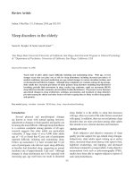
Figure 4.1 illustrates the biology of aging and its
ultimate clinical consequences, and suggests ways
of assessing an individual’s physiologic age. A pro-
gressive exhaustion of functional reserve of multiple
organ systems occurs as a result of genetically deter-
mined programs (a very reasonable, albeit never
conclusively proven, hypothesis), environmental
impact, and disease. Both disease and reduced func-
tional reserve conspire in reducing a person’s life
expectancy and tolerance of stress, and in increas-
ing the risk of disease and functional dependence.
A number of systemic changes, such as increased
concentration of cytotoxic cytokines in the circulation,
Blood Disorders in the Elderly, ed. Lodovico Balducci, William Ershler, Giovanni de Gaetano.
Published by Cambridge University Press. © Cambridge University Press 2008.
parameters. In the following discussion we will
describe three forms of geriatric assessment –
clinical, functional, and biochemical – and we will
explore ways to integrate the geriatric assessment
into a reproducible clinical classifi cation of older
individuals.
Clinical assessment of aging
Aging is multidimensional and involves decline in
functional reserve as well as increased prevalence of
chronic diseases, including a number of conditions,
called “geriatric syndromes,” that become more
common with age. In addition, age involves emo-
tional and social changes, such as increased preva-
lence of depression, waning economic resources,
and social isolation, that may be associated with
reduced access to care and poor nutritional and
health habits. Not surprisingly, the most common
and time-honored evaluation of the older aged per-
son is a multidimensional assessment.
Comprehensive geriatric assessment (CGA)
Though the CGA has not been standardized, there is
general agreement on its main components (Table 4.1)
[23–31].
Function
Function is assessed as performance status (PS),
activities of daily living (ADL), and instrumental
activities of daily living (IADL). ADLs include trans-
ferring, bathing, dressing, eating, toileting, and
continence; dependence in one or more of these
activities, with the exception of incontinence, indi-
cates that the person needs a home caregiver, and
is associated with a two-year mortality rate of 27%.
ADL dependence may prompt admission to an
assisted living facility [32–35]. IADLs are necessary
to maintain an independent life and include use of
transportation, shopping, ability to take medica-
tions, provide for one’s meals, use the telephone,
and manage fi nances. Dependence in one or more
endocrine, immune, and proliferative senescence,
effect and catalyze the decline in functional reserve
and the susceptibility to stress and disease [4,5].
Infl ammatory cytokines are responsible in part for sar-
copenia [6–8], osteoporosis [9,10], and dysfunction of
multiple organ systems [4,11–13], including the central
nervous system [14–17] and the hematopoietic system
[18,19]. Endocrine senescence involves decreased pro-
duction of sexual hormones and chronic hypersecre-
tion of adrenal corticosteroids [20] that together may
lead to sarcopenia, osteoporosis, fatigue, and func-
tional dependence. Immunosenescence involves pro-
gressive loss of cell-mediated immunity, which may
predispose to infection by intracellular organisms,
especially viruses [21], and to highly immunogenic
tumors [21]. Proliferative senescence, best described
in stromal cells, involves the loss of a cell’s self-
replicative capacity, associated with production of
growth factors and lytic enzymes that may in the
meantime destroy normal tissues and promote the
growth of neoplastic ones [22].
Figure 4.1 suggests a number of ways of assessing
a person’s physiologic aging, including evaluation of
function, of medical conditions, and of laboratory
40 Lodovico Balducci, Claudia Beghe
Figure 4.1 The biology of aging and its clinical
consequences.
Genome Environment
Reduced
life expectancy
Disease
Loss of independent living
Declining
functional
reserve
Reduced
stress
tolerance
Catabolic cytokines
Endocrine, Immune
and Proliferative
Senescence
From fi tness to frailty 41
IADLs is associated with a 16% two-year mortality
rate and indicates that the person cannot survive
alone for a long period of time and needs the sup-
port of a caregiver, albeit not necessarily a home
caregiver. In addition, dependence in one of more
IADLs is harbinger of dementia in approximately
50% of cases [34, 35] and of complications from cyto-
toxic chemotherapy, especially neutropenic infec-
tions [36, 37]. Two studies found a poor correlation
between functional dependence and PS, and rec-
ommended that both be evaluated [26, 27]. Though
they are not part of the CGA, the advanced activi-
ties of daily living (AADL) are generating increasing
interest. The AADLs are those that make life pleasur-
able and include leisure as well as professional and
other working activities. Seemingly AADLs may rep-
resent an indirect measurement of the quality of life
of the older person [38, 39].
Comorbidity
In the CGA, comorbidity refers mainly to chronic
diseases. It is important to remember, however, that
the mortality from acute conditions, especially
infections and emergency surgery, increases with
age [40,41]. Comorbidity is associated with decreased
survival and function, and may affect hematopoiesis
and hemostasis. For example, anemia of chronic
infl ammation and anemia of chronic renal insuf-
fi ciency are among the most common forms of
anemia in older individuals [42]. The assessment
of comorbidity has not been standardized and is a
subject of ongoing geriatric research. From a prac-
tical standpoint it is helpful to recognize that some
comorbidities are independent risk factors of death.
These include congestive heart failure and chronic
renal insuffi ciency [43,44]. Of special interest to the
readers of this book, anemia was also found to be an
independent risk factor of mortality for individuals
aged 65 and older [45–49], but it is not clear whether
anemia itself is a cause of mortality or simply a marker
of underlying diseases. After compiling a list of con-
ditions associated with decreased survival in the
general population, two approaches have been taken
for the assessment of comorbidity. One approach
is to sum the number of comorbid conditions [44].
The other utilizes comorbidity scales, accounting
for the severity as well as the number of these condi-
tions. The Charlson scale and the Cumulative Illness
Rating Scale for Geriatrics (CIRS-G) have been used
Table 4.1. Comprehensive geriatric assessment and clinical implications.
Functional status Dependence in one or more of these activities is associated with
Activities of daily living (ADL) and instrumental decreased life expectancy and with functional dependence
activities of daily living (IADL)
Comorbidity Comorbidity is associated with reduced life expectancy and with
Number of comorbid conditions and comorbidity functional dependence. In addition, comorbidity may be
indices associated with polypharmacy and may affect hematopoiesis
and hemostasis
Emotional conditions Depression has been associated with decreased life expectancy
Geriatric Depression Scale (GDS) and function. It may reduce motivation for health care
Nutritional status Reversible condition. Possible relationship to survival. May
Mini Nutritional Assessment (MNA) affect hematopoiesis
Polypharmacy Risk of drug interactions and hematopoietic suppression.
Risk of drug-induced hemolytic anemia and bleeding
Geriatric syndromes Virtually all geriatric syndromes are associated with reduced
Delirium, dementia, depression, falls, incontinence, life expectancy and with functional dependence
spontaneous bone fractures, neglect and abuse,
failure to thrive, vertigo
42 Lodovico Balducci, Claudia Beghe
in the majority of studies [44]. The Charlson scale is
suitable for epidemiologic studies, as it is simpler to
use and may be scored based on data derived from
medical and insurance records, whereas the CIRS-
G appears more appropriate for individual assess-
ment of comorbidity in clinical studies [41]. The
CIRS-G is more cumbersome and time-consuming,
but is more sensitive [44]. Another advantage of the
CIRS-G is that its score may be translated into a
Charlson score.
In addition to providing an estimate of physiologic
aging, the assessment of comorbidity reveals con-
ditions that may be reversed or arrested, at least in
part, and whose management may delay aging. For
the non-geriatrician this emphasis on comorbidity
assessment may appear redundant, as it should be
part of all good practice. The fact is, however, that
disease manifestations in the elderly may often be
neglected or misinterpreted by the patients them-
selves or by the healthcare provider, because they
are attributed to a pre-existing condition or are
wrongly considered normal manifestations of aging.
For example, the diagnosis of bone cancer may be
delayed as bone pain may be ascribed to pre-existing
arthritis or to pain and ache typical of age. For this
reason a careful medical history with special empha-
sis on new symptoms is recommended at each
encounter with older patients. Atypical presentation
of diseases is another reason why comorbidity may
be under-diagnosed. Coronary ischemia in individu-
als over 70 may present as fatigue as commonly as it
does with chest pain [50], and delirium is a harbinger
of underlying organic disorders, such as infections,
electrolyte imbalance, pain, and medication-related
problems [51].
Geriatric syndromes
These conditions are typical of aging, if not specifi c,
and include dementia, depression, delirium, incon-
tinence, falls, spontaneous bone fractures, failure
to thrive, neglect and abuse, and vertigo. They are
associated with reduced life expectancy and almost
always with some degree of functional dependence
[34,35,51–57]. Effective management may reverse
depression, falls, and osteoporosis, and may arrest
the progression of other geriatric syndromes,
including dementia. Screening older individuals
for dementia, depression, osteoporosis, and risks of
falling may be benefi cial by allowing early diagnosis
and timely management [51,58,59].
Failure to thrive, the inability to gain weight despite
adequate food intake, is a sign of advanced aging and
is seldom reversible. The cause is unknown in most
cases and the mechanism may include overwhelm-
ing concentration of catabolic cytokines in the cir-
culation leading to progressive sarcopenia [60].
Neglect and abuse is the least defi nable of the geriat-
ric syndromes and is recognizable because patients
are poorly kept and withdrawn. This is also a sign of
advanced aging and of inadequate caregiving.
Geriatric syndromes are recognized as such when
they interfere with a person’s daily life. Dementia
must be severe enough to disconnect an individual
from daily activities; delirium must occur as a result
of medications or organic diseases that do not com-
monly affect the central nervous system (e.g., uri-
nary or upper respiratory infections); incontinence
must cause a restriction of one’s social life; depres-
sion must prevent pleasurable interactions and be
associated with eating or sleeping disorders; falls
must occur at least three times a month or the fear
of falling must prevent regular activities, such as
walking; vertigo must be continuous and so annoy-
ing as to cause a restriction in mobility.
Social resources
The adequacy of social resources is determined by
individual needs. Those who are dependent in one
or more ADLs do need a home caregiver, at least part
of the time; those dependent in one or more IADLs
do need a caregiver that is reachable and available
on a short-time notice. Even for individuals who
are fully independent and with negligible comor-
bidity it may be useful to identify a potential care-
giver, as any acute disease or strenuous treatment,
such as cancer chemotherapy, may precipitate func-
tional dependence. Generally the caregiver or pro-
spective caregiver is an older spouse with health
From fi tness to frailty 43
problem of his/her own or an adult child, more often
a daughter, who has to manage competing requests,
from parents, from her/his family and from her/his
profession. In addition to improving the quality of
caregiving, appropriate planning may minimize the
emotional stress [61,62].
Living conditions, access to transportation and
to food, and income are interrelated and determine
the quality of health even for individuals who are
functionally independent. It is clear that a person in
a wealthy retirement community, with close neigh-
bors and shopping centers, and a choice of public
transportations, has a better chance to survive an
acute problem, causing momentary loss of func-
tion, than a person living in a run-down and unsafe
neighborhood or one living alone in the countryside
far from shops or public transportation.
Simple adjustments in home environment may
go a long way in preventing common complications
of aging. Good illumination, removal of carpets or
obstacles, creation of a walking pathway where an
individual can always fi nd a support, prevent falls
and allow the older person rapid access to the phone
in case of emergency. In addition, changes in home
environment, such as bathroom bars, may avoid the
transformation of disability into handicap [63].
Nutrition
The prevalence of protein/calorie malnutrition
increases with age [64]. Isolation, depression, eco-
nomic restriction, reduced appreciation of hunger,
may all contribute to insuffi cient food intake, while
chronic diseases, infl ammatory cytokines, and lack
of exercise may impede the synthesis of new proteins
[65]. The Mini Nutritional Assessment (MNA) is a sim-
ple nutritional screening test of worldwide use that
identifi es patients who are malnourished and those
at risk of becoming malnourished, and allows the
prevention and early reversal of malnutrition [66].
Polypharmacy
The prevalence of polypharmacy increases with age,
and among cancer patients aged 70 and older was
found as high as 41% [44,67]. Polypharmacy may
include redundant prescriptions as well as danger-
ous drug interactions, and highlights a common
problem of older individuals in developed countries:
the absence of a primary care provider responsible
for supervising the various medications. According
to a recent study, more than 50% of individuals aged
70 and older in the USA, Canada, and Israel, while
attending multiple specialty clinics, lacked a pri-
mary care physician [68].
Clinical application of the CGA
In general geriatric practice, the CGA has generated
interventions able to preserve the health and inde-
pendence of older individuals, resulting in a decline
in admissions to hospital and to assisted living
facilities. According to early studies, the CGA also
improved the survival of older individuals [27–30].
In addition, the CGA may be used to estimate a
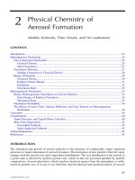
person’s life expectancy [69]. Walter and Covinsky
integrated the results of the CGA with the US life
tables. The life expectancy of each age cohort was
subdivided into quartiles and the CGA determined
to which quartile each individual belonged (Fig.
4.2). The same group of investigators established
criteria to estimate the one-year mortality rate for
older individuals discharged from the hospital
(Table 4.2) [43] and the two-year mortality rate for
home-dwelling older individuals based on function
and comorbidity (Table 4.3) [70]. The benefi ts of the
CGA extend beyond the realm of general geriatrics.
In the management of cancer in older patients, the
geriatric assessment has allowed the identifi cation
of a number of conditions including comorbidity,
cognitive disorders, depression, and malnutrition
that would have remained otherwise unrecognized
[71–73], and it has identifi ed risk factors for chemo-
therapy-related toxicity [37].
Of special interest to the readers of this book,
the geriatric assessment may allow a nosologic
classifi cation of age based on physiologic rather
than chronologic parameters. Hamerman has pro-
posed a frame of reference for this classifi cation
(Table 4.4) [74].
44 Lodovico Balducci, Claudia Beghe
Limitation of the geriatric assessment
The CGA has allowed a formal, systematic, and
largely reproducible exploration of aging and has
demonstrated that aging is multidimensional,
highly individualized, and poorly refl ected in chron-
ologic age. The clinical repercussions of the CGA
include improved management of older individuals
with preservation of function and quality of life and
possibly improvement of comorbidity and of sur-
vival. The CGA may thus be considered the gold-
standard geriatric evaluation and the reference for
the development of new instruments. Several areas
of geriatric assessment need improvement and fi ne-
tuning, as suggested by its current limitations:
• Originally the CGA was designed to improve
the management of patients with advanced
Figure 4.2 Estimate of life expectancy using the life tables: upper, middle, and lower quartiles for women (A) and men (B)
at selected ages. From Walter & Covinsky, 2001 [69], with permission.
25
20
15
10
5
0
Years
70 75 80 85 90 95
Life expectancy for women
21.3
15.7
9.5
17
11.9
6.8
13
8.6
4.6
9.6
5.9
2.9
6.8
3.9
1.8
4.8
2.7
1.1
Top 25th Percentile
Lowest 25th Percentile
50th Percentile
(A)
25
20
15
10
5
0
Years
Life expectancy for men
70 75 80 85 90 95
18
12.4
6.7
14.2
9.3
4.9
10.8
6.7
3.3
7.9
4.7
2.2
5.8
3.2
1.5
4.3
2.3
1
(B)
Age, y
From fi tness to frailty 45
functional impairment and multiple comorbidi-
ties, such as those living in assisted living facilities
and nursing homes, or attending outpatient geri-
atric clinics. As the majority of individuals over 65
enjoy good health and independence it is legiti-
mate to ask two questions: Is a full CGA necessary
and benefi cial for these individuals? Is the CGA
able to identify those healthy older individuals
who are at risk of more rapid functional decline
and for whom immediate management would be
benefi cial?
• The CGA has not been standardized, which makes
it diffi cult to compare research and clinical data
from different institutions and different prac-
tices. Its multidimensional nature makes stand-
ardization problematic. The two major variables
include the number of different tools available for
the assessment of each domain, and the person(s)
performing the assessment. In many cases the
CGA is based on patients’ self-reports; in others
it is performed by a nurse or a research assistant;
and in others it involves different professionals
(nurse, dietitian, social worker, pharmacist).
• The CGA may be redundant in the sense that
it provides an excess of information. It is well
Table 4.2. Estimate of one-year mortality risk for
individuals aged 70 and older discharged from hospital [43].
Scoring system
Risk factor Odds ratio p-value Score
Male 1.4 (1.1–1.8) Ͻ0.01 1
ADL
1–4 2.1 (1.6–2.8) Ͻ0.0001 2
all 5.7 (4.2–7.7) Ͻ0.0001 5
Comorbidity
CHF 2.0 (1.5–2.5) Ͻ0.001 2
Early cancer 2.2 (1.2–3.2) Ͻ0.001 3
Metastatic cancer 13.4 (6.2–39.0) Ͻ0.001 8
Creatinine Ͼ3.0 1.7 (1.2–2.5) Ͻ0.01 1
Serum albumin
3.0–3.4 1.7 (1.2–2.3) Ͻ0.001 1
Ͻ3.0 2.1 (1.4–3.0) Ͻ0.001 2
One-year mortality risk
Score Mortality risk
0–1 Ͻ10%
2–3 18%
4–6 31%
Ͼ6 62%
Table 4.3. Estimate of two-year mortality rate for home-
dwelling individuals aged 70 and older [70].
Scoring system
Risk factor p-value Score
Male Ͻ0.01 2
Age
76–80 Ͻ0.05 1
Ͼ80 Ͻ0.01 2
Function
Bathing Ͻ0.01 1
Shopping Ͻ0.0001 2
Walking more than 3 blocks Ͻ0.001 2
Pulling or pushing Ͻ0.05 1
Two-year mortality risk
Score Mortality risk
0–2 3%
3–6 13%
Ͼ6 34%
Table 4.4. A nosologic classifi cation of aging based
on the geriatric functional continuum proposed by
Hamerman [74].
Group Characteristics
Primary No functional dependence
Negligible comorbidity
Intermediate Dependence in one or more IADLs
Stable comorbidity (for example
stable angina, chronic
renal insuffi ciency, etc.)
Secondary or frailty One of the following criteria:
• Dependence in one or more ADLs
• Three or more comorbid
conditions or one poorly
controlled comorbid conditions
• One or more geriatric syndrome
Tertiary Near death
46 Lodovico Balducci, Claudia Beghe
known that a correlation exists among the differ-
ent parameters of the CGA (function and comor-
bidity, function and cognitive decline, function
and depression, etc.) [35,75,76]. Ideally one would
like to be able to compress the wealth of informa-
tion into a small number of indexes predicting
life expectancy and risk of functional decline, and
identifying patients in need of special medical,
nutritional, and social interventions.
• The CGA is complex, time-consuming, resource-
intense and costly.
In the last ten years a number of short instruments
have been developed to screen older individuals and
identify those who may benefi t from a CGA. Some of
these instruments have also identifi ed individuals
at risk for functional decline, hospitalization, and
death.
Shortened forms of assessment
There are several shortened forms of assessment that
may be used to identify individuals in need of a full
CGA. A review of all tests proposed to screen older
individuals is beyond the scope of this chapter. We
will provide three examples of tests that are widely
used in clinical practice and in clinical studies.
In the “get up and go” test an individual is asked to
get up from an armchair, walk 3 m (10 feet) forward
and back, and sit down again. The performance
requires less than a minute, and is scored from 0
(the best), to 3 (the worst). One point is assigned for
using the arms in getting up, for taking more than 10
seconds to complete the exercise, and for unstable
gait [77]. The higher the score, the higher is the risk
of mortality and functional dependence. It appears
reasonable to limit the full CGA to those individu-
als who score 1 or higher. This test, which has been
validated in a prospective study, has the advantage
of being very simple, but it may not be sensitive
enough to identify healthy older individuals at risk
for functional deterioration.
The Vulnerable Elders Survey (VES-13) is a
13-item questionnaire concerning age, self-reported
health, selected ADL/IADL, and the performance
of common activities (Table 4.5) [78]. In a group of
290 individuals aged 70 and over a score of 4 or
higher indicated a fourfold increased risk of mor-
tality or functional decline during the following fi ve
years. The main advantage of the VES-13 is that it is
self-administered; the main disadvantage is the fact
that it is age-weighted, that is chronologic age heav-
ily infl uences the fi nal score. Like the “get up and
go” the VES-13 may not be sensitive enough to
identify healthy individuals at risk for functional
deterioration.
In the Cardiovascular Health Study (CHS), approx-
imately 8500 home-dwelling individuals aged 65
and older have been followed yearly for 11 years.
The primary goal of the CHS was to identify factors
of risk for coronary artery disease and congestive
heart failure in the elderly. At the same time data on
mortality, hospitalization, and functional decline
were collected. Of approximately 200 variables
examined, fi ve were independent factors of risk for
mortality and functional decline (Table 4.6). Based
Table 4.5. The Vulnerable Elders Survey (VES-13)
questionnaire for the defi nition of vulnerability [78].
Element of assessment Score
Age
75–84 1
у85 3
Self-reported health
Good or excellent 0
Fair or poor 1
ADL/IADL. Needs helps in
Shopping 1
Money management 1
Light housework 1
Transferring 1
Bathing 1
Activities. Needs help in
Stooping, crouching, or kneeling 1
Lifting or carrying 10 lb (4.5 kg) 1
Writing or handling small objects 1
Reaching or extending arm above shoulder 1
Walking 1/4 mile (0.4 km) 1
Heavy housework 1
From fi tness to frailty 47
Table 4.6. Independent risk factors for mortality and functional decline in the Cardiovascular Health Study (CHS) [79].
Evaluation of frailty according to the CHS
1. Weight loss. Unintentional weight loss of у10 lb (4.5 kg) in prior year, by direct measurement of weight
2. Grip strength Ͻ20% below standard for BMI, measured with Jamar Hydraulic Dynamometer (see below)
3. Walk time below a cutoff point for sex and height (see below)
4. Exhaustion, measured by two statements from the CES-D depression scale (see below)
5. Physical activity, measured on the short version of the Minnesota Leisure Time activity (see below). Men Kcal/week
Ͻ383; women Ͻ270
Grip strength by body mass index (BMI) derived from height and body surface
BMI Cutoff grip strength (kg)
Man
р24 р29
24.1–26 р30
26.1–28 р30
Ͼ28 р32
Woman
р23 р17
23.1–26 р17.3
26.1–29 р18
Ͼ29 р21
Walk time
Height (cm) Cutoff point (seconds)
Man
р173 у7
Ͼ173 у6
Woman
р159 у7
Ͼ159 у6
Exhaustion: score 2 or 3 on two questions of the Center of Epidemiologic Studies Depression Scale (CES-D)
a. I felt everything I did was an effort
b. I could not get going
Score: 0 ϭ never; 1 ϭ 1–2 days a week; 2 ϭ 3–4 days a week; 3 ϭ most of the time
Physical activity. Patients are asked whether they engaged in any of the following activities in the past two weeks
High-intensity activities Moderate or light-intensity activities
Swimming Gardening
Hiking Mowing
Anaerobics Raking
Tennis Golfi ng
Jogging Bowling
Racquetball Biking
Walked for exercise for Dancing
at least 1 hour Ͼ4 miles/hour
Calisthenics
Exercise cycle
Walked for exercise for at least one hour at a strolling pace
Patients who did not engage in any of these activities over the past two weeks will be considered at low physical activity
48 Lodovico Balducci, Claudia Beghe
on the presence of these variables, three groups of
individuals were identifi ed: fi t (those for whom all
parameters were normal); pre-frail (those with one or
two abnormal parameters), and frail (those who had
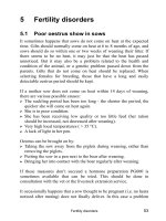
three or more abnormal variables). Over 11 years,
the three groups showed different risks of mortality
(Fig. 4.3), of hospitalization, and of functional
dependence [79]. As it has been validated in a large
number of patients, for more than a decade, and
is simple to perform, the CHS assessment appears
almost ideal for screening apparently healthy older
individuals for the risk of death and functional
dependence. It has been proposed that the CHS
classifi cation be adopted as the offi cial functional
classifi cation of older individuals. The CHS assess-
ment is accurate in predicting which healthy older
individuals are at risk of functional decline and
therefore need an “in-depth” geriatric assessment.
In its present form, however, it cannot be used for a
nosologic classifi cation of the whole older popula-
tion. A large portion of older individuals, and more
than 50% of the oldest old (that is, those 85 and over),
present some degree of functional dependence and
of comorbidity that causes disability, shortens their
life expectancy, and enhances their vulnerability to
minimal stress. These individuals are not accounted
for by the CHS assessment.
Practical applications of the geriatric
assessment
From the discussion of geriatric assessment it is rea-
sonable to conclude:
• Aging is multidimensional and its assessment
should be multidimensional.
• A CGA is the most exhaustive form of evaluation
of an older person.
• A CGA is clearly indicated in individuals present-
ing some degree of functional dependence or
comorbidity, or one or more geriatric syndrome.
• For all other individuals, a CGA may be
indicated if they are at increased risk of functional
deterioration.
Figure 4.3 Survival of fi t, pre-frail, and frail populations in the CHS study.
0
20
40
(%)
60
80
100
0 24487290
Group
No frailty
Intermediate
Frail
n Deaths
2469
2480
368
260
474
130
Months after study entry
From fi tness to frailty 49
• Of the screening tests for risk of functional dete-
rioration, the CHS assessment appears the best
validated and probably the most practical; pre-
frail and frail individuals should undergo a CGA.
• A nosologic classifi cation of older individuals is
still wanted. The CHS assessment offers the best-
validated classifi cation, but the frail subgroup
encompasses a wide array of conditions and
requires fi ne-tuning, based on functional depend-
ence, comorbidity, nutrition, and other variables
included in the CGA.
Other forms of geriatric assessment
In addition to the CGA, aging has been assessed with
physical performance and laboratory tests.
Tests of physical performance
These tests evaluate the ability of a person to per-
form one or more simple physical activities. They
may assess the actual performance of the activity
or the individual’s self-reports. The get-up-and-go
tests, or the measurement of grip strength and walk-
ing speed in the CHS assessment, are examples of
directly evaluated physical performances, while the
VES-13 is an example of self-report [77,78]. Both
approaches have proved reliable.
A list and description of all tests of physical per-
formance is beyond the scope of this chapter. As
a general rule these tests may be used to screen
healthy individuals for risk of disability and func-
tional dependence, and are not a substitute for geri-
atric assessment.
Laboratory assessment
Several studies have demonstrated that aging is
associated with an increased concentration of
infl ammatory cytokines [5,16,60] and other mark-
ers of infl ammation, such as the C-reactive protein
and D-dimer, in the circulation. The concentration
of these substances in the circulation is increased
in most geriatric syndromes as well as in common
diseases of aging, including dementia [16], oste-
oporosis [9,10], anemia [49], cardiovascular diseases
[80], disability [7], and depression [81]. Interleukin 6
(IL-6) has probably been the best characterized of
these substances.
A recent study in more than 1000 home-dwelling
individuals aged 70 and older showed that the con-
centration of IL-6 and D-dimer in the circulation
may be used to predict the risk of mortality and
functional decline [5]. Those individuals in whom
the concentration of both substances was below
the upper quartile had a two-year risk of mortality
or functional dependence less than 10%; for those
in whom the concentration of either substance was
in the upper quartile the risk was 20%; for those in
whom the concentration of both substances was ele-
vated the risk was approximately 40%. These results
are very encouraging, and suggest that laboratory
tests may become a routine part of the geriatric
assessment in the near future. Any study involving
older individuals should consider assessing IL-6 and
D-dimer as part of the patient evaluation.
Frailty, real and elusive
Frail and frailty are recurrent terms in both geriatric
and gerontology literature; for some, frail is almost
synonymous with aged [79,82]. If asked to defi ne a
frail person, most of us would probably think of a
curved older person, moving very slowly with the
help of a walker and at risk of falling at any moment.
The translation of this literary description into a
clinical entity is lacking, however, and the clinical
meaning of frailty remains elusive.
From the studies we have summarized one can
see that the term frailty has been used by different
authors in at least two different senses. In the clas-
sifi cation proposed by Hamerman frailty means an
almost complete exhaustion of functional reserve,
that is a person unable to withstand even negligible
stress [74]. In clinical terms this may be seen as a
person dependent in one or more ADLs, with one
or more geriatric syndromes and affected by severe
life-limiting comorbidity [82]. In this construct,
50 Lodovico Balducci, Claudia Beghe
frailty is largely irreversible, and the main goal of
management is to prevent further functional dete-
rioration. For the investigators of the CHS, frailty
means a predisposition to functional decline, that is
the frail persons represent a subgroup of independ-
ent persons at increased risk of developing func-
tional dependence. Seemingly, frailty may then be
reversed by proper interventions including rehabili-
tation and treatment or prevention of diseases. This
concept of frailty is predominant in the most recent
literature [79].
Irrespective of the term being used, both condi-
tions described as frailty are real and deserve to be
recognized, but the reader of this book should be
aware that a consensual defi nition of frailty is still
wanted.
Functional dependence and disability
Prevention of functional dependence has been
enounced as one of the goals of geriatrics, and func-
tional dependence has been defi ned as inability to
survive safely alone. Another common concept of
geriatrics, linked to functional dependence but not
to be confused with it, is disability.
Three terms related to disability have been well
defi ned by the World Health Organization: functional
impairment, disability, and handicap [83]. Functional
impairment involves the deterioration of a specifi c
function, such as walking or performing fi ne hand
movements. Disability is the loss of a certain activity,
such as climbing stairs, using the silverware, or driv-
ing, due to functional impairment. Clearly, not all
forms of functional impairment are severe enough
to cause disability. A disability becomes a handicap
in the absence of environmental arrangements able
to compensate for individual disability. For example,
inability to walk or to climb stairs due to loss of the
function of the lower extremities becomes a handi-
cap in the absence of a wheelchair or an elevator, or
a ramp allowing wheelchairs to climb to or descend
from different levels of a building.
The prevalence of functional impairment, disabil-
ity, and handicaps increases with age, and clearly
these conditions may limit a person’s ability for
independent living. One of the goals of the tests of
physical performance is to identify individuals at
risk of disability and to prevent its development. Of
special interest to the hematologist is the fact that
anemia, even mild anemia, is associated with an
increased risk of disability [48,49].
For the purpose of a classifi cation of older individu-
als, however, it is important to distinguish functional
dependence and disability and to realize that disabil-
ity does not always cause functional dependence.
Toward a nosologic classifi cation of aging
Though an offi cial and consensual classifi cation of
aging is still wanted, the discussion related to the
geriatric assessment allows us to distinguish some
broad categories of older individuals. The outline
proposed by Hamerman (Table 4.4) encompasses
all different states of aging, but probably needs to be
fi ne-tuned for clinical applications. In particular:
• The primary state should be subdivided accord-
ing to the risk of functional deterioration. The
CHS assessment [79], as well as the evaluation of
circulating markers of infl ammation, may allow
this distinction.
• The intermediate state should include individuals
with initial functional dependence (for example,
IADL dependence) and disability who are ame-
nable to rehabilitation, those with early geriatric
syndromes (memory loss, depression, osteoporo-
sis) that may be arrested with proper intervention,
and those with a comorbidity that is function-
impairing (for example, osteoarthritis), but not
life-limiting.
• Whether we decide to call it frailty or not, the sec-
ondary state should include individuals who are
dependent in one or more ADLs, those with more
advanced geriatric syndromes, and those with
life-limiting diseases (for example, congestive
heart failure or some form of metastatic cancer).
• The third state should include individuals who
have an average life expectancy of six months or
less, for whatever reason.
From fi tness to frailty 51
The classifi cation of aging in different states is under-
going continuous remodeling with the emergence
of new data and the interpretation of existing data.
Seemingly this process will never be concluded.
Current information allows us to frame the hematol-
ogy of aging in a context that is not purely chronologic
and that takes into account function, comorbidity,
the presence or absence of geriatric syndromes, as
well as the social context of the older aged person.
REFERENCES
1. Lipsitz LA. Age-related changes in the “complexity” of
the cardiovascular dynamics: a potential marker of vul-
nerability to disease. Chaos 1995; 5: 102–9.
2. Marineo G, Marotta F. Biophysics of aging and thera-
peutic interventions by entropy-variation systems.
Biogerontology 2005; 6: 77–9.
3. Lipsitz LA. Physiological complexity, aging, and the path
to frailty. Sci Aging Knowl Environ 2004; 16: pe16.
4. Ferrucci L, Corsi A, Lauretani F, et al. The origins of age-
related proinfl ammatory state. Blood 2005; 105: 2294–9.
5. Cohen HJ, Harris T, Pieper CF. Coagulation and activa-
tion of infl ammatory pathways in the development of
functional decline and mortality in the elderly. Am J Med
2003; 114: 180–7.
6. Payette H, Roubenoff R, Jacques PF, et al. Insulin-like
growth factor 1 and interleukin 6 predict sarcopenia
in very-old community-living men and women: the
Framingham Heart Study. J Am Geriatr Soc 2003; 51:
1237–43.
7. Ferrucci L, Penninx BW, Volpato S, et al. Change in mus-
cle strength explains accelerated decline of physical
function in older women with high interleukin-6 serum
levels. J Am Geriatr Soc 2002; 50: 1947–54.
8. Roubenoff R, Parise H, Payette HA, et al. Cytokines,
insulin-like growth factor 1, sarcopenia, and mortal-
ity in very old community dwelling men and women:
the Framingham Heart Study. Am J Med 2003; 115:
429–35.
9. Abrahamsen B, Bonnevie-Nielsen V, Ebbesen EN, Gram J,
Beck-Nielsen H. Cytokines and bone loss in a 5-year
longitudinal study-hormone replacement therapy
suppresses serum soluble interleukin 6 receptor and
increases interleukin-1-receptor antagonist: the Danish
Osteoporosis Prevention Study. J Bone Miner Res 2000;
15: 1545–54.
10. Moffett SP, Zmuda JM, Cauley JA, et al. Association of
the G-174C variant in interleukin-6 promoter region
with bone loss and fracture risk in older women. J Bone
Miner Res 2004; 19: 1612–18.
11. Brunsgaard H, Pedersen BK. Age-related infl ammatory
cytokines and disease. Immunol Allergy Clin North Am
2003; 23: 15–39.
12. Fernandez-Real JM, Vayreda M, Richart C, et al.
Circulating interleukin 6 levels, blood pressure, and
insulin sensitivity in apparently healthy men and
women. J Endocrinol Metab 2001; 86: 1154–9.
13. Pai JK, Pischon T, Ma J, et al. Infl ammatory markers and
risk of coronary heart disease in men and women. N
Engl J Med 2004; 351
: 2599–610.
14. Alesci S, Martinez PE, Kelkar S, et al. Major depression is
associated with signifi cant diurnal elevations in plasma
interleukin-6 levels, a shift of its circadian rhythm,
and loss of physiological complexity in its secretion:
clinical implications. J Clin Endocrinol Metab 2005; 90:
2522–30.
15. Yaffe K, Lindquist K, Penninx BW, et al. Infl amma-
tory markers and cognition in well-functioning
African American and white elders. Neurology 2003;
61: 76–8.
16. Wilson CJ, Cohen HJ, Pieper CF. Cross-linked fi brin
degradation products (D-Dimer), plasma cytokines,
and cognitive decline in community-dwelling elderly
persons. J Am Geriatr Soc 2003; 51: 1374–81.
17. Wilson CJ, Finch CE, Cohen HJ. Cytokines and cogni-
tion: the case for a head-to-toe infl ammatory para-
digm. J Am Geriatr Soc 2002; 50: 2041–56.
18. Rothstein G. Disordered hematopoiesis and myelodys-
plasia in the elderly. J Am Geriatr Soc 2003; 51 (3 Suppl):
S22–S26.
19. Balducci L, Hardy CL, Lyman GH. Hemopoiesis and
aging. In Balducci L, Extermann M, eds, Biological
Basis of Geriatric Oncology (New York, NY: Springer,
2005), 111–34.
20. Duthie EH. Physiology of aging: relevance to symp-
toms, perceptions, and treatment tolerance. In
Balducci L, Lyman GH, Ershler WB, Extermann M, eds,
Comprehensive Geriatric Oncology, 2nd edn (London:
Taylor and Francis, 2004), 207–22.
21. Burns EA, Goodwin JS. Immunological changes of aging.
In Balducci L, Lyman GH, Ershler WB, Extermann M, eds,
Comprehensive Geriatric Oncology, 2nd edn (London:
Taylor and Francis, 2004), 158–70.
22. Hornsby PJ. Replicative senescence and cancer. In
Balducci L, Extermann M, eds, Biological Basis of
52 Lodovico Balducci, Claudia Beghe
Geriatric Oncology (New York, NY: Springer, 2005),
53–74.
23. Balducci L, Cohen HJ, Engstrom P, et al. Senior adult
oncology clinical practice guidelines in oncology. J Natl
Compr Canc Netw 2005; 3: 572–90.
24. Balducci L, Extermann M. Assessment of the older
patient with cancer. In Balducci L, Lyman GH,
Ershler WB, Extermann M, eds, Comprehensive Geriatric
Oncology, 2nd edn (London: Taylor and Francis, 2004),
223–35.
25. Rao AV, Seo PH, Cohen HJ. Geriatric assessment and
comorbidity. Semin Oncol 2004; 31: 149–59.
26. Rockwood K, Mogilner A, Mitnitsky A. Changes with
age in the distribution of a frailty index. Mech Aging
Dev 2004; 125: 517–19.
27. Cohen HJ, Feussner JR, Weinberger M, et al. A control-
led trial of inpatient and outpatient geriatric evaluation
and management. N Engl J Med 2002; 346: 905–12.
28. Rubinstein LN. Comprehensive geriatric assessment:
from miracle to reality. J Gerontol A Biol Sci Med Sci
2004; 59: 473–7.
29. Kuo HK, Scandrett KG, Dave J, Mitchell SL. The infl u-
ence of outpatient geriatric assessment on survival: a
meta-analysis. Arch Gerontol Geriatr 2004; 39: 245–54.
30. Caplan GA, Williams AJ, Day B, Abraham K. A ran-
domized controlled trial of comprehensive geriat-
ric assessment and multidisciplinary intervention
after discharge of elderly patients from emergency
department: the DEED II study. J Am Geriatr Soc 2004;
52: 1417–23.
31. Balducci L. New paradigms for treating elderly patients
with cancer: the comprehensive geriatric assessment
and guidelines for supportive care. J Support Oncol
2003; 1 (4 Suppl 2): 30–7.
32. Reuben DB, Rubenstein LV, Hirsch SH, Hays RD. Value
of functional status as predictor of mortality. Am J Med
1992; 93: 663–9.
33. Inouye SK, Peduzzi PN, Robison JT, Hughes JS,
Horwitz RI, Concato J. Importance of functional meas-
ures in predicting mortality among older hospitalized
patients. JAMA 1998; 279: 1187–93.
34. Ramos LR, Simoes EJ, Albert MS. Dependence in activi-
ties of daily living and cognitive impairment strongly
predicted mortality in older urban residents in Brazil. J
Am Geriatr Soc 2001; 49: 1168–75.
35. Barberger-Gateau P, Fabrigoule C, Helmer C, Rouch I,
Dartigues JF. Functional impairment in instrumental
activities of daily living: an early clinical sign of demen-
tia? J Am Geriatr Soc 1999; 47: 456–62.
36. Zagonel V, Fratino L, Piselli P, et al. The comprehensive
geriatric assessment predicts mortality among elderly
cancer patients. Proc Am Soc Clin Oncol 2002; 21: 365a,
abs 1458.
37. Extermann M, Chen A, Cantor AB, et al. Predictors
of tolerance to chemotherapy in older cancer patients:
a prospective pilot study. Eur J Cancer 2002; 38:
1466–73.
38. Katz P. Function, disability, and psychological well
being. Adv Psychosom Med 2004; 25: 41–62.
39. Avlund K, Vass M, Hendriksen C. Onset of mobility
disability among community-dwelling old men and
women: the role of tiredness in daily activities. Age
Ageing 2003; 32: 579–84.
40. Lloyd H, Ahmed I, Taylor S, Blake JR. Index for predict-
ing mortality in elderly surgical patients. Br J Surg 2005;
92: 487–92.
41. Arenal JJ, Bengoechea-Beeby M. Mortality associated
with emergency abdominal surgery in the elderly. Can
J Surg 2003; 46: 111–16.
42. Weiss G, Goodnough LT. Anemia of chronic disease.
N Engl J Med 2005; 352: 1011–23.
43. Walter LC, Brand RJ, Counsell RS, et al. Development
and validation of a prognostic index for 1 year mor-
tality in older adults after hospitalization. JAMA 2001;
285: 2987–93.
44. Extermann M. Biological basis of the association of
cancer and aging comorbidity. In Balducci L,
Extermann M, eds, Biological Basis of Geriatric
Oncology (New York, NY: Springer, 2005), 173–88.
45. Izaks GJ, Westendorp RGJ, Knook DL. The defi nition of
anemia in older persons. JAMA 1999; 281: 1714–17.
46. Kikuchi M, Inagaki T, Shinagawa N. Five-year survival of
older people with anemia: variation with hemoglobin
concentration. J Am Geriatr Soc 2001; 49: 1226–8.
47. Anía BJ, Suman VJ, Fairbanks VF, Rademacher DM,
Melton JL. Incidence of anemia in older people: an
epidemiologic study in a well defi ned population. J Am
Geriatr Soc 1997; 45: 825–31.
48. Chaves PH, Xue QL, Guralnik JM, Ferrucci L, Volpato S,
Fried LP. What constitutes normal hemoglobin
concentration in community-dwelling disabled older
women? J Am Geriatr Soc 2004; 52: 1811–16.
49. Woodman R, Ferrucci L, Guralnik J. Anemia in older
adults. Curr Opin Hematol 2005; 12: 123–8.
50. Tresch DD. Management of the older patient with acute
myocardial infarction: difference in clinical presenta-
tions between older and younger patients. J Am Geriatr
Soc 1998; 46: 1157–62.
From fi tness to frailty 53
51. Weber JB, Coverdale JH, Kunik ME: Delirium: current
trends in prevention and treatment. Intern Med J 2004
34: 115–21.
52. Stump TE, Callahan CM, Hendrie HC. Cognitive impair-
ment and mortality in older primary care patients. J Am
Geriatr Soc 2001; 49: 934–40.
53. Blazer DG, Hybels CF. What symptoms of depression
predict mortality in community-dwelling elderly? J Am
Geriatr Soc 2004; 52: 2052–6.
54. Tinetti ME, Williams CS. The effect of falls and fall
injuries on functioning in community-dwelling
older persons. J Gerontol A Biol Sci Med Sci 1998; 53:
M112–M119.
55. Kao AC, Nanada A, Williams CS, Tinetti ME. Validation
of dizziness as a possible geriatric syndrome. J Am
Geriatr Soc 2001; 49: 72–5.
56. Verdery RB. Failure to thrive in old age: follow-up
on a workshop. J Gerontol A Biol Sci Med Sci 1997;
52: M333–M336.
57. Pavlik VN, Hyman DJ, Festa NA. Quantifying the
problem of abuse and neglect in adults: analysis of a
statewide data base. J Am Geriatr Soc 2001; 49: 45–8.
58. Green AD, Colon-Emeric CS, Bastian L, Drake MT,
Lyles KW. Does this woman have osteoporosis? JAMA
2004; 292: 2890–900.
59. Fortinsky RH, Iannuzzi-Sucich M, Baker DI, et al. Fall-
risk assessment and management in clinical practice.
J Am Geriatr Soc 2004; 52: 1522–6.
60. Hamerman D. Frailty, cancer cachexia and near death.
In Balducci L, Lyman GH, Ershler WB, Extermann M, eds,
Comprehensive Geriatric Oncology, 2nd edn (London:
Taylor and Francis, 2004), 236–49.
61. Carreca I, Balducci L, Extermann M. Cancer in the older
person. Cancer Treat Rev 2005; 31: 380–402.
62. Haley WE, Burton AM, Lamonde LA. Family care-
giving issues for older cancer patients. In Balducci L,
Lyman GH, Ershler WB, Extermann M, eds,
Comprehensive Geriatric Oncology, 2nd edn (London:
Taylor and Francis, 2004), 843–52.
63. Baker DI, King MB, Fortinsky RH, et al. Dissemination
of an evidence-based multicomponent fall risk-assess-
ment and management strategy throughout a geo-
graphic area. J Am Geriatr Soc 2005; 53: 675–80.
64. Fisher A. Of worms and women: sarcopenia and its role
in disability and mortality. J Am Geriatr Soc 2004; 52:
1185–90.
65. Goldspink G. Age-related muscle loss and progressive
dysfunction in mechanosensitive growth factor signal-
ing. Ann NY Acad Sci 2004; 1019: 294–8.
66. Guigoz Y, Vellas B, Garry PJ. Mini nutritional assess-
ment: a practical assessment tool for grading the
nutritional state of elderly patients In Facts, Research,
Interventions in Geriatrics (New York: Serdi, 1997), 15–60.
67. Corcoran MB. Polypharmacy in the senior adult patient.
In Balducci L, Lyman GH, Ershler WB, Extermann M, eds,
Comprehensive Geriatric Oncology, 2nd edn (London:
Taylor and Francis, 2004), 502–9.
68. Clarfi eld AM, Bergman H, Kane R. Fragmentation of
care for frail older people: an international problem.
Experience from three countries: Israel, Canada, and
the United States. J Am Geriatr Soc 2001; 49: 1714–21.
69. Walter LC, Covinsky KE. Cancer screening in elderly
patients: a framework for individual decision making.
JAMA 2001; 285: 2750–6.
70. Carey EC, Walter LC, Lindquist K, Covinsky KE.
Development and validation of a functional morbidity
index to predict mortality in community-dwelling eld-
erly. J Gen Intern Med 2004; 19: 1027–33.
71. Repetto L, Fratino L, Audisio RA, et al. Comprehensive
geriatric assessment adds information to the Eastern
Cooperative group Performance Status in Elderly can-
cer patients. An Italian Group for Geriatric Oncology
Study. J Clin Oncol 2002; 20: 494–502.
72. Ingram SS, Seo PH, Martell RE, et al. Comprehensive
assessment of the elderly cancer patient: the feasibility
of self-report methodology. J Clin Oncol 2002; 20: 770–5.
73. Extermann M, Overcash J, Lyman GH, Parr J, Balducci L.
Comorbidity and functional status are independent in
older cancer patients. J Clin Oncol 1998; 16: 1582–7.
74. Hamerman D. Toward an understanding of frailty. Ann
Intern Med 1999; 130: 945–50.
75. Lyness JM, King DA, Cox C, Yoediono Z, Caine ED. The
importance of subsyndromal depression in older pri-
mary care patients. Prevalence and associated func-
tional disability. J Am Geriatr Soc 1999; 47: 647–52.
76. Kivela SL, Pahkala K. Depressive disorder as predictor
of physical disability in old age. J Am Geriatr Soc 2001;
49: 290–6.
77. Podsiadlo D, Richardson S. The timed “up & go”: a test
of basic functional mobility for frail elderly persons.
J Am Geriatr Soc 1991; 39: 142–8.
78. Saliba D, Elliott M, Rubenstein LZ, et al. The Vulnerable
Elders Survey: a tool for identifying vulnerable older
people in the community. J Am Geriatr Soc 2001; 49:
1691–9.
79. Fried LP, Tangen CM, Walston J, et al. Frailty in older
adults: evidence for a phenotype. J Gerontol A Biol Sci
Med Sci 2001; 56: M146–M156.
54 Lodovico Balducci, Claudia Beghe
80. Ikeda U. Infl ammation and coronary artery disease.
Curr Vasc Pharmacol 2003; 1: 65–70.
81. Illman J, Corringham R, Robinson D, et al. Are infl am-
matory cytokines the common link between cancer-
associated cachexia and depression? J Support Oncol
2005; 3: 37–50.
82. Balducci L, Stanta G. Cancer in the frail patient: a com-
ing epidemic. Hematol Oncol Clin North Am 2000; 14:
235–50.
83. Warshaw GA, Murphy JB. Rehabilitation and the aged. In
Reichel W, ed, Care of the Elderly: Clinical Aspects of Aging
(Baltimore, MD: Williams & Wilkins, 1995), 187–97.
Hematopoiesis
PART II
Do hematopoietic stem cells show
age-related loss of function?
The production of over 4 ϫ 10
15
erythrocytes, lym-
phocytes, and myeloid cells during the lifetime of an
individual rests on the shoulders of the hematopoi-
etic stem cell (HSC) [1]. While the demand placed
upon the HSC may seem Sisyphean in its magnitude,
it is hardly a futile endeavor. For instance, a single
HSC can repopulate the entire hematopoietic system
of a lethally irradiated mouse, and engraftment levels
after secondary transplantation mirror those of the
primary recipients [2,3]. While other transplantation
protocols show that the numbers of primitive cells in
the bone marrow (BM) of recipients remain depressed
permanently, circulating blood cell numbers are
not signifi cantly different from non-transplanted
mice [4]. This is a profound statement of the ability
of these pluripotent stem cells to proliferate, differ-
entiate, and perhaps most importantly, self-renew.
Furthermore, BM cells can be serially transplanted
up to fi ve times before the marrow grafts fail to sus-
tain hematopoiesis [5,6]. The transplantation process
places extreme demand on the HSC population that
is not encountered during normal aging, which leads
to the suggestion that mouse BM cells have suffi cient
proliferative capacity to sustain hematopoiesis over
multiple mouse lifespans [7,8]. Even more confound-
ing is the fi nding by our laboratory and others that
the absolute number of HSCs does not decrease but
actually increases during the lifetime of the widely
used C57BL/6 (B6) mouse strain [9–11]. Indeed, even
human studies have shown that the ability of HSCs
5
Stem cell exhaustion and aging
Jeffrey Yates, Gary Van Zant
to support hematopoiesis throughout life is refl ected
by the constancy of mature blood cell counts [12,13].
In light of this evidence it would seem pointless to
suggest that HSCs become impaired as a result of the
aging process. However, we now know that at the cel-
lular level HSCs show aging-associated changes in
processes integral for proper hematopoiesis. Here we
present a brief yet comprehensive gathering of data
that support the hypothesis that HSCs are signifi cant
targets of the aging process, which in turn result in
the impairment and subsequent exhaustion of their
functional capacity to maintain tissue homeostasis.
What do we mean by “exhaustion”? In the scope
of this chapter, we refer to exhaustion as one of two
outcomes: (1) the decreased hematopoietic capac-
ity of HSCs, or (2) a decline in the number of HSCs
to a threshold level that results in the impairment
of steady-state and/or stress hematopoiesis. In this
chapter we take a point-by-point approach to iden-
tify the parameters of stem cell function that may
serve as substrates for the aging-associated decline
of their function, while integrating putative mecha-
nisms of aging, such as oxidative stress, DNA dam-
age, and replicative senescence. Specifi cally, we will
examine the processes of self-renewal, proliferation,
and multi-lineage differentiation, a combination
of characteristics that uniquely defi ne a pluripo-
tent stem cell. Furthermore, we will discuss aging-
associated changes in stem cell mobilization and
homing, processes that are required not only dur-
ing BM transplantation but also during steady-state
hematopoiesis. We will also explore how aging may
lead to alterations in the integrity of the HSC genome
57
5
Blood Disorders in the Elderly, ed. Lodovico Balducci, William Ershler, Giovanni de Gaetano.
Published by Cambridge University Press. © Cambridge University Press 2008.
58 Jeffrey Yates, Gary Van Zant
as well as the role that apoptosis plays in the regula-
tion of the stem cell pool. Finally, we will summarize
the recent developments in our lab relating to the
genetic regulation of HSC aging.
Identifi cation and study of the
pluripotent HSC
The hematopoietic system is arguably the best-
studied and most well-defi ned stem-cell-driven
tissue in mammalian physiology. However, a con-
sensus defi nition of the HSC, whether by functional
assays or by cell surface phenotype, has been diffi -
cult to attain within the scientifi c community. This
diffi culty arises because most assays used to study
HSCs rely on their clonogenic capacity, e.g., colony
formation in spleen and methylcellulose or periph-
eral blood cell production after BM transplantation.
In other words, the very cells being studied are lost
due to the induction of proliferation and differentia-
tion necessary for colony formation. Recent investi-
gations have thus focused on applying these assays
to BM subpopulations that are enriched and/or
depleted for cell surface proteins. These cell sur-
face antigens commonly consist of the c-kit recep-
tor and stem cell antigen 1 (Sca-1) on a background
that is devoid of lineage markers for differentiated
cells, such as granulocytes, B cells, T cells, etc. (Lin-
Sca1ϩ ckitϩ or LSK). However, this paradigm of
HSC identifi cation has recently been challenged
by the fi nding that cells expressing CD150 but not
CD48 receptors of the signaling lymphocytic acti-
vation molecule family show remarkable purity for
HSCs as defi ned by long-term repopulating ability
[14]. Other approaches have targeted the ability of
HSCs to effl ux fl uorescent dyes, such as Rhodamine
123 and Hoechst 33342 [15,16]. Indeed, it appears
the most stringent defi nition of murine HSC activity
may be found in the CD34-LSK fraction within the
side population phenotype as assessed by Hoechst
33342 staining [17]. One caveat to studies using cell
surface markers or vital dyes, however, is the fact
that we do not yet know the full extent of how aging
may affect the staining profi les of HSCs and their
progeny. Evidence suggests that this may not be the
case, with several studies showing unaltered stain-
ing profi les of the ckit, Sca-1, and lineage antigens
in old mice [9,11,18].
Systemic versus cellular aging
What is aging? When does it begin? What are its tar-
gets? These are questions for which there are no easy
answers. For instance, does aging begin at birth, at
which point development has culminated in an
independently functioning individual, or does it
begin at puberty, when the individual has attained
reproductive maturity, the putative endpoint of nat-
ural selection [19]? Furthermore, we can ask at what
level the aging process occurs.
It has been proposed that there are two
separate yet not necessarily mutually exclusive
general levels of organismal aging – systemic and
cell-autonomous. Systemic aging has been more
formally proposed as the hormonal control of aging,
where changes in humoral factors with age can cause
system-wide changes in the homeostatic condition
[20]. Support for this idea has gained traction from
studies of mice expressing a mutant form of the
KLOTHO gene product encoding a protein hormone
that leads to phenotypic changes characteristic of
accelerated aging [21]. Conversely, when the wild-
type KLOTHO gene is overexpressed in mice it leads
to a modest yet signifi cant increase in both male
and female lifespan [22].
The cell-autonomous theory on the other hand
posits that individual cells are the targets of the
aging process, via a time-dependent increase in
homeostatic dysfunction. The potential mecha-
nisms include increases in the production of reac-
tive oxygen species, telomere shortening, and, not
surprisingly, genomic instability. An implication of
this theory is that long-lived cells in the organism,
such as neurons, muscle, and importantly stem
cells, would be the predominant substrates of aging,
while those cells that undergo rapid and continuous
turnover would be removed before they could exert
an effect on tissue function. Here we take the view
Stem cell exhaustion and aging 59
that aging targets the cell-intrinsic processes neces-
sary for maintenance of tissue and thus organismal
function. Specifi cally, we defi ne aging as the detri-
mental and irreversible changes that occur during a
cell’s lifetime that lead to the inability of the resident
tissue to maintain homeostasis both at steady-state
levels and in response to stress. Importantly, this def-
inition could also apply to the cellular changes that
lead to carcinogenesis, a process bearing some of the
common principles of aging. The fact that the inci-
dence of most types of cancer escalates rapidly after
the age of 65 and arises from accumulated genomic
lesions is evidence that cancer is a manifestation of
the aging process [23]. Thus, the changes in cellular
biology that occur during oncogenesis should also
be evident, in part, during successful aging.
Model systems of stem cell aging
The fi eld of hematology has benefi ted immensely
from the study of a wide variety of organisms.
Studies of invertebrate systems such as C. elegans
and D. melanogaster have yielded keen insight into
stem cell biology and mechanisms of aging, but it
has predominantly been the study of the mam-
malian hematopoietic system that has led to the
current understanding of the physiology of hemat-
opoiesis. The utilization of mouse genetics has only
recently been fully realized as a tool, as it was this
mammalian model that yielded the breakthrough
discoveries of Till and McCulloch [24]. Most studies
on the aging of HSCs have used the B6 strain due
to its utility as a model for transplantation studies
via the polymorphic CD45 locus. However, we now
know that the B6 mouse strain is not necessarily rep-
resentative of all other inbred mouse strains. We and
others have shown that the HSCs of B6 mice differ
markedly from other strains in proliferative kinetics,
homing and engraftment properties, and pool size
with age [25–27]. In addition, the B6 mouse strain
is one of the longest-lived mouse strains, with a
mean lifespan of 3 years, versus other mouse strains
with mean lifespans of 1.5 to 2 years. Therefore, it is
evident that the genetic background of a particular
mouse strain can have a profound effect on the
biology of the HSC population as well as organismal
longevity. Indeed, it is for this reason that it is diffi cult
to compare fi ndings from various laboratories where
different mouse strains are used. Furthermore, cau-
tion must be exercised when attempting to extrap-
olate fi ndings in homozygous laboratory mice to
genetically heterogeneous humans.
The identifi cation and study of human HSCs have
lagged behind that of mouse and other mammalian
HSCs primarily due to the diffi culty in obtaining
signifi cant amounts of BM, particularly from very
old donors. Furthermore, the fi eld was hampered
early on by the reliance on in-vitro clonogenic
assays of putative HSC function in the absence of
reliable in-vivo model systems such as those used
by mouse researchers. A signifi cant development
in this regard has been the creation of severe com-
bined immune defi cient (SCID) mice that are able
to support human HSC-derived hematopoiesis fol-
lowing BM transplant [28]. These mice have yielded
key insights into the structure of the human hemat-
opoietic hierarchy as well as the conservation of
hematopoietic regulation between mouse and man.
However, study of the long-term repopulating and
self-renewal ability ascribed to HSCs, particularly
as they relate to aging, is hampered by the large
cell doses necessary for engraftment, the delayed
time course of engraftment, and the relatively short
repopulating period [28,29].
Regulation of aged HSC proliferation
A current model of HSC-directed hematopoiesis
is based on the principle that one or at most a few
HSCs of a highly quiescent population divide to pro-
duce highly proliferative progenitors with restricted
developmental potential. These lineage-restricted
transit-amplifying cells bear the proliferative load
necessary for the production of the repertoire of cell
types found in the peripheral blood. Thus, the ability
of HSCs to carry out the demands of hematopoiesis
hinges on their ability to proliferate in response to
both intrinsic and extrinsic cues. The clonal selection
60 Jeffrey Yates, Gary Van Zant
theory of hematopoiesis [30] is supported by studies
showing that when retrovirally marked HSCs were
transplanted into a conditioned host, only a few
clones contributed to mature blood cell produc-
tion [31,32]. This observation was confi rmed by Van
Zant et al. [33], who, using the same retroviral mark-
ing strategy in B6-D2 chimeric mice, also showed
the involvement of only a few clones in carrying
out hematopoiesis. However, recent evidence sug-
gests that the integration of these retroviral vectors
into the DNA is not necessarily neutral in their effect
on the fi tness of the transformed cells. For example,
the integration sites of clonally dominant HSCs often
encode regulatory regions involved in the processes
of HSC self-renewal and survival [34]. Furthermore,
the transplantation studies that demonstrate oligo-
clonal hematopoiesis may not be representative of
steady-state hematopoiesis in an unperturbed ani-
mal. Finally, when mice were continuously admin-
istered BrdU in their drinking water, the entire
population of HSCs completed at least one round of
replication within a two-month time period [35–37].
This fi nding implies that all HSCs in the BM con-
tribute to steady-state hematopoiesis, thus arguing
against an oligoclonal process.
The idea that the proliferative nature of HSCs may
change during aging is consistent with the observa-
tion that the incidence of myeloproliferative disor-
ders markedly increases with age in both mice and
humans. One study showing that the frequency of
HSCs in cycle old B6 mice was three times higher
than in young animals seems to corroborate this
fi nding. If true, this means that with an HSC fre-
quency seven times higher in old mice, the increase
in the absolute numbers of proliferating HSCs is
quite profound [9]. Furthermore, studies using
serial administration of hydroxyurea or irradiation
of BM cells have shown no evidence for a decline in
the capacity of HSCs to proliferate.
It should come as no surprise that most factors
responsible for regulation of the cell cycle were
discovered in the study of cancer, a disease of dys-
regulated cellular proliferation. A classic example
is the retinoblastoma protein (pRb), which was fi rst
discovered as the affected gene product responsible
for the development of retinal tumors during child-
hood. It has since been shown that members of the
pRb family act to suppress entry into the active cell
cycle and, upon their phosphorylation, allow for the
assembly of the replicative machinery. Their phos-
phorylation is governed by the concerted actions
of the cyclin-dependent kinases (cdks) and the cdk
inhibitors (ckis). Chief among the ckis are p16
INK4a
,
p21
cip-1
, and p27
kip-1
. Their role in hematopoietic
progenitor cell (HPC) proliferation was fi rst shown
by Mantel et al. [38], who demonstrated that p21
levels rise while those of p27 decrease after stimu-
lation by the hematopoietic cytokines Steel factor
and granulocyte colony-stimulating factor (G-CSF).
Additionally, it has been shown that p27 has no
effect on proliferation in the stem cell compart-
ment, yet has a dramatic effect on the progenitor
cell compartment [39]. It has also been shown that
p21 plays a role in both stem cell proliferation and
self-renewal [40]. Lewis et al. [41] demonstrated that
mice null for the p16
INK4a
locus exhibit increased
proliferation in the progenitor compartment. It was
recently shown that transcriptional repressors, such
as the Gfi -1 gene product, promote HSC quiescence
and thus maintain the HSC in its pluripotent state
[42,43]. While no studies have reported whether
there are age-related alterations in the levels of these
mitotic factors, it is tempting to hypothesize that
these same molecular changes that contribute to
tumorigenesis occur during “normal” aging as well.
Self-renewal of HSCs is altered during aging
In demonstration of the diffi culty in parsing out
changes in HSC proliferation and self-renewal, it
has been shown that, while the self-renewal ability
of murine HSCs undergoes progressive decline with
serial transplantation [44], there are alterations in the
frequency of cycling HSCs with age [9,10]. Moreover,
many factors that play a role in the regulation of stem
cell cycling also regulate self-renewal, particularly at
the genomic level. Much excitement has been gen-
erated recently with the identifi cation of the homeo-
box domain (Hox) family of transcription factors as
Stem cell exhaustion and aging 61
potent regulators of stem cell function. The family
member HOXB4 can promote self-renewal as well as
proliferation of HSCs while still allowing for effective
differentiation. Other Hox family members impli-
cated in HSC renewal, however, show pronounced
effects on the differentiation pathways, often result-
ing in acute myeloid leukemia (AML). For example,
the pro-leukemic HOXB6 promotes HSC expansion
and myeloid-directed differentiation at the expense
of lympho- and erythropoiesis when overexpressed
in mouse HSCs [45]. Similar results have also been
shown with HOXA9 and HOXA10 [46,47].
Histone modifi cation may play a role in the self-
renewal of HSCs by modulating the transcriptional
accessibility of the chromatin. Histones are targets
of multiple classes of enzymes involved in acetyla-
tion, methylation, and phosphorylation whose
function is to modify the DNA binding properties
of the histones. In HSCs, members of the Polycomb
gene family have been shown to play a key role in
regulating self-renewal. The archetype of this group
of chromatin modifi ers is the BMI-1 gene, whose
function is to serve as a scaffold for the assembly
of multimeric protein complexes consisting of his-
tone methylases and deacetylases. In mice null for
the BMI-1 gene, the pool of HSCs shows accelerated
exhaustion both in unperturbed and transplant
settings [48]. The mel-18 gene product is another
member of this group, and has been shown to result
in the expansion of competitive repopulating units
(CRUs) when overexpressed in mice [49].
The gold standard of proof for changes with age
in the ability to self-renew comes from CRU stud-
ies of serially transplanted mice where the CRU
frequency of young and old donors can be reliably
measured in BM recipients. However, studies of B6
mice, where HSC numbers increase with age, have
traditionally been the only available model to study
competitive repopulation. Recently, Kamminga
et al. [50] compared the renewal capacity of HSCs
from B6 and D2 mice and showed that HSCs from
D2 have a 1000-fold less capacity for expansion
compared to B6 mice. It is tempting to speculate
that changes in factors involved in self-renewal are
altered during aging. In fact, a recent profi ling of the
transcriptional changes that occur in HSCs of B6
mice during aging found that 16 genes were upregu-
lated with age that regulate hematopoiesis, including
self-renewal [51].
Telomeres shorten with age
Telomeres have been postulated to serve as the
mitotic clock underlying Hayfl ick’s limit of replica-
tive capacity [52]. Telomeres, the repetitive, non-
coding DNA sequences at the ends of DNA strands,
are the molecular solution to the end-replication
problem of DNA synthesis. With each round of cell
division, portions of these “dispensable” sequences
are lost until a point is reached when they have
contracted to a critically short length termed “cri-
sis” [53]. This phenomenon commonly precedes
cell death and/or senescence as well as oncogenic
transformation. In renewing cell populations, it is
believed that telomeres are resistant to replication-
induced erosion through the activity of telomerase,
the enzyme responsible for adding new sequences
to the ends of telomeres. In fact, HSCs exhibit sig-
nifi cant activity of this enzyme, thus potentially
extending their proliferative capacity [54]. However,
telomere shortening does indeed occur in the HSC
compartment during aging and after HSC transplan-
tation [55,56]. That telomeres also serve as docking
regions for proteins regulating DNA integrity, such
as TRF1, TRF2, and Ku, testifi es to the impact of
telomere shortening with age. Whether telomere
shortening represents a molecular factor for the
aging of HSCs or is merely correlative remains to
be seen (for review see Greider [57] and Blackburn
[58]). Furthermore, inbred mouse models may not
be representative of telomere dynamics among
mammals because inbred mice exhibit signifi cantly
longer telomeres than outbred mice [59] and thus
they are not limiting in the proliferative potential of
mouse HSCs during a typical lifespan [60]. However,
because human telomeres are signifi cantly shorter
than mouse telomeres, telomere shortening may
play a role in human HSC aging, especially after HSC
transplantation.
62 Jeffrey Yates, Gary Van Zant
Differentiation of stem cells
The ability of HSCs to provide adequate numbers
of differentiated progeny is critical for the essential
processes of oxygen transport, immune response,
and blood coagulation. Indeed, in humans, aging
is often accompanied by increased platelet activ-
ity, decreased immune responses, and changes in
erythrocytes such as membrane deformability and
oxygen carrying capacity. Whether these changes
can be traced back to alterations in the HSCs is not
yet clear. However, changes in the differentiation
capacity of aged HSCs are supported by evidence
that shows skewing of the ratios of the mature blood
cell types. For instance, in older humans, as well as in
mice, blood cell production is often skewed toward
the myeloid lineage at the expense of both T- and
B-cell production [11]. Furthermore, this phenom-
enon arises from a qualitative change in the HSC, as
evidenced by an age-associated increase in pheno-
typic HSCs with increased myeloid potential. This
fi nding was recently corroborated in experiments
studying the homing of aged HSCs, where homed
HSCs showed an aging-associated myeloid skewing
with a concomitant defi ciency in T-cell production
in animals transplanted with old CRUs compared
to young CRUs [9,61]. While a potential mechanism
has yet to be defi ned, it is interesting that the cellular
basis of acute myeloid leukemia is the production of
myeloid progenitors arrested at the blast stage from
a population of leukemic stem cells. It is feasible
that the molecular changes during aging that cause
myeloid skewing may also serve as one hit in the
two-hit model of leukemogenesis [62].
Mobilization of stem cells
The frequency of stem cells in BM is determined not
only by their proliferation and self-renewal but also
by the balance of mobilization and homing. Here
mobilization refers to the detachment of stem cells
from their supporting stroma and subsequent entry
into the systemic circulation. In clinical practice,
this process has been manipulated using cytokines
such as G-CSF in order to obtain suffi cient num-
bers of cells for BM transplantation. However, it has
become apparent that mobilization is a signifi cant
process during steady-state hematopoiesis as well.
For instance, peripheral blood of mice contains
low levels of BM progenitor cells. Furthermore,
using parabiotic mice, these progenitor cells in the
peripheral blood engraft non-conditioned BM of the
partner mouse [63]. However, no studies have con-
clusively determined whether aging has an effect on
either homeostatic or cytokine-induced HSC mobi-
lization. Dose-response studies of G-CSF-induced
mobilization of hematopoietic progenitors showed
that 60% fewer CFU-GM-forming cells entered the
circulation in old adults aged 70 to 80 years com-
pared to young adults aged 20 to 30 years [64].
Furthermore, de la Rubia et al. [65] showed that the
mobilization effi ciency of CD34ϩ cells in response
to G-CSF is two fold higher in patients younger than
18 versus those at older ages. In contrast, Boiret
et al. [66] showed that the numbers of long-term
culture-initiating cells in peripheral blood after G-CSF
treatment do not differ between children and adults.
Thus, more studies are needed to determine whether
HSC mobilization is affected by the aging process,
especially during steady-state hematopoiesis.
HSC homing during aging
The ability of transplanted BM cells to rescue hemat-
opoiesis in lethally irradiated recipients requires
that the injected cells migrate to the appropriate BM
niche. This process of homing and ensuing engraft-
ment is highly dependent on factors intrinsic to the
HSCs and the BM microenvironment. Prior studies
showing a decreased ability of aged cells to repopu-
late serially transplanted hosts compared to young
cells have failed to adequately address the age-
related changes in the ability of the cells to home to
the proper microenvironment. While Morrison et al.
[9] observed that old BM cells may not home as well
to the BM as young cells, the study was not able to
distinguish between the homing and actual engraft-
ment of the HSCs.