Clinical Pancreatology for Practising Gastroenterologists and Surgeons - part 3 doc
Bạn đang xem bản rút gọn của tài liệu. Xem và tải ngay bản đầy đủ của tài liệu tại đây (516.65 KB, 56 trang )
PART I
90
or only weakly active metabolites. Thus it may be used
safely in cases of renal failure. It does not cause seizures.
Nevertheless, it has an important emetic effect that is
sometimes difficult to manage. When used sublingually
the dose is 0.2–0.4 mg every 6–8 hours. The usual par-
enteral dose is 0.3–0.6 mg intramuscularly or intra-
venously every 6 hours or 0.002 mg/kg per hour as an
intravenous perfusion.
4 Tramadol: although it has agonist effects on opioid
receptors, it also shows analgesic activity due to other
mechanisms. It is a weaker analgesic than morphine
(about eight times). Since its half-life is slightly longer, it
is used parenterally at a dose of 100–150 mg every 6–8
hours (0.17 mg/kg per hour in perfusion). In cases of
renal failure the drug accumulates in the bloodstream
and it is advisable to increase the interval between
doses. It favors the development of seizures in the con-
ditions described for meperidine. Unlike most opiates it
does not cause addiction.
5 Hydromorphone is eight times more potent as an
analgesic than morphine. The recommended dose is
0.5 mg every 3 hours intravenously or 1–2 mg intra-
muscularly or subcutaneously. A dose of 0.2–1 mg/
hour may be given as a perfusion.
6 Fentanyl is 80 times more potent than morphine. It is
hardly used parenterally in pancreatitis but the trans-
dermal route, which allows slow drug release, is used
especially to treat chronic pain. Recently, this treatment
has also been used successfully in acute pancreatitis
(see below).
Effect on the sphincter of Oddi Traditionally, several
opioids, including morphine, have been rejected as
treatments for pain in acute pancreatitis on the assump-
tion that they increase biliary pressure. This was based
on the findings of preliminary studies that indirectly
measured biliary pressure after the use of these drugs.
However, opioids such as meperidine did not cause
pressure changes and consequently it has become the
narcotic of choice in acute pancreatitis. However, as
commented before, morphine has several advantages
over meperidine in the management of this disorder: it
is more potent, its management is more widely known,
and it is safer in cases of renal failure with less risk of
seizures.
Direct manometric studies of the sphincter of Oddi
have not fully confirmed the initial hypothesis (Table
9.2). In these studies both morphine and meperidine
significantly increased the frequency of the phasic
waves of the sphincter, whereas buprenorphine and tra-
madol did not seem to have any effect. The increase in
frequency of the phasic waves causes a reduction in pas-
sive filling of the sphincter segment and results in an in-
crease in biliary pressure (confirming the result of the
preliminary studies). However, only high cumulative
doses of morphine cause a significant increase in the
basal pressure of the sphincter of Oddi. Furthermore,
no study has yet shown that the increased basal pres-
sure of the sphincter caused by this dose of morphine
has a deleterious effect on patients with acute pancre-
atitis. Therefore it is possible to use morphine (or any
Table 9.2 Effect of opioids on sphincter of Oddi dynamics (direct measurement).
Drug Study Dose Results
Morphine Helm et al. (1988) Successive dose: 2.5, 2.5, 5, 2.5–5 mg/kg: increased frequency
10 mg/kg every 5 min i.v. 10–20 mg/kg: increased basal
pressure, frequency and amplitude
Thune et al. (1990) Cumulative dose: 2.5, 5, Increased frequency of phasic waves
10 mg/kg every 2 min i.v.
Meperidine Elta & Barnett (1994) 1 mg/kg i.v. Increased frequency of phasic waves
Thune et al. (1990) Cumulative dose: 25, 25, Decreased frequency of phasic waves
50 mg/kg every 2 min i.v.
Sherman & Lehman (1996) 1 mg/kg to 75 mg i.v. Increased frequency of phasic waves
Buprenorphine Staritz et al. (1986) 0.3 mg i.v. No changes
Cuer et al. (1989) 0.3 mg i.v. No changes
Tramadol Staritz et al. (1986) 50 mg i.v. No changes
i.v., intravenous.
CHAPTER 9
91
other opioid) in the management of pain in acute pan-
creatitis, although more studies are still necessary to
confirm this hypothesis.
Controlled studies Despite the number of therapeutic
drugs used to treat pain in acute pancreatitis, there are
few published controlled studies that compare these
drugs with each other or with a placebo (Table 9.3).
In 1984, Blamey and colleagues compared the use
of intramuscular buprenorphine with intramuscular
meperidine in 32 patients with acute pancreatitis.
These authors found similar analgesic responses to
these drugs in both the intensity and duration of pain
relief. Adverse effects were minimal (nausea and vomit-
ing) and occurred in the same proportion in both types
of treatment. A year later, Ebbehoj et al. studied the
analgesic effect of rectal indomethacin (indometacin)
compared with a placebo in 30 patients with acute pan-
creatitis. In this study, treatment with indomethacin
significantly reduced the number of days with pain and
the amount of other analgesics (opiates) given. In 1995
Patankar et al. reported another controlled study com-
paring the use of pancreatic enzymes with a placebo in
23 patients with acute pancreatitis. No difference was
found in the analgesia obtained by these patients. The
main adverse effect seen was nausea, which occurred
in approximately half the patients in both groups. Re-
cently, Jakobs and colleagues compared the analgesic
effects of intravenous buprenorphine and procaine.
In 40 patients with acute pancreatitis or acute bouts
of chronic pancreatitis, buprenorphine produced
higher pain relief and reduced the need for addi-
tional analgesics. Apart from slight sedation of the
buprenorphine-treated group, the secondary effects
were few and comparable. Another recent German
controlled trial confirmed the lower analgesic effects
Table 9.3 Controlled studies with analgesics in acute pancreatitis.
No. of Pain Adverse
Study patients Drugs assessment Outcome effects
Blamey et al. 32 Buprenorphine 0.3 mg i.m. Standard Similar relief Similar (nausea,
(1984) lineal scale Similar duration of vomiting)
Meperidine 100 mg i.m. Categories pain relief
scale
Ebbehoj et al. 30 Indomethacin 50 mg twice Visual analog Indomethacin group: None
(1985) (rectal) scale less number of days
Placebo with pain and opiate
administration
Patankar et al. 23 Oral pancreatic enzymes Visual analog Similar pain relief and Similar (nausea)
(1995) (7800 U protease daily) scale analgesic requirements
Placebo
Jakobs et al. 40 Buprenorphine 0.3 mg Visual analog Buprenorphine group: Buprenorphine
(2000) (bolus i.v.) + 2.4 mg scale higher pain relief and group: higher
(infusion i.v.) per 24 hours less additional analgesic sedation rate
Procaine 2 g (infusion i.v.) requirements
per 24 hours
Stevens et al. 32 TTS fentanyl + meperidine Self-reported Fentanyl group: less None reported
(2002) Placebo + meperidine pain intensity pain intensity at 36, 45,
and 60 hours from
admission
Kahl et al. 107 Pentazocine 30 mg (bolus Visual analog Pentazocine group: lower None
(2004) i.v.) per 6 hours scale pain scores over 72
Procaine 2 g (infusion i.v.) hours
per 24 hours
i.m., intramuscular; i.v., intravenous; TTS, transdermal therapeutic system.
PART I
92
of procaine. Finally, Stevens et al. reported that trans-
dermal fentanyl (plus meperidine for further relief)
failed as compared with placebo (plus meperidine) in
obtaining significant pain relief during the first 24
hours in hospital in 32 patients with acute pancreatitis.
However, fentanyl was more effective for pain relief
after the first 36 hours in hospital.
Thus although there is scanty evidence, we must con-
clude that the use of certain opioids such as meperidine
and buprenorphine is safe and effective for pain control
in patients with acute pancreatitis. Further controlled
studies are needed to confirm whether opioids in gen-
eral are more effective than theoretically less potent but
more widely used drugs such as NSAIDs and to clarify
the role of morphine (more potent and safer than
meperidine) in pain management in this condition.
Epidural analgesia
Epidural analgesia is becoming widely used in delivery
and in the immediate postoperative period after ab-
dominal or gynecologic surgery. When this route of ad-
ministration is used, the drug is concentrated where the
painful impulses enter the spinal cord (i.e., on the spinal
nerve roots). This permits the use of doses substantially
lower than those required for oral or parenteral admin-
istration. Systemic adverse effects are thus decreased.
The procedure involves the insertion of a catheter 3 cm
into the epidural space between T5 and T9 (usually T8)
and analgesia is instituted by injection of an analgesic
drug through the catheter. Because dural puncture is
not intended, the site of entry may be at any vertebral
level that permits a segmental blockade approximately
limited to the chosen region. Usually local anesthetics
such as bupivacaine or opioids such as fentanyl or mor-
phine, or a combination of both types of drugs, are
used. The association of both agents permits the use
of lower doses, minimizing local anesthetic-induced
complications of motor blockade and opioid-induced
complications. The dose of local anesthetic used can
produce high concentrations in blood following ab-
sorption from the epidural space, which is rich in ve-
nous plexuses. On the other hand, since conduction in
autonomic, sensory, and motor nerves is not affected by
opioids, blood pressure, motor function, and nocicep-
tive sensory perception typically are not influenced by
epidural opioids. Pruritus, nausea, vomiting, and uri-
nary retention may appear. Delayed respiratory depres-
sion and sedation, presumably from cephalad spread of
opioid within the cerebrospinal fluid, occurs infre-
quently with the doses of opioids currently used.
The technique may involve a single dose but to
achieve analgesia over a prolonged period a catheter
should be placed for either intermittent dosage or
continuous perfusion. As previously mentioned, PCA
pumps can be applied. If continuous perfusion is
administered, stable analgesic levels are obtained.
Therefore, early patient mobilization, improvement in
muscular tone, and fewer episodes of hypotension are
expected. After correct placing of the epidural catheter,
it is necessary to administer a single dose; if adverse ef-
fects do not develop, a continuous perfusion should be
programmed with variable rate according to the anal-
gesic level obtained. Table 9.4 shows some examples of
epidural administration of analgesic drugs.
This type of analgesia has reduced postoperative
morbidity and mortality. Recently, a systematic review
reported that in patients undergoing laparotomy
epidural administration of local anesthetics and opioids
provided higher postoperative analgesia than the use
of local anesthetics alone. However, local anesthetics
were found to be associated with less gastrointestinal
Table 9.4 Epidural administration of opioids and local anesthesics.
Loading dose Infusion (per hour) Bolus
Morphine 1–2 mg 0.2–0.4 mg 0.1–0.2mg/hour
Meperidine 25–50 mg 10–15 mg 20–25 mg/hour
Fentanyl 100 mg 50–75 mg 25–50 mg/hour
Fentanyl + bupivacaine (0.0625%) 75 mg + 3.75 mg 50 mg + 2.5 mg 12.5 mg + 0.0625 mg/30 min
(6 mL) (4 mL/hour) (1 mL)
Morphine + bupivacaine (0.0625%) 1mg + 5 mg 0.15 mg + 1.8mg 0.15mg +1.8mg/30 min
(3 mL) (3 mL)
CHAPTER 9
93
paralysis than when systemic or epidural opioids were
used.
In patients with acute pancreatitis, this type of anal-
gesia has many theoretical advantages. Firstly, it per-
mits a reduction in high doses of opioids when these are
excessive and/or associated with adverse effects (as pre-
viously mentioned, opioids facilitate the occurrence or
aggravation of respiratory failure and some show in-
creased neurotoxicity in the presence of renal failure).
Also, it allows severely ill patients to achieve a sitting or
semi-sitting position readily and therefore improves
gas exchange and reduces the incidence of respiratory
infections. Intestinal blood flow and motility is also
said to improve. Finally, in postoperative patients,
epidural analgesia reduces the metabolic response and
improves catabolism. All these beneficial effects favor
mobilization, reduce the incidence of complications,
and permit early resumption of oral feeding. Unfortu-
nately, there are still no controlled studies of patients
with acute pancreatitis which confirm the theoretical
benefits of this type of analgesia.
Nevertheless, this type of analgesia may have adverse
effects, such as hypotension (due to involvement of
the sympathetic nervous system when the catheter is in-
serted or medication administered), headache, urinary
retention, radicular damage, or catheter migration.
The most serious, though infrequent, complication is
the development of epidural hematoma or abscess.
Epidural analgesia is contraindicated in hypovolemic
shock, severe coagulopathy, infection, or radiculopa-
thy at the level of catheter insertion. As previously men-
tioned, since variable amounts of the drugs reach the
peripheral blood, systemic adverse effects of local anes-
thetics or opioids might develop.
Large series of patients with acute pancreatitis
treated by epidural anesthesia have been reported to
have had excellent pain control, with no neurologic or
septic complications. Finally, there have been sporadic
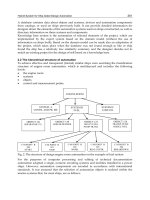
Patient with
acute pancreatitis
Without organ failure
Metamizol i.v. or tramadol i.v.*
(+ meperidine s.c. between dose if necessary)
Adequate pain relief No pain relief
Metamizol or tramadol if necessary
Adequate pain relief
Meperidine s.c.*
or buprenorphine i.v. i.m.*
No pain relief
Epidural analgesia*
(+ parenteral opioids)
With organ failure
Figure 9.1 Guidelines for the
treatment of pain in acute pancreatitis.
*, Patient-controlled analgesia, if
possible; i.m., intramuscular; i.v.,
intravenous; s.c., subcutaneous.
domethacin treatment of acute pancreatitis. A controlled
double-blind trial. Scand J Gastroenterol 1985;20:
788–800.
Elta GH, Barnett JL. Meperidine need not be proscribed dur-
ing sphincter of Oddi manometry. Gastrointest Endosc
1994;40:7–9.
Helm JF, Venu RP, Geenen JE et al. Effects of morphine on the
human sphincter of Oddi. Gut 1988;29:1402–1407.
Holte K, Kehlet H. Epidural anaesthesia and analgesia: effects
on surgical stress responses and implications for postopera-
tive nutrition. Clin Nutr 2002;21:199–206.
Isenhower HI, Mueller BA. Selection of narcotic analgesics for
pain associated with pancreatitis. Am J Health Syst Pharm
1998;55:480–486.
Jakobs R, Adamek MU, von Bubnoff AC, Riemann JF.
Buprenorphine or procaine for pain relief in acute
pancreatitis. A prospective randomized study. Scand J
Gastroenterol 2000;35:1319–1323.
Jorgesen H, Wetterslev J, Moiniche S, Dahl JB. Epidural local
anaesthesics versus opioid-based analgesic regimens for
postoperative gastrointestinal paralysis, PONV and pain
after abdominal surgery. Cochrane Database Syst Rev
2003;4:CD001893.
Kahl S, Zimmerman S, Pross M et al. Procaine hydrochloride
fails to relieve pain in patients with acute pancreatitis.
Digestion 2004;69:5–9.
Patankar BV, Chand R, Johnson CD. Pancreatic enzyme
supplementation in acute pancreatitis. HPB Surg 1995;8:
159–162.
Rodgers A, Walker N, Schung S et al. Reduction of postopera-
tive mortality and morbidity with epidural or spinal anaes-
thesia: results from overview of randomised trials. BMJ
2000;321:1–12.
Sherman S, Lehman G. Opioids and the sphincter of Oddi.
Gastrointest Endosc 1996;44 :239–242.
Staritz M, Poralla T, Manns M et al. Effect of modern anal-
gesic drugs (tramadol, pentazocine and buprenorphine) on
the bile duct sphincter in man. Gut 1986;27:567–569.
Stevens M, Esler R, Asher G. Transdermal fentanyl for the
management of acute pancreatitis pain. Appl Nurs Res
2002;15:102–110.
Thompson DR. Narcotic analgesic effects on the sphincter of
Oddi: a review of the data and therapeutic implications in
treating pancreatitis. Am J Gastroenterol 2001;96:1266–
1272.
Thune A, Baker RA, Saccone GT et al. Differing effects of
pethidine and morphine on human sphincter of Oddi motil-
ity. Br J Surg 1990;77:992–995.
PART I
94
reports of good pain relief following percutaneous
pharmacologic blockade of the celiac plexus.
Guidelines for the management of pain
in acute pancreatitis
Pain due to acute pancreatitis should be treated from
the very onset of the disease by regular analgesic
administration. In general terms, PCA pumps are
recommended (see Table 9.1). Staged treatment should
be given (Fig. 9.1). Thus we may use metamizol
(2000 mg every 6–8 hours intravenously) or tramadol
(100 mg every 8 hours intravenously), with meperidine
(50–100 mg subcutaneously as a single dose) for rescue
between doses. When pain control is satisfactory or
the pain disappears, the same dosage may be used on
demand by the patient. However, if the pain is not
controlled, opioids become necessary. Until studies
confirm the safety of morphine and its derivatives, the
use of meperidine (50–100 mg every 4 hours subcuta-
neously) or buprenorphine (0.3–0.6 mg every 6 hours
parenterally; 0.2–0.4 mg every 6 hours sublingually;
0.002 mg/kg per hour as intravenous continuous perfu-
sion) is recommended.
Patients who require high doses of opioids for ade-
quate pain control, and especially those with organ fail-
ure (mainly renal and/or respiratory failure), should be
treated with epidural anesthesia using either local anes-
thesics alone or, better, local anesthesics plus opioids
(see Table 9.4). This kind of analgesia may be adminis-
tered in addition to systemic opioids, the dose of which
can then be reduced, or can be used as the sole
treatment.
Recommended reading
Blamey SL, Finlay IG, Carter DC, Imrie CW. Analgesia in
acute pancreatitis: comparison of buprenorphine and
pethidine. BMJ 1984;288:1494–1495.
Cuer JC, Dapoigny M, Ajmi S et al. Effects of buprenorphine
on motor activity of the sphincter of Oddi in man. Eur J Clin
Pharmacol 1989;36:203–204.
Ebbehoj N, Friis J, Svendsen B, Bülow S, Madsen P. In-
95
Acute pancreatitis is a disease with a wide spectrum of
clinical courses, ranging from the mild form with mini-
mum morbidity and almost zero mortality, to the severe
form with a high percentage of complications and high
risk for a lethal outcome.
In about 80% of patients, the inflammatory process
is self-limited, involving only the pancreas and immedi-
ate pancreatic tissues, and resolves spontaneously
within less than a week. These mild cases require only a
short period of fasting, intravenous hydration, elec-
trolytes, and analgesia. Patients can usually start an
oral low-fat diet within 3–7 days of the onset of their
pain, resulting in minor and usually easily reversible
nutritional defects.
This is not the case in severe acute pancreatitis,
which is characterized by various degrees of necrosis of
pancreatic parenchyma as well as local and systemic
complications such as systemic inflammatory response
syndrome (SIRS) and multiple organ failure (MOF).
This form of the disease represents a typical hypermeta-
bolic septic model, with increased resting energy re-
quirements and considerable protein catabolism that
leads to severe malnutrition.
As a result nutritional support in acute pancreatitis
should be one of the main therapeutic aims and nutri-
tional management should depend on the underlying
pancreatic disease.
Malnutrition and metabolic changes in
acute pancreatitis: why?
Regardless of the etiology, all cases of acute pancreatitis
share a common pathogenetic pathway that involves
the premature activation of trypsinogen to trypsin,
after which a cascade of pancreatic enzyme activation
begins that leads to autodigestion of the pancreas and
peripancreatic tissues. At the same time, a number of
powerful inflammatory mediators are produced locally
and systemically, with cytokines being the most impor-
tant because they initiate or amplify an inflammatory
cascade and induce the development of SIRS and re-
mote organ failure. Later in the course of the disease, in-
fective complications may occur, particularly infected
pancreatic necrosis, consequent sepsis, and sepsis-
related MOF, that further increase energy requirements.
The release of inflammatory mediators, particularly
tumor necrosis factor (TNF)-a and interleukin (IL)-6,
and in cases of sepsis the release of catabolic hormones
(catecholamines, cortisol, glucagon), change protein
and energy metabolism in ways that increase both
energy demands and urinary nitrogen excretion, which,
in parallel with the reduction of food intake, result in
the development of protein–energy malnutrition.
Clinical studies have shown that patients with acute
pancreatitis have a resting energy expenditure (REE)
that is 1.2–1.5 times that predicted by the Harris–
Benedict equation, depending on the severity of the
disease. Septic patients are the ones with the greater
protein–energy needs, since they are in marked meta-
bolic stress. These patients exhibit accelerated catabo-
lism and protein breakdown and have a decreased
blood supply to vital organs due to hypovolemia or de-
creased cardiac performance during the inflammatory
process.
As already mentioned, nitrogen loss during severe
disease is increased. While a healthy adult loses ap-
proximately 12 g of nitrogen daily in the urine in the
10
Nutrition in the acute phase of
pancreatitis: why, when, how,
and how long?
Konstantina Paraskeva, Costas Avgerinos, and Christos Dervenis
fasting state, patients with acute pancreatitis compli-
cated by sepsis commonly lose up to 40 g of nitrogen
daily, with most of this loss coming from the skeletal
muscle. Negative nitrogen adversely affects host de-
fenses and immune competence balance and is asso-
ciated with increased morbidity and mortality.
Another metabolic response to severe inflammation
and energy deprivation is endogenous gluconeogenesis
from protein degradation, which can only partially be
inhibited by exogenous glucose. Intravenous adminis-
tration of high doses of glucose carries the risk of hy-
perglycemia as the insulin response is often impaired.
Furthermore, insulin release is also frequently impaired
as a result of the inflamed pancreas, rendering the pa-
tient susceptible to hyperglycemia in 40–90% of cases.
It has been suggested that transient hyperglycemia may
impair complement fixation, evoking an immunosup-
pressive state. Parenteral nutrition is associated with an
additional risk for hyperglycemia and careful monitor-
ing of blood glucose levels is necessary in these patients.
Finally, lipid metabolism is also altered in acute pan-
creatitis via a mechanism that is not entirely clear. In-
creased serum triglycerides may either be the cause or the
result of acute pancreatitis. Increase in cholesterol and
free fatty acids in serum have also been reported. After
the acute phase subsides, serum lipids tend to return to
normal. Infusion of exogenous fat does not seem to inter-
fere with the development or the course of acute pan-
creatitis and is therefore not contraindicated, provided
that patients are monitored for hypertriglyceridemia.
Energy supply in acute pancreatitis
Patients with severe acute pancreatitis manifest in-
creased basal energy requirements, accentuated pro-
tein catabolism, and endogenous gluconeogenesis. The
goals of nutritional support in this setting are (i) to
lessen nitrogen wasting, (ii) to support organ structure
and function, and (iii) to positively affect the clinical
course of the disease if possible.
Individual protein–calorie needs vary widely de-
pending mostly on the severity of the disease, as well as
the age, body size (height and weight), and sex of the
patient. The most accurate method of measuring
caloric requirement is indirect calorimetry, which is
also useful for determining the fuel mix being oxidized
and for assessing the metabolic stress level. Unfortu-
nately, it is not often available, and therefore the most
commonly used method for estimation of REE is the
equation devised by Harris and Benedict. The formulas
for calculating REE (in kcal/day), using the four vari-
ables age, height, weight, and sex, are as follows:
BMR
women
= 655 + 9.5W + 1.8H - 4.7A
BMR
men
= 66 + 13.7W + 5H - 6.8A
where W is the actual or usual weight (kg), H is height
(cm), and A is age (years). In patients with acute pan-
creatitis, REE as determined by indirect calorimetry
varies from 77 to 158% of the energy expenditure pre-
dicted by the Harris–Benedict equation, being higher in
patients with pancreatitis complicated by sepsis or
MOF. These results make the Harris–Benedict equa-
tion a very rough method for estimating the energy
demands of these patients.
Even simpler REE equations are often used in clinical
practice and it should be remembered that these may
overestimate or underestimate the measured values by
20 or even 30% for any individual. In severely ill pa-
tients, REE is usually about 25–35 kcal/kg daily and
1.2–1.5 g of protein per kilogram dry body weight, ad-
justing for obesity. With increasing metabolic stress,
calories and protein should be increased, except in
critically ill patients. During the early catabolic stage,
15–25 kcal/kg and 1.5 g/kg of protein are more suitable
in nonsurgical patients with MOF.
During artificial nutrition, energy should be pro-
vided in the form of mixed fuel, with 60–70% given as
glucose and 30–40% as lipid emulsion. Patients with
severe disease and MOF often have high serum glucose
and triglyceride levels. Intravenous infusion of glucose
and fat does not suppress endogenous production and
may therefore result in further elevations of blood glu-
cose and triglycerides. Hyperglycemia predisposes to
fluid retention (due to increased insulin requirements)
and immunosuppression. High-dose lipid emulsion is
also immunosuppressive and hypertriglyceridemia
may exacerbate pancreatitis; therefore blood glucose
levels should be monitored and should not exceed
10 mmol/L, while serum triglyceride concentrations
should not exceed 1.5–2 times normal. Requirements
for protein can be adjusted by performance of a nitro-
gen balance study.
Hypocalcemia is the most frequent mineral aber-
ration seen in patients with acute pancreatitis, and
a marked reduction of serum calcium is associated
with a poor prognosis. Systemic endotoxin exposure
appears to play a significant role in the development of
PART I
96
hypocalcemia in severe attacks. In cases where ionized
calcium is low and this is not a false reduction due to
hypoalbuminemia, an attempt to correct this reduc-
tion should be made. Excessive calcium infusion may
induce pancreatitis.
Patients with pancreatitis may also benefit from
glutamine supplementation, as it is an important fuel
for the gastrointestinal tract (pancreatic islets, acinar
cells, and enterocytes). The oxidation of one molecule
of glutamine produces 30 mmol of ATP, which makes
this amino acid a very rich energy source. It appears
that although enterocytes are rich in glutamine and
may even synthesize it endogenously, this amino acid is
an essential nutrient in stressed patients.
Attempts to favorably modulate the immune and
inflammatory responses of severely ill patients led to
efforts to enrich nutrition with various immune-
enhancing nutrients. This has become known as im-
munonutrition. Of the various nutrients that have been
suggested as beneficial, glutamine, arginine, w-3 fatty
acids, and nucleotides have been introduced into clini-
cal use in the form of several standard formulas, often
in combination preparations. There are a number of re-
ports, mainly in severely injured patients, dealing with
the role of immune-enhanced enteral diets in these
cases. A metaanalysis of 1009 patients from 11 trials
showed that immune-modulated regimens resulted in a
significant reduction of infective complications and
length of hospital stay, but with no effect on survival.
Only one study dealt with the use of glutamine in acute
pancreatitis, as a supplement in standard total par-
enteral nutrition (TPN). This investigation found that
glutamine improved leukocyte activity and reduced
proinflammatory cytokine release in acute pancreatitis.
No conclusions can be drawn from these studies and al-
though it seems possible that immune-enriched diets
could play a role, further studies are needed to clarify
this issue.
In the light of the emerging evidence regarding the
primary role of the intestine in the pathophysiology of
acute pancreatitis, enteral feeding is now considered
the preferred mode of nutritional support in these pa-
tients. Enteral feeding has proved to be safe and in the
majority of patients may cover caloric needs. Due to its
beneficial effect on gut integrity, it should be started
very early in the course of the disease (during the first 24
hours) and should be continued until the patient toler-
ates oral feeding. In cases where the caloric goal cannot
be achieved by enteral nutrition, combined parenteral
nutrition should be used. Even a low volume of low-
residue enteral diet given in cases where TPN is used is
sufficient to protect the intestinal mucosa. Recently, it
was suggested that gastric feeding may be feasible in
patients with severe pancreatitis. The optimal feeding
formula has yet to be determined, but an elemental
or immune-enhancing diet (10–30 mL/hour) con-
tinuously perfused to the jejunum is suggested.
Total parenteral nutrition in
acute pancreatitis
Traditionally, TPN has been the only nutrient-provid-
ing treatment in patients with acute pancreatitis and
prolonged starvation. TPN achieves energy and protein
provision without stimulating pancreatic exocrine se-
cretion. Although Feller et al. in 1974, in an uncon-
trolled retrospective study, showed a decrease in the
mortality rate of patients with acute pancreatitis who
received intravenous hyperalimentation, several other
similar retrospective uncontrolled clinical trials have
failed to reproduce these results. On the contrary, other
authors observed a higher incidence of catheter-related
sepsis among TPN groups but no difference in total
mortality.
Two prospective nonrandomized trials have been
published on this subject. In 1989, Sitzmann et al. di-
vided 73 patients with acute pancreatitis into three
groups depending on their ability to tolerate glucose-
free, lipid-based, and lipid-free nutrition. Within 15
days most patients in all groups achieved improvement
in nutritional status. A higher mortality was observed
in the fat-free group as well as among patients with
persistent negative nitrogen balance. A high incidence
of catheter sepsis was also documented. In 1991,
Kalfaretzos et al. divided 67 patients with severe acute
pancreatitis (more than three Ranson criteria) into two
groups of early (within 72 hours after admission) and
late (after 72 hours) onset of TPN. They noted a signifi-
cantly lower incidence of complications and mortality
in the early group but a high incidence of catheter-
related sepsis as well.
The only prospective randomized controlled trial on
the effects of early parenteral nutrition versus no nutri-
tional support in patients with acute pancreatitis was
published by Sax et al. in 1987. During this study, 54
patients were randomized to receive either supporting
treatment alone or supportive treatment with early
CHAPTER 10
97
TPN (within 24 hours of admission). TPN had no
significant effect on clinical outcome, duration, and
pancreatitis-related complications, but patients in the
TPN group had a ninefold increase in the incidence of
catheter sepsis. A significant drawback of this study is
the fact that all patients studied had mild pancreatitis
(mean Ranson score 1) and hence had low complica-
tion and mortality rates with conventional treatment.
In conclusion, it can be stated that there is no strong
information regarding the role of TPN in acute pancre-
atitis and more trials are needed in order to establish
any benefit. The use of TPN does not seem to interfere
with the progress of the disease but indicates a trend in
improvement of morbidity and mortality in patients
with severe pancreatitis who achieve a state of positive
nitrogen balance and in those who require prolonged
starvation (i.e., persistent pancreatic inflammation,
abscess, and pancreatic fistula). TPN is associated
with certain disadvantages, such as an increased rate of
catheter-related infections, metabolic disturbances
such as hyperglycemia, effects on gut permeability, and
increased cost.
Role of the gut in acute pancreatitis
Contamination of pancreatic necrosis and consequent
sepsis is the main cause of death in severe pancreatitis,
although in the early period of the disease SIRS remains
the main fatal cause. The organisms responsible for sec-
ondary pancreatic infection are usually Gram-negative
bacteria of the same type that colonize the gastroin-
testinal tract. This suggests gut barrier dysfunction,
increased intestinal permeability, and subsequent
bacterial translocation through the gut wall.
Indeed, changes in intestinal permeability have been
proven to occur in acute pancreatitis and are directly re-
lated to the severity of the disease. Patients with severe
acute pancreatitis have increased intestinal permeabil-
ity compared with healthy controls or those with mild
attacks, and patients who develop MOF have even
greater changes compared with those with severe dis-
ease and more favorable outcome. Intestinal perme-
ability changes occur within 72 hours of the onset of
pancreatitis and normalize during recovery.
It has been proposed that intestinal permeability may
allow bacteria and bacterial components to migrate
from the intestinal lumen to extraintestinal sites. In
fact, bacterial translocation from the lumen to the pan-
creas and mesenteric lymph nodes is well documented
in animal models but has not been convincingly demon-
strated in humans. Nevertheless there are some data
that support the hypothesis. Firstly, it has been demon-
strated that 50% of patients with pancreatic necrosis
have gut-origin bacteria colonizing the pancreas, and
that colonization is maximal during the second to third
week after the onset of the disease. Secondly, intestinal
colonization with Gram-negative organisms precedes
pancreatic infection and represents an early risk factor
for developing a pancreatic infection. Thirdly, clinical
studies indicate an association between gut dysfunction
and infection, acute respiratory distress syndrome,
and MOF. However, studies in patients with acute
pancreatitis have demonstrated that the changes in gut
permeability occur early, whereas pancreatic infection
usually occurs during the second to third week after
the onset of the disease, and patients with increased
permeability do not necessarily have more septic
complications.
The early changes in intestinal permeability have
been also correlated with corresponding levels of
endotoxemia. Endotoxins derive from Gram-negative
bacteria and have systemic toxic effects, such as
tachycardia, hypotension, and pyrexia, and also de-
range the immune system. Endotoxemia appears to
correlate with the severity, incidence of systemic com-
plications, and mortality of patients with acute pancre-
atitis. Patients with severe attacks have higher serum
concentrations of endotoxin compared with those with
mild disease, and the same was found in nonsurvivors
compared with survivors and in patients with MOF as
opposed to those without it. Nevertheless, in a study
conducted by Moore et al. on severely injured trauma
patients, it was not possible to document bacteria or en-
dotoxin in the portal blood, even in patients with MOF.
Selective gut decontamination seems to reduce infec-
tion complications, but it does not increase patients’
survival.
Overall, the maintenance of intestinal structure and
function is a complicated and multifactorial process
that requires the adequate delivery of energy and oxy-
gen. Enterocytes use glutamine and short-chain fatty
acids as primary fuel. The presence of these nutrients in
the lumen stimulates the proliferation of mucosal cells
and enhances gut integrity. Fasting leads to mucosal
atrophy, increased rate of enterocyte apoptosis, de-
creased glutamine and arginine transport, and altered
mucin composition of goblet cells. These changes may
PART I
98
develop as early as the first week and intestinal perme-
ability changes occur within 48–72 hours of the disease
onset. Furthermore, the impairment of gut motility that
occurs within 12 hours of the onset of acute pancreati-
tis favors bacterial overgrowth and contributes to en-
dotoxemia and bacterial translocation. Enteral feeding
repairs the mucosal damage caused by fasting and, if
given very early, preserves epithelial integrity and bac-
terial ecology, therefore helping to maintain gut barrier
function.
The intestinal barrier is particularly susceptible to is-
chemia and therefore an adequate blood supply is of
great importance for its function. Severe acute pancre-
atitis produces hypovolemia and third-space fluid
losses that induce splanchnic vasoconstriction and
subsequent intestinal ischemia. The hypoxia that oc-
curs early in patients with acute pancreatitis may
further contribute to mucosal ischemia. The ischemic
effect is also enhanced by the local production of
various inflammatory mediators. Intestinal reperfusion
causes further damage through the production of oxy-
gen free radicals and inflammatory mediators. Severe
acute pancreatitis is associated with priming and subse-
quent overactivation of leukocytes, which may be the
main cause of intestinal injury, by inducing gut is-
chemia, amplifying inflammation, and releasing oxy-
gen free radicals. Fluid replacement and resuscitation
is essential in order to maintain microcirculation and
prevent ischemia and reperfusion injury.
Recently, the role of the gut in acute pancreatitis has
expanded beyond the bacterial translocation and endo-
toxin phenomenon, as emerging evidence has indicated
that the gut may be a source of cytokines and a site of
neutrophil priming. It appears that intestinal ischemia
and reperfusion injury results in the overactivation of
gut macrophages and gut-associated lymphoid tissue,
which in turn release excessive cytokines and other
mediators. The release of cytokines contributes to
SIRS and MOF.
Enteral nutrition
Based on the above, efforts have been made to find a
more natural way of delivering nutrients in patients
with pancreatitis. Despite concerns for the possible
stimulatory effect of oral feeding on pancreatic secre-
tion and for disease exacerbation, several experimental
and clinical trials have shown that delivery of nutrients
to the jejunum does not increase pancreatic secretion
and is well tolerated with no increase in complications.
More specifically, although administration of lipid into
the duodenum is a strong stimulatory factor for pancre-
atic exocrine secretion, jejunal delivery of the same
amount of lipid causes minimal pancreatic reaction.
Similar minor effects of intravenous lipid infusion have
been shown in human studies. Gastric or duodenal pro-
tein or carbohydrate administration is also a strong
stimulus for pancreatic secretion, whereas jejunal de-
livery of the same nutrients is harmless to the pancreas.
Additionally, it has been confirmed that enteral feed-
ing is technically feasible and clinically safe even in
critically ill patients with severe disease, and provides
efficient nutrition support. Severe paralytic ileus is not a
contraindication to nasojejunal feeding, but in rare
cases it may prevent adequate calorie intake. From the
practical point of view, enteral feeding is achieved by
the insertion of a nasojejunal feeding tube, usually
placed endoscopically or under radiologic screening,
distal to the ligament of Treitz. Occasionally, correct
feeding tube location and maintenance of its patency
may be troublesome.
Five randomized controlled studies have been pub-
lished that compare enteral nutrition (EN) with TPN.
Kalfaretzos et al. randomized 38 patients, all with se-
vere acute pancreatitis, in two groups (EN vs. TPN).
They found a significant reduction in total, including
septic, complications in the EN group. The cost was
three times lower in the EN than the TPN group, and
the authors suggested that the use of EN is preferable in
all patients with severe disease. In another other study,
by Windsor et al., 34 patients were randomized in EN
and TPN groups. In this study patients with moderate
and severe disease were included. Patients who received
EN fared better after 7 days with respect to APACHE II
score and C-reactive protein (CRP) levels compared
with the TPN group. The authors also reported an in-
crease in serum IgM anti-endotoxin antibodies in the
TPN group, levels of which remained unchanged in the
EN group. The total antioxidant capacity was less in
the former group. They concluded that patients on EN
were exposed to less endotoxin levels. This was proba-
bly related to preserved host defense.
More recently, Abou-Assi and O’Keefe demon-
strated earlier recovery, shorter hospital stay and shorter
duration of nutritional support, better tolerance to
restarting oral feeding, and much cheaper cost for nu-
trition in a group of 17 enterally fed patients with acute
CHAPTER 10
99
pancreatitis compared with 16 patients who received
TPN. Catheter-related sepsis and hyperglycemia neces-
sitating insulin were significantly more common in the
TPN group but overall mortality was no different.
Olah et al. compared conventional parenteral nutrition
with early jejunal nutrition in 89 patients admitted with
acute pancreatitis. The rate of septic complications,
need for surgery, MOF, and death was higher in the
TPN group but differences were not statistically signifi-
cant. Conversely, Powell et al. have published the only
randomized controlled study that compared EN with
no nutritional support and which studied the effect of
early EN on markers of the inflammatory response in
predicted severe pancreatitis. Serum IL-6, TNF recep-
tor 1, and CRP were used as inflammatory markers.
Despite previous findings the authors documented that
early EN did not ameliorate the inflammatory response
in patients with severe acute pancreatitis compared
with no nutritional intervention. An ongoing random-
ized study by our group is trying to identify the role of
early EN, compared with standard TPN, in reducing
the need for surgery in patients with predicted severe
acute pancreatitis. We have reported preliminary re-
sults in which we showed that early EN seemed to re-
duce surgical interventions in the EN group by reducing
the incidence of sepsis (9% vs. 33%).
The above studies provide compelling evidence that
enteral feeding is safe and most probably beneficial in
patients with severe acute pancreatitis. Enteral jejunal
feeding can be started during the first 24 hours after ad-
mission and be continued until the patient is able to feed
orally. At present there is no definite evidence that arti-
ficial nutrition support, either TPN or EN, alters the
outcome in patients with mild or moderate acute pan-
creatitis, unless malnutrition is also a problem. Diagno-
sis of acute pancreatitis is not itself an indication for
instituting artificial nutrition, unless severity of the dis-
ease is the case. EN is safe, well tolerated, and does not
stimulate the pancreas, and therefore should be used
preferably in the treatment or prevention of malnutri-
tion and probably immunosupression and infection in
patients with severe acute pancreatitis.
Finally, larger, well-conducted trials are needed be-
fore any conclusive statement about the benefits of nu-
tritional support on outcome can be made. These trials
should recruit only patients with severe pancreatitis
and should stratify them for disease severity, nutri-
tional status, and etiology of pancreatitis before
randomization.
PART I
100
Recommended reading
Abou-Assi S, O’Keefe SJD. Nutrition support during acute
pancreatitis. Nutrition 2002;18:938–943.
Ammori BJ. Role of the gut in the course of severe acute pan-
creatitis. Pancreas 2003;26:122–129.
Ammori BJ, Leeder PC, King PF et al. Early increase in intesti-
nal permeability in patients with severe acute pancreatitis:
correlation with endotoxemia, organ failure and mortality.
J Gastrointest Surg 1999;3:252–262.
Beaux AC, O’Riordain MG, Ross JA et al. Glutamine-
supplemented total parenteral nutrition reduces blood
mononuclear cell interleukin-8 release in severe acute
pancreatitis. Nutrition 1998;14:261–265.
Dervenis C, Johnson CD, Bassi C et al. Diagnosis, objective as-
sessment of severity and management of acute pancreatitis:
Santorini consensus conference. Int J Pancreatol 1999;
25:195–210.
Dickerson RN,Vehe KL, Mullen JL et al. Resting energy
expenditure in patients with pancreatitis. Crit Care Med
1991;19:484–490.
Eatock FC, Brombacher GD, Steven A et al. Nasogastric feed-
ing in severe acute pancreatitis may be practical and safe. Int
J Pancreatol 2000;28:23–29.
Edelmann K, Valenzuela JE. Effect of intravenous feeding on
human pancreatic secretion. Gastroenterology 1983;85:
1063–1068.
Flint RS, Windsor JA. The role of the intestine in the patho-
physiology and management of severe acute pancreatitis
HPB Surg 2003;5:69–85.
Hernandez G, Velasco N, Wainstein C et al. Gut mucosal
atrophy after a short enteral fasting period in critically
ill patients. J Crit Care 1999;14:73–77.
Heys SD, Walker LG, Smith I et al. Enteral nutrition supple-
mentation with key nutrients in patients with critical illness
and cancer: a metaanalysis of randomized controlled trials.
Ann Surg 1999;229:467–477.
Imrie CW, Carter CR, McKay CJ. Enteral and parenteral nu-
trition in acute pancreatitis. Best Pract Res Clin Gastroen-
terol 2002;16:391–397.
Kalfarentzos FE, Karavias DD, Karatzas TM, Alevizatos BA,
Androulakis LA. Total parenteral nutrition in severe acute
pancreatitis. J Am Coll Nutr 1991;10:156–164.
Kalfarentzos F, Kehagias J, Mead N et al. Enteral nutrition is
superior to parenteral nutrition in severe acute pancreatitis:
results of a randomised prospective trial. Br J Surg 1997;
83:349–353.
Luiten EJ, Hop WC, Endtz HP et al. Prognostic importance of
Gram negative intestinal colonization preceding pancreatic
infection in severe acute pancreatitis. Results of a controlled
clinical trial of selective decontamination. Intensive Care
Med 1998;24:438–445.
Meier R, Beglinger C, Layer P et al. ESPEN guidlines on
Fischer JE. Early total parenteral nutrition in acute pancre-
atitis: lack of beneficial effects. Am J Surg 1987;153:
117–124.
Sitzmann JV, Steinborn PA, Zinner MJ, Cameron JN. Total
parenteral nutrition and alternate energy substrates in treat-
ment of severe acute pancreatitis. Surg Gynecol Obstet
1989;168:311–317.
Vu MK, Van Der Veek P, Frolich M et al. Does jejunal feeding
activate exocrine pancreatic secretion? Eur J Clin Invest
1999;29:1053–1056.
Windsor AC, Kanwar S, Li AG et al. Compared with par-
enteral nutrition, enteral feeding attenuates the acute phase
response and improves disease severity in acute pancrea-
titis. Gut 1998;42:431–435.
CHAPTER 10
101
nutrition in acute pancreatitis. Clin Nutr 2002;21:173–
183.
Olah A, Pardavi G, Belagyi T, Nagy A, Issekutz A, Mohamed
GE. Early nasojejunal feeding in acute pancreatitis is associ-
ated with a lower complication rate. Nutrition 2002;18:
259–262.
Powell JJ, Murchison JT, Feavon KCH et al. Randomized
controlled trial of the effect of early enteral nutrition on
markers of the inflammatory response in predicted severe
acute pancreatitis. Br J Surg 2000;87:1357–1381.
Pupelis G, Austrums E, Jansone A et al. Randomized trial
of safety and efficacy of postoperative enteral feeding in
patients with severe pancreatitis. Preliminary report. Eur J
Surg 2000;166:383–387.
Sax AC, Warner BW, Talamini MA, Hamilton FN, Bell RH Jr,
102
Introduction
Acute pancreatitis is characterized by a wide range of
clinical manifestations, ranging from mild self-limiting
to severe life-treatening. The gold standard for treat-
ment of acute pancreatitis is conservative management
with fluid balance correction and administration of
opiates. Patients with the more severe forms may also
be kept in intensive care. In severe pancreatitis, progno-
sis is strictly related to the extension of glandular necro-
sis as the risk of infection depends on the extent of
pancreatic necrosis. The aim of antibiotic prophylaxis
is to prevent superinfection of necrotic tissues. The in-
dication for the prophylactic schedule includes the
presence of glandular necrosis as demonstrated by
computed tomography (CT) or a serum value of C-
reactive protein (CRP) that surpasses 150 mg/dL in a
sample obtained at least 48 hours after onset of disease.
The accepted antibiotic protocols advocate the use of
broad-spectrum antibacterial agents such as imipenem,
which are particularly active against Gram-negative
bacteria of intestinal origin.
Rationale
The presence of infected necrosis is the single most im-
portant negative prognostic index during the course of
severe acute pancreatitis and is the major factor respon-
sible for mortality and morbidity. The infection rate is
related to the amount of necrosis, and infection is pre-
sent in about 30–40% of patients with more than 30%
necrosis. The infectious organisms able to reach the
necrotic parenchyma are mostly Gram-negative bacte-
ria of intestinal origin (Table 11.1). They access the
pancreatic necrosis through the intestinal mucosal bar-
rier, which may have been previously damaged during
acute pancreatitis by several factors, including cytokine
activation and ischemia. Data from experimental
models and early microbiologic cultures of necrotic tis-
sue have demonstrated that infection is an initial conse-
quence of severe pancreatitis. Therefore, the efficacy of
antibiotic prophylaxis (or, as we prefer, early antibiotic
treatment) is strictly dependent on the pharmacologic
therapy used, as well as its appropriate timing. Initial
efforts to demonstrate the efficacy of prophylactic ther-
apy in the 1970s failed due to the use of ampicillin, an
antibiotic not able to penetrate into pancreatic tissue.
The different pattern of tissue penetration demon-
strated in clinical/microbiologic studies by other anti-
biotics (Table 11.2) led to a new series of prospective
randomized trials in the 1990s. From those studies, it
was concluded that early antibiotic treatment reduces
morbidity, and in one instance mortality was also de-
creased (Table 11.3). The metaanalyses by Golub et al.
and Sharma and Howden revealed that antibiotic
prophylaxis also reduces the rate of mortality.
In our experience, imipenem–cilastatin reduced the
incidence of bacterially infected necrosis compared
with a homogeneous control group of patients without
treatment (12.2% vs. 30.3%; P<0.01, Mann–Whitney
U-test). No significant reduction in overall mortality
was observed in the treated group with respect to con-
trols, possibly due to the relatively small number of pa-
tients (n =74) and to the number of deaths in the treated
patients who had early surgery for multiorgan failure
without pancreatic sepsis. Moreover, the number of pa-
tients who either died or underwent surgical interven-
11
Antibiotic prophylaxis for acute
pancreatitis in clinical practice:
rationale, indications, and protocols
for clinical practice
Giovanni Butturini, Roberto Salvia, Nora Sartori,
and Claudio Bassi
CHAPTER 11
103
tion for infected necrosis or abscess was twice that in
the group not receiving antibiotic therapy with respect
to the group of patients treated with prophylactic
imipenem. In 35.7% of cases with severe necrosis
(>50% of glandular volume), imipenem did not pre-
vent superinfection.
We have also compared the efficacy of imipenem
(500 mg three times daily) with pefloxacin (400 mg
twice daily) in patients suffering from severe necrosis
(>50% of glandular volume) using a multicenter,
prospective, randomized study involving 60 patients.
Patients treated with pefloxacin had a significantly
higher infection rate compared with the imipenem-
treated group (37% vs. 10%), despite its theoretic po-
tential. Thus, the latter antibiotic is still the therapy of
choice for prophylactic treatment. Again, no significant
differences in mortality rates between the different
treatment groups were observed, most likely due to the
relatively low number of patients.
Indications
Early antibiotic treatment is indicated in all patients
suffering from necrotizing pancreatitis, although there
is still wide debate about the criteria that should be used
to identify this subgroup of patients with acute pancre-
atitis. The need to select only patients with necrosis for
early therapy is related to the broad-spectrum antibiot-
ic nature of the administered drugs and their potential
capacity to select for multiresistant strains. Our current
Table 11.1 Infectious organisms found in over 1100 cases of
infected necrotizing pancreatitis.
Escherichia coli 35%
Klebsiella pneumoniae 24%
Enterococcus spp. 24%
Staphylococcus spp. 14%
Pseudomonas spp. 11%
Table 11.2 Antibacterial agents and penetrative capacity in
pancreatic tissue.
Good penetrators
Clindamycin
Fluoroquinolone
Imipenem
Metronidazole
Mezlocillin
Poor penetrators
Aminoglycosides
Ampicillin
Cephalosporins
Moxalactam
Tetracyclines
Table 11.3 Pancreatic infection and mortality rate in six randomized controlled trials of antibiotic prophylaxis.
Pancreatic infection
No. of Antimicrobial
rate (%) Mortality (%)
Study patients agents Control Case Control Case
Pederzoli et al. (1993) 74 Imipenem 30 12* 12 7
Luiten et al. (1995) 102 SDD and i.v. cefotaxime 38 18** 35 22
Sainio et al. (1995) 60 Cefuroxime 40 30 23 3***
Delcenserie et al. 23 Ceftazidime, amikacin, 58 0** 25 9
(1996) metronidazole
Schwarz et al. (1997) 26 Ofloxacin, metronidazole 53 61 15 0
Bassi et al. (1998) 60 Pefloxacin vs. imipenem 34 0** 24 10
i.v., intravenous; SDD, selective digestive decontamination (see text).
* P < 0.01; ** P = 0.03; *** P = 0.028.
policy is to determine CRP after 48 hours from the
onset of acute pancreatitis, and a serum level greater
than 150 mg/dL is considered a reliable cutoff for
necrosis. CT is also performed after 48–72 hours to de-
tect and quantify the amount of necrosis. Furthermore,
in our experience, other measurements taken during
the first 24 hours of hospital admission, such as serum
creatinine (values > 2 mg/dL) and pulmonary involve-
ment (pleural effusions or parenchymal densifications),
may be of prognostic significance and have been
successfully tested in combination to predict severity
in a multicenter study. Although all patients with pan-
creatic necrosis might benefit from early antibiotic
treatment on the basis of available clinical data, some
experienced pancreatic surgeons believe that this
therapy should be abandoned or at least limited to
highly selected cases. In a recent editorial, Beger and
Imrie underlined the increasing problem of antibiotic
resistance and fungal infection. This was also revealed
by a survey conducted in the UK and Ireland in 1999.
In our experience the microbiologic findings in pa-
tients with infected necrosis in the latest trial were
rather different from those of the first clinical trial; in
particular, higher rates of infection with Staphylococ-
cus aureus (methicillin-resistant), Candida glabrata,
and Pseudomonas aeruginosa were observed. As previ-
ously reported, this observation is in agreement with
several recent reports and represents a grave problem,
since methicillin-resistant species and fungal infection,
even when appropriately treated, leads to a high mor-
tality rate.
Protocols
The antibiotic of choice for early prophylactic treat-
ment in necrotizing pancreatitis is imipenem, as
demonstrated in our two randomized trials. This find-
ing was recently confirmed by Mitchell and colleagues
in an article published in Lancet. Imipenem must be
started early at a dose of 500 mg intravenously every 8
hours and administered for 2 weeks. In order to avoid
the development of multiresistant infective agents, pa-
tients with acute pancreatitis requiring prophylactic
therapy should be carefully selected. As soon as
possible, the administration of total enteral nutrition
through a nasoenteric feeding tube placed beyond the
ligament of Treitz (rather than total parenteral nutri-
tion) should also be combined with antibiotics. As it is
well demonstrated that enteral nutrition is able to pre-
vent gut mucosal damage and bacterial translocation,
this is the most rational therapeutic strategy proposed
to date. The decision to implement antifungal therapy
with fluconazole in addition to the antibiotic prophy-
laxis appears to give rise to other problems, such as the
development of multiresistant Candida species, al-
though definitive data are not yet available. Patients
should be selected for antibiotic therapy based on the
extent of necrosis. When the necrosis is over 50%, the
infection rate is significantly higher, while in the sub-
group with less than 30% necrosis, the rate of infection
is only about 20%. Careful clinical monitoring may
avoid antibiotic therapy or at least limit its use to 5–7
days as opposed to the conventional 2 weeks. As soon
as possible, fine-needle aspiration of pancreatic necro-
sis has to be done in the subgroup with worsening clini-
cal conditions in order to obtain early data about the
infectious organisms present. The choice between
surgical débridement or antibiotic therapy in infected
necrosis is a matter of debate, even if surgery still
remains the preferred standard.
Summary
The rationale for early antibiotic treatment in necrotiz-
ing pancreatitis is based upon the evidence that mor-
tality in this pathology is strictly correlated with
superinfection. The most common infectious agents are
Gram-negative bacteria of intestinal origin, whose
transmission is facilitated by the damage to the gut bar-
rier and subsequent translocation. Several prospective
randomized trials have demonstrated that prophylaxis
reduces the rate of infection of the necrotic areas and
leads to additional advantages in terms of morbidity
and, in metaanalysis, of mortality.
The indications for antibiotic prophylaxis are all
forms of severe necrotizing pancreatitis; the assessment
and classification of early pancreatitis is imperative in
order for prophylaxis to be undertaken as soon as
possible.
The protocols are mainly based on antibiotics able to
penetrate both the necrotic and viable tissues of the
pancreas (imipenem 500 mg three times daily for 2
weeks or 1 g three times daily for 10 days). It is reason-
able to assume that in necrotizing pancreatitis limited
to less than 30% of the glandular parenchyma, patients
able to start early enteral nutrition with a good
PART I
104
Isenmann R, Rau B, Beger HG. Bacterial infection and extent
of necrosis are determinants of organ failure in patients
with acute necrotizing pancreatitis. Br J Surg 1999;86:
1020–1024.
Kalfarentzos F, Kehagias J, Mead N, Kokkinis K, Gogos CA.
Enteral feeding is superior to parenteral nutrition in severe
acute pancreatitis: results of a randomized prospective trial.
Br J Surg 1997;84:1665–1669.
Luiten EJ, Hop WC, Lange JF, Bruining HA. Controlled clini-
cal trial of selective decontamination for the treatment of se-
vere acute pancreatitis. Ann Surg 1995;222:57–65.
Lumsden A, Bradley EL III. Secondary pancreatic infections.
Surg Gynecol Obstet 1990;170:459–467.
Mitchell RMS, Byrne MF, Baillie J. Pancreatitis. Lancet
2003;361:1447–1455.
Nordback I, Sand J, Saaristo R, Paajanen H. Early treatment
with antibiotics reduces the need of surgery in acute
necrotizing pancreatitis. A single centre randomized study.
J Gastrointest Surg 2001;5:113–118.
Pederzoli P, Bassi C, Vesentini S, Campedelli A. A randomized
multicenter clinical trial of antibiotic prophylaxis of septic
complications in acute necrotizing pancreatitis with
imipenem. Surg Gynecol Obstet 1993;176:480–483.
Powell JJ, Campbell E, Johnson CD, Siriwardena AK. Survey
of antibiotic prophylaxis in acute pancreatitis in the UK and
Ireland. Br J Surg 1999;86:320–322.
Robbins EG, Stollman NH, Bierman P et al. Pancreatic fungal
infections: a case report and review of the literature.
Pancreas 1996;12:308–312.
Sainio V, Kemppainen E, Puolakkainen P et al. Early anti-
biotic treatment in acute necrotising pancreatitis. Lancet
1995;346:663–667.
Schwarz M, Isenmann R, Meyer H, Beger HG. Antibiotic use
in necrotizing pancreatitis. Results of a controlled study.
Dtsch Med Wochenschr 1997;122:356–361.
Sharma VK, Howden CW. Prophylactic antibiotic administra-
tion reduces sepsis and mortality in acute necrotizing pan-
creatitis: a meta-analysis. Pancreas 2001;22:28–31.
Talamini G, Bassi C, Falconi M et al. Risk of death from acute
pancreatitis. Role of early, simple “routine” data. Int J
Pancreatol 1996;19:15–24.
Talamini G, Uomo G, Pezzilli R et al. Serum creatinine and
chest radiographs in the early assessment of acute pancre-
atitis. Am J Surg 1999;177:7–14.
Windsor AJC, Kanwar S, Li AJK et al. Compared with
parenteral nutrition, enteral feeding attenuates the acute
phase response and improves disease severity in acute
pancreatitis. Gut 1998;42:431–435.
CHAPTER 11
105
response (decrease in CRP) may benefit by antibiotic
prophylaxis lasting only 5–7 days, thereby avoiding
fungal infection.
Acknowledgments
We are grateful to Dr Patrick Moore, senior researcher
at our university, for his review of the English version of
this chapter.
Recommended reading
Ammori BJ. Role of the gut in the course of severe acute pan-
creatitis. Pancreas 2003;26:122–129.
Bassi C, Falconi M, Talamini G et al. Controlled clinical trial
of pefloxacin versus imipenem in severe acute pancreatitis.
Gastroenterology 1998;115:1513–1517.
Beger HG, Rau B, Mayer J, Pralle U. Natural course of acute
pancreatitis. World J Surg 1997;21:130–135.
Beger HG, Isenmann R, Imrie CW. Diagnosis, objective as-
sessment of severity, and management of acute pancreatitis.
Santorini Consensus Conference by C. Dervenis et al. Int J
Pancreatol 1999;26:1–3.
Buchler M, Malfertheiner P, Friess H et al. Human pancreatic
tissue concentration of bactericidal antibiotics. Gastroen-
terology 1992;103:1902–1908.
Buchler MW, Gloor B, Muller CA, Friess H, Seiler CA, Uhl W.
Acute necrotizing pancreatitis: treatment strategy accord-
ing to the status of infection. Ann Surg 2000;232:619–626.
Butturini G, Salvia R, Bettini R, Falconi M, Pederzoli P, Bassi
C. Infection prevention in necrotizing pancreatitis: an old
challenge with new perspectives. J Hosp Infect 2001;49:
4–8.
Delcenserie R, Yzet T, Ducroix JP. Prophylactic antibiotics in
treatment of severe acute alcoholic pancreatitis. Pancreas
1996;13:198–201.
Golub R, Siddiqi F, Pohl D. Role of antibiotics in acute pancre-
atitis: a meta-analysis. J Gastrointest Surg 1998;2:496–
503.
Grewe M, Tsiotos GG, Luque de-Leon E, Sarr MG. Fungal in-
fection in acute necrotizing pancreatitis. J Am Coll Surg
1999;188:408–414.
Howard TJ, Temple MB. Prophylactic antibiotics alter the
bacteriology of infected necrosis in severe acute pancreati-
tis. J Am Coll Surg 2002;195:759–767.
106
Background
In the past decade, increased understanding of the
pathophysiology of acute pancreatitis has led to an in-
terest in the potential of cytokines or cytokine antago-
nists to prevent or treat the systemic complications of
the disease. In this chapter, the importance of the innate
inflammatory response to the outcome from acute pan-
creatitis will be explored and potential therapeutic
targets discussed.
Natural history of acute pancreatitis
Before examining the possible benefit of any treatment
in acute pancreatitis, we need first to consider the
natural history of the disease. Regardless of etiology, the
majority of cases of acute pancreatitis are self-limiting
and require no treatment other than intravenous fluid
and appropriate analgesia. Severe attacks occur in
10–20% of cases and are characterized by varying
degrees of systemic organ dysfunction. The most
common clinical manifestation of this is respiratory
insufficiency, which is seen to some extent in almost all
patients with severe acute pancreatitis. Some, although
by no means all, of these patients will have evidence of
pancreatic necrosis on contrast-enhanced computed
tomography and are therefore at risk of developing late
septic complications. Two phases of mortality are rec-
ognized: (i) early deaths occur within the first week and
are usually caused by overwhelming multiple organ
failure; (ii) later deaths are more commonly associated
with infected pancreatic necrosis, although this is also
complicated by multiple organ failure in fatal cases.
While there is continuing debate about the relative im-
portance of early and late mortality to overall outcome
from acute pancreatitis, there can be no doubt that the
key event in patients at risk of death from acute pancre-
atitis is the development of multiple organ dysfunction
syndrome (MODS).
Recent prospective studies in patients with severe
acute pancreatitis have demonstrated that in those
patients who go on to develop systemic complications
some evidence of systemic organ dysfunction is present
at the time of hospital admission in 70% of cases, and
develops within 48 hours of admission in the remain-
der. Worsening organ dysfunction during the first week
of illness is associated with mortality approaching
50%. A clinically useful system for prediction of those
patients who will develop MODS, or for the identi-
fication of those patients with MODS in whom
early resolution is unlikely, has yet to be developed.
Multifactorial predictive systems, such as the widely
used Ranson and Glasgow criteria, have proved insuffi-
ciently accurate to influence decision-making in acute
pancreatitis, and use of the Acute Physiology and
Chronic Health Evaluation (APACHE) II scoring sys-
tem is limited to selection of patients for clinical trials
and monitoring of patient progress. Careful observa-
tion of patients for the development of systemic com-
plications and appropriate supportive care remain the
basis of management.
Despite advances in supportive care and improved
understanding of the natural history of the disease,
there is little evidence that mortality from acute pancre-
atitis has reduced. In a population study over a 12-year
period in Scotland, we found no evidence of a reduction
in case mortality from acute pancreatitis. Some special-
12
Modulation of the inflammatory
response in acute pancreatitis:
what can be expected?
Colin J. McKay
ist units have recently reported that early deaths from
MODS can be largely prevented by appropriate sup-
portive care, but outside specialist units such deaths
continue to account for up to 50% of total mortality
from acute pancreatitis.
It is clear from these data that if we are to improve
overall mortality in acute pancreatitis, the patients to
whom specific treatment should be targeted are those
with MODS. It is here that modulation of the inflam-
matory response is most likely to be of value.
Role of the inflammatory response
in the development of MODS
in acute pancreatitis
The inflammatory response is mediated by a complex
system of cytokines and cytokine inhibitors and has
been widely studied in many acute and chronic ill-
nesses. In the early stages of acute pancreatitis, proin-
flammatory cytokines such as tumor necrosis factor
(TNF), interleukin (IL)-8, IL-6, and IL-1 are released by
mononuclear phagocytes. These cytokines induce mar-
gination and infiltration of neutrophil polymorphs,
neutrophil priming and degranulation, and induction
of the hepatic acute-phase response. Clinically, this is
manifested as the systemic inflammatory response syn-
drome (SIRS), characterized by fever, tachycardia, and
leukocytosis. Under most circumstances, this process is
tightly regulated and self-limiting but in a small number
of patients there is an overwhelming inflammatory re-
sponse that results in MODS. Although this process is
far better understood than was the case a decade ago,
the precise mechanisms leading to this overwhelming,
dysregulated inflammatory response remain unclear.
Cytokine response in acute pancreatitis
Tumor necrosis factor and interleukin-1
TNF and IL-1 are both produced predominantly by
monocytes and macrophages and not only have direct
effects on endothelial cells but can also induce produc-
tion of most other cytokines, resulting in amplification
and prolongation of the inflammatory response.
Studies in experimental acute pancreatitis have identi-
fied IL-1 and TNF as the earliest mediators of the in-
flammatory response. These are detectable within the
pancreatic parenchyma within 30 min of the onset of
acute pancreatitis and are produced by infiltrating
leukocytes, and possibly also pancreatic acinar cells. It
has proven difficult to assess the role of these cytokines
in clinical acute pancreatitis as their action is mainly at
a paracrine level and the quantity in tissue is therefore
of considerably more importance than serum levels.
TNF can be detected in the serum of one-third of pa-
tients with severe acute pancreatitis, but IL-1 is rarely
found in the systemic circulation. Increased production
of TNF, and to a lesser extent IL-1, has been demon-
strated in circulating mononuclear cells taken from
patients with severe acute pancreatitis. This finding
demonstrates that mononuclear cells are primed in vivo
and may be induced to release proinflammatory
cytokines in response to a systemic trigger. Systemic
production of these cytokines is associated with the
development of pulmonary injury in experimental
models but the factors responsible for the induction of
TNF and IL-1 release in the lungs and other systemic
organs are unknown.
The release of TNF and IL-1 is normally tightly con-
trolled, although the mechanisms are at present only
partly understood. Soluble TNF receptors are released
and may serve to regulate the local and systemic effects
of TNF. Similarly, soluble IL-1 receptor antagonist (IL-
1ra) is released in tandem with IL-1. In addition, TNF
and IL-1 induce the release of antiinflammatory cy-
tokines, of which IL-10 is perhaps the most important.
There are therefore mechanisms in place that serve to
“mop-up” cytokines released by inflammatory cells
and also to rapidly downregulate the inflammatory re-
sponse. The failure of these mechanisms is presumed to
be central to the pathophysiology of MODS in acute
pancreatitis and other acute illnesses such as sepsis.
Certain pancreatic enzymes (elastase, carboxypepti-
dase A, and lipase) have been demonstrated to induce
TNF production by monocytes in vitro, although other
mechanisms may well be involved.
In the absence of TNF and IL-1, it appears that
the subsequent inflammatory response is greatly atten-
uated. Inhibition of TNF and IL-1 translation reduces
the severity of pancreatic damage in experimental acute
pancreatitis and prevents the induction of later cy-
tokines such as IL-6. In fact, because of the synergistic
action of TNF and IL-1, inhibition of either cytokine
greatly decreases the magnitude of the subsequent in-
flammatory response and ameliorates the effect of ex-
perimental pancreatitis. However, by the time patients
with acute pancreatitis present to hospital, the inflam-
CHAPTER 12
107
matory response is well established. This is clearly seen
in those studies that have examined systemic serum
cytokine levels, mainly with a view to their use as prog-
nostic indices. Secondary cytokines, such as IL-6, IL-8,
and IL-10, are frequently elevated at the time of hospi-
tal admission and, as will be discussed later in this chap-
ter, most patients who develop systemic complications
have evidence of organ dysfunction at this early stage.
Interleukin-6
IL-6 is produced by monocytes, macrophages, en-
dothelial cells, T cells, and neutrophil polymorphs in
response to various stimuli including TNF and IL-1. It
is responsible for induction of the hepatic acute-phase
response, resulting in the induction of C-reactive pro-
tein (CRP), fibrinogen, and a
1
-antitrypsin. Many of
these acute-phase proteins have important roles in con-
trolling hemostasis (as with fibrinogen) or modulating
the potentially toxic effects of enzymes derived from in-
flammatory cells (as with a
1
-antitrypsin). IL-6 levels
correlate with levels of CRP in peripheral blood but
peak levels precede those of CRP by 24 hours, leading
to the investigation of IL-6 as a possible early predictor
of severe acute pancreatitis. Most patients with severe
attacks have elevated IL-6 levels at admission to hospi-
tal. IL-6 levels correlate with objective measurements
of systemic illness and are also linked to mortality. One
study has demonstrated a fivefold increased risk of
death with early IL-6 levels greater than 1000 pg/mL
and others have reported significant differences in ad-
mission IL-6 levels when patients with mild and severe
pancreatitis are compared. However, although high
levels of IL-6 correlate with disease severity and mor-
tality, it is entirely possible that this represents an
adaptive process designed to control the inflammatory
response and initiate the regenerative process.
Interleukin-8
IL-8 was originally discovered as a chemokine respon-
sible for activating neutrophils after stimulation of
monocytes by lipopolysaccharide. Its main role in acute
pancreatitis is the induction of neutrophil priming, ag-
gregation, and activation. Neutrophils are key effector
cells of the inflammatory response, responsible for the
release of free oxygen radicals at tissue level that induce
endothelial damage and the widespread capillary leak
typical of MODS. Although less widely studied than IL-
6, raised levels of IL-8 are seen in patients with severe
acute pancreatitis. IL-8 levels peak within 24 hours of
symptom onset and remain raised in those patients with
systemic complications.
Platelet-activating factor
Platelet-activating factor (PAF) is a phospholipid
released from cell membranes in response to a variety
of physiologic stimuli. It is released from many of the
key cells involved in MODS, including monocytes,
macrophages, neutrophils, platelets, and endothelial
cells. PAF is capable of inducing the release of many
proinflammatory cytokines and acts on other inflam-
matory cells to induce its own production, thereby
amplifying the inflammatory response. PAF itself also
increases endothelial permeability and primes and acti-
vates neutrophils. Experimental pancreatitis is associ-
ated with increased levels of PAF in peritoneal exudates
and blood. When injected into the gastroduodenal
artery or intraperitoneally, PAF can induce the changes
of acute pancreatitis and PAF inhibitors ameliorate the
effects of experimental acute pancreatitis. For these
reasons, PAF was seen as an ideal target for therapeutic
intervention and the PAF antagonist lexipafant has
been studied in several large clinical trials.
Interleukin-10
IL-10 is a potent antiinflammatory cytokine produced
by monocytes and macrophages and inhibits the
transcription of proinflammatory cytokines such as
TNF and IL-1. Higher levels of IL-10 are seen in pa-
tients with severe acute pancreatitis and sustained high
levels are associated with the most severe episodes. This
demonstrates that, in parallel with the proinflamma-
tory cytokine response, there is a compensatory anti-
inflammatory response (CARS). It is unclear why there
is continuing proinflammatory activity despite an ap-
parently adequate CARS in severe acute pancreatitis.
One suggestion is that in severe acute pancreatitis,
although the antiinflammatory response is activated,
there may be a relative deficiency of such cytokines as
IL-10. Evidence supporting this comes from a study
from New Zealand in which a reduced IL-10/IL-8 ratio
was observed in severe attacks compared with mild
acute pancreatitis. Similar findings have been reported
in patients with severe sepsis. Another explanation may
be a failure of the antiinflammatory response at a key
PART I
108
stage early in the development of MODS. There is
evidence that the capacity of an individual to produce
IL-10 may, like other cytokines, be genetically deter-
mined, leading to the recent suggestion that low IL-10
productive capacity may be associated with more
severe attacks of acute pancreatitis.
Chemokines
Chemokines are inflammatory mediators involved in
recruitment and activation of inflammatory cells and
an increasing number have been studied in acute pan-
creatitis. Monocyte chemoattractant protein (MCP)-1
levels are increased in the serum of patients with acute
pancreatitis and correlate with the severity of systemic
complications. Similar finding have been reported
with other chemokines, including macrophage inhibi-
tory factor, growth-related oncogene, and epithelial
neutrophil-activating protein-78.
Potential therapeutic targets
Tumor necrosis factor and interleukin-1
Given the pivotal role of TNF and IL-1 in the patho-
physiology of acute pancreatitis, these cytokines would
seem the most obvious candidates for appropriate ther-
apeutic targeting. Although there have been no clinical
studies to date, a number of experimental studies have
been reported. Pretreatment of rats with a polyclonal
anti-TNF antibody reduced the biochemical severity
of acute pancreatitis. In a separate study in a similar
model, anti-TNF antibody reduced pancreatic histo-
logic damage and significantly improved survival. An-
tagonism of TNF using recombinant TNF receptor also
improved survival in a murine model of acute pancre-
atitis. Interestingly, this effect was most marked when
administration of TNF receptor was delayed until pan-
creatitis was established, but before the maximal peak
in serum cytokine levels. Similarly, pretreatment with
recombinant IL-1ra reduced amylase release and the
extent of pancreatic necrosis in a rat model of acute
pancreatitis. Both pretreatment and delayed treatment
with IL-1ra were associated with reduced mortality
in a murine model. This effect was associated with a
marked reduction in cytokine levels.
Another approach to IL-1 inhibition has been the use
of inhibitors of IL-1 converting enzyme. This enzyme is
responsible for the cleavage of IL-1 into its biologically
active form and its inhibition has been reported to
improve outcome if given before or after induction
of experimental acute pancreatitis.
Although none has been tested in the clinical setting
of acute pancreatitis, large-scale trials of anti-TNF
antibody, TNF receptor, and IL-1ra have been carried
out in patients with sepsis. Unfortunately, none of these
agents has improved outcome in severe sepsis, perhaps
because any therapeutic window that may exist in these
patients has long passed by the time the clinical mani-
festations of MODS are apparent.
Interleukin-10
IL-10 is a potent antiinflammatory cytokine and
evidence from experimental models suggests that
augmenting IL-10 production may improve outcome in
acute pancreatitis. Prophylactic and therapeutic IL-10
gene therapy have been demonstrated to reduce se-
verity of experimental acute pancreatitis. IL-10 itself
reduces the severity of experimental acute pancreatitis,
even if given 2 hours after onset. Of considerable inter-
est is the randomized placebo-controlled trial from
Belgium demonstrating that a single dose of recombi-
nant human IL-10 can reduce the incidence of acute
pancreatitis following endoscopic retrograde cholan-
giopancreatography. Unfortunately, a study from Ohio
failed to confirm this finding and there have been no
therapeutic studies carried out in the treatment of acute
pancreatitis. There is little evidence to suggest that the
IL-10 response in acute pancreatitis is anything other
than an adaptive homeostatic response and the poten-
tial effects of augmentation of this response are unclear.
It has been suggested that increased susceptibility to
secondary septic complications may result from the
balance of the inflammatory response swinging toward
CARS.
Other cytokine targets
Antibodies against intracellular adhesion molecule
(ICAM)-1 have been assessed in two experimental
studies. ICAM-1 is upregulated by proinflammatory
cytokines and mediates leukocyte adhesion and
infiltration. In both studies, monoclonal anti-ICAM-1
antibody was associated with beneficial effects. In the
second study, reduced capillary leakage was also
demonstrated using antibodies to the receptor
of another vasoactive mediator, endothelin-A. Met-
CHAPTER 12
109
RANTES, a chemokine antagonist, reduced the extent
of lung injury in a murine model of acute pancreatitis.
Similar effects have been reported with antibodies to
another cytokine, macrophage inhibitory factor.
PAF antagonism
In the 1990s, it was hoped that lexipafant, a potent PAF
antagonist, would lead to reduced mortality from
MODS in severe acute pancreatitis. Pretreatment with
PAF antagonists reduced the local and systemic mani-
festations of acute pancreatitis in experimental models.
Lexipafant is a potent PAF receptor antagonist and was
shown to reduce the effects of experimental pancreati-
tis when given before or shortly after induction. These
findings led to four randomized trials of this agent in
patients with acute pancreatitis.
Phase II studies
In a phase II randomized study from Liverpool,
Kingsnorth and colleagues reported the effect of
lexipafant on biochemical markers of severity in 83
patients with acute pancreatitis of all severity grades.
Patients admitted to five UK hospitals were recruited if
they had pain of less than 48 hours’ duration. Patients
were given 15 mg of lexipafant by intravenous bolus for
a maximum of 12 doses. Biochemical markers, serum
cytokines, and organ failure scores were monitored.
Lexipafant treatment was associated with a reduction
in IL-8 levels at day 1 and nonsignificant reductions in
IL-6 and E-selectin. There was also a reduction in organ
failure scores and no patient receiving lexipafant devel-
oped new organ dysfunction after admission. These en-
couraging results were reinforced by a study from our
unit in Glasgow. In this study, 100 mg lexipafant was
given by continuous intravenous infusion over 24
hours and continued for up to 7 days. Of 188 patients
admitted to 11 participating hospitals, 50 were recruit-
ed to the study on the basis of an admission APACHE II
score of more than 5 (although 43/50 had an APACHE
II score > 8). The primary end point of this study was re-
duction in organ failure scores. Overall mortality was
18%, and 62% had evidence of organ failure. There
was a significant reduction in organ failure scores at the
completion of the 7-day treatment period. Five patients
in the placebo group developed new organ failure after
entering the study compared with two in the lexipafant
group. In both of the lexipafant-treated patients the
organ failure was transient. On the basis of these en-
couraging data, a large multicenter study was con-
ducted within the UK.
UK multicenter study
Between 1994 and 1996, a multicenter trial was con-
ducted in 78 UK hospitals. The aim of this study was to
examine the effect of lexipafant on the development of
organ failure in severe acute pancreatitis. The phase II
studies had demonstrated an effect on organ failure
scores, but for this study a firm clinically relevant end
point was required. The study was powered on the
basis of demonstrating a 40% reduction in the inci-
dence of systemic complications. As with the Glasgow
study, 100 mg lexipafant was administered over 24
hours for up to 7 days. From a total of more than 2000
patients screened, 290 were eventually recruited; 44%
of patients had evidence of organ failure at the time of
admission to hospital, with only 14% developing new
organ failure after admission. Therefore, in 75% of pa-
tients who had systemic complications, evidence of this
was present at study onset, thereby invalidating the pri-
mary end point. Of further concern was the fact that,
unlike the two previous studies, there was no reduction
in the incidence of new organ failure in patients receiv-
ing lexipafant. In addition, unlike the Glasgow study,
there was no significant effect of lexipafant on organ
failure scores at 7 days (although a significant reduction
at 3 days was observed). However, in a post-hoc analy-
sis, the lexipafant-treated group had a reduction in
mortality if treated within 48 hours of symptom onset.
The potential reduction in mortality was given further
reinforcement by a metaanalysis of the Glasgow and
UK studies, which came close to demonstrating a sig-
nificant reduction in mortality with lexipafant treat-
ment. When patients in the two studies were combined,
mortality in the lexipafant-treated patients was 9.8%
compared with 16.8% in the placebo groups (P=0.06).
In addition, the results from the combined patient
group demonstrated a marked effect on organ failure
scores (Fig. 12.1 & Table 12.1).
International study
The suggestion of a reduction in mortality in those
treated within 48 hours led to a large-scale interna-
tional study that aimed to recruit 1500 patients with
predicted severe acute pancreatitis. In this study, only
those with symptoms of less than 48 hours’ duration
were eligible for recruitment, compared with 72 hours
in the previous studies. Patients were randomized to
PART I
110
CHAPTER 12
111
one of two doses of lexipafant (10 or 100 mg daily) or to
placebo, with the primary end point being all-cause 28-
day mortality. Secondary end points were 7-day and
90-day mortality, the development of MODS, local
complications, and various physiologic and biochemi-
cal markers of severity. A total of 1518 patients were
randomized, of whom 1501 were included in the final
analysis. There were 121 deaths within 28 days, result-
ing in a surprisingly low mortality rate of only 8%. This
figure is similar to the mortality rate for acute pancre-
atitis overall, and is the lowest reported in any series of
patients with predicted severe attacks. The mortality
rates in the placebo, lexipafant 10 mg, and lexipafant
100 mg groups were 8.1%, 8.3%, and 7.7% respec-
tively. Not only was there no difference in mortality be-
tween groups, but the incidence of local complications,
length of intensive care stay, hospital stay, and change
in organ failure scores were similar in all three study
groups. Following these disappointing results, further
development of lexipafant in acute pancreatitis was
abandoned.
Enteral nutrition
Recent years have seen a change in the nutritional man-
agement strategy for patients with acute pancreatitis.
Previous algorithms involving total gut rest and total
parenteral nutrition (TPN) have been largely replaced
by an enthusiasm for enteral routes of feeding. This fol-
lows several randomized trials demonstrating a reduc-
tion in septic complications when compared with TPN.
In parallel, there has been interest in the role of the in-
testine in the pathophysiology of multiple organ failure
in critical illness, with loss of gut barrier function po-
tentially leading to endotoxemia and SIRS. Enteral nu-
trition is associated with improved gut barrier function
but there is evidence that supplementing the enteral for-
mula with key nutrients may have additional effects on
the immune system. There have been many trials com-
paring so-called “immunonutrition” with standard
enteral feed in critically ill patients, and the majority
demonstrate significant reductions in septic complica-
tions with the supplemented feeds. In acute pancreati-
tis, nasojejunal feeding has been shown to reduce the
incidence of septic complications when compared with
TPN, although these findings relate mainly to chest and
urinary tract infections and there is no evidence that the
incidence of infected pancreatic necrosis is reduced.
In the Leeds study, enteral feeding was associated with
a reduction in SIRS scores and attenuation of the rise
in IgM antibodies to endotoxin. One small study has
assessed the effect of early jejunal feeding compared
with no feeding in a group of patients with acute
pancreatitis. This study was designed to assess the
effect of feeding on the inflammatory response and
measured serum cytokines sequentially during the
first week of illness. No difference in the inflammatory
response was observed in the enterally fed patients.
The effects of immunonutrition on early organ failure
Day 0 Day 3
Day after admission
Day 7
2.5
2
1.5
1
0.5
0
Median organ failure score
Placebo
Lexipafant
Figure 12.1 Effect of lexipafant on organ failure scores,
Glasgow and UK multicenter studies combined. P = 0.01
(day 3) and P = 0.03 (day 7).
Table 12.1 Phase II and III UK studies of lexipafant in acute pancreatitis.
No. of Effect on organ
Study patients Selection failure scores Effect on MODS
Kingsnorth et al. (1995) 83 None Less at 3 days No new MODS
McKay et al. (1997) 50 APACHE II > 5 Less at 7 days No new MODS
Johnson et al. (2001) 290 APACHE II > 6 Less at 3 days No effect
APACHE, Acute Physiology and Chronic Health Evaluation; MODS, multiple organ dysfunction syndrome.
Conclusion
Despite a decade of enthusiastic research and huge
financial investment in clinical trials, there is no im-
mediate prospect of cytokine or anticytokine therapy in
the clinical management of patients with acute pancre-
atitis. In the immediate future, the only likely develop-
ment is the use of early enteral nutrition but this
approach has not yet been clearly shown to reduce
the incidence or severity of systemic complications.
Improved supportive care, avoidance of unnecessary
or ill-timed surgical intervention, and the involvement
of a dedicated multidisciplinary team are the best hopes
for improving outcome at the present time. It seems
likely that unless modulation of the inflammatory re-
sponse is demonstrated to improve outcome in the
more common setting of sepsis, it is unlikely to become
a clinical reality in patients with acute pancreatitis.
Recommended reading
Brivet F, Emilie D, Galanaud P et al. Pro- and anti-
inflammatory cytokines during acute severe pancreatitis:
an early and sustained response, although unpredictable of
death. Crit Care Med 1999;27:749–755.
Buter A, Imrie CW, Carter CR, Evans S, McKay CJ. Dynamic
nature of early organ dysfunction determines outcome in
acute pancreatitis. Br J Surg 2002;89:298–302.
Johnson CD, Kingsnorth AN, Imrie CW et al. Double blind,
randomised, placebo controlled study of a platelet activat-
ing factor antagonist, lexipafant, in the treatment and
prevention of organ failure in predicted severe acute
pancreatitis. Gut 2001;48:62–69.
Kingsnorth AN, Galloway SW, Formela LJ. Randomized,
double-blind phase II trial of Lexipafant, a platelet-
activating factor antagonist, in human acute pancreatitis.
Br J Surg 1995;82:1414–1420.
McKay CJ, Curran F, Sharples C et al. Prospective placebo-
controlled randomized trial of lexipafant in predicted
severe acute pancreatitis. Br J Surg 1997;84:1239–1243.
Norman J. The role of cytokines in the pathogenesis of acute
pancreatitis. Am J Surg 1998;175:76–83.
PART I
112
and the inflammatory response have not been as-
sessed in acute pancreatitis but such a study seems
justified given the results from studies in other acute
illnesses.
Future studies
It is now clear that some patients with early organ
failure have progressive deterioration whereas others
have rapid resolution. The reasons behind this re-
main obscure but for a therapeutic agent to be clini-
cally useful it must be capable of both preventing
organ failure and limiting its progression. In the lexi-
pafant studies, more than 70% of patients who devel-
oped organ failure had evidence of this at admission
to hospital or shortly thereafter. It is therefore likely
that the “therapeutic window” for intervention is
short or even nonexistent. Evidence from studies in
established organ failure due to sepsis effectively
rules out therapy with anti-TNF, anti-endotoxin
antibody, or IL-1ra as being of likely benefit in acute
pancreatitis.
It is also clear that any future study will need to be on
a very large scale and focused on those with the most
severe attacks. Smaller studies have proven misleading
and the use of surrogate markers of outcome, such as
organ failure scores, has led to inappropriate optimism.
Clear, clinically relevant end points are necessary and,
of these, the only one likely to lead to a change in prac-
tice is mortality. Unfortunately, as demonstrated in the
largest lexipafant study, very large numbers of patients
need to be recruited. In this study, despite restriction
to patients with predicted severe acute pancreatitis,
overall mortality was less than 10%. In the absence of
an accurate early predictive tool it continues to be diffi-
cult to identify those patients to whom future trials
should be targeted, but it is clear from this study that
currently available predictive systems are insufficiently
accurate.
113
Background
Gallstones are the leading etiology of acute pancreatitis
in Western and Asian countries. Although most patients
will recover from an attack of acute pancreatitis,
15–25% of patients will have significant morbidity. Se-
vere acute pancreatitis can carry up to a 13% risk of
mortality. Investigators have hypothesized that gall-
stones, through mechanical means, initiate pancreatitis
as they pass through the distal common bile duct
(CBD). It is also believed that persistent obstruction due
to a CBD stone causes more severe pancreatic injury.
Early surgical doctrine recommended aggressive re-
moval of gallstones in all patients with suspected acute
biliary pancreatitis (ABP), while endoscopic retrograde
cholangiopancreatography (ERCP) was avoided due to
concern for procedure-related complications. How-
ever, a few case reports suggested a benefit from imme-
diate endoscopic removal of CBD stones in ABP. These
early studies led to the performance of a number of
randomized clinical trials.
Four randomized controlled trials have evaluated the
impact of early ERCP with or without endoscopic
sphincterotomy (ES) in patients with ABP. These stud-
ies involved over 800 patients in Western and Asian
countries. Overall, the data suggested a benefit from
early ERCP in patients with biliary obstruction or in-
dices predicting severe pancreatitis, although the re-
sults from one study were contradictory. Due to the
conflicting findings, two metaanalyses have been per-
formed in an attempt to clarify the controversy. Impor-
tant conclusions can be drawn from the available data
and are useful for guiding clinical practice. In order to
devise and employ a treatment algorithm in ABP, it is
important to discuss the potential mechanism of pan-
creatic injury and how to distinguish biliary pancreati-
tis from other etiologies of pancreatitis.
Gallstones and pancreatitis
A relationship between gallstones and pancreatitis was
first described over 100 years ago by Opie. He detailed
a patient who died from severe pancreatitis and who,
on autopsy, was found to have a stone impacted at the
ampulla of Vater. Opie proposed that ampullary ob-
struction led to bile reflux into the pancreatic duct,
which precipitated pancreatic injury. More recent
animal studies suggest that reflux of bile into the pan-
creatic duct may not be sufficient to initiate pancreatic
injury, but obstruction of the pancreatic and bile ducts
may be required to cause pancreatic injury.
Although the exact mechanism is not yet understood,
there is extensive evidence in the literature for a link
between gallstones and acute pancreatitis. Gallstones
can be recovered from the stool in 85–95% of patients
with ABP. Conversely, only 10% of patients with sym-
ptomatic cholelithiasis without pancreatitis have gall-
stones in their stool. Approximately 60–70% of
patients with ABP have CBD stones found on ERCP or
at surgery performed within 48 hours of admission.
A few published studies suggest that even very small
stones or biliary sludge are associated with pancreatitis.
Diagnosis of acute gallstone pancreatitis
It is important to distinguish between biliary (gall-
13
Early endoscopic sphincterotomy in
acute pancreatitis: is it indicated,
advisable, not indicated, or
contraindicated? A proposal for
clinical practice
Jennifer Barro, Roy M. Soetikno, and David L. Carr-Locke
stone) pancreatitis and other etiologies of pancreatitis.
A diagnosis of ABP is established by combining the pa-
tient history and clinical presentation with laboratory
and radiographic findings.
There are a number of biochemical parameters and
radiographic studies that are useful in predicting a bil-
iary etiology for pancreatitis. Amylase levels tend to be
higher in patients with biliary pancreatitis compared to
those with alcoholic pancreatitis; in particular, an amy-
lase level greater than 1000 U/L suggests a biliary etiol-
ogy. Abnormal liver biochemistries, specifically an
alanine aminotransferase (ALT) level greater than three
times normal, are predictive of a biliary etiology. Ele-
vated bilirubin and alkaline phosphatase are not neces-
sarily specific for ABP.
Documenting gallstones can help suggest a biliary
etiology. Although abdominal imaging may be useful in
detecting gallbladder stones, ultrasound and computed
tomography (CT) can often fail to detect stones, espe-
cially CBD stones or microlithiasis. Also, an absence of
biliary dilation on ultrasound or CT may not be a pre-
dictive finding early in the course of ABP. Neoptolemos
et al. reported that ultrasound within 72 hours of ad-
mission did not detect gallstones in 18.5% of patients
later diagnosed with a biliary etiology for acute pancre-
atitis. Recent studies have suggested that endoscopic
ultrasound has a sensitivity and specificity of 84–
98% and 95–100% for detecting choledocholithiasis.
This is much more sensitive than transabdominal ultra-
sound, estimated at 25–63%. Performance characteris-
tics for magnetic resonance cholangiopancreatography
(MRCP) are similar to endoscopic ultrasound with
slightly lower specificity. However, it is not clear
how the newer imaging modalities of endoscopic ultra-
sound and MRCP play into the algorithm of ABP
management.
Grading the severity of pancreatitis
Approximately 75–80% of patients with ABP will have
a mild attack and recover. Criteria have been developed
to identify patients who are likely to develop severe
pancreatitis. Ranson developed an 11- factor system to
predict severity on admission based on age over 55
years, white blood cell count greater than 16 000/mm
3
,
blood glucose greater than 200 mg/dL, serum lactate
dehydrogenase (LDH) greater than 350 U/L, and as-
partate aminotransferase (AST) greater than 250 U/L.
Additional factors were evaluated at 48 hours: decrease
in hematocrit greater than 10%, increase in blood urea
nitrogen greater than 5 mg/dL, serum calcium less than
8 mg/dL, arterial oxygen tension less than 60 mmHg,
base deficit greater than 4 mEq/L, and fluid sequestra-
tion greater than 6 L. If a patient has three or more cri-
teria within the first 48 hours, they are predicted to have
a 28% risk of mortality compared with a 0.9% risk for
patients with less than three criteria. A modified and
simplified form of Ranson’s criteria (Glasgow or Imrie)
uses patient age, white blood cell count, glucose, blood
urea nitrogen, LDH, albumin, calcium, serum transam-
inases, and arterial oxygen tension within 48 hours of
admission to predict outcome. Hemoconcentration
(admission hematocrit > 44%) may be an important
risk factor by itself for predicting poor outcome. Some
investigators have employed the Acute Physiology and
Chronic Health Evaluation (APACHE) system for
grading pancreatitis severity. This system includes vari-
ables from seven major organ systems and can be used
to grade other disease processes as well as pancreatitis.
CT findings can be used to predict severity of pancreati-
tis. The Balthazar CT grading system uses signs of pan-
creatic edema, the presence of retroperitoneal fluid
collections, and/or pancreatic necrosis early in the hos-
pital course to predict prognosis, with the highest score
predicting 92% morbidity and 17% mortality rate
compared with 2% and 0% respectively for patients
with a low severity score.
The use of standardized scoring systems for grading
pancreatitis assists in comparing studies performed at
different institutions. Unfortunately, prior published
studies examining ERCP outcomes in ABP have not all
employed the same method of predicting pancreatitis
severity and one study used criteria that have not been
validated. This potentially confounds the interpreta-
tion of the studies discussed below.
Early surgical studies
Most of the early studies were retrospective and did not
standardize the type of surgical procedure performed.
Most surgical procedures involved a cholecystectomy
with bile duct exploration as indicated and occasional
transduodenal sphincterotomy. Some investigators
report better outcomes with early surgical inter-
vention and others found a significant increase in
mortality when operating on patients with early acute
PART I
114