Clinical Pancreatology for Practising Gastroenterologists and Surgeons - part 6 pptx
Bạn đang xem bản rút gọn của tài liệu. Xem và tải ngay bản đầy đủ của tài liệu tại đây (445.88 KB, 56 trang )
Pathological changes in pancreatic ducts from patients with
chronic pancreatitis. Int J Pancreatol 1997;21:119–126.
Clain JE, Pearson RK. Diagnosis of chronic pancreatitis. Is a
gold standard necessary? Surg Clin North Am 1999;79:
829–845.
di Mola FF, Friess H, Martignoni ME et al. Connective tissue
growth factor is a regulator for fibrosis in human chronic
pancreatitis. Ann Surg 1999;230:63–71.
Di Stasi M, Lencioni R, Solmi L et al. Ultrasound-guided fine
needle biopsy of pancreatic masses: results of a multicenter
study. Am J Gastroenterol 1998;93:1329–1333.
Ebert MP, Ademmer K, Muller-Ostermeyer F et al.
CD8
+
CD103
+
T cells analogous to intestinal intraepithelial
lymphocytes infiltrate the pancreas in chronic pancreatitis.
Am J Gastroenterol 1998;93:2141–2147.
Emmrich J, Weber I, Nausch M et al. Immunohistochemical
characterization of the pancreatic cellular infiltrate in
normal pancreas, chronic pancreatitis, and pancreatic
carcinoma. Digestion 1998;59:192–198.
Etemad B, Whitcomb DC. Chronic pancreatitis: diagnosis,
classification, and new genetic developments. Gastroen-
terology 2001;120:682–707.
Freeny PC. Radiology. In: HG Beger, AL Warshaw, MW
Büchler, DL Carr-Locke, JP Neoptolemos, C Russell, MG
Sarr (eds) The Pancreas. Oxford: Blackwell Science,
1998:728–739.
Friess H, Cantero D, Graber H et al. Enhanced urokinase
plasminogen activation in chronic pancreatitis suggests a
role in its pathogenesis. Gastroenterology 1997;113:904–
913.
Fritscher-Ravens A, Brand L, Knöfel WT et al. Comparison of
endoscopic ultrasound-guided fine needle aspiration for
focal pancreatic lesions in patients with normal paren-
chyma and chronic pancreatitis. Am J Gastrenterol 2002;
97:2768–2775.
Hollerbach S, Klamann A, Topalidis T, Schmiegel WH. Endo-
scopic ultrasonography (EUS) and fine-needle aspiration
(FNA) cytology for diagnosis of chronic pancreatitis.
Endoscopy 2001;33:824–831.
Imdahl A, Nitzsche E, Krautmann F et al. Evaluation of
positron emission tomography with 2-[
18
F]fluoro-2-deoxy-
D-glucose for the differentiation of chronic pancreatitis and
pancreatic cancer. Br J Surg 1999;86:194–199.
Jaskiewicz K, Nalecz A, Rzepko R, Sledzinski Z. Immuno-
cytes and activated stellate cells in pancreatic fibrogenesis.
Pancreas 2003;26:239–242.
Kasbay K, Tarnasky PR, Hawes RH, Cotton PB. Increased
TGF beta in the pure pancreatic juice in pancreatitis.
Gastroenterology 1999;116:A1136–A1137.
Lee MS, Gu DL, Feng LL et al. Accumulation of extracellular-
matrix and developmental dysregulation in the pancreas
by transgenic production of transforming growth-factor-
beta-1. Am J Pathol 1995;147:42–52.
Malfertheiner P, Büchler M. Correlation of imaging and
function in chronic pancreatitis. Radiol Clin North Am
1989;27:51–64.
Mallery JS, Centeno BA, Hahn PF, Chang Y, Warshaw AL,
Brugge WR. Pancreatic tissue sampling guided by EUS,
CT/US, and surgery: a comparison of sensitivity and speci-
ficity. Gastrointest Endosc 2002;56:218–224.
Mori T, Kawara S, Shinozaki M et al. Role and interaction of
connective tissue growth factor with transforming growth
factor-beta in persistent fibrosis: a mouse fibrosis model. J
Cell Physiol 1999;181:153–159.
Müller MW, McNeil PL, Büchler MW, Friess H, Beger HG,
Bockman DE. Membrane wounding and early ultrastructur-
al findings. In: MW Büchler, W Uhl, H Friess, P Malfertheiner
(eds) Acute Pancreatitis: Novel Concepts in Biology and
Therapy. Oxford, Berlin: Blackwell Science, 1999:27–34.
Qi Z, Atsuchi N, Ooshima A, Takeshita A, Ueno H. Blockade
of type beta transforming growth factor signaling prevents
liver fibrosis and dysfunction in the rat. Proc Natl Acad Sci
USA 1999;96:2345–2349.
Sanvito F, Nichols A, Herrera PL et al. TGF-beta-1 overexpres-
sion in murine pancreas induces chronic-pancreatitis and to-
gether with TNF-alpha, triggers insulin-dependent diabetes.
Biochem Biophys Res Commun 1995;217:1279–1286.
Slater SD, Williamson RC, Foster CS. Expression of trans-
forming growth factor-beta(1) in chronic pancreatitis.
Digestion 1995;56:237–241.
Sparchez Z. Ultrasound-guided percutaneous pancreatic
biopsy. Indications, performance and complications. Rom J
Gastroenterol 2002;11:335–341.
Sparmann G, Merkord J, Jaschke A et al. Pancreatic fibrosis in
experimental pancreatitis induced by dibutyltin dichloride.
Gastroenterology 1997;112:1664–1672.
Van Laethem J-L, Deviere J, Resibois A et al. Localization
of transforming growth factor b-1 and its latent binding
protein in human chronic pancreatitis. Gastroenterology
1995;108:1873–1881.
Van Laethem JL, Robberecht P, Resibois A, Deviere J. Trans-
forming growth factor beta promotes development of fibro-
sis after repeated courses of acute pancreatitis in mice.
Gastroenterology 1996;110:576–582.
Vogelmann R, Ruf D, Wagner M et al. Development of pan-
creatic fibrosis in a TGFb1 transgenic mouse. Gastroen-
terology 1999;116:A1174.
Werz O, Brungs M, Steinhilber D. Purification of transforming
growth factor beta 1 from human platelets. Pharmazie
1996;51:893–896.
Yamanaka Y, Friess H, Büchler, Beger HG, Gold LI, Korc M.
Synthesis and expression of transforming growth factor
beta-1, beta-2, and beta-3 in the endocrine and exocrine
pancreas. Diabetes 1993;42:746–756.
Zech CJ, Helmberger T, Wichmann MW, Holzknecht N,
Diebold J, Reiser MF. Large core biopsy of the pancreas
under CT fluoroscopy control: results and complications. J
Comput Assist Tomogr 2002;26:743–749.
PART II
258
259
Because histology is usually not available for the diag-
nosis of chronic pancreatitis, this is based on the
demonstration of the morphologic and/or functional
changes that typically develop over time in the course of
the disease. Exocrine pancreatic function is impaired
progressively as chronic pancreatitis develops. Thus,
exocrine pancreatic dysfunction refers to a mild, mod-
erate, or severe reduction of exocrine pancreatic func-
tion. Finally, pancreatic function becomes insufficient
to maintain normal digestive processes. Exocrine
pancreatic insufficiency thus refers to the presence of
maldigestion and malabsorption of nutrients as a con-
sequence of primarily and/or secondarily impaired ex-
ocrine pancreatic function. Thus the terms “exocrine
pancreatic insufficiency” and “severe exocrine pan-
creatic dysfunction” are synonymous.
Exocrine pancreatic dysfunction is a frequent finding
not only in chronic pancreatitis but also in most other
diseases of the exocrine and endocrine pancreas, i.e.,
cystic fibrosis, pancreatic tumors, after acute necrotiz-
ing pancreatitis and insulin-dependent diabetes
mellitus. In addition, secondary exocrine pancreatic
dysfunction frequently develops after gastrointestinal
surgery (partial or total gastrectomy, duodenectomy).
Functional evaluation of the exocrine pancreas may
be important for supporting the diagnosis of pancreatic
disease in cases of inconclusive morphologic findings
on imaging methods. However, the most relevant role
for functional evaluation of the pancreas is the detec-
tion of primary or secondary pancreatic insufficiency in
patients with known pancreatic disease or after gas-
trointestinal surgery in order to aid in the indication of
enzyme substitution therapy and to control the efficacy
of this therapy.
Exocrine pancreatic function may be evaluated by
means of direct methods requiring duodenal intubation
and noninvasive indirect methods (Table 31.1). The
clinical usefulness of each of the available methods is
related to factors like diagnostic accuracy, applicability
to clinical routine, and cost. Direct pancreatic function
tests, mainly the secretin–cholecystokinin test, are the
gold standard for evaluation of exocrine pancreatic
function. However, these tests are invasive, cumber-
some, time-consuming, and expensive and thus limited
to some specialized centers. Indirect pancreatic func-
tion tests are more easily applicable to clinical routine
and therefore more widely used. Among these are oral
and breath tests that, together with fecal fat quantifica-
tion, evaluate the digestive ability of the exocrine
pancreas, and fecal tests that measure the activity or
concentration of pancreatic enzymes in feces. The sen-
sitivity and specificity of these indirect tests are variable
and lower than those of the direct tests. Since the infor-
mation provided by each test is different, it is important
to select the optimal test to be performed in each clinical
situation.
In patients with clinical suspicion of chronic pancre-
atitis but normal imaging, only the secretin–cerulein
test is sufficiently sensitive to support the diagnosis of
the disease. The development of endoscopic ultra-
sonography, which has a very high sensitivity for the
diagnosis of chronic pancreatitis, has further limited
31
Pancreatic function tests for
diagnosis and staging of chronic
pancreatitis, cystic fibrosis, and
exocrine pancreatic insufficiency of
other etiologies: which tests are
necessary and how should they be
performed in clinical routine?
J. Enrique Domínguez-Muñoz
PART II
260
the clinical usefulness of direct pancreatic function
tests. Conversely, the diagnosis of primary or
secondary exocrine pancreatic insufficiency and, in this
context, the indication for or control of the efficacy of
enzyme substitution therapy require a test able to detect
maldigestion. It is easy to understand that in these two
clinical situations the test to be used should have a very
different sensitivity, highest in the former case, lowest
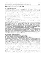
in the latter (Fig. 31.1). In transitional situations, tests
with an intermediate sensitivity may be useful for the
screening of chronic pancreatitis in patients with a
compatible clinical picture and for the long-term
follow-up of patients with known chronic pancreatitis
(Fig. 31.1).
Direct tests
Invasive pancreatic function tests are based on the di-
rect measurement of pancreatic enzymes and bicarbon-
ate output in samples of duodenal juice obtained after
stimulation of the gland by intravenous administration
of secretin and cholecystokinin (CCK) or cerulein
(secretin–cholecystokinin test). Simple stimulation by
intravenous secretin (secretin test) is used in the
so-called endoscopic test, which is based on the mea-
surement of bicarbonate concentration in endoscopy-
guided aspirates of duodenal juice (see below). Finally,
endogenous stimulation by a test meal (Lundh test) is
no longer used because of a lower diagnostic accuracy.
Since direct pancreatic function tests are invasive,
cumbersome, time-consuming, nonstandardized, and
expensive, and since the development of novel sensitive
imaging methods (i.e., endoscopic ultrasonography)
has markedly improved the diagnosis of chronic pan-
creatitis, the usefulness of the secretin–cholecystokinin
test is nowadays limited to its use as gold standard in
the validation of new pancreatic function tests.
Secretin–cholecystokinin test
Method
The secretin–cholecystokinin test protocol differs
among centers. A double-lumen nasoduodenal tube
should be placed for constant aspiration of gastric juice
and complete and fractionated collection of duodenal
juice on ice during continuous intravenous infusion of
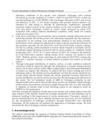
secretin and CCK or cerulein. The protocol recom-
mended by our group is summarized in Fig. 31.2.
Despite duodenal juice being continuously aspirated,
collection may be incomplete. The amount of juice lost
toward the jejunum may be calculated by constant duo-
denal perfusion of a nonabsorbable dilution marker,
usually polyethylene glycol. However, this requires a
triple-lumen tube and further complicates the perfor-
mance of the test.
An additional problem is the variable inactivation of
pancreatic enzymes within the collected duodenal juice
despite the use of antiproteases and collection on ice.
This may be overcome by the single quantification of
zinc instead of bicarbonate and enzymes. Zinc secre-
tion is linked to pancreatic proteases; it is easily quan-
tifiable and very stable in duodenal juice. Our group
has recently demonstrated that the secretin–cerulein
test based on single quantification of zinc output is as
Table 31.1 Pancreatic function tests.
Direct tests
Secretin–cholecystokinin test
Endoscopic test
Indirect tests
Fecal fat quantification
Fecal levels of pancreatic enzymes
NBT-PABA test
Pancreolauryl test
Amino acid consumption test
Breath tests (
13
C-labeled substrates)
HIGH
LOW
Sensitivity of the test to be used
• Diagnosis of chronic pancreatitis in cases of
inconclusive morphologic changes
• Sereening of chronic pancreatitis in patients
with compatible clinical symptoms
• Long-term follow-up of patients with known
chronic pancreatitis
• Diagnosis of primary or secondary pancreatic
insufficiency
• Indication for and control of the efficacy of oral
enzyme substitution therapy
Figure 31.1 Indications for evaluation of exocrine pancreatic
function. The sensitivity of the function test to be used varies
according to the indication.
CHAPTER 31
261
accurate as the test based on quantification of bicar-
bonate and enzymes for evaluation of exocrine pan-
creatic function.
Interpretation
The secretin–cholecystokinin test allows classification
of the severity of exocrine pancreatic dysfunction
(Table 31.2). The sensitivity and specificity of this test
for the diagnosis of chronic pancreatitis both exceed
90% (Table 31.3).
Endoscopic test
The endoscopic pancreatic function test has been devel-
oped in order to avoid the problems associated with the
secretin–cholecystokinin test, i.e., intubation, dura-
tion, and clinical applicability. It is based on the
measurement of bicarbonate concentration and/or
pancreatic enzyme activity in samples of duodenal juice
obtained during upper gastrointestinal endoscopy after
intravenous secretin stimulation.
Method
The protocol for the endoscopic pancreatic function
test is based on the following four steps.
1 Standard endoscopy to the descending duodenum
with the patient under conscious sedation.
2 Intravenous administration of secretin (1 U/kg or
0.2 mg/kg).
3 Endoscopic duodenal fluid collection at 0, 15, 30,
45, and 60 min after secretin injection. A short version
of the test is based on the collection of duodenal juice
for only 10 min.
4 Fluid analysis for bicarbonate concentration and/or
pancreatic enzyme activity.
Interpretation
The peak bicarbonate concentration over 60 min is
lower in patients with advanced chronic pancreatitis
than in those with abdominal pain of extrapancreatic
origin. Measurement of lipolytic activity in duodenal
juice collected for 10 min after intravenous secretin is
also significantly lower in patients with chronic pancre-
atitis compared with patients with normal pancreas,
but it is not accurate enough for routine clinical use.
Calculation of bicarbonate and enzyme output
Quantification of volume, bicarbonate concentration, and
amylase, lipase and protease (trypsin, chymotrypsin,
and/or elastase) activities
Overnight fasting
Placement of a double-lumen tube under
fluoroscopic control, with the tip at the
ligament of Treitz
Continuous aspiration of gastric and duodenal juice
Continuous intravenous infusion of secretin
(1 U/kg per hour) and cerulein (100ng/kg per hour)
over 90 min
Sampling of duodenal juice in 10-min aliquots over the
last 60 min of hormone infusion
Figure 31.2 Secretin–cerulein test protocol.
Table 31.2 Severity of exocrine pancreatic dysfunction based
on the secretin–cholecystokinin test.
Normal Normal output of enzymes and
bicarbonate
Mild dysfunction Secretion of enzymes and
bicarbonate ≥ 75% of the lower
limit of normal
Moderate dysfunction Secretion of enzymes and
bicarbonate 30–75% of the
lower limit of normal
Severe dysfunction Secretion of enzymes and
bicarbonate < 30% of the lower
limit of normal
Table 31.3 Mean accuracy of exocrine pancreatic function
tests for the diagnosis of chronic pancreatitis.
Sensitivity (%) Specificity (%)
Secretin–cholecystokinin 90 94
test
Fecal chymotrypsin 57 88
Fecal elastase 70 85
Optimized serum 82 90
pancreolauryl test
Although the endoscopic pancreatic function test is a
promising procedure, it is far from being the current
standard. Whether the peak bicarbonate concentra-
tion, instead of output over time, is a reliable marker of
exocrine pancreatic function is questionable. In fact,
most experts in this field support bicarbonate and
enzyme output and not concentration as the most
reliable marker of exocrine pancreatic function. This is
due to the inverse relationship between bicarbonate
concentration and rate of juice secretion in response to
secretin. In addition, the endoscopic pancreatic func-
tion test requires the endoscope to be maintained in the
duodenum for 1 hour, which is at least as uncomfort-
able for patients as nasoduodenal tubing. Because of
this, pharmacologic conscious sedation is required in
the context of the endoscopic test, although the effect of
these drugs on exocrine pancreatic function has not
been specifically evaluated. All these facts hinder the
clinical usefulness of the endoscopic pancreatic func-
tion test.
Indirect tests
Indirect tests evaluate exocrine pancreatic function by
quantifying either the digestive ability of the gland or
levels of pancreatic enzymes in feces (Table 31.1). From
a methodologic point of view, these tests can thus be
classified as oral tests and fecal tests. In oral tests, a
substrate is orally given together with a test meal.
Pancreatic enzymes hydrolyze the substrate within the
duodenum; the released metabolites are absorbed from
the gut and can then be measured in serum, urine, or
breath. Oral tests include the pancreolauryl test and
different breath tests, mainly using
13
C-labeled sub-
strates. Other tests like the NBT-PABA test and the
amino acid consumption test are no longer commer-
cially available and/or have insufficient diagnostic ac-
curacy to be recommended for clinical use.
Several extrapancreatic factors are known to limit
the accuracy of oral pancreatic function tests, mainly
those interfering with normal digestion (slow gastric
emptying rate, decreased bile acid secretion) and in-
testinal absorption (intestinal diseases) as well as those
affecting the elimination of digestion products (renal
insufficiency). Variability in gastric rate can be avoided
to some extent by administration of metoclopramide or
any other prokineticum in the context of the test. The
potential negative role of renal disturbances is avoided
by the quantification of digestion products in serum
instead of urine.
Fecal tests are based on the quantification of pan-
creatic enzyme concentration (elastase) or activity
(chymotrypsin) in feces. Enzymes are deactivated and
diluted or concentrated to a variable degree during
intestinal passage, which must be taken into account
when interpreting test results. Exocrine pancreatic
function can also be measured indirectly in feces by
means of fecal fat quantification. The amount of fat
eliminated within the feces indirectly reflects fat diges-
tion and therefore pancreatic lipase secretion.
Fecal tests
Fecal fat quantification
Fecal fat quantification using the classical Van de
Kamer test is the gold standard for the diagnosis of
steatorrhea. However, this test has several important
disadvantages that limit its clinical applicability. Pa-
tients must eat a standard diet containing 80–120 g of
fat daily for five consecutive days. This is an important
handicap since the majority of patients with chronic
pancreatitis are alcoholics and thus have limited com-
pliance. Furthermore, patients should collect the total
amount of feces produced over the last 3 days of the
diet. Again this is not easy for alcoholic patients. A 3-
day collection is needed to reduce errors and variability
that may occur if a shorter collection period is used.
Patient compliance is not the only limitation of fecal
fat quantification; so is the handling of stool samples
in the laboratory. Stool samples collected over 3 days
must be first homogenized and then processed man-
ually, making this test unpleasant and cumbersome. A
new methodology based on near-infrared reflectance
analysis (NIRA) has greatly simplified the quantifica-
tion of fat in stool and thus could make feasible the
wide application of this test in clinical routine. Never-
theless, the difficulties associated with patient com-
pliance remain the same.
Method In our laboratory, patients are instructed to
eat a diet containing 92 g of fat for 5 days. Stool from
the last three consecutive days is collected in three dif-
ferent containers. The daily amount of fat excreted
(g/day) is quantified based on fat concentration meas-
ured by NIRA (g/100 g stool) and the total weight of
the stool on each day. The mean of the three values
obtained is considered as the result.
PART II
262
Interpretation Following the test protocol described
above, a fecal fat excretion below 7.5 g/day is consid-
ered normal. Fat maldigestion indicating exocrine pan-
creatic insufficiency is defined by a fecal fat excretion
greater than 7.5 g/day. Interpretation of the test may be
improved by keeping a record of all dietary intake over
the 5-day period. In this way, fat intake can be deter-
mined and thus the fractional fat absorption can be cal-
culated. It should be noted that fecal fat quantification
is a nonspecific pancreatic function test since any other
cause of maldigestion (i.e., obstructive jaundice) or
malabsorption (i.e., sprue, Crohn’s disease) may also
induce abnormal fecal fat excretion.
Fecal chymotrypsin activity
Quantification of fecal chymotrypsin is a simple test
and easy to apply to the clinical routine. This test is
based on the enzymatic quantification of chymotrypsin
activity in an isolated small stool sample. Because of
this, fecal chymotrypsin has been widely introduced in
clinical routine as an exocrine pancreatic function test.
However, chymotrypsin is variably inactivated during
intestinal passage in such a way that fecal chymotrypsin
activity does not accurately reflect pancreatic secretion
of the enzyme. In addition, dilution of the enzyme in
patients with diarrhea of any etiology will also decrease
the fecal activity of the enzyme.
Because of this, and in order to maintain adequate
specificity of the test, a low cut-off (3 U/g of stool) is
generally accepted as the definition of an abnormal test.
Patients with fecal chymotrypsin activity of less than
3 U/g of stool are thus considered as suffering from ex-
ocrine pancreatic dysfunction, although the sensitivity
obtained with the test is too low to recommend it for
clinical practice. In fact, the test is not able to detect a
single case of mild exocrine pancreatic dysfunction,
and can only detect slightly more than half of those
patients with moderate or severe dysfunction (Table
31.3).
Last but not least, orally administered exogenous
pancreatic enzymes as a treatment for exocrine pan-
creatic insufficiency interact with the determination
of chymotrypsin in stool and thus this therapy should
be interrupted for at least the 48 hours preceding
stool sample collection. This is not always easy to
accomplish for patients with exocrine pancreatic
insufficiency.
In conclusion, and after taking into consideration all
the aspects mentioned above, fecal chymotrypsin quan-
tification should no longer be considered adequate
for evaluating exocrine pancreatic function in clinical
routine.
Fecal elastase concentration
Compared with chymotrypsin, pancreatic elastase is
highly stable during gastrointestinal transit and the
fecal concentration of this enzyme correlates signifi-
cantly with the amount of enzyme secreted by the ex-
ocrine pancreas. Furthermore, since the methodology
used to quantify this enzyme is based on human-
specific monoclonal antibodies, oral enzyme substitu-
tion therapy does not interfere with the test. Therefore,
interruption of this therapy previous to stool collection
is not needed, which is an important advantage.
Method Quantification of fecal elastase is performed
in a single small stool sample by a specific enzyme
immunoassay.
Interpretation A fecal elastase concentration higher
than 200 mg/g is considered normal. Concentrations
lower than 50 mg/g are related to exocrine pancreatic
insufficiency. Although fecal elastase quantification is
not sensitive enough to detect patients with mild ex-
ocrine pancreatic dysfunction, its sensitivity in cases of
moderate to severe dysfunction is very high, reaching
values close to 100%. The specificity of fecal elastase
is also high, only limited by dilution in cases of watery
diarrhea.
Fecal elastase based on the use of human-specific
monoclonal antibodies is therefore an excellent test for
the diagnosis of exocrine pancreatic dysfunction in the
context of chronic pancreatitis. Since this test is easy to
apply to the clinical routine, it may be used as a first
step in the study of patients with clinically suspected
chronic pancreatic disease and for the follow-up of
patients with known chronic pancreatitis. In situations
of secondary exocrine pancreatic dysfunction (i.e.,
after gastrointestinal surgery), fecal elastase is useful
for evaluating pancreatic secretion but not for detect-
ing maldigestion.
Oral tests
Pancreolauryl test
Fluorescein dilaurate is administered orally together
with a standardized breakfast. A pancreas-specific
cholesterol ester hydrolase acts on this compound
CHAPTER 31
263
and water-soluble fluorescein is released and absorbed
from the gut. Fluorescein can thus be measured in
serum or urine after renal excretion. The advantage of
this test is that it is easily applicable to the clinical
routine and can be used not only for supporting the
diagnosis of chronic pancreatitis but also for the
follow-up of patients with this disease. The major dis-
advantages of the test are the limiting factors of oral
tests described above and a limited sensitivity for the
early diagnosis of chronic pancreatitis.
Method The standard test requires collection of urine
over 10 hours after the ingestion of the standard break-
fast and fluorescein dilaurate. The diuresis should be in-
creased by the ingestion of at least 1500 mL of water
during the test. In order to compensate for the variable
intestinal absorption and renal excretion of the sub-
strate, the test should be repeated 3 days later by giving
fluorescein sodium as substrate. On both test days,
urine fluorescein concentration is measured.
Repetition of the test is not necessary if fluorescein
concentration is measured in serum. Our group has op-
timized the serum pancreolauryl test by administering
intravenous metoclopramide just after the ingestion
of the test meal in order to avoid potential problems
related to gastric emptying. In addition, intravenous
administration of secretin just before ingestion of the
test meal significantly increases the sensitivity of the test
by inducing a washout of stored pancreatic enzymes
accumulated overnight. Finally, we have optimized the
measurement of serum fluorescein concentration. The
optimized serum pancreolauryl test protocol is summa-
rized in Fig. 31.3. Potential adverse effects of metoclo-
pramide and secretin present very rarely after a single
dose. In our experience, transient mouth dryness is ob-
served occasionally after metoclopramide administra-
tion. A few patients suffer from nausea after secretin,
which can be prevented by slow injection of the drug
(over 2–3 min).
Interpretation Results of the urine pancreolauryl test
are expressed as the quotient between the urine fluores-
cein concentration at day 1 (when fluorescein dilaurate
is given as substrate) and that at day 2 (when fluorescein
sodium is given as substrate). A quotient is considered
as normal if higher than 30 and abnormal if lower than
20. Values between 20 and 30 are inconclusive.
The peak serum fluorescein concentration is consid-
ered as the result of the serum test. A peak greater than
PART II
264
4.5 mg/mL indicates normal exocrine pancreatic func-
tion. Mild to moderate exocrine pancreatic dysfunc-
tion is defined by a peak between 2.5 and 4.5 mg/mL.
A result below 2.5 mg/mL is observed in patients with
severe pancreatic dysfunction.
The accuracy of the optimized serum pancreolauryl
test is much higher than the accuracy of the standard
test in urine (Table 31.3). The sensitivity of the opti-
mized serum test for the diagnosis of mild exocrine pan-
creatic dysfunction is 75%, and for moderate or severe
dysfunction is 100%. False-positive results can be ob-
tained in patients with gastrointestinal extrapancreatic
diseases leading to maldigestion of fluorescein dilau-
rate (e.g., partial gastric resection with Billroth II anas-
tomosis, obstructive jaundice) or to malabsorption of
released fluorescein (e.g., sprue).
13
C-substrate breath tests
Several substrates, mainly
13
C-labeled, have been used
to evaluate exocrine pancreatic function by means of
breath tests. In these tests, the labeled substrate is given
orally together with a test meal. After intraduodenal
hydrolysis of the substrate by specific pancreatic en-
zymes,
13
C-marked metabolites are released, absorbed
from the gut, and metabolized within the liver. As a con-
sequence of hepatic metabolism,
13
CO
2
is released and
Overnight fast
Placement of an indwelling cannula in an antecubital vein
Take basal blood sample (10 mL)
Intravenous administration of secretin (1 U/kg body
weight) over 2–3 min
Ingestion of the test meal (40 g of white bread, 20g of
butter, 200 mL of tea) together with 1 mmol fluorescein
dilaurate spread on the bread together with the butter
Intravenous metoclopramide administration (10 mg)
Take blood samples (5 mL each) at 120, 150, 180 and
240 min after test meal ingestion
Measurement of serum fluorescein concentration
in all samples
Figure 31.3 Optimized serum pancreolauryl test protocol.
CHAPTER 31
265
thereafter eliminated with expired air (Fig. 31.4). The
amount of
13
CO
2
expired, which indirectly reflects ex-
ocrine pancreatic function, can be measured by means
of mass spectrometry or infrared analysis.
Most substrates used in breath tests, among them
mixed
13
C-triglyceride, cholesteryl
13
C-octanoate,
13
C-
hiolein, and
13
C-triolein, are hydrolyzed by pancreatic
lipase. In this way pancreatic function breath tests
should be seen as fat digestion tests and thus considered
as an alternative to fecal fat quantification.
The only breath test that has been optimized is the
mixed
13
C-triglyceride (
13
C-MTG) breath test. In our
experience, this is the optimal substrate for the diagno-
sis of fat maldigestion and thus the
13
C-MTG breath
test has been developed as a simple alternative to fecal
fat quantification.
Method According to the protocol developed by our
group, a total of 250 mg of
13
C-MTG is spread on a
solid test meal containing 16 g of fat. Before the meal
(basal sample) and in 30-min intervals for 6 hours after
ingestion of the meal, breath samples are collected in
10-mL tubes. A single dose of a prokineticum (i.e.,
metoclopramide) is given orally 20–30 min before the
meal in order to avoid potential problems related to
gastric emptying. The amount of
13
CO
2
in breath
samples is measured by mass spectrometry. The result
of the test is expressed as the total amount of recovered
13
CO
2
over the 6 hours.
Interpretation A
13
CO
2
below 58% indicates the pres-
ence of fat maldigestion, with a sensitivity and specific-
ity higher than 90%. The test is also highly accurate
for the diagnosis of maldigestion in clinical situations
of secondary exocrine pancreatic insufficiency, such
as partial or total gastrectomy or duodenectomy.
The
13
C-MTG breath test is a simple, noninvasive,
and accurate method for the diagnosis of exocrine pan-
creatic insufficiency. It is easily applicable to the clinical
routine and can be repeated as often as necessary. In this
way, the utility of the test is not only limited to the diag-
nosis of exocrine pancreatic insufficiency but can also
be extended to control of the efficacy of oral enzyme
substitution therapy in these patients. Therefore, the
13
C-MTG breath test may play a relevant role in the
management of patients with maldigestion secondary
to chronic pancreatitis, cystic fibrosis, pancreatic can-
cer, and after acute necrotizing pancreatitis or gastric or
duodenal surgery.
Summary
A wide variety of tests are nowadays available for
the evaluation of exocrine pancreatic function. The
secretin–cholecystokinin test is still the gold standard,
but its use is presently limited to the evaluation of new
function tests in specialized centers. Quantification of
pancreatic zinc output as a single marker may simplify
the clinical applicability of this direct test.
The optimized serum pancreolauryl test is the most
sensitive tubuless pancreatic function test and probably
13
C-substrate
13
CO
2
13
CO
2
13
C-metabolites
Figure 31.4 Basis of pancreatic
function breath tests.
Domínguez-Muñoz JE, Martínez S, Leodolter A,
Malfertheiner P. Quantification of pancreatic zinc output as
pancreatic function test: making the secretin–caerulein test
applicable to clinical practice. Pancreatology 2004;4:57–
62.
Gullo L. Value and clinical role of intubation tests in chronic
pancreatitis. In: HG Beger, M Buchler, H Ditschuneit, P
Malfertheiner (eds) Chronic Pancreatitis. Berlin: Springer-
Verlag, 1990: 287–290.
Lembcke B. Present and future of breath test in the diagnosis
of pancreatic insufficiency. In: P Malfertheiner, JE
Dominguez-Muñoz, HU Schulz, H Lippert (eds) Diagnostic
Procedures in Pancreatic Disease. Berlin: Springer-Verlag,
1997: 261–271.
Lembcke B, Grimm K, Lankish PG. Raised fecal fat concentra-
tion is not valid indicator of pancreatic steatorrhea. Am J
Gastroenterol 1987;82:526–531.
Lembcke B, Braden B, Caspary WF. Exocrine pancreatic
insufficiency: accuracy and clinical value of the uniformly
labeled
13
C-hiolein breath test. Gut 1996;39:668–74.
Leodolter A, Kahl S, Domínguez-Muñoz JE, Gerard C,
Glasbrenner B, Malfertheiner P. Comparison of two tube-
less function tests in the assessment of mild to moderate
exocrine pancreatic insufficiency Eur J Gastroenterol
Hepatol 2000;12:1335–1338.
Löser C, Möllgaard A, Fölsch UR. Faecal elastase 1: a novel,
highly sensitive and specific tubeless pancreatic function
test. Gut 1996;39:580–586.
Löser C, Brauer C, Aygen S, Hennemann O, Fölsch UR. Com-
parative clinical evaluation of the
13
C-mixed triglyceride
breath test as an indirect pancreatic function test. Scand J
Gastroenterol 1998;33:327–334.
Malfertheiner P, Büchler M. Correlation of imaging and
function in chronic pancreatitis. Radiol Clin North Am
1989;27:51–64.
Stein J, Purschian B, Bieniek U, Caspary WF, Lemcke B. Near-
infrared reflectance analysis (NIRA): a new dimension in
the investigation of malabsorption syndromes. Eur J Gas-
troenterol Hepatol 1994;6:889–894.
Stein J, Jung M, Sziegoleit A, Zeuzem S, Caspary F, Lembcke
B. Immunoreactive elastase 1: clinical evaluation of a new
noninvasive test of pancreatic function. Clin Chem 1996;
42:222–226.
Vantrappen GR, Rutgeerts PJ, Ghoos YF, Hiele MI. Mixed
triglyceride breath test: a noninvasive test of pancreatic
lipase activity in the duodenum. Gastroenterology 1989;
96:1126–1134.
Ventrucci M, Cipolla A, Ubalducci GM, Roda A, Roda E.
13
C-labelled cholesteryl octanoate breath test for assessing
pancreatic exocrine insufficiency. Gut 1998;42:81–87.
PART II
266
the most appropriate for the screening of chronic pan-
creatitis in patients with clinical suspicion of the dis-
ease. The urine pancreolauryl test can no longer be
recommended because of its low sensitivity and the
need to repeat the test twice three days apart.
Fecal elastase quantification is the most adequate
fecal test. It is clearly more accurate than fecal chy-
motrypsin for evaluation of exocrine pancreatic func-
tion and is easy to apply to the clinical routine.
Therefore, fecal elastase may be applied as a first step in
the study of patients with suspected chronic pancreati-
tis and to aid in the differential diagnosis of chronic
diarrhea. Fecal chymotrypsin activity is a nonsensi-
tive pancreatic function test and can no longer be
considered for clinical routine.
The
13
C-MTG breath test appears to be an accurate
alternative to fecal fat quantification for the diagnosis
of maldigestion of any etiology. This is a simple and
noninvasive method, easily applicable to the clinical
routine, that can be repeated as frequently as needed
and that is useful for the diagnosis of maldigestion as
well as for optimization of enzyme substitution therapy
in patients with primary or secondary exocrine pan-
creatic insufficiency.
Recommended reading
DiMagno EP, Go VLW, Summerskill HJ. Relations between
pancreatic enzyme outputs and malabsorption in severe
pancreatic insufficiency. N Engl J Med 1973;288:813–815.
Domínguez-Muñoz JE. Noninvasive pancreatic function
tests. In: MW Büchler, H Friess, W Uhl, P Malfertheiner
(eds) Chronic Pancreatitis: Novel Concepts in Biology and
Therapy. Oxford, Berlin: Blackwell Publishing, 2002:
225–232.
Domínguez-Muñoz JE, Malfertheiner P. Optimized serum
pancreolauryl test for differentiating patients with and
without chronic pancreatitis. Clin Chem 1998;44:869–
875.
Domínguez-Muñoz JE, Pieramico O, Büchler M,
Malfertheiner P. Clinical utility of the serum pancreolauryl
test in diagnosing and staging of chronic pancreatitis. Am J
Gastroenterol 1993;88:1237–1241.
Domínguez-Muñoz JE, Hyeronimus C, Sauerbruch T,
Malfertheiner P. Fecal elastase test: evaluation of a new
noninvasive pancreatic function test. Am J Gastroenterol
1995;90:1834–1837.
267
In the initial stages of chronic pancreatitis, which
generally last about 5 or 6 years, the disease is charac-
terized by attacks of abdominal pain that recur at vari-
able intervals during which the patient is pain-free.
When the disease is more advanced, the pain tends to
disappear, either spontaneously or following surgery,
but other symptoms or complications may develop that
can alter the course of the disease. In this chapter
we discuss the role of the physician in management of
this disease, particularly as regards follow-up and
complications.
What to do in the follow-up
The clinical onset of chronic pancreatitis most com-
monly occurs when the patient is in his thirties or for-
ties. A typical patient with chronic pancreatitis is a male
who is employed in a job that requires heavy labor
and who generally (70–80% of cases) drinks alcohol
to excess. In Italy, alcohol is by far the most frequent
etiologic factor, present in 75–80% of patients with
chronic pancreatitis who have an average daily con-
sumption of 120–140 g of pure alcohol. Thus, the first
and most important task for the physician is to con-
vince the patient to stop drinking alcohol, informing
him that if he does not do so there is little or no chance
that his condition will improve, and that he may well
also develop unpleasant complications. It should also
be explained that if he ceases to drink, the attacks may
become less frequent and eventually disappear. Unfor-
tunately, not all patients quit drinking, some resuming
once a painful attack has subsided (Table 32.1).
A majority of individuals with chronic pancreatitis
also smoke, and so another duty of the physician is to
persuade the patient to quit this habit as well, even
though it has not been clearly demonstrated that smok-
ing has a pathogenetic role in chronic pancreatitis or
that it can negatively influence progression of the
disease.
Pain, the most important symptom in chronic pan-
creatitis, particularly in its initial stage, must be care-
fully assessed and monitored in each patient. If the
frequency and intensity of the painful attacks are re-
duced by cessation of alcohol ingestion, the attacks are
likely to eventually disappear, generally within the first
5 or 6 years of the disease; for these patients, surgical
intervention is not indicated. Among our patients,
roughly 50% fall into this category. If, on the contrary,
the frequency and intensity of the painful attacks in-
crease or remain high, surgery or, for a few selected
patients, endoscopic intervention should certainly be
considered, which is the case for about 50% of our
patients. For most of the patients who undergo surgery,
this generally occurs within 5 or 6 years of clinical onset.
It is important to study exocrine and endocrine pan-
creatic function from the initial stages of the disease,
both to support the clinical diagnosis of chronic pan-
creatitis and to guide its treatment. In studies that uti-
lized duodenal intubation and prolonged maximal
pancreatic stimulation, we showed that exocrine pan-
creatic function is impaired in almost all patients with
chronic pancreatitis, starting in the initial stages of the
disease, at which point the functional impairment is
generally mild or moderate. Although duodenal intu-
bation is the more sensitive means of assessing exocrine
pancreatic function, it is time-consuming and trouble-
some and is no longer used in clinical practice. At pre-
32
Follow-up of patients with chronic
pancreatitis: what to do and which
complications can be expected
Lucio Gullo and Raffaele Pezzilli
not stop drinking, often do not keep their scheduled
appointments but may show up only after an attack
of severe pain.
In the nonalcoholic forms of chronic pancreatitis
(about 20–30% of cases), the most important measure
is to determine the cause of the disease and to eliminate
it, which usually leads to improvement in the clinical
picture. With regard to the follow- up, the measures are
essentially the same as those for alcoholic pancreatitis.
The patient with advanced chronic pancreatitis, who
has had the disease for longer than 5 or 6 years, gener-
ally presents with different clinical problems. In the ma-
jority of studies on chronic pancreatitis, it is reported
that pain, the principal clinical manifestation in the
early stages of the disease, is generally no longer present
in the more advanced stages. The patient may have had
surgery for the pancreatitis, or the pain may have
resolved on its own. Those for whom pain continues
to be a significant problem are generally either those
who continue to drink, and for these patients it is diffi-
cult to find a definitive solution for the pain, or they are
patients who have developed a complication, most
often a pseudocyst. We should mention, however, that
in one study pain has been reported to be frequent even
in advanced stages of the disease.
In the advanced stages of chronic pancreatitis, gener-
ally after 8–10 years from clinical onset, exocrine
pancreatic insufficiency may become severe (< 10% of
normal enzyme production) and steatorrhea develops,
necessitating the administration of pancreatic extracts.
It is very important to establish the correct daily dose of
the extracts, which must be adequate to prevent the loss
of fat in the feces; a dose of 30 000 U per meal is gener-
ally sufficient. If steatorrhea does not disappear com-
pletely, this dose can be increased. In patients with
gastric acid hypersecretion it can be helpful to adminis-
ter H
2
blocking agents or proton pump inhibitors with
the extracts in order to prevent their inactivation by
gastric acid. Steatorrhea develops in about 50–60% of
patients with advanced chronic pancreatitis.
In advanced stages of chronic pancreatitis, usually
after 7 or 8 years from clinical onset, diabetes can de-
velop as a result of the destruction of islet cells by pan-
creatic fibrosis. It usually starts in a mild form that is
treatable with oral antidiabetic agents or low doses of
insulin but often progresses to a more severe form with
higher insulin requirements. The complications of dia-
betes due to chronic pancreatitis are similar to those of
primary diabetes. In particular, we studied the fre-
PART II
268
sent, it has been substituted by indirect tests of pancre-
atic function that often show normal results when
the chronic pancreatitis is mild. We now use the fecal
elastase test, which has good sensitivity particularly
in patients who have moderate or severe pancreatic
insufficiency.
Patients who have mild or moderate pancreatic
insufficiency do not have steatorrhea and therefore do
not require the use of pancreatic extracts. However,
some authors have advocated the use of extracts in
patients with mild to moderate insufficiency as well,
for the purpose of preventing attacks of pain. In this
regard, various studies have been carried out but the re-
sults have been conflicting, possibly due to the different
types of enzyme preparations that have been used. The
preparations that seemed to be useful in preventing at-
tacks of pain were those administered in tablet form.
Endocrine pancreatic function is generally normal in
the initial phases of chronic pancreatitis and clinically
evident diabetes usually appears in the advanced stages
of the disease, generally 8–10 years after onset. Thus, in
the early stages of the disease blood glucose determina-
tion and a glucose tolerance test every 6–12 months are
generally sufficient for monitoring endocrine function.
For optimal management of patients with chronic
pancreatitis, especially in the initial stages of the dis-
ease, it is essential to have frequent follow-up visits, at
least once every 6 or 12 months. This serves to monitor
the frequency of episodes of pain as well as the appear-
ance of other disturbances, and especially to determine
whether the patient has stopped drinking. Many pa-
tients quit drinking alcohol and these same individuals
generally keep their appointments for follow-up visits.
Others, who are typically heavier drinkers and who do
Table 32.1 What to do in follow-up.
Ascertain whether the patient has stopped drinking
In nonalcoholic forms, determine the cause and eliminate it
Evaluate pain; if the attacks are frequent, consider surgery or,
in select patients, endoscopy
Assess exocrine and endocrine pancreatic function; if
impaired, treat accordingly
Assess for complications and treat accordingly
Activity limitations and dietetic rules: this is pertinent mainly
to patients with severe steatorrhea or advanced diabetes
Arrange for check-up visits at least every 6–12 months, when
possible, with a specialist in pancreatic diseases
quency of diabetic retinopathy in chronic pancreatitis
and found that it is similar to that of patients with type
I diabetes. Diabetes develops in about 50–60% of
patients with advanced chronic pancreatitis.
Another task for the physician responsible for pa-
tients with chronic pancreatitis is to educate them re-
garding their diet. Prior to onset of the disease these
patients are often hearty eaters and drinkers. In the
early stages of the disease, when steatorrhea and dia-
betes are not yet present, there is no need for particular
dietetic measures; it is important, however, that the diet
be well balanced and that it meets the nutritional needs
of the individual. It is generally advised to reduce fat
intake, although there is no clear evidence that this is
useful. Obviously, if there is decreased glucose toler-
ance, carbohydrate and sugar intake should be re-
duced. In more advanced stages of the disease, when
steatorrhea may be present, the diet should be hyper-
caloric but, other than a reduction of fat intake, the
patient should not be subjected to other restrictions
unless diabetes is also present.
Regarding restrictions on activity, none should be
imposed except in the case of patients who have severe
steatorrhea or advanced diabetes; if their job entails
heavy labor, they should be advised to seek less
strenuous employment.
Complications
Of the various complications that can develop in the
course of chronic pancreatitis, pseudocysts and steno-
sis (generally mild) of the retropancreatic portion of the
common bile duct are the most frequent. Several less
common ones can also be encountered (Table 32.2).
Pancreatic pseudocysts
Pseudocysts most commonly develop during the initial
stages of chronic pancreatitis; their reported frequency
varies from study to study, but they are fairly frequent,
occurring in about 25–30% of cases. In surgical series
their frequency is higher (about 50–60%). Most often
pseudocysts present as a single lesion, but sometimes
two or more can be seen; their size is variable, they are
often symptomatic (persistent pain being the most
frequent symptom), and they are occasionally com-
plicated by rupture or infection. In our experience, in
the great majority of cases the pseudocysts derive from
CHAPTER 32
269
dilated ducts, and thus are true cysts; as they dilate,
the epithelial lining can be lost, at which point they no
longer appear to be true cysts. Among our patients with
chronic pancreatitis, postnecrotic pseudocysts are rare,
the main reason being that we see few patients (about
10%) who have had an acute necrotic attack.
As far as treatment is concerned, if the pseudocysts
are asymptomatic and without complications they can
be left untreated, but repeat ultrasound is recom-
mended every 6–12 months to control their size. If they
become painful or develop a complication, treatment
becomes obligatory. Years ago the only treatment
available was surgery; more recently this has been
abandoned in favor of endoscopic intervention.
Another possibility in the treatment of painful
pseudocysts in chronic pancreatitis is the administra-
tion of octreotide, a synthetic analog of somatostatin,
which causes the cysts to shrink and eventually disap-
pear. We have shown that this treatment (100 mg every
8 hours) is effective mainly when the cysts do not com-
municate with the Wirsung duct and if the drug is ad-
ministered when their size is increasing. When these
criteria are met, the pain disappears completely and
definitively after 3–4 days of octreotide treatment,
as the cysts begin to shrink in size; the cysts then dis-
appear completely after 6–8 weeks of treatment.
We would like to point out that size alone is not an
indication for treatment. Although it has been a guiding
principle that cysts of greater than 5–6 cm in diameter
should be treated, we feel that if the cysts are asympto-
matic treatment is not necessary, regardless of their
Table 32.2 Complications, associated diseases, and
mortality in chronic pancreatitis.
Pancreatic pseudocysts 25–30%
Stenosis (usually mild) of the retropancreatic 40–50%
common bile duct
Pancreatic cancer 1–3%
Extrapancreatic cancer 10–15%
Splenic vein thrombosis 2–5%
Pseudoaneurysm 2–3%
Duodenal obstruction 4–5%
Pancreatic fistula 2–3%
Pancreatic abscess 2–3%
Alcoholic liver diseases 25–40%
Cardiovascular diseases 20–30%
Mortality 20–35%
size. We have followed several patients whose cysts
have been larger than 5 or 6 cm and stable in size for
many years, during which they have also been pain-
free; we believe that intervention is not necessary in
these cases. Generally speaking, once cysts are treated
they are no longer problematic.
Stenosis of the retropancreatic common bile duct
Stenosis of the distal portion of the common bile duct is
a complication seen in both initial and advanced stages
of chronic pancreatitis and can be observed in up to
40–50% of cases; it is generally mild and does not ob-
struct the flow of bile. The stenosis is caused by pancre-
atic fibrosis in the area of the duct. It can contribute to a
generally mild and transient (lasting 3–10 days) form of
jaundice that can occur during attacks of pain, when
the already stenotic bile duct is compressed by pancre-
atic edema. This transient jaundice during attacks of
abdominal pain occurs in about 30–40% of cases.
In about 5–10% of the patients with chronic pancre-
atitis the jaundice persists and requires treatment. It is
due to complete obstruction of the retropancreatic
common bile duct, most often by pancreatic fibrosis,
although sometimes it is due to compression of the
duct by a cyst of the pancreatic head or, more rarely, by
cancers of the pancreatic head, which can complicate
chronic pancreatitis. In these cases, it is necessary to
perform a choledochojejunostomy. A stent is some-
times placed endoscopically, but these tend to become
occluded, and the procedure is often complicated by the
development of cholangitis; for this reason, it should
only be used in carefully selected cases, such as in pa-
tients awaiting surgery or in those who present a high
surgical risk.
Pancreatic cancer
Many studies have been published on the risk of pan-
creatic cancer in patients with chronic pancreatitis, but
results have been conflicting: some have concluded that
there is a risk, others that there is not or that it is very
low. We believe that chronic pancreatitis is a risk factor
for pancreatic cancer but that this risk is low, on the
order of 1–3%. Lowenfels et al. reported a cumulative
risk of pancreatic cancer in subjects with chronic
pancreatitis who were followed for 10 and 20 years
after the diagnosis of pancreatitis of 1.8 and 4%
respectively.
The risk of pancreatic cancer has been reported to be
very much higher in patients with hereditary chronic
pancreatitis. Lowenfels et al. have shown that the esti-
mated cumulative risk of pancreatic cancer to age 70
years in patients with this disease approaches 40%.
Extrapancreatic cancer
Patients with chronic pancreatitis have a high incidence
(10–15%) of extrapancreatic cancer, the commonest
sites being the upper and lower airways as well as the
gastrointestinal tract. The reason for this increased
incidence is not clear, but the abuse of tobacco and
alcohol in these patients is thought to be responsible.
Splenic vein thrombosis
While splenic vein thrombosis is well known as a com-
plication of chronic pancreatitis, its incidence is not.
Bradley reported an incidence of 2% among his pa-
tients with this disease. In our experience, the incidence
has been a little higher (about 5%). Thrombosis of the
splenic vein is due to involvement of the vein by the
chronic inflammatory process in the pancreas. It can
result in the development of gastric or esophageal
varices; although there are no precise data regarding
the frequency with which these varices bleed, the
percentage is generally low.
Pseudoaneurysm
This is a rare complication of chronic pancreatitis, seen
in about 2–3% of cases. It usually occurs in association
with pancreatic pseudocysts, the mechanism of forma-
tion being erosion of an expanding pseudocyst into a
nearby artery. The vessels most commonly involved are
the splenic, gastroduodenal, pancreaticoduodenal, and
hepatic arteries. Pancreatic pseudoaneurysms cause
bleeding that may be slow and intermittent or acute and
massive. Treatment is necessary even if they are not ac-
tively bleeding because untreated pseudoaneurysms
have a very high mortality rate. Bleeding can be suc-
cessfully controlled by arteriographic transcatheter
embolization or surgery.
Duodenal obstruction
This complication occurs in about 4–5% of patients
with chronic pancreatitis. It is generally due to marked
PART II
270
fibrosis of the head of the pancreas that involves the
duodenum or to a pseudocyst; treatment consists of
surgery for the former and endoscopic drainage or sur-
gical treatment for the latter.
Pancreatic fistula
Pancreatic fistulas are a rare complication of chronic
pancreatitis (2–3%). External fistulas generally
develop after surgical procedures on the pancreas or
after attacks of necrotic pancreatitis. Internal pan-
creatic fistulas are generally due to rupture of the main
pancreatic duct or leakage from a pseudocyst. The
main complications of internal fistulas are pancreatic
ascites or pleural effusions.
Treatment consists of fasting, parenteral nutrition,
octreotide (100 mg every 8 hours), or endoscopic stent
placement; if the fistula persists surgery is indicated.
Pancreatic abscess
This is a rare complication that involves only about
2–3% of patients with chronic pancreatitis. The ab-
scess often develops at the site of a previous pseudocyst.
Treatment with antibiotics is generally unsuccessful,
leaving surgery as the only viable alternative.
Alcoholic liver disease
Based on clinical studies, it was long believed that in pa-
tients with chronic alcoholic pancreatitis the damage
from alcohol was limited to the pancreas and that liver
involvement was rare. However, with histologic studies
of the liver we and others have shown that a high per-
centage of these patients have alcoholic damage to the
liver as well. In particular, in a study done on surgical
biopsies of the liver taken from 50 patients with chronic
alcoholic pancreatitis undergoing surgery for pancre-
atitis, we showed that 22 (44%) had associated
alcoholic liver disease; of these 22, 13 had alcoholic
hepatitis, 7 cirrhosis, and 2 steatosis. The percentage
of alcoholic liver disease in this series of patients
was similar to that found in the general alcoholic
population.
We have seen that patients with chronic alcoholic
pancreatitis who develop hepatic disease are most often
those who drink larger quantities of alcohol (> 200 g of
pure alcohol daily) and for a longer period of time (> 20
years). It is therefore important that patients with
chronic alcoholic pancreatitis, especially those whose
alcohol consumption is to the above-described extent,
are periodically monitored with liver function and
imaging tests for early recognition and timely treat-
ment of any associated liver disease.
Cardiovascular lesions
Several investigators have reported an increased
frequency of vascular lesions in patients with chronic
pancreatitis, but while some have assumed that this
was simply a coincidence, others have suggested that a
causal relationship may exist. In a study of 54 patients
with chronic pancreatitis (mean age 44 years, range
26–66), we found evidence of vascular involvement in
18 (33%) of the patients and in 5 (9%) of the controls.
In particular, we found electrocardiographic signs of
coronary artery disease in eight patients, as well as
peripheral signs and symptoms of obliterative athero-
sclerotic disease in the lower extremities in 12 patients.
No significant differences in the prevalence of the major
vascular risk factors were noted between patients with
vascular lesions and those without, or between the pa-
tients and the control subjects.
In another study, we showed that in 57 patients with
chronic pancreatitis there was radiologic evidence of
aortic calcification in 35 (41.4%), but only in 12 of 40
(30%) smoker controls. Interestingly, these patients
had a mean age of 44 years (range 26–59), whereas in
the general population aortic calcifications are rarely
seen in persons under the age of 50–60 years. None of
these patients with chronic pancreatitis had conditions
associated with atherosclerosis, such as diabetes,
arterial hypertension, obesity, or hyperlipidemia. It
should be mentioned that aortic calcification is associ-
ated with a marked increase in risk of death by cardio-
vascular disease. These two studies indicate that,
compared with the general population, patients with
chronic pancreatitis have more frequent cardiovascu-
lar lesions and the lesions tend to develop at an earlier
age; the reason for these findings is not clear.
Mortality
All the studies published on mortality in chronic pan-
creatitis have concluded that it is high. Ammann et al. in
their study of 245 patients with chronic pancreatitis
reported that 86 (35%) died; the mean age at death
was 54 years in 54 patients with alcoholic pancreatitis
CHAPTER 32
271
lence of aortic calcification in chronic pancreatitis. Am J
Gastroenterol 1996;91:759–761.
Gullo L, Ventrucci M, Tomassetti P, Migliori M, Pezzilli R.
Fecal elastase 1 determination in chronic pancreatitis. Dig
Dis Sci 1999;44:210–213.
Gullo L, Tomassetti P, Migliori M, Casadei R, Marrano D. Do
early symptoms of pancreatic cancer exist that can allow an
earlier diagnosis? Pancreas 2001;22:210–213.
Hansen TH, Laursen M, Christensen E et al. Chronic pancre-
atitis and extrapancreatic cancer. Int J Pancreatol 1995;
18:235–240.
Hayakawa T, Kondo T, Shibata T, Sugimoto Y, Kitagawa M.
Chronic alcoholism and evolution of pain and prognosis in
chronic pancreatitis. Dig Dis Sci 1989;34:33–38.
Karlson BM, Ekbom A, Josefsson S et al. The risk of pan-
creatic cancer following pancreatitis: an association due
to confounding? Gastroenterology 1997;113:587–592.
Lankisch PG, Lohr-Happe A, Otto J, Creutzfeldt W. Natural
course in chronic pancreatitis. Pain, exocrine and endocrine
pancreatic insufficiency and prognosis of the disease. Diges-
tion 1993;54:148–155.
Layer P, Yamamoto H, Kalthoff L, Clain JE, Bakken LJ,
DiMagno EP. The different courses of early and late onset
idiopathic and alcoholic chronic pancreatitis. Gastroen-
terology 1994;107:1481–1487.
Levy P, Milan C, Pignon JP, Baetz A, Bernades P. Mortality fac-
tors associated with chronic pancreatitis. Unidimensional
and multidimensional analysis of a medical–surgical series
of 240 patients. Gastroenterology 1988;96:1165–1172.
Lowenfels AB, Maisonneuve P, Cavallini G et al. Pancreatitis
and the risk of pancreatic cancer. N Engl J Med 1993;
328:1433–1437.
Lowenfels AB, Maisonneuve P, DiMagno EP et al. Hereditary
pancreatitis and the risk of pancreatic cancer. J Natl Cancer
Inst 1997;89:442–446.
Miyake H, Harada H, Kunichik K, Ochi K, Kimura I. Clinical
course and prognosis of chronic pancreatitis. Pancreas
1987;2:378–385.
Pradeep B, Sonnenberg A. Pancreatitis is a risk factor for pan-
creatic cancer. Gastroenterology 1995;109:247–251.
Saed ZA, Ramirez FC, Hepps KS. Endoscopic stent placement
for internal and external pancreatic fistulas. Gastroenterol-
ogy 1993;105:1213–1217.
Segal I, Parekh D, Lipschitz J et al. Treatment of pancreatic
ascites and external pancreatic fistulas with a long-acting
somatostatin analogue. Digestion 1993;54:53–58.
Woods MS, Traverso LW, Kozarek RA et al. Successful treat-
ment of bleeding pseudoaneurysms of chronic pancreatitis.
Pancreas 1995;10:22–26.
PART II
272
and 66 years in 32 with nonalcoholic pancreatitis. In a
study by Levy et al. of 240 patients with chronic pan-
creatitis, of whom 210 were drinkers, it was reported
that after a mean of 20 years from the clinical onset of
the disease, 57 patients (23.7%) were dead and that the
average age at the time of death was 52 years.
The mortality rate for chronic alcoholic pancreatitis
is higher than it is for idiopathic or other forms of
chronic pancreatitis; as would be expected, among
patients with alcoholic pancreatitis the mortality rate
is higher for those who continue to drink. The main
causes of death for these patients are cardiovascular
disease, hepatic cirrhosis, extrapancreatic or pan-
creatic cancer, postoperative complications, com-
plications of chronic pancreatitis or of diabetes, and
alcoholism. In general, less than 20% of deaths are
directly related to chronic pancreatitis.
Recommended reading
Ammann RW, Akovbiantz A, Largiader F, Schueler G. Course
and outcome of chronic pancreatitis. Longitudinal study of
a mixed medical–surgical series of 245 patients. Gastroen-
terology 1984;86:820–828.
Bender JS, Bouwman DL, Levison MA et al. Pseudocysts and
pseudoaneurysms: surgical strategy. Pancreas 1995;10:
143–145.
Bradley EL III. The natural history of splenic vein thrombosis
due to chronic pancreatitis: indications for surgery. Int J
Pancreatol 1987;2:87–92.
Gullo L, Barbara L. Treatment of pancreatic pseudocysts with
octreotide. Lancet 1991;338:540–541.
Gullo L, Stella A, Labriola E et al. Cardiovascular lesions in
chronic pancreatitis. A prospective study. Dig Dis Sci
1982;27:716–722.
Gullo L, Barbara L, Labò G. Effect of cessation of alcohol
use on the course of pancreatic dysfunction in alcoholic
pancreatitis. Gastroenterology 1988;95:1063–1068.
Gullo L, Parenti M, Monti L, Pezzilli R. Diabetic retinopathy
in chronic pancreatitis. Gastroenterology 1990;98:1577–
1581.
Gullo L, Casadei R, Campione O, Grigioni W, Marrano D.
Alcoholic liver disease in alcoholic chronic pancreatitis:
a prospective study. Ital J Gastroenterol 1995;27:69–
72.
Gullo L, Tassoni U, Mazzoni G, Stefanini F. Increased preva-
273
Introduction
Chronic pancreatitis is an inflammatory, often painful dis-
ease of the exocrine pancreas that leads to exocrine insuf-
ficiency. Its incidence is about 8.2 new cases per 100 000
inhabitants per year in western Europe. Complications
encountered in chronic pancreatitis include biliary
(10–30%) and duodenal (10–25%) obstruction and, as
the disease progresses, maldigestion and diabetes mellitus.
However, the most disturbing complication of chronic
pancreatitis is abdominal pain. Surgical management is
often indicated in cases with medically intractable pain
and this has also repercussions on the economic manage-
ment of these patients. Three typical pain profiles that
occur during the evolution of chronic pancreatitis have
been described: (i) repeated episodes of acute pancreati-
tis (acinar necrosis) in early stages; (ii) spontaneous last-
ing pain relief in association with severe pancreatic
dysfunction in the late stage of uncomplicated chronic
pancreatitis; and (iii) persistent severe pain (or frequent
recurrent episodes of pain) usually in association with
local complications such as pseudocysts, ductal hyper-
tension or extrapancreatic complications such as partial
obstruction of the common bile duct, peptic ulcer, and
opiate addiction. Clearly, the actual pathophysiology of
abdominal pain remains elusive and several hypotheses
have been postulated over the last few years.
At present, pancreatic and extrapancreatic mecha-
nisms are implicated in the development of pain in
chronic pancreatitis.
Pancreatic causes of pain
Acute inflammation of the pancreas
Acute inflammation is readily apparent when there is
severe abdominal pain and tenderness, elevation of
serum amylase and lipase, and evidence of acute pan-
creatic inflammation on computed tomography. The
causes are likely to be the same as for inflammation
associated with acute pancreatitis, involving activated
enzymes and other injurious substances.
Increased pressure in the pancreatic ducts and tissue
Intrapancreatic ductal pressure might be related to
pancreatic secretion itself and to the presence of an ob-
struction in the pancreatic duct. Therefore, many inves-
tigators have related the origin of pain to increased
pressure in the pancreatic ducts and tissue. This ductal
hypertension hypothesis as an explanation for pain in
chronic pancreatitis is derived from observations that
decompression of a dilated pancreatic duct or pseudo-
cyst frequently relieves pain in patients with chronic
pancreatitis. Pancreatic enzyme supplementation may
also relieve pain in some patients with chronic pan-
creatitis. It is believed that the beneficial effect of pan-
creatic enzymes is explained by regulation involving
cholecystokinin-mediated feedback between pancreat-
ic exocrine secretion and the activity of proteases in
the lumen of the small intestine. According to this
hypothesis, administration of enzymes reduces hyper-
cholecystokininemia in patients with chronic pancre-
atitis, thus resulting in less stimulation of the pancreas
and subsequently lowered intraductal pressure and
pain. Interestingly, different studies have shown that
progressive pancreatic insufficiency, which appears
several years after the first diagnosis, is often associated
with a reduction in, and sometimes complete relief of,
pain in patients with chronic pancreatitis, thus indicat-
ing that disease progression might “burn out” the pan-
creas itself, as mentioned before. In contrast, we have to
33
Conservative treatment of pain in
chronic pancreatitis: guidelines for
clinical routine
Pierluigi Di Sebastiano, Markus A. Weigand, Jörg Köninger,
Fabio F. di Mola, Helmut Friess, and Markus W. Büchler
consider that often pain in chronic pancreatitis is not
related to the consumption of food, and even pain
intensity, radiation, and duration are not constant. In
addition, other studies have calculated that around
30% of the patients treated with decompressive
surgery exhibit recurrent attacks of pain. On one hand,
when patients are pain-free after surgery, this could
often be due to the reduction of alcohol ingestion or to
progressive pancreatic insufficiency. At present, the
relationship between pancreatic parenchymal pressure
and pain in chronic pancreatitis is still controversial.
Neurogenic inflammation
Recent concepts have focused on the possible involve-
ment of the nervous system in chronic pain and the in-
flammatory process in chronic pancreatitis. Supporting
this fascinating hypothesis, Keith et al. postulated that
neural and perineural alteration might be important in
the pathogenesis of pain in chronic pancreatitis. They
demonstrated that pain severity correlated with the
duration of alcohol consumption, pancreatic calcifica-
tion and, more interestingly, with the percentage of
eosinophil number in the perineural infiltrate, but not
with duct dilatation.
A subsequent study demonstrated that there is an
increase in both number and diameter of pancreatic
nerve fibers in the course of chronic pancreatitis
compared with normal pancreas. Also, there is an
altered pattern of intrinsic and possibly extrinsic
innervation of the pancreas in chronic pancreatitis,
leading to upregulation of neuropeptides such as sub-
stance P and calcitonin gene-related peptide. Because
both of these peptides are generally regarded as pain
transmitters, these findings provided evidence that
changes in pancreatic nerves themselves might be in-
volved in the long-lasting pain syndrome in chronic
pancreatitis.
Another interesting finding of these studies was the
observation of close contacts between neuronal struc-
tures and immune cells in chronically inflamed pan-
creas, which led to the concept that neuroimmune
mechanisms play a role in the pathogenesis of chronic
pancreatitis and the accompanying abdominal pain. To
confirm this interesting hypothesis, in a subsequent
report the presence of growth-associated protein
(GAP)-43, an established marker of neuronal plasticity,
correlated with individual pain scores in patients with
chronic pancreatitis.
Extrapancreatic causes of pain
Bile duct stenosis and duodenal stenosis due to exten-
sive pancreatic fibrosis and inflammation are often
considered putative extrapancreatic causes of pain.
However, only a few authors are in accordance with
this belief. Recently, Becker and Mischke described a
pathologic condition named “groove pancreatitis” in
19.5% of 600 patients with chronic pancreatitis. This
form of chronic pancreatitis is characterized by the for-
mation of a scar plate between the head of the pancreas
and the duodenum. Scars in the groove lead to compli-
cations also determined by the topography: distur-
bance in the motility of the duodenum, stenosis of the
duodenum, and tubular stenosis of the common bile
duct, which occasionally leads to obstructive jaundice.
These alterations might be responsible for several
symptoms present in chronic pancreatitis and also for
postprandial pain, probably due to compression of
several critical structures, such as nerves and ganglia,
present between the pancreatic head and the duodenum.
Characteristics of pain in
chronic pancreatitis
Pain is the leading symptom in chronic pancreatitis and
should be treated to improve quality of life and to pre-
vent excessive weight loss in these patients. Pain can be
mild, moderate, or severe and increase or decrease over
time. Multifactorial elements are involved and this may
explain why all patients do not respond to the same
treatment modality.
Pain in chronic pancreatitis is usually elicited by the
activation of specific nociceptive receptors (nocicep-
tors) and is thus referred to as nociceptive pain. How-
ever, it may also result from injury to sensory fibers or
from damage to the central nervous system itself (neuro-
pathic pain). Inflammation due to neural activity is
called neurogenic inflammation. In healthy conditions,
nociceptors in the pancreas are silent and are not
activated by noxious stimulation. In chronic pancreati-
tis, however, inflammation, ischemia, elevated pres-
sures, and release of substances such as prostaglandins,
bradykinin, leukotrienes, and substance P sensitize no-
ciceptors to the generation of action potentials and in-
duce nociceptive pain. In addition, pain is also based on
neuropathic changes such as proliferation of unmyeli-
nated nerve fibers, destruction of the perineurium, neur-
PART II
274
CHAPTER 33
275
al edema, and damage to nerve fibers. Thus, nociceptive
pain and neurogenic inflammation in chronic pancreati-
tis provide the rationale for medical, analgesic, and
antiinflammatory treatment. In the beginning, patients
should be examined for obvious abnormalities and
extrapancreatic causes of pain. Surgical intervention
should seriously be considered if long-term use of potent
opioids is required for pain relief, before the develop-
ment of narcotic addiction. In addition, surgery is
recommended if pain no longer responds to analgesics
or if complications in adjacent organs occur.
Conservative treatment of pain
Abstinence from alcohol
The first step in managing pain in chronic pancreatitis
consists of the complete avoidance of alcohol. Alcohol
abstinence can achieve pain relief in up to 50% of pa-
tients, but its effect seems to be restricted to patients
with mild or moderate disease.
Analgesics
The treatment of pain in chronic pancreatitis is per-
formed according to the three-step ladder of the World
Health Organization for the relief of cancer pain (Fig.
33.1). The first step is for mild to moderate pain and
consists of nonopioid analgesics. The second step is for
moderate to severe pain and a nonopioid analgesic is
combined with a mild opioid, which is titrated until
pain relief is satisfactory. The third step is for severe
pain and requires the use of a potent opioid such as
morphine. Adjuvant drugs like tricyclic antidepres-
sants may be used at each step on the ladder. However,
as already mentioned above, when pain treatment
in chronic pancreatitis requires the long-term use
of potent opioids, surgical intervention should be
considered.
Although the application of reasonable levels of
analgesics is the first-line therapy for pain in chronic
pancreatitis, there are no randomized studies compar-
ing different drugs for pain therapy in chronic pancre-
atitis. Regular application of analgesic drugs should be
preferred to drug intake on demand in order to provide
consistent analgesia. Pain intensity should be regularly
estimated by visual analog scale and the lowest drug
dose for sufficient pain relief prescribed. Pain therapy
should be coordinated and provided by one physician
to avoid overprescription and to reduce the risk of
abuse or addiction.
Acetaminophen (paracetamol) and metamizol
According to a consensus report from the German
Society of Gastroenterology, acetaminophen or
metamizol are the drugs of first choice for pain treat-
ment in chronic pancreatitis. Acetaminophen is a drug
with good analgesic and antipyretic properties and
minimal adverse effects. In particular, no relevant gas-
trointestinal adverse effects of the drug in the recom-
Step 1
Step 2
Step 3
Step 4
Nonopioid Analgesic
Acetaminophen
Metamizol
NSAIDs
Selective COX-2
inhibitors
Mild opioids
Tramadol
Tilidin/naloxone
(Dihydro)codeine
Dextropropoxyphene
Adjuvant drugs (tricyclic antidepressants, anticonvulsants, and steroids)
Physiotherapy, psychotherapy
Surgery
and other
interventional
procedures
Potent opioids
Morphine
Buprenorphine
Transdermal patches
of fentanyl and
buprenorphine
Change in opioids or application mode
Treatment of opioid-induced advevse effects
Neurolytic
procedures
Figure 33.1 World Health
Organization analgesic ladder for
cancer pain adapted for pain
management in chronic pancreatitis.
COX, cyclooxygenase; NSAIDs,
nonsteroidal antiinflammatory drugs.
PART II
276
mended dose is known. Acetaminophen may also have
synergistic effects with other nonopioid analgesics.
Metamizol has potent analgesic and antipyretic
effects and is the standard nonopioid analgesic in
many countries. In addition to its analgesic properties,
metamizol also has spasmolytic effects, which can be
beneficial for pain treatment in chronic pancreatitis. A
major adverse effect of metamizol is the risk of agranulo-
cytosis. Because of this, metamizol is not approved in the
USA and UK. When metamizol is used on a regular basis,
a white blood cell count has to be performed regularly.
Unfortunately, acetaminophen and metamizol have
only weak inhibitory activity against cyclooxygenase
(COX) and therefore possess no antiinflammatory
effects. Since inflammation is a major cause of the
nociceptive and neuropathic pain during chronic
pancreatitis, antiinflammatory analgesic agents may be
beneficial under these conditions.
Nonsteroidal antiinflammatory drugs
Nonsteroidal antiinflammatory drugs (NSAIDs), with
the exception of aspirin, inhibit COX-1 and COX-2 in
a reversible manner and thus provide consistent analge-
sia and antiinflammatory action. In addition, they have
antipyretic properties. NSAIDs are recommended as
first-line drugs for the treatment of nociceptive pain
(Table 33.1).
Although the inhibition of inflammation by NSAIDs
is an attractive hypothesis because (neurogenic) inflam-
mation is a key pathogenic factor in chronic pancreati-
tis, there are no studies confirming the superiority of
antiinflammatory analgesics in chronic pancreatitis.
Therefore, the potential adverse effects of NSAID
treatment must be balanced with the potential benefits.
The adverse effects of NSAIDs range from trivial, such
as skin irritation or dyspepsia, to life-threatening, such
as gastric ulceration and renal toxicity. In addition
Table 33.1 Drug overview for the management of pain in chronic pancreatitis.
Dosing interval Maximum
Drug class Generic name Single dose (mg) (hours) dosage (mg) COX-2 selectivity*
Nonacidic nonopioid Acetaminophen 500–1000 6 4000 (6000)
analgesic Metamizol 500–1000 6 4000 (6000)
Acidic nonopioid Diclofenac 50–100 8–12 150–200 4
analgesic/NSAIDs Ibuprofen 400–800 6–8 2400 0.4
Naproxen 250–500 12 1000 0.3
Selective COX-2 Celecoxib 100–200–400 12 800 9
inhibitors Rofecoxib 12.5–25–50† 24 50 80
Valdocoxib 20–40–80† 24 80
Mild opioids Tramadol/(retard) 50–100/(100) 6/(8–12) 400
Tilidin/(retard) 50–200/(50–100) 8/(12) 600
Potent opioids Morphine/(retard) 5–10/(10) 4/(8–12) As required
Buprenorphine 0.2–0.4 6–8 4–5 Partial m agonist
Transdermal fentanyl 25–50 mg/hour (48)–72
Transdermal 35–52.5 mg/hour 72
buprenorphine
Tricyclic Amitriptyline 25–50–100 24 In the evening Sedating
antidepressants Clomipramine 20–50–100 24 In the morning Mild stimulating
Anticonvulsants Gabapentin 300–800 8 2400–(3600) Start with low dosage
Carbamazepine 200–400 6–8 1200–(1600) Sedating
* The 80% inhibitory concentration ratios of COX-2 relative to COX-1 in human whole blood assays (Warner et al. 1999).
† Highest dosage only approved for treatment of acute pain.
COX, cyclooxygenase; NSAIDs, nonsteroidal antiinflammatory drugs.
to direct nephrotoxicity, patients with reduced
glomerular filtration rate (especially when already
taking diuretics), cirrhosis, and heart failure may be
seriously affected by NSAIDs. Risk factors for gas-
trointestinal bleeding include old age, cirrhosis, and
coagulation and platelet disorders. Thus, due to these
adverse effects of NSAIDs, acetaminophen or metami-
zol may be the safer drugs for long-term use in chronic
pancreatitis, at least in at-risk patients. If NSAIDs are
used on a regular basis, proton pump inhibitors should
be added.
COX-2 inhibitors
Recent data demonstrate that COX-2 is overexpressed
in chronic pancreatitis and correlates with the stage of
disease and diabetes mellitus. These data point to the
treatment of pain in chronic pancreatitis with selective
COX-2 inhibitors such as celecoxib, rofecoxib, and
valdecoxib. However, the contribution of the constitu-
tive COX-1 enzyme to pronociceptive pools of
prostaglandins should not be discounted. Therefore,
under certain conditions COX-2-selective drugs are
suspected to be less analgesic than unselective COX in-
hibitors such as diclofenac. In addition, treatment with
COX-2-selective analgesics turned out to be a double-
edged sword. Although studies indicate a lower inci-
dence of ulcer complications/symptomatic ulcers and a
decreased rate of lower gastrointestinal clinical events
with coxib treatment of rheumatoid arthritis compared
with nonselective NSAIDs, the total incidence of non-
gastrointestinal serious adverse events was increased.
In particular, renal adverse events, blood pressure, an-
nualized myocardial infarction rate, and mortality
were higher with coxib use. Thus, the potential benefits
of COX-2-selective inhibitors have to be weighed
against potential harm. Because gastric ulcer complica-
tions are only reduced but not eliminated by selective
COX-2 inhibitors, proton pump inhibitors cannot al-
ways be omitted when patients are treated with these
drugs. At present, COX-2 inhibitors are not approved
for pain treatment in chronic pancreatitis.
Opioids
If no sufficient pain relief is achieved by nonopioid
analgesics, mild opioids should be added (Table 33.1).
Treatment with opioids can be started with immediate-
release formulations in order to titrate the opioid, and
later can be continued with depot preparations to
reduce the frequency of administration. Nausea and
vomiting either settle with time or can be treated with
standard antiemetics. In case of constipation laxatives
may be prescribed.
Morphine is the standard potent opioid. However,
injection of morphine increases constriction of the
sphincter of Oddi, resulting in a 10–15 fold elevation in
pressure in the common bile duct. This may worsen
pain symptoms in chronic pancreatitis. In contrast,
fentanyl is much less likely to cause this problem and
buprenorphine has no undesireable effect on the biliary
tract. Transdermal fentanyl and buprenorphine are
effectively absorbed from a patch by the transdermal
route, with a long duration of onset and offset suitable
for providing stable blood levels over days.
Adjuvant drugs
Patients with chronic pancreatitis may often be
depressed due to their pain syndrome. In addition,
tricyclic antidepressants are classical drugs for treat-
ment of neuropathic pain. Therefore, the addition of
tricyclic antidepressants as adjuvant therapy is helpful
in many cases. Depending on the patient’s condition,
either a sedating (amitriptyline) or a mild stimulating
(clomipramine) tricyclic antidepressant has to be
prescribed.
According to recent data for treatment of pain in dia-
betic neuropathy, anticonvulsants are now frequently
recommended as drugs of first choice for neuropathic
pain. In particular, gabapentin is now widely used
for this indication. Gabapentin should be started in
low doses such as 100mg t.i.d. and daily increased to
300mg t.i.d. or higher.
Secretory inhibition
Inhibition of gastric acid secretion by proton pump in-
hibitors such as omeprazole and pantoprazole leads to
a higher duodenal pH and might therefore lessen
stimulus-driven pancreatic secretion. In addition,
patients with chronic pancreatitis show an enhanced
risk of duodenal ulcer formation. Although no formal
studies have demonstrated the effectiveness of proton
pump inhibitors, they are used in many institutions
especially when NSAIDs are used, because this ap-
proach is relatively easy and safe.
As mentioned above, application of high-dose pan-
creatic enzymes reduces hypercholecystokininemia in
CHAPTER 33
277
patients with chronic pancreatitis and thus may reduce
pancreatic secretion and pain. Six studies and one
metaanalysis investigating the effects of pancreatic en-
zyme therapy for pain in chronic pancreatitis have been
performed, with controversial results. Only two studies
using enzyme preparations in tablet form, which are be-
lieved to release pancreatic enzymes in the duodenum,
showed a benefit. Thus, the role of pancreatic enzymes
in reducing pain in chronic pancreatitis remains con-
troversial. However, because high-dose pancreatic
enzyme therapy carries only minor risks and may some-
times be helpful in pain management in chronic pan-
creatitis, a 6–8 week trial with preparations releasing
enzymes in the duodenum seems to be worth trying.
Octreotide, a somatostatin analog, strongly inhibits
pancreatic secretion. Although some patients may
show some pain relief with high doses of octreotide,
most patients do not. Since octreotide injections are
painful and expensive and the effects very controver-
sial, octreotide is not recommended for general use in
chronic pancreatitis.
Other treatment options
The pancreas is innervated by sympathetic, parasym-
pathetic, motor, and sensory fibers. The sensory fibers
transmitting pain travel from the pancreas without
synapsing in the splanchnic nerves to the dorsal root,
transversing the celiac plexus and the sympathetic
chain. Thus, blocking these nerve fibers anywhere
along this path may theoretically provide pain relief in
chronic pancreatitis.
Celiac plexus block is achieved with 25mL of 50%
alcohol on each side, which should be preceded by a
positive diagnostic block with long-acting local anes-
thetics at least 1 day before. However, the results of this
procedure have been disappointing. Leung et al. found
that 12 of 23 patients with chronic pancreatitis had
initially complete, and six patients partial, pain relief
with celiac plexus block. However, the mean pain-free
interval was only 2 months and repeated blocks were
ineffective.
The role of transcutaneous electrical nerve stimula-
tion, often used in other pain syndromes, is not yet
determined for pain in chronic pancreatitis. In cases
of severe pain in chronic pancreatitis which do not
respond to medical or surgical therapy, celiac plexus
block or transthoracic splanchniectomy or individual
therapeutic options such as epidural local anesthetics
or intrathecal administration of opioids via a subcuta-
neous infusion pump should be discussed. However,
conservative treatment is not always successful in pa-
tients with chronic pancreatitis. Pain that significantly
reduces the quality of life, especially if it requires mor-
phine on a regular basis, may represent an indication
for surgical treatment. Surgery for chronic pancreatitis
is indicated in patients with pain refractory to medical
measures. It can be performed with low mortality
and morbidity and pain relief is achieved in the vast
majority of patients.
Conclusions
For decades, physicians have been trying different ap-
proaches to the problem of pain in chronic pancreatitis.
There is still disagreement about the mechanisms that
contribute to the generation of pain and about the best
method of managing pain in chronic pancreatitis.
Earlier pain hypotheses, for example those postulat-
ing increased intraductal and intraparenchymal pres-
sure or postprandial pancreatic hyperstimulation by
decreased enzyme secretion and insufficient function-
ing of the so-called negative feedback mechanism, are
now seriously questioned as reliable explanations of
abdominal pain in patients with chronic pancreatitis.
In the last 10 years we have learned about the role of
altered nerve patterns in the inflamed pancreas and the
overproduction of different neuromediators in the
enlarged pancreatic nerves. In the light of these con-
siderations, it is now clear that we must explore the
pathophysiology of pain generation in order to develop
new drugs to control pain in chronic pancreatitis. In ad-
dition, the lack of a good animal model of chronic pan-
creatitis remains a limiting factor in understanding the
complete cascade of event that generate and sustain the
long-lasting pain syndrome in the natural history of
chronic pancreatitis.
Based on current knowledge there are no gold
standards for the therapy of chronic pancreatic pain. A
multidisciplinary approach based on individual pain
history is recommended.
Recommended reading
AGA technical review: treatment of pain in chronic pancre-
atits. Gastroenterology 1998;15:765–776.
PART II
278
CHAPTER 33
279
Andrén-Sandberg A, Hoem D, Gislason H. Pain management
in chronic pancreatitis. Eur J Gastroentereol Hepatol
2002;14:957–970.
Beger HG, Krautzberger W, Bittner R, Büchler M. Duodenum-
preserving resection of the head of the pancreas in patients
with severe chronic pancreatitis. Surgery 1985;97:467–
473.
Beger HG, Büchler M, Malfertheiner P (eds) Standards in
Pancreatic Surgery. New York: Springer-Verlag, 1993:
41–46.
Bockman DE, Buchler M, Malfertheiner P, Beger HG. Analy-
sis of nerves in chronic pancreatitis. Gastroenterology
1988;94:1459–1469.
Brown A, Hughes M, Tenner S, Banks PA. Does pancreatic
enzyme supplementation reduce pain in patients with
chronic pancreatitis: a meta-analysis. Am J Gastroenterol
1997;92:2032–2035.
Buchler M, Weihe E, Friess H et al. Changes in peptidergic
innervation in chronic pancreatitis. Pancreas 1992;7:
183–192.
Büchler MW, Friess H, Müller M, Wheatley AM, Beger HG.
Randomized trial of duodenum-preserving pancreatic head
resection versus pylorus-preserving Whipple in chronic
pancreatitis. Am J Surg 1995;169:65–69.
Di Sebastiano P, Fink T, Weihe E et al. Immune cell infiltration
and growth-associated protein 43 expression correlate with
pain in chronic pancreatitis. Gastroenterology 1997;112:
1648–1655.
Ditschuneit H. Treatment of pain in chronic pancreatitis
by inhibition of pancreatic secretion with octreotide. Gut
1995;36:450–454.
Ebbehoj N, Borly L, Bulow J et al. Pancreatic tissue fluid
pressure in chronic pancreatitis. Relation to pain, mor-
phology, and function. Scand J Gastroenterol 1990;25:
1046–1051.
Hacker JF, Chobanian SJ. Pain of chronic pancreatitis:
etiology, natural history, therapy. Dig Dis 1987;5:41–48.
Halgreen H, Pederson NT, Worning H. Symptomatic effect
of pancreatic enzyme therapy in patients with chronic pan-
creatitis. Scand J Gastroenterol 1986;21:104–108.
Hiraoka T, Watanabe E, Katoh T et al. A new surgical ap-
proach for control of pain in chronic pancreatitis: complete
denervation of the pancreas. Am J Surg 1986;152:549–551.
Ihse I, Borch K, Larsson J. Chronic pancreatitis: results of
operations for relief of pain. World J Surg 1990;14:53–58.
Isaksson G, Ihse I. Pain reduction by an oral pancreatic
enzyme preparation in chronic pancreatitis. Dig Dis Sci
1983;28:97–102.
Jansen JB. Pain in chronic pancreatitis. Scand J Gastroenterol
1995;212:117–125.
Kahl ST, Glasbrenner B, Schulz HU, Malfertheiner P. An
integrated approach to the non-operative treatment of
pain in chronic pancreatitis. In: MW Büchler, H Friess,
W Uhl, P Malfertheiner (eds) Chronic Pancreatitis:
Novel Concepts in Biology and Therapy. Oxford, Berlin:
Blackwell Publishing, 2002: 409–419.
Khalid A, Whitcomb DC. Conservative treatment of chronic
pancreatitis. Eur J Gastroentereol Hepatol 2002;14:943–
949.
Kloppel G. Pathology of chronic pancreatitis and pancreatic
pain. Acta Chir Scand 1990;156:261–265.
Koliopanos A, Friess H, Roggo A, Zimmermann A, Büchler
MW. Cyclooxygenase-2 expression in chronic pancreatitis:
correlation with stage of the disease and diabetes mellitus.
Digestion 2001;64:240–247.
Lankisch PG, Lohr-Happe A, Otto J, Creutzfeldt W. Natural
course of chronic pancreatitis. Pain, exocrine and endocrine
pancreatic insufficiency and prognosis of the disease. Diges-
tion 1993;54:148–155.
Leung JW, Bowen-Wright M, Aveling W, Shorvon PJ, Cotton
PB. Coeliac plexus block for pain in pancreatic cancer and
chronic pancreatitis. Br J Surg 1983;70:730–732.
Malesci A, Gaia E, Fioretta A et al. No effect of long-term
treatment with pancreatic extract on recurrent abdominal
pain in patients with chronic pancreatitis. Scand J Gas-
troenterol 1995;30:392–398.
Malfertheiner P, Pieramico O, Buchler M, Ditschuneit H.
Relationship between pancreatic function and pain in
chronic pancreatitis. Acta Chir Scand 1990;156:267–271.
Manes G, Buchler M, Pieramico O, Di Sebastiano P,
Malfertheiner P. Is increased pancreatic pressure related to
pain in chronic pancreatitis? Int J Pancreatol 1994;15:
113–117.
Mössner J, Secknus R, Meyer J, Niederau C, Adler G. Treat-
ment of pain with pancreatic extracts in chronic pancreati-
tis: results of a prospective placebo-controlled multicenter
trial. Digestion 1992;53:54–66.
Mössner J, Keim V, Niederau C et al. Guidelines for therapy of
chronic pancreatitis. Consensus Conference of the German
Society of Digestive and Metabolic Diseases. Z Gastroen-
terol 1998;6:359–367.
Pitchumoni CS. Chronic pancreatitis: pathogenesis and
management of pain. J Clin Gastroenterol 1998;27:101–
107.
Prinz RA, Aranha GV, Greenlee HB, Kruss DM. Common
duct obstruction in patients with intractable pain of
chronic pancreatitis. Am Surg 1982;48:373–377.
Schlosser W, Schlosser S, Ramadani M, Gansauge F, Gansauge
S, Beger HG. Cyclooxygenase-2 is overexpressed in chronic
pancreatitis. Pancreas 2002;25:26–30.
Uhl W, Anghelacopoulos SE, Friess H, Büchler MW. The role
of octreotide and somatostatin in acute and chronic pancre-
atitis. Digestion 1999;60(Suppl 2):23–31.
Warner TD, Guiliano F, Vojuovic I et al. Nonsteroid drug
selectivities for cyclo-oxygenase-1 rather than cyclo-
oxygenase-2 are associated with human gastrointestinal
toxicity: a full in vitro analysis. Proc Natl Acad Sci USA
1999;96:7563–7568.
280
Chronic pancreatitis is a progressive disease without a
curative treatment. Therapeutic efforts have therefore
centered on palliative treatment of pain, which is
present in about 90% of cases. Abdominal pain is the
predominant symptom of chronic pancreatitis that
initially brings most of the patients to the physician’s
attention.
The typical epigastric pain radiating to the back is
common, but any type of abdominal pain may occur.
Pain is often worsened by eating or by the supine posi-
tion. The pain presents a heterogeneous pattern, from
relapsing episodes to persistent pain of varying inten-
sity. The usual pattern is initial episodes of acute
abdominal pain and recurrent episodes of acute “non-
biliary” pancreatitis; with progression of the disease
the attacks become more frequent and severe, ultima-
tely culminating in continuous pain requiring narcotic
analgesic and frequent hospital admissions. In some
patients the pain improves with time, particularly dur-
ing the end stage of the disease, which often coincides
with deterioration of pancreatic function. A small pro-
portion of patients have painless disease or minimal
pain throughout the course of their illness, and in these
the clinical emphasis is usually on endocrine or
exocrine insufficiency.
Pathogenesis of pain in
chronic pancreatitis
The pathogenesis of pancreatic pain is often multifac-
torial and may vary at different stages of the disease,
explaining why not all patients respond to the same
treatment. These factors may include the following.
• Increased pancreatic parenchymal pressures (compart-
ment syndrome) secondary to increased ductal pressure
resulting from outflow obstruction caused by strictures,
pancreatic stone, or compressing pseudocyst and to re-
duced parenchymal compliance caused by fibrosis.
• Inflammatory infiltration with fibrotic encasement
of sensory nerves, and a neuropathy characterized by
both increased number and size of intrapancreatic sen-
sory nerves and by inflammatory injury to the nerve
sheaths allowing exposure of the neural elements to
toxic substances such as activated pancreatic enzymes,
calcitonin gene-related peptide, and substance P.
• Pancreatic ischemia secondary to lack of compliance
of the pancreatic gland that impairs blood flow leading
to hypoxia and acidosis.
• Release of oxygen-derived free radicals associated
with a round cell inflammatory response and tissue
damage. Oxidative stress is responsible for mediating
pain especially during acute exacerbations of chronic
pancreatitis.
• Complications such as pseudocysts.
Endoscopic treatment of pain in
chronic pancreatitis
The history of the endoscopic management of chronic
pancreatitis begins in November 1976, when Cremer
performed the first pancreatic sphincterotomy to treat
an impacted pancreatic stone in the major papilla that
was causing acute cholangitis. Subsequent historical
steps in this field include pancreatic stenting in 1985
and extracorporeal shock-wave lithotripsy (ESWL) for
pancreatic stones in 1987.
34
Endoscopic treatment of pain in
chronic pancreatitis: really useful or
only feasible?
Guido Costamagna and Andrea Tringali
When major pain episodes due to chronic pancreati-
tis cannot be controlled by acceptable maintenance
analgesics, intervals of narcotics, or reasonable and
brief period of hospitalization, interventional therapy
can be justified. Whether immediate treatment can
change the natural history of progressive loss of ex-
ocrine and endocrine function is still not known, even
though experimental and clinical evidence suggests
that early ductal decompression may be beneficial in
modulating the outcome of chronic pancreatitis.
Selection of candidates for endoscopic pancreatic
duct drainage
In addition to standard laboratory tests and routine
plain films of the pancreatic area for detection of
pancreatic calcifications, magnetic resonance imaging
(MRI) is currently the noninvasive modality of choice
for selection of patients who might benefit from
endoscopic treatment. Intravenous administration of
secretin, which stimulates the secretion of fluid and
bicarbonate, enhances visualization of the pancreatic
ducts and acts as an endogenous contrast medium, a
technique known as secretin-enhanced magnetic reso-
nance cholangiopancreatography (S-MRCP). It gives
appropriate information on the presence of downstream
ductal obstruction or a cystic lesion and on pancreatic
exocrine function by quantification of duodenal filling.
S-MRCP provides a diagnostic pancreatogram with
which to identify those patients with a single obstruc-
tion in the head of the pancreas caused by an impacted
ductal stone or a fibrotic stricture (or both). Those cases
with Cremer type IV chronic pancreatitis are the best
candidates for endotherapy.
Pancreatic sphincterotomy
Endoscopic pancreatic sphincterotomy is generally
performed as the first step toward improving access to
the pancreatic duct before pancreatic stone extraction
or endoprosthesis insertion. Minor papilla sphinctero-
tomy may be needed in up to 20% of patients in cases of
dominant dorsal duct anatomy (complete or incom-
plete pancreas divisum or ansa pancreatica). In a subset
of patients, pancreatic sphincterotomy by itself may be
sufficient to resolve papillary stenosis causing upstream
dilatation and/or to extract small nonobstructive pan-
creatic stones.
The early complication rate of pancreatic sphinc-
terotomy seems to be lower in chronic pancreatitis than
in other indications, perhaps because of the periductal
fibrosis and limited amounts of nearby acinar tissue as-
sociated with chronic pancreatitis. Early complications
occur in 4.1–16% of cases and include the exacerbation
of mild pancreatitis (1.8–9%), bleeding (1.3–3.6%),
cholangitis (0–4.3%) and, in rare cases, retroduodenal
perforation (0.6%).
Endoscopic manometry using a variety of techniques
has yielded conflicting results when sphincter of Oddi
and main pancreatic duct pressures in patients with
chronic pancreatitis were compared with controls. In
patients with chronic pancreatitis, sphincter of Oddi
function varied from normal to gross disturbances of
basal and phasic contractions. Laugier and colleagues
observed a significantly increased pressure response of
the sphincter of Oddi and pancreatic duct to secretin
stimulation in patients with early disease when com-
pared with advanced disease associated with pan-
creatic duct dilatation. These changes may be
construed as evidence of increased volume responses
with early disease. The role of increased viscosity of
pancreatic juice in chronic pancreatitis and the hypoth-
esis that protein plugs may impact on the sphincter
leading to obstruction and pain is yet to be confirmed.
The results of pancreatic sphincterotomy alone in the
treatment of chronic pancreatitis was retrospectively
assessed in 55 patients followed for a median time of
16 months (range 3–52): 34 patients (62%) reported
significant improvement in their pain assessed using a
numeric rating scale (P < 0.01).
Endoscopic treatment could be regarded as the initial
management of choice for patients with early-onset
(before the age of 35) idiopathic chronic pancreatitis.
In our clinic, 11 patients with pain due to early-onset
chronic pancreatitis were treated by endoscopic
sphincterotomy (major and/or minor papilla) and
stone extraction. One patient had a dominant pancre-
atic duct stricture on the head and underwent pan-
creatic stenting; the other cases were treated by
pancreatic sphincterotomy (major and/or minor papil-
la) with or without stone extraction after ESWL. Seven
patients (64%) remained free of pain relapses after a
mean follow-up of 6.5 years (range 3–9.5). Causes of
recurrent pain included stenosis of the pancreatic
sphincterotomy, new pancreatic stricture formation,
pancreatic stone migration, and pancreatic stent
occlusion. All these complications were successfully
CHAPTER 34
281
retreated endoscopically. The frequency of hospitaliza-
tion before and 1, 3, and 6 years after endoscopic treat-
ment was significantly reduced.
In selected cases, such as early stage of chronic pan-
creatitis and early-onset idiopathic chronic pan-
creatitis, pancreatic sphincterotomy in the absence of
pancreatic duct stricture and dilation can be proposed
as a treatment for reducing the frequency of pain and
recurrence of pancreatitis. Patients with an attack of
“nonbiliary” pancreatitis are studied with S-MRCP
and those who show signs of initial chronic pancreatitis
(side-branch dilation, tortuous pancreatic duct) under-
go clinical follow-up. In cases of a second attack of pan-
creatitis within 1–2 years pancreatic sphincterotomy
can be proposed.
ESWL of pancreatic stones
Approximately one-third of patients with chronic pan-
creatitis have pancreatic stones and half of these will
have the main portion of their stone burden within the
main duct in the pancreatic head or body. Successful en-
doscopic stone extraction after pancreatic sphinctero-
tomy depends on the size (< 10 mm), number (< 3), and
pancreatic location (head or body) of the stones, and
may not be possible if strictures are present or if the
stones are impacted in the ductal wall. ESWL is neces-
sary to fragment stones prior to endoscopic extraction
in 36–44% of patients with chronic pancreatitis.
ESWL-related complications (organ damage or
acute pancreatitis) are rare (0–12.5%) and mortality
was absent in the largest published series and in our ex-
perience (300 pancreatic ESWL). Mild adverse effects
of ESWL include petechiae on the skin in the area of
shock-wave penetration, and in the gastric antrum.
The best fragmentation of pancreatic stones is ob-
tained using a two-dimensional radiologic targeting
system under conscious sedation or general anesthesia.
In two series where ultrasound was used to localize
the stones, the fragmentation rate was much lower. In
cases where calcified pancreatic stones are present,
radiologic targeting is easy and ESWL can be per-
formed as a first procedure before therapeutic endo-
scopic retrograde cholangiopancreatography (ERCP).
Results from the literature show that ESWL and en-
dotherapy achieve stone fragmentation in 54–100% of
cases, complete duct clearance in 44–74%, and com-
plete or partial pain relief in 48–85% after a mean
follow-up of 7–40 months; surgery is necessary due to
persistence or recurrence of pain in 3–20% of patients
(Table 34.1).
PART II
282
Table 34.1 Results of extracorporeal shock-wave lithotripsy (ESWL) and endotherapy for chronic calcific pancreatitis.
Complete Complete or Need for
No. of Fragmentation clearance partial pain surgery Mean follow-up
Study Year patients (%) (%) relief (%) (%) (months)
ESWL and endotherapy
Delhaye 1992 123 99 59 85 8 14
Schneider 1994 50 86 60 62 12 20
Johanns 1996 35 100 46 83 14 23
Costamagna et al. 1997 35 100 74 72 3 27
Adamek et al. 1999 80 54 ND 76 10 40
Brand et al. 2000 48 60 44 82 4 7
Farnbacher et al. 2002 125 85 64 48* 13 29
Kozarek et al. 2002 40 100 ND 80 20 30
ESWL alone
Ohara 1996 32 100 75 86 3 44
Karasawa 2002 10 NA NA NA NA 12
* Patients with complete pain relief during follow-up.
NA, not addressed.