Clinical Pancreatology for Practising Gastroenterologists and Surgeons - part 7 pdf
Bạn đang xem bản rút gọn của tài liệu. Xem và tải ngay bản đầy đủ của tài liệu tại đây (585.58 KB, 56 trang )
head of the pancreas: a randomized study. Chirurg1997;68:
369–377.
Izbicki JR, Bloechle C, Broering DC, Kuechler T, Broelsch CE.
Longitudinal V-shaped excision of the ventral pancreas for
small duct disease in severe chronic pancreatitis: prospec-
tive evaluation of a new surgical procedure. Ann Surg
1998;227:213–219.
Jalleh RP, Aslam M, Williamson RC. Pancreatic tissue and
ductal pressures in chronic pancreatitis. Br J Surg 1991;
78:1235–1237.
Kahl S, Zimmermann S, Genz I et al. Risk factors for failure of
endoscopic stenting of biliary strictures in chronic pancre-
atitis: a prospective follow-up study. Am J Gastroenterol
2003;98:2448–2453.
Lowenfels AB, Maisonneuve P, Cavallini G et al. Pancreatitis
and the risk of pancreatic cancer. International Pancreatitis
Study Group. N Engl J Med 1993;328:1433–1437.
Martin RF, Rossi RL, Leslie KA. Long-term results of pylorus-
preserving pancreatoduodenectomy for chronic pancreati-
tis. Arch Surg 1996;131:247–252.
Nealon WH, Matin S. Analysis of surgical success in prevent-
ing recurrent acute exacerbations in chronic pancreatitis.
Ann Surg 2001;233:793–800.
Nealon WH, Thompson JC. Progressive loss of pancreatic
function in chronic pancreatitis is delayed by main pan-
creatic duct decompression. A longitudinal prospective
analysis of the modified Puestow procedure. Ann Surg
1993;217:458–466.
Nealon WH, Townsend CMJ, Thompson JC. Operative
drainage of the pancreatic duct delays functional impair-
ment in patients with chronic pancreatitis. A prospective
analysis. Ann Surg 1988;208:321–329.
Rattner DW, Fernandez-Del CC, Warshaw AL. Pitfalls of dis-
tal pancreatectomy for relief of pain in chronic pancreatitis.
Am J Surg 1996;171:142–145.
Rosch T, Daniel S, Scholz M et al. Endoscopic treatment of
chronic pancreatitis: a multicenter study of 1000 patients
with long-term follow-up. Endoscopy 2002;34:765–771.
Sakorafas GH, Sarr MG, Farley DR, Farnell MB. The signifi-
cance of sinistral portal hypertension complicating chronic
pancreatitis. Am J Surg 2000;179:129–133.
Strasberg SM, Drebin JA, Soper NJ. Evolution and current
status of the Whipple procedure: an update for gastroen-
terologists. Gastroenterology 1997;113:983–994.
Trede M, Carter DC. Preoperative assessment and indications
for operation in chronic pancreatitis. In: M Trede,
DC Carter (eds) Surgery of the Pancreas. Edinburgh:
Churchill Livingstone, 1997: 313–328.
PART II
314
Chronic pancreatitis is an irreversible patchy inflam-
mation of the pancreatic tissue that progresses to
fibrosis due to duct changes subsequent to the
necrotic–inflammatory processes in the pancreas. In
industrialized countries, chronic alcoholic pancreatitis
is the most frequent etiology; in Asian countries, the
nutritional tropical pancreatitis prevails (Table 39.1).
From a pathomorphologic point of view, patients with
an inflammatory mass in the head of the pancreas fre-
quently show focal necrotic lesions, small pseudocystic
cavities, calcifications of the pancreatic parenchyma,
and duct stones in the head area. Considering the head
of the pancreas as the pacemaker, but not the cause, of
chronic pancreatitis, the inflammatory mass in the head
of the gland is the result of a variety of factors deriving
from the anatomy (Table 39.2). In epidemiologic terms,
chronic pancreatitis is a risk factor for the development
of ductal pancreatic cancer. In the subset of patients suf-
fering from chronic pancreatitis with an inflammatory
mass, ductal pancreatic cancer was found in the pan-
creatic head in 6% of patients undergoing pancreatic
head resection for long-lasting chronic pancreatitis (see
Fig. 39.1).
In addition to the main abdominal symptoms of
chronic pancreatitis, such as exocrine insufficiency
and, in 20–40% of patients, endocrine insufficiency,
pain is the decisive symptom, causing discomfort
and limitations in daily life. Pain is considered to
be a multifactorial process in chronic pancreatitis.
Ductal and tissue hypertension, as well as chronic pan-
creatitis-associated neuritis with perineural inflamma-
tion and increased sensory neurotransmitters in the
tissue–nerve environment, are the main factors (see
Fig. 39.2).
Indications for surgery
The most important clinically relevant local complica-
tion in patients with chronic pancreatitis is stenosis
of the main pancreatic duct, frequently caused by pan-
creatic duct stones. On the basis of investigations with
endoscopic retrograde cholangiopancreatography
(ERCP), common bile duct stenosis in the intrapancre-
atic segment of the duct is observed in about every
second patient. One-third of these patients suffer some
degree of cholestasis and around 15% develop clinical
jaundice. Pseudocystic lesions are frequent in chronic
pancreatitis; however, the indication for surgical
drainage is mandatory in persistent large pseudocystic
lesions not responding to interventional or endoscopic
drainage. Severe duodenal stenosis has been document-
ed in about 5–10%; portal vein compression, some-
times with the consequence of portal vein and/or
splenic vein thrombosis, is observed in 12–20% of pa-
tients (Table 39.3). A difficult indication for surgery is
inflammatory mass in the head of the pancreas which is
not discriminable from a malignant process. Patients
who suffer daily pain with the need for analgesic treat-
ment should have surgical treatment (Table 39.4; see
also Fig. 39.3).
There are three surgical principles for treatment:
duct drainage, local excision of the pancreatic head
using duodenum-preserving pancreatic head resection,
and the major surgical procedure pylorus-preserving
head resection. Only a minority of patients benefit from
total pancreatectomy, in cases where exocrine and en-
docrine insufficiency are found in combination with a
severe pain syndrome without an inflammatory mass in
the head of the pancreas. The Whipple procedure, a
315
39
Surgical approaches to chronic
pancreatitis: technical implications
and outcome
Hans G. Beger, Bernd Mühling, Naoki Hiki, Zhengfei Zhou,
Zhanbing Liu, and Bertram Poch
PART II
316
Table 39.1 Etiology of chronic pancreatitis.
Alcoholic (60–90%)
Idiopathic (20%)
Hereditary (< 10%)
Tropical
Associated with hyperparathyroidism
Pancreas divisum (< 1%)
Table 39.2 Head of the pancreas is the pacemaker of chronic
pancreatitis: factors likely to be involved in causing
inflammatory mass of the pancreatic head (IMH).
Anatomy of the pancreatic head: 40–50% of the pancreatic
tissue
Embryologically two parts: dorsal and ventral pancreas
Two ductal systems with different drainage capacities: duct of
Santorini, duct of Wirsung
Pancreas divisum
Development of IMH has been observed combined with a
marked alteration of the ducts up to the confluence
(“knee”) of the ducts
Papilla–duct connections
Pancreaticobiliary maljunctions
Table 39.3 Chronic pancreatitis: frequency of local
complications.
Results in the Authors’
literature experience*
Common bile 23% (Frey 1990) 43%
duct stenosis
Main pancreatic < 90% (Nagai 1989) 20%
duct stones
Pseudocysts 40–60% (Grace 1993) 32%
Necroses 49% (Amman 1996) 9%
Obstruction of 0.8% (Frey 1990) 23%
duodenum
Portal vein and 10–20% (Warshaw 1997) 16%
superior
mesenteric vein,
splenic vein
obstruction/
thrombosis
* Department of General Surgery, University of Ulm,
Germany, 1972–1998.
Table 39.4 Surgical options in chronic pancreatitis.
Duct drainage
Partington–Rochelle procedure
Coring-out modification of Frey
Gastrointestinal drainage of pseudocysts
Local resection
Duodenum-preserving pancreatic head resection
Spleen-preserving left resection
Major resection
Pylorus-preserving head resection
Total pancreatectomy
Historical
Whipple resection
Bypass procedure
Sphincteroplasty
Resection of splanchnic nerves
bypass operation, or sphincteroplasty are historical. A
Whipple resection of the pancreatic head is an
overtreatment of this benign disease and results in long-
lasting disadvantages regarding maintenance of en-
docrine function and late morbidity. In case of a
suspected malignancy, a pylorus-preserving head resec-
tion is indicated. The most frequently used duct
drainage procedure is pancreatic duct drainage accord-
ing to Partington–Rochelle, with a duct opening from
the prepapillary area of both papillas up to the tail of
the pancreas. The coring-out technique, described by
Frey as a modification of the Partington–Rochelle/Frey
procedure, removes a minor part of the ventral pan-
creas, but is different from duodenum-preserving
pancreatic head resection, which results in subtotal re-
section of the pancreatic head.
The aims of surgical treatment for chronic pancreati-
tis are (i) pain relief, (ii) control of pancreatitis-associ-
ated complications of adjacent tissue, (iii) preservation
of exocrine and endocrine pancreatic function, (iv) so-
cial and occupational rehabilitation, and (v) improve-
ment of quality of life. The frequency of the surgical
techniques currently used in the first author’s institu-
tion are given in Table 39.5.
Drainage procedure
Pancreatic duct drainage using the Partington–
Rochelle modification of the Puestow technique results
in a ventral incision of the dilated main pancreatic duct.
A drainage procedure is most beneficial in patients with
chronic pancreatitis who have a dilated main pancrea-
tic duct without multiple stenosis of the side branches
and who lack an inflammatory mass in the head of the
pancreas. A critical point of the Partington–Rochelle
modification is the excision of the ventral pancreatic
tissue in the head of the pancreas at the level of the
prepapillary ducts. The Frey modification of coring-out
is similar to the Partington–Rochelle drainage proce-
dure. The excised tissue has a wet weight of about 5 g.
The Izbicki–Frey modification of the coring-out tech-
nique is equivalent to a duodenum-preserving subtotal
head resection if the coring-out results in subtotal
excision of the pancreatic head tissue. Long-lasting
pain relief after pancreatic duct drainage using the
Partington–Rochelle procedure (i.e., pancreaticoje-
junostomy) is achieved in only about 50% of patients.
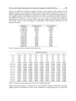
The figures given in Table 39.6 show that in patients un-
dergoing a duct drainage procedure, 20% failed to gain
relief from pain while 25% suffered further pain but a
little less than preoperatively. Failure to control pain
with the use of a duct drainage procedure is caused by
tissue changes outside the duct system, mostly in
patients with chronic pancreatitis and an inflammatory
process in the head of the pancreas. Duct drainage into
the jejunum is an inadequate treatment in these cases. It
has been demonstrated that reappearance of pain after
a duct drainage procedure is caused by an inflamma-
tory mass; resection of this mass leads to a long-lasting
pain-free status and improvement of the quality of life if
the head of the pancreas is resected in a second surgical
procedure. Furthermore, lateral duct-to-jejunum
anastomosis is ineffective in chronic pancreatitis with
side-duct stenosis through the pancreas.
Duodenum-preserving pancreatic
head resection
The rationale for duodenum-preserving pancreatic
head resection in chronic pancreatitis is removal of the
main inflammatory process, considered to be the pace-
maker of the disease, while preserving the upper gas-
trointestinal tract. The surgical procedure preserves the
stomach, duodenum, and biliary tree. Preservation of
the duodenum is superior to Whipple-type resection,
which includes duodenectomy. Preservation of the
duodenum has been shown to be very important
because the duodenum is essential for the regulation
of glucose metabolism and gastric emptying.
The duodenum-preserving head resection is based on
two principal steps: (i) subtotal resection of the pancre-
atic head with removal of the inflammatory mass, while
preserving the duodenum, extrahepatic common bile
duct, gallbladder, and stomach, as well as the pancrea-
tic parenchyma to a large extent; and (ii) restoration of
the flow of pancreatic juice from the left pancreas, in-
cluding neck, body, and tail, to the upper gastrointesti-
nal tract by the use of a Roux-en-Y excluded upper
jejunal loop. There are three technical steps in the pro-
cedure, starting with exposure of the head of the pan-
creas (Fig. 39.1). After tunneling of the pancreatic neck
ventrally to the portal vein along the portal groove,
CHAPTER 39
317
Table 39.5 Surgery in chronic pancreatitis: Ulm experience
(905 patients).
Duct drainage: 121 patients (13%)
Left resection: 83 patients (9%)
Duodenal-preserving pancreatic head resection*: 548 patients
(61%)
Pylorus-preserving head resection: 78 patients (9%)
Kausch–Whipple: 12 patients (1%)
Others: 63 patients (7%)
* Department of General Surgery, Free University of Berlin,
November 1972 to April 1982, and Department of General
Surgery, University of Ulm, May 1982 to September 2000.
Table 39.6 Pain relief after pancreatic duct drainage by
pancreaticojejunostomy: results after more than 5 years of
follow-up of 582 patients.*
Complete pain relief in 55%
Pain, but improved in 25%
Failure of pain control in 20%
Unsatisfactory long-term results in 25 + 20 = 45%
* Sources: Leger (1974), White (1979), Prinz
(1981), Morrow (1984), Bradley (1987), Drake
(1999), Greenie (1990), Wilson (1992), Adams
(1994), Kestens (1996), Gonzales (1997), Shama
(1998), Sidhu (2001).
transection of the pancreas along the duodenal border
of the portal vein is performed. After transection of the
pancreas, the pancreatic head and the duodenum are
rotated by 90° to a ventral–dorsal position (Fig. 39.2).
Subtotal resection of the pancreatic head along the in-
trapancreatic segment of the common bile duct leads in
most cases to decompression of the narrowed common
bile duct without opening the duct. The wet weight of
an operative specimen after subtotal head resection
with duodenum-preserving head resection is between
25 and 45 g; in 54 patients, the median was 28 g. After
completion of the subtotal resection, a shell-like rest of
the pancreatic head along the duodenal C-line is pre-
served. The dorsal pancreaticoduodenal arteries are
preserved, whereas in most cases the ventral branch of
the gastroduodenal artery has to be ligated. To restore
flow of pancreatic juice into the upper intestine, a jeju-
nal loop is separated and interposed (Fig. 39.3). Two
pancreatic anastomoses have to be performed. In cases
with stenosis of the common bile duct, due to inflam-
mation of the duct wall, an additional internal biliary
anastomosis between the bile duct and jejunal loop has
to be carried out (Fig. 39.4). In cases with a biliary anas-
tomosis, three connections to the jejunal loop are estab-
lished: two pancreatic and one biliary anastomoses. In
patients with multiple stenosis and dilatation of the
main pancreatic duct in the body and tail, a side-to-side
PART II
318
Figure 39.1 Duodenum-preserving pancreatic head
resection: the first step is transection of the neck of the
pancreas; resection line is along the mass of the pancreatic
head.
Figure 39.2 Duodenum-preserving pancreatic head
resection: dorsal view of the pancreas. The dorsal capsule of
the head of the pancreas is preserved. The dorsal
pancreaticoduodenal arcades are intact.
Figure 39.3 Duodenum-preserving head resection:
reconstruction with a jejunal loop.
CHAPTER 39
319
Figure 39.4 Duodenum-preserving pancreatic head resection
with intrapancreatic stenosis of the common bile duct:
additional biliary drainage into the jejunal loop has to be
performed.
Table 39.7 Duodenum-preserving pancreatic head resection
in chronic pancreatitis: early postoperative results (504
patients).*
Hospitalization (postoperative): 14.5 (7–87) days
Relaparotomy: 28 patients (5.6%)
Hospital mortality: 4 patients (0.8%)
* Department of General Surgery, Free University
of Berlin, November 1972 to April 1982, and
Department of General Surgery, University of
Ulm, May 1982 to December 1998.
Figure 39.5 Multiple stenosis of the main pancreatic duct in
the body and tail: duodenum-preserving pancreatic head
resection is combined with a pancreaticojejunostomoses
lateral-lateral.
anastomosis has to be performed additionally (Fig.
39.5). Early postoperative results after duodenum-pre-
serving head resection in chronic pancreatitis are given
in Table 39.7.
Using the duodenum-preserving head resection, con-
trol of pain is achieved in about 90% of patients in the
late follow-up (Table 39.8). With regard to endocrine
function, the duodenum-preserving head resection re-
sults in improvement of glucose metabolism in 8–15%
of patients. In the long-term follow-up, however, an
Table 39.8 Late postoperative pain after duodenum-preserving pancreatic head resection in chronic pancreatitis (Beger 1999).
* Median years of follow-up.
Late postoperative follow-up
1984 1988 1997
2.0 years* 3.6 years* 5.7 years*
57 patients 109 patients 303 patients
Pain-free 92.8% 89% 91.3%
Continuing abdominal pain 7.2% 11% 8.7%
Abdominal complaints
—
12% 12%
Hospitalization due to attacks of pancreatitis 14% 11% 9%
increase in insulin-dependent diabetes occurs; after a
median follow-up of 5.7 years, about 50% of patients
are diabetic (Table 39.9). Table 39.10 shows the results
of randomized clinical trials that compared duodenum-
preserving pancreatic head resection with pylorus-
preserving (Kausch–Whipple) resection and with the
Izbicki–Frey modification.Duodenum-preserving head
resection is superior or equal to the other procedures
with regard to postoperative morbidity, postoperative
maintenance of glucose metabolism, delay of gastric
emptying, and low level of rehospitalization, as well as
restoration of quality of life. In the long-term outcome
after surgical treatment, it has been convincingly
demonstrated that in a small group of patients the
Partington–Rochelle procedure (i.e., duct drainage
procedure) using the modification of Izbicki–Frey
PART II
320
Table 39.9 Duodenum-preserving pancreatic head resection changes the natural course of chronic pancreatitis.
1972–83 1972–87 1972–94 1982–96
58 patients* 128 patients† 298 patients‡ 368 patients§
Follow-up 2.8 years (median) 3.6 years 6.0 years 5.7 years (median)
57 patients 109 patients 258 patients 303 patients
Follow-up rate 100% 96% 87% 94%
Pain-free 93% 89% 88% 91%
Continuing abdominal pain 7% 11% 12% 9%
Hospitalization for acute episode 14% 11% 10% 9%
Late death rate 3.6% 4.7% 8.9% 13%
Endocrine functions improved 15.8% 5.5% 11%
Professional rehabilitation 89% 67% 63% 69%
Quality of life/Karnorfsky 80–100 82%
* November 1972 to October 1983.
† November 1972 to December 1987.
‡ November 1972 to December 1994.
§ May 1982 to October 1996.
Table 39.10 Duodenum-preserving pancreatic head resection (DPPHR) versus Whipple resection in chronic pancreatitis: results
of randomized trials.
Study Procedures compared Results
Buechler et al. (1995) DPPHR vs. pylorus-preserving DPPHR much superior with regard to postoperative
Whipple resection morbidity, glucose metabolism, gastric emptying,
and rehospitalization
Klempa et al. (1995) DPPHR vs. Whipple resection DPPHR superior with regard to postoperative
morbidity, glucose metabolism, and rehospitalization
Izbicki et al. (1995) DPPHR vs. Beger–Frey DPPHR* Both methods equal with regard to pain control,
glucose metabolism, postoperative morbidity, and
quality of life
Izbicki et al. (1998) Frey DPPHR* vs. pylorus-preserving DPPHR superior with regard to postoperative
Whipple resection morbidity, gastric emptying, and quality of life
Witzigmann et al. (2003) DPPHR vs. Whipple resection DPPHR superior with regard to postoperative
morbidity, maintenance of endocrine function,
rehospitalization, and quality of life
* Frey, modified by Izbicki.
delays deterioration of function (Table 39.10).
Duodenum-preserving pancreatic head resection is the
surgical technique of choice in patients suffering chron-
ic pancreatitis with an inflammatory mass in the head.
Pylorus-preserving pancreatic head
resection in chronic pancreatitis
A complete pancreatic head resection is mandatory in
chronic pancreatitis suspected to be associated with
pancreatic cancer. In patients suffering long-lasting
chronic pancreatitis, a malignant lesion is observed
in 4–6%. The cancer risk in chronic pancreatitis is
predicted to be increased 16-fold after 20 years.
The criteria for malignancy inlude the double-duct
sign and continuously increasing CA-19-9 and/or CEA
in the peripheral blood after biliary stenting of jaun-
diced patients. Most suggestive of cancer is positive
cancer cell staining of biopsy material or of intraopera-
tively obtained frozen sections. Infiltration of the portal
or superior mesenteric vein wall develops rarely in
chronic pancreatitis but is more frequent in cancer.
Increased mutations of K-ras, p53, p16, and DPC4 can
be used as markers of carcinogenic process in the
pancreas.
Conclusion
In chronic pancreatitis complicated by medically
intractable pain, common bile duct stenosis, main
pancreatic duct stenosis, portal vein compression,
and duodenal stenosis, and in pancreas divisum, the
application of duodenum-preserving pancreatic head
resection with or without lateral duct drainage offers
the benefits of low postoperative morbidity, pain-free
status in 90% of patients, reduction in pancreatitis-re-
lated hospitalization to less than 5%, postoperative
maintenance of endocrine status, professional rehabili-
tation in more than 60% of patients, and significant im-
provement in quality of life. In patients with main
pancreatic duct dilatation without multiple main- and
side-duct stenoses and without an inflammatory mass
in the head, a Partington–Rochelle procedure or a Frey
modification is the first choice for surgical treatment. In
patients with a mass in the head of the pancreas, sus-
pected to be an association of chronic pancreatitis with
pancreatic cancer, a pylorus-preserving resection has to
be performed once the diagnosis is confirmed by posi-
tive frozen section.
Recommended reading
Barton CM, Hall PA, Hughes CM, Gullick WJ, Lemoine NR.
Transforming growth factor alpha and epidermal growth
factor in human pancreatic cancer. J Pathol 1991;163:
111–116.
Beger HG, Büchler M. Duodenum-preserving resection of the
head of the pancreas in chronic pancreatitis with inflamma-
tory mass in the head. World J Surg 1990;14:83–87.
Beger HG, Büchler M, Bittner R, Oettinger W, Röscher R.
Duodenum-preserving resection of the head of the pancreas
in severe chronic pancreatitis: early and late results. Ann
Surg 1989;209:273–278.
Birk D, Schoenberg MH, Gansauge F, Formentini A, Fortnagel
G, Beger HG. Carcinoma of the head of the pancreas arising
from the uncinate process: what makes the difference? Br J
Surg 1998;85:498–501.
Bockman DE, Buchler M, Malfertheiner P, Beger HG. Analy-
sis of nerves in chronic pancreatitis. Gastroenterology
1988;94:1459–1469.
Bordalo O, Bapista A, Dreiling D, Noronha M. Early patho-
morphological pancreatic changes in chronic alcoholism.
In: KE Gyr, MV Singer, H Sarles (eds) Pancreatitis: Concepts
and Classification. Amsterdam: Elsevier/North-Holland,
1984: 642.
Büchler M, Malfertheiner P, Friess H, Senn T, Beger HG.
Chronic pancreatitis with inflammatory mass in the head of
the pancreas: a special entity? In: HG Beger, M Büchler, H
Ditschuneit, P Malfertheiner (eds) Chronic Pancreatitis.
Berlin: Springer-Verlag, 1990: 41–46.
Caldas C, Hahn SA, da Costa LT et al. Frequent somatic
mutations and homozygous deletions of the p16 (MTS1)
gene in pancreatic adenocarcinoma. Nat Genet 1994;8:27–
32.
D’Ardenne AJ, Kirkpatric P, Sykes BC. Distribution of
laminin, fibronectin, and interstitial collagen type III in soft
tissue tumours. J Clin Pathol 1984;37:895–904.
Ebbehoj N. Pancreatic tissue fluid pressure and pain in chron-
ic pancreatitis. Dan Med Bull 1992:39:128–133.
Friess H, Yamanaka Y, Büchler M et al. Increased expression
of acidic and basic fibroblast growth factors in chronic
pancreatitis. Am J Pathol 1994;144:117–128.
Friess H, Yamanaka Y, Büchler M, Kobrin MS, Tahara E,
Köre M. Cripto, a member of the epidermal growth factor
family, is overexpressed in human pancreatic cancer and
chronic pancreatitis. Int J Cancer 1994;56:668–674.
Friess H, Yamanka A, Büchler M et al. A subgroup of patients
with chronic pancreatitis overexpress the c-erbB-2
protooncogene. Ann Surg 1994;220:183–192.
CHAPTER 39
321
Gansauge S, Gansauge F, Beger HG. Molecular oncology in
pancreatic cancer. J Mol Med 1996;74:313–320.
Gansauge S, Schmid RM, Gansauge F et al. Genetic alterations
in chronic pancreatitis: evidence for early occurrence of
p53 but not K-ras mutations. Br J Surg 1998;85:337–
340.
Gress TM, Müller-Pillasch F, Lerch MM et al. Balance of ex-
pression of genes coding for extracellular matrix proteins
and extracellular matrix degrading proteases in chronic
pancreatitis. Z Gastroenterol 1994;32:221–225.
Klöppel G, Maillet B. Pseudocysts in chronic pancreatitis: a
morphological analysis of 57 resection specimens and 91
autopsy pancreata. Pancreas 1991;6:266–274.
Korc M, Friess H, Yamanaka Y, Kobrin MS, Büchler M, Beger
HG. Chronic pancreatitis is associated with increased con-
centrations of epidermal growth factor receptor, transform-
ing growth factor a, and phospholipase C-gamma. Gut
1994;35:1468–1473.
Lowenfels AB, Maisonneuve P, Cavallini G et al. Pancreatitis
and the risk of pancreatic cancer: International Pancreatitis
Study Group. N Engl J Med 1993;328:1433–1437.
Matsubara T, Sakurai Y, Funabiki T et al. K-ras point muta-
tions in cancerous and noncancerous biliary epithelium in
patients with pancreaticobiliary maljunction. Cancer
1996;77:1752–1757.
Oertel JE, Heffess CS, Oertel YC. Pancreas. In: SS Sternberg
(ed.) Diagnostic Surgical Pathology. New York: Raven
Press, 1989: 1057–1093.
Sarles H, Dagorn JC, Giorgi D, Bernard JP. Remaining pancre-
atic stone protein as “lithostatin.” Gastroenterology
1990;99:900–905.
Schlosser W, Schoenberg MH, Siech M, Gansauge F, Beger
HG. Development of pancreatic cancer in chronic pancre-
atitis. Z Gastroenterol 1996;34:3–8.
Shimoyama S, Gansauge F, Gansauge S, Oohara T, Beger HG.
Altered expression of extracellular matrix molecules and
their receptors in chronic pancreatitis and adenocarcinoma
of the pancreas in comparison to normal pancreas. Int J
Pancreatol 1995;18:227–234.
van Laethem JL, Deviere J, Resibois A et al. Localization of
transforming growth factor beta l and its latent binding pro-
tein in human chronic pancreatitis. Gastroenterology
1995;108:1873–1881.
Warshaw AL. Pancreas divisum: a case for surgical treatment.
Adv Surg 1988:21:93–109.
Watanabe M, Tanaka J, Masauji N et al. Detection of point
mutation of K-ras gene codon 12 in biliary tract and am-
pullary carcinoma by modified two-step polymerase chain
reaction. Nippon Shokakibyo Gakkai Zasshi 1993;90:
789–794.
Weihe E, Nohr D, Müller S, Büchler M, Friess H, Zentel HJ.
The tachykinin neuroimmune connection in inflammatory
pain. Ann NY Acad Sci 1991;632:283–295.
Widmaier U, Schmidt A, Schlosser W, Beger HG. Die duode-
numerhaltende Pankreaskopfresektion in der Therapie des
Pancreas divisum. Chirurg 1997;68:180–186.
PART II
322
Introduction
Pancreatic pseudocysts are chronic inflammatory fluid
collections associated with pancreatitis. Pseudocysts
are the most common complication of acute and chron-
ic pancreatitis and nearly one-third of patients with
pancreatitis will develop a pseudocyst. Because the
fluid cavities are not lined with an epithelium, they are
not true cysts. The cavities are instead lined with a
reactive granulation tissue that surrounds a collection
of enzyme-rich fluid, debris, and necrotic tissue.
The treatment of pancreatic pseudocysts is highly
variable, ranging from observation to surgical
drainage. Drainage procedures via radiologic or endo-
scopic approaches are also an important option. An
understanding of the pathogenesis of pseudocysts
associated with chronic pancreatitis will aid the
clinician in the selection of proper treatment.
Pathophysiology of pancreatic fluid
collections and pseudocysts
Pseudocysts associated with chronic pancreatitis
Pseudocysts are chronic fluid collections that consist of
pancreatic secretions and inflammatory debris and
contain large concentrations of active proteolytic
enzymes. The fluid collections may develop after an
episode of acute pancreatitis or insidiously in the set-
ting of chronic pancreatitis. Small pancreatic pseudo-
cysts are usually intrapancreatic and have a thin wall.
Large pseudocysts may be so large that they occupy
areas remote from the pancreas. The histologic features
of pseudocyst walls are similar in all types of pseudo-
cysts, consisting of fibrosis and inflammatory tissue.
Most pancreatic pseudocysts originate from large or
small leaks from the ductal system and this feature can
be demonstrated with endoscopic retrograde cholan-
giopancreatography (ERCP).
Focal fluid collections arising in the setting
of acute pancreatitis
Acute peripancreatic fluid collections commonly arise
during episodes of acute pancreatitis. The fluid collec-
tions may accumulate as a result of ductal disruptions
or the liquefaction of necrotic pancreatic tissue. Simple
fluid collections as a result of ductal leaks are usually
unilocular and filled with pancreatic secretions that
contain high concentrations of enzymes. Early in the
formation of these fluid collections, the fluid is not well
contained in the peripancreatic space and may spread
throughout the peritoneal and retroperitoneal spaces.
Early fluid collections located adjacent to organs such
as the stomach, colon, liver, and mesentery are the
source of older mature pseudocysts. Chronic fluid col-
lections are contained by thick walls of fibrotic inflam-
matory tissue that often include the serosa of adjacent
organs.
Complex fluid collections often originate from pan-
creatic tissue necrosis during acute pancreatitis. These
focal fluid collections or phlegmons contain semisolid
debris, inflammatory fluid, and high concentrations of
pancreatic enzymes and can be divided into loculations
by fibrotic septations. Complex fluid collections are
particularly prone to infection and often require
sampling and drainage.
323
40
Management of chronic pancreatic
pseudocyst: when to observe, when
and how to drain
William R. Brugge
Focal fluid collections arising from a duct leak
Leakage and accumulation of fluid from a disrupted
pancreatic duct may occur without evidence of pancre-
atitis or tissue necrosis. Most commonly, simple leaks
take place in the postoperative setting when an incom-
plete anastomosis breaks down and allows the escape
of fluid from a duct. Nonsurgical injuries such as ab-
dominal trauma and endoscopic instrumentation may
also be responsible for ductal defects. These fluid
collections are often unilocular and respond readily to
closure of ductal defects.
Clinical manifestations
Most pancreatic pseudocysts cause mild symptoms.
The most common symptom is early satiety and
distension; these symptoms occur in 76–94% of
patients. In general, the size of the pseudocyst and
the duration of the clinical course are the most impor-
tant predictors of symptoms. With large pseudocysts,
there may be a palpable fullness or mass that is sensed
by the patient or an examining physician. However,
small pseudocysts and pseudocysts located behind
the stomach or in the retroperitoneum are rarely
detected by physical examination. Related to gastric
compression, weight loss is observed in 20% of
patients and is a result of poor intake as well as
maldigestion. Jaundice, as manifest by icterus, dark
urine, pruritus, and acholic stools, may be noted in
10% of patients. The onset of jaundice is usually
slow, as a result of bile duct compression by the pseudo-
cyst or the inflamed pancreas itself. Fever is unusual in
chronic uncomplicated pseudocysts and its presence
should raise the suspicion of an occult infection of a
pseudocyst.
Pain from distension and compression
Gastric compression and poor emptying is commonly
observed in large pseudocysts located in the head of the
pancreas. Patients often complain of early satiety, nau-
sea, and vomiting, particularly after meals. Duodenal
obstruction may arise from the presence of the pancre-
atic pseudocyst or the fibrotic process in the pancreas.
Large pseudocysts in the body and tail of the pancreas
may also compress the stomach and cause early satiety.
After attempted drainage, pseudocysts may dissect into
the gastric wall and cause an intramural inflammatory
process.
Bleeding
Acute or chronic gastrointestinal bleeding from a vari-
ety of sources is seen in 10–20% of patients with chron-
ic pseudocysts. The most serious potential source of
bleeding is from gastric varices that arise from splenic
vein thrombosis. However, bleeding from varices that
arise from splenic vein obstruction is unusual. A com-
mon associated lesion is portal hypertensive gastropa-
thy, which may cause chronic gastrointestinal bleeding
from the stomach.
Occasionally, bleeding may originate from within
the pseudocyst cavity or necrotic pancreatic tissue.
Since bleeding in a pseudocyst cavity usually originates
from an arterial source, the bleeding may result in sud-
den and massive distension of the pseudocyst. Bleeding
from pseudoaneurysms is the most common cause of
significant bleeding and may be responsible for bleed-
ing within pancreatic tissue as well as within the
pseudocyst cavity. Spontaneous bleeding into a
pseudocyst and communication with the main pancre-
atic duct results in hemosuccus pancreaticus, a rare
form of upper gastrointestinal bleeding associated with
pseudocysts. Blood-filled pseudocysts may also rupture
into the stomach and cause bleeding within the lumen
of the stomach. There are reports of hemobilia as a
result of bleeding from a pseudocyst and subsequent
erosion into the bile duct. Lower gastrointestinal
bleeding may result from spontaneous erosion of a
pseudocyst into the colon.
Infection
Infection of pseudocysts usually takes place within the
protein-rich fluid of the pseudocyst cavity. Infections
may be mild and transient, but more commonly are
severe and result in a sepsis syndrome. Spontaneous
infections are caused by proliferation of enteric
organisms in the protein-rich fluid of the pseudocyst.
Instrumentation combined with inadequate drainage
may result in infection of a pseudocyst, particularly if
there is necrotic tissue present within the pseudocyst
cavity. Patients with an infected pseudocyst will present
with abdominal pain, fever, or sepsis. Computed to-
mography (CT) may reveal the presence of air in the
pseudocyst cavity, a highly specific sign of infection.
PART II
324
Similar findings have been reported with transabdomi-
nal ultrasound. Percutaneous CT- or ultrasound-
guided aspirations are used to sample fluid for culture
and Gram staining. False-positive diagnostic fluid
aspirations with Gram stains are rare. In selected pa-
tients with signs of infection, aspiration studies reveal
evidence of infection in more than 30% of patients.
Despite the use of CT-guided aspiration for the diagno-
sis of early pancreatic pseudocyst abscess, the long-
term mortality rate of treated pancreatic abscesses
remains significant at 17%. Severe systemic infections
occur when infected fluid communicates with the peri-
toneal cavity or the bloodstream, often in the setting of
pancreatic necrosis. Long-term percutaneous drainage
of infected pseudocysts is successful in 60–70% of pa-
tients but is less successful if the infection is associated
with areas of tissue necrosis and complex infected fluid
collections. Drainage of multiple infected fluid collec-
tions requires prolonged hospitalization and a combi-
nation of surgery and percutaneous drainage. The
initial treatment of infected pseudocysts and fluid col-
lections is percutaneous drainage, followed by surgery
in patients with a poor response to drainage. Long-term
use of external catheters for drainage is often compli-
cated by the development of a cutaneous–pancreatic
fistula. Endoscopic drainage of infected pseudocysts
using ERCP avoids the complications of fistula forma-
tion but may contribute to the introduction of bacteria
into a pseudocyst cavity. Internal drainage of infected
pseudocysts has also been performed using endoscopic
ultrasound (EUS) guidance and the approach avoids
the risk of fistula formation. EUS-guided drainage of
infected pseudocysts may be improved by prolonged
drainage using nasocystic drains placed across the
gastric wall. Surgical drainage of infected pseuodocysts
should be performed in patients not responding to en-
doscopic or radiologic drainage therapy. Despite these
aggressive therapeutic approaches, the mortality rate
of treated infected pseudocysts remains quite high
(~ 10%).
Vascular injury associated with pseudocysts
Splenic vein thrombosis, the common vascular injury
associated with pseudocysts, occurs in about 13% of
patients with pseudocysts, particularly when the
pseudocyst is located in the body or tail of the pancreas
and is associated with chronic pancreatitis. The throm-
bosis presumably occurs in the lumen of the splenic vein
compressed by the pancreas and/or the pseudocyst.
Splenic vein thrombosis will also result in dilation of
the short gastric veins, splenomegaly, and the forma-
tion of gastric varices. There are few symptoms related
to this complication, except for the occasional bleeding
from gastric varices and portal hypertensive gastropa-
thy. At times, the patient with a pseudocyst may present
solely with splenomegaly found on physical examina-
tion or hypersplenism. On rare occasions, splenic vein
thrombosis may be complicated by extension of the
thrombus into the portal vein.
Thrombocytopenia and leukopenia may arise from
“hypersplenism,” but it is rare for the patient to present
with any symptoms relating to sequestration of
platelets and white blood cells. The treatment of splenic
vein thrombosis, splenectomy, is indicated in those
patients with complications such as bleeding. The
long-term results of splenectomy for the treatment of
recurrent gastric variceal bleeding is excellent and is the
treatment of choice.
Pseudoaneuryms are potentially the most lethal com-
plication of chronic pancreatitis and pseudocysts. These
focal inflammatory weaknesses of an arterial wall most
commonly occur in the splenic and gastroduodenal
arteries. The low-attenuation lesions are readily seen on
cross-sectional imaging studies as dilated fluid-filled
structures and may be confused with pancreatic fluid
collections. The average size of pseudoaneurysms that
require intervention is quite large, nearly 14 cm. How-
ever, the use of Doppler ultrasound and EUS can readily
differentiate between fluid collections and pseudo-
aneurysms. EUS with color Doppler may also diagnose
ruptured pseudoaneurysms. Prospective studies have
demonstrated that 10% of patients with pancreatic
pseudocysts have pseudoaneurysms as demonstrated by
angiography. Pseudoaneurysms are the most common
source of bleeding in pancreatic pseudocyst cavities.
The treatment of bleeding from pseudoaneurysms in-
cludes surgical resection and angiographic techniques.
Surgical techniques for the control of acute bleeding
from pseudoaneurysms consist of arterial ligation and
surgical resection. The reported surgical mortality rate
for the control of bleeding from a pseudoaneurysm is
high (> 10%). The angiographic control of bleeding
from pseudoaneurysms consists of arterial emboliza-
tion and is successful in 40–50% of patients with acute
bleeding. Percutaneous arterial embolization may
be used prophylactically to prevent bleeding from
pseudoaneurysms.
CHAPTER 40
325
Pancreatic biliary duct obstruction
Obstruction of the pancreatic biliary ducts is relatively
common, particularly when the pseudocyst is located
within the head of the pancreas. Local compression is
the most common cause of ductal obstruction, al-
though direct fibrotic involvement of the ducts is occa-
sionally seen. Obstruction of the distal bile duct with
resulting obstructive jaundice is the most common sce-
nario. Although the degree of obstruction of the bile
duct may be impressive, it is rare for the patient’s symp-
toms to be related to biliary obstruction. Drainage of a
pseudocyst associated with bile duct compression re-
solves the obstruction of the duct, particularly the bile
duct. With long-term obstruction of the pancreatic bil-
iary ducts, stones, sludge, and debris will commonly ac-
cumulate and may be responsible for episodes of
cholangitis.
Diagnostic testing
Ultrasound/CT
Pseudocysts are readily seen with CT and appear as
low-attenuation lesions within or adjacent to the
pancreas (Fig. 40.1). Chronic pseudocysts are most
commonly round in shape and surrounded by a thick
dense wall. Large pseudocysts may appear in the medi-
astinum or pelvis, or involve the mesentery. Prominent
vessels, as depicted with color Doppler, adjacent to the
pseudocyst wall are common and may represent para-
gastric varices and thrombosis of the splenic vein.
Although pseudocysts are most commonly unilocular,
fibrotic strands within the cavity may cause multiple
septations, commonly encountered in patients with
postpancreatitis, complex fluid collections. The
pseudocyst cavity may also contain debris, blood, or in-
fections that appear as high-attenuation areas within
the fluid-filled cavity. It may be difficult to distinguish
between pseudocysts and true pancreatic mucinous
cysts associated with malignancy without the use of
aspiration fluid analysis. Choledochocysts, as they
appear on CT, may also be confused with pancreatic
pseudocysts.
ERCP
Pancreatography in the setting of a pseudocyst often re-
veals a diffusely abnormal duct with changes of chron-
ic pancreatitis evident. The main pancreatic duct may
be partially or completely obstructed by compression
of the pseudocyst. In more than half of patients, the
pseudocyst will fill with contrast during retrograde
pancreatography. Pancreatic ductal leaks are common
and may originate from the pancreatic duct or may be
small and originate from a secondary radicle. A normal
pancreatogram should suggest the possibility of a cystic
neoplasm rather than a pseudocyst. Retrograde injec-
tion of contrast into a pseudocyst may result in infec-
tion of the pseudocyst. Contamination of a pseudocyst
cavity may be prevented by the use of antibiotics and
minimizing the amount of contrast injected.
EUS
Using EUS, pseudocysts appear as anechoic fluid-filled
structures adjacent to the upper gastrointestinal tract
and pancreas (Fig. 40.2). Fluid collections associated
with acute pancreatitis will not be surrounded with a
wall, whereas pseudocysts are often surrounded by a
thick hyperechoic rim. Calcifications in a cyst wall are
highly suggestive of a mucinous cystadenoma rather
than a pseudocyst. Within the pseudocyst cavity, EUS
will readily demonstrate the presence of fluid. Debris in
the dependent portion of the cavity is common and may
PART II
326
Figure 40.1 Computed tomography scan of a pancreatic
pseudocyst in the body of the pancreas.
represent blood, infection, or necrotic material. Color
Doppler of the wall often reveals multiple prominent
vessels, including paragastric varices. EUS-directed
fine-needle aspiration (FNA) with cyst fluid analysis
will differentiate between pseudocysts and neoplastic
cysts in more than 90% of patients.
Cyst aspiration
FNA of pseudocysts is performed for diagnostic or
therapeutic purposes using CT/ultrasound or EUS for
guidance. Since pseudocysts may be confused with true
cysts of the pancreas in nearly 20% of patients, aspira-
tion is performed to differentiate between pseudocysts
and a wide variety of benign and malignant cystic neo-
plasms of the pancreas. If infection of a pseudocyst is
suspected, the cyst should be aspirated for culture.
FNA of pseudocysts can be performed with a variety
of techniques. The most common approach is to use
CT/ultrasound guidance. A needle is placed through
the abdominal wall and into the cystic cavity; small
amounts of fluid are aspirated for cytology and tumor
markers such as carcinoembryonic antigen (CEA). EUS
can also be used to guide aspiration through the gastric
or duodenal wall and is ideal for small cystic lesions.
The aspirated fluid from a cystic lesion is examined
cytologically for evidence of inflammatory cells. The
presence of pigmented histiocytes is diagnostic of a
pseudocyst, but this finding may be absent in a signifi-
cant number of patients with a pseudocyst. If there
is cytologic evidence of epithelial cells with the cyst
fluid, this should raise the suspicion of a cystic
neoplasm rather than a pseudocyst. The presence of
granulocytes in the aspirated fluid is suggestive of an
acute infection. A high concentration of amylase in as-
pirated fluid is predictive of a connection with the main
pancreatic duct and helps confirm the diagnosis of a
pseudocyst.
The cytologic analysis of a cystic lesion may not pro-
vide diagnostic material because of the low cellularity
of cyst fluid. Cyst fluid tumor markers are often used to
assist in differentiating between pseudocysts and cystic
neoplasms. CEA is the most commonly used marker
because mucinous cystic neoplasms secrete CEA into
cystic fluid, whereas pseudocysts should have relatively
low levels of CEA.
If there is any concern about an infected pseudocyst,
the aspirated fluid should be sent for culture and
sensitivity. Although Gram-negative enteric organisms
are the most common organism, occasionally Gram-
positive bacteria may infect a pseudocyst. Viral and
mycobacterial infections of pseudocysts are very rare
but Candida species may infect secondarily.
Treatment
Natural history
Small pseudocysts of less than 4 cm in diameter often
resolve spontaneously and are rarely associated with
clinical symptoms. In long-term observation studies,
9% of patients experience a complication of a pseudo-
cyst. Spontaneous resolution of pseudocysts takes
place through drainage into the gastrointestinal tract or
the pancreatic duct. Of pseudocysts less than 6 cm,
40% will require drainage because of complications or
persistence. Small pseudocysts located in the tail of the
pancreas and arising from acute biliary pancreatitis
have a very high rate of spontaneous drainage. In a
large series, the overall mortality of pseudocyst
drainage by any method was 9–14%. Prior to drainage,
it is critical to confirm the diagnosis of a pseudocyst
using fluid analysis and cytology. Mistakenly diag-
nosed pseudocysts that are drained with percutaneous
drainage often do not resolve and may harbor occult
malignancy.
CHAPTER 40
327
Figure 40.2 Endoscopic ultrasound image of a thick-walled
pancreatic pseudocyst.
Drainage
Pancreatic pseudocysts may be drained using a variety
of approaches. Simple, one-time aspiration of pseudo-
cysts rarely provides lasting resolution because of com-
munication with the pancreatic ductal system. External
drainage using CT/ultrasound guidance is the most
common approach and should include the use of
Doppler or contrast injection to order to differentiate
between pockets of fluid and vascular structures. With
CT/ultrasound guidance, a single pigtail drainage
catheter is placed percutaneously into the fluid cavity
and fluid is drained into an external collection system
carried by the patient (Fig. 40.3). The short-term suc-
cess rate for this relatively simple technique is very high
(> 90%). Patients with communicating pseudocysts, as
evidenced by high concentration of amylase in cyst
fluid, and pancreatic duct strictures should not have
percutaneous drainage because of the high risk of
prolonged drained and fistula formation. Acutely ill
patients with evidence of infection in the pseudocyst
should be urgently aspirated and drained under ultra-
sound guidance at the bedside. Long-term success rates
of external drainage are much lower than immediate
success rates and a significant percentage of patients
with external drainage will require surgical drainage.
However, the placement of an external drain will not
interfere with surgical drainage or excision if external
drainage is not successful. The placement of the
catheter across the stomach reduces the risk of subse-
quent external fistula formation and allows for subse-
quent transgastric stenting. Peripancreatic fluid collec-
tions complicated by infection are also easily drained
with CT-guided catheters and external drainage.
Pseudocyst fluid drained externally is collected over an
average of 3 weeks into an external collection bag.
When the drainage output becomes minimal, the
catheter is removed. Contrast injection into the cyst
cavity will demonstrate the size of the cavity and this
finding can be used to monitor the progress of the
chronic drainage. This technique is highly successful at
resolving pseudocysts, but is plagued by infections and
the need for an external bag. The average duration of
external drainage is 24 days.
Surgical drainage of pseudocysts is performed by
providing a large anastomosis between the pseudocyst
wall and the stomach or small bowel. The anastomosis
should be placed in the most dependent portion of the
cystic cavity in order to maximize the chances of com-
plete drainage. The cyst-gastrostomy or -enterostomy
usually remains patent and functional for several
months. ERCP is often performed prior to surgical
drainage in order to evaluate the main pancreatic duct
for evidence of strictures, fistula, and leaks. Surgical
drainage is probably the best approach when the
pseudocyst is complicated by areas of necrosis or infec-
tion, or involves adjacent organs such as the spleen.
However, maturation of a pseudocyst wall over 4–6
weeks allows the formation of a thick wall that will
provide a more secure anastomosis. Cyst-gastrostomy
is the easiest approach and requires less operating time
than cyst-jejunostomy. However, the risk of bleeding is
greater with cyst-gastrostomy. A jejunal anastomosis is
indicated in giant pseudocysts because drainage with a
cyst-gastrostomy is often inadequate and the recur-
rence rate is quite high. An alternative to a cyst anasto-
mosis is a longitudinal pancreaticojejunostomy, which
has been reported to provide high rates of successful
drainage and with fewer complications such as anasto-
motic bleeding. Pancreaticojejunostomy can be per-
formed if there is an associated pancreatic duct
diameter of more than 7mm and is more likely to be
successful if there is communication between the
pseudocyst cavity and the main pancreatic duct. When
the pseudocyst has resolved, the low output of fluid al-
lows spontaneous closing of the anastomosis. Old re-
ports of surgical drainage suggested high mortality and
complication rates. More recent series report overall
success rates for surgical drainage of 90%, with major
complication rates of 9% and recurrence rates of 3%.
PART II
328
Figure 40.3 Percutaneous drainage of a pancreatic
pseudocyst.
Laparoscopic techniques are being developed that
will enable the surgeon to provide internal drainage
and resection of necrotic tissue. However, these tech-
niques are also associated with similar complications as
reported with endoscopic drainage, i.e., cyst wall
bleeding and postoperative cyst infection. If areas of
pancreatic necrosis are encountered during the laparo-
scopic procedure, laparoscopy may enable surgeons to
noninvasively remove areas of necrotic tissue.
Endoscopic drainage is the newest approach for the
eradication of pseudocysts. It is used most commonly
for uncomplicated unilocular pseudocysts. Drainage is
accomplished either with a transpapillary approach
with ERCP or with direct endoscopic drainage across
the stomach or duodenal wall. A transpapillary ap-
proach with drainage is used when the pseudocyst com-
municates with the main pancreatic duct, usually in the
head of the pancreas. The transpapillary approach has
also proven successful in the drainage of infected
pseudocysts or pseudocysts associated with strictures
or leaks of the main pancreatic duct. The transgastric or
duodenal approach is used when the pseudocyst is di-
rectly adjacent to the gastroduodenal wall. EUS is used
to determine the size, location, and thickness of the
pseudocyst wall (Fig. 40.2). A wall thickness of more
than 1 cm or the presence of large intervening vessels or
varices on EUS precludes the possibility of endoscopic
drainage. Endoscopic-guided drainage may be per-
formed using direct endoscopic or EUS imaging. With
the presence of a visible bulge in the wall of the stomach
or the duodenum, endoscopic drainage is successful by
the placement of transmural catheters or stents (Fig.
40.4). EUS guidance is required if a bulge is not evident
during the endoscopic evaluation prior to drainage.
One-step EUS-directed drainage is now possible with
pseudocyst drainage catheters that can be used in
therapeutic echoendoscopes. This approach has
proven highly successful and may be feasible for infect-
ed pseudocysts. Recently, endoscopic drainage of
necrotic pancreatic tissue through an endoscopic cyst-
gastrostomy has been reported in three patients. The
success rate within 3 months of endoscopic drainage
is reported to be more than 80%, with lower rates re-
ported with alcoholic pancreatic disease. The improve-
ment in endoscopic skills necessary for the successful
performance of pseudocyst drainage takes place over
about 20 cases and results in significant decreases in
complications and the length of hospital stay. Overall,
the complication rate of elective endoscopic drainage is
about 13%, with success rates of more than 90% and
recurrence rates of 10–20%.
Conclusions
Small asymptomatic pseudocysts associated with
chronic pancreatitis should be monitored. If there is
any question about the diagnosis or the presence of an
infection, cyst aspiration should be performed. Simple,
chronic, uninfected, unilocular pseudocysts should be
drained endoscopically when the expertise is available;
otherwise this type of pseudocyst should be drained
radiologically with an external drainage catheter.
Complex, postnecrotic, multilocular pseudocysts
should be managed with surgical drainage and/or
resection.
Recommended reading
Andersson R, Cwikiel W. Percutaneous cystogastrostomy in
patients with pancreatic pseudocysts. Eur J Surg 2002;
168:345–348.
Baril NB, Ralls PW, Wren SM et al. Does an infected peripan-
creatic fluid collection or abscess mandate operation? Ann
Surg 2000;231:361–367.
CHAPTER 40
329
Figure 40.4 Endoscopic gastric bulge as a result of a
pancreatic pseudocyst.
Beckingham IJ, Krige JE, Bornman PC, Terblanche J. Long
term outcome of endoscopic drainage of pancreatic pseudo-
cysts. Am J Gastroenterol 1999;94:71–74.
Bender JS, Bouwman DL, Levison MA, Weaver DW. Pseudo-
cysts and pseudoaneurysms: surgical strategy. Pancreas
1995;10:143–147.
Bhattacharya D, Ammori BJ. Minimally invasive approaches
to the management of pancreatic pseudocysts: review of
the literature. Surg Laparosc Endosc Percutan Tech 2003;
13:141–148.
Boerma D, van Gulik TM, Obertop H, Gouma DJ. Endoscop-
ic stent placement for pancreaticocutaneous fistula after
surgical drainage of the pancreas. Br J Surg 2000;
87:1506–1509.
Boggi U, Di Candio G, Campatelli A, Pietrabissa A, Mosca F.
Nonoperative management of pancreatic pseudocysts.
Problems in differential diagnosis. Int J Pancreatol 1999;
25:123–133.
Brugge WR. EUS-guided pancreatic fine needle aspiration:
instrumentation, results, and complications. Tech Gastro-
intest Endosc 2000;2:149–154.
Byrne MF, Mitchell RM, Baillie J. Pancreatic pseudocysts.
Curr Treat Options Gastroenterol 2002;5:331–338.
Carr JA, Cho JS, Shepard AD, Nypaver TJ, Reddy DJ.
Visceral pseudoaneurysms due to pancreatic pseudocysts:
rare but lethal complications of pancreatitis. J Vasc Surg
2000;32:722–730.
Cohen-Scali F, Vilgrain V, Brancatelli G et al. Discrimination
of unilocular macrocystic serous cystadenoma from pan-
creatic pseudocyst and mucinous cystadenoma with CT:
initial observations. Radiology 2003;228:727–733.
Deviere J, Bueso H, Baize M et al. Complete disruption of the
main pancreatic duct: endoscopic management. Gastroin-
test Endosc 1995;42:445–451.
Fockens P, Johnson TG, van Dullemen HM, Huibregtse K,
Tytgat GN. Endosonographic imaging of pancreatic
pseudocysts before endoscopic transmural drainage.
Gastrointest Endosc 1997;46:412–416.
Frossard JL, Amouyal P, Amouyal G et al. Performance of en-
dosonography-guided fine needle aspiration and biopsy in
the diagnosis of pancreatic cystic lesions. Am J Gastroen-
terol 2003;98:1516–1524.
Giovannini M, Pesenti C, Rolland AL, Moutardier V, Delpero
JR. Endoscopic ultrasound-guided drainage of pancreatic
pseudocysts or pancreatic abscesses using a therapeutic
echo endoscope. Endoscopy 2001;33:473–477.
Hammel P. Diagnostic value of cyst fluid analysis in cystic
lesions of the pancreas: current data, limitations, and
perspectives. J Radiol 2000;81:487–490.
Harewood GC, Wright CA, Baron TH. Impact on patient
outcomes of experience in the performance of endoscopic
pancreatic fluid collection drainage. Gastrointest Endosc
2003;58:230–235.
Heider R, Behrns KE. Pancreatic pseudocysts complicated by
splenic parenchymal involvement: results of operative and
percutaneous management. Pancreas 2001;23:20–25.
Heider R, Meyer AA, Galanko JA, Behrns KE. Percutaneous
drainage of pancreatic pseudocysts is associated with a
higher failure rate than surgical treatment in unselected pa-
tients. Ann Surg 1999;229:781–787; discussion 787–789.
Mori T, Abe N, Sugiyama M, Atomi Y. Laparoscopic pancre-
atic cystgastrostomy. J Hepatobiliary Pancreat Surg 2002;
9:548–554.
Nealon WH, Walser E. Duct drainage alone is sufficient in the
operative management of pancreatic pseudocyst in patients
with chronic pancreatitis. Ann Surg 2003;237:614–620;
discussion 620–622.
Parks RW, Tzovaras G, Diamond T, Rowlands BJ. Manage-
ment of pancreatic pseudocysts. Ann R Coll Surg Engl
2000;82:383–387.
Seifert H, Dietrich C, Schmitt T, Caspary W, Wehrmann T.
Endoscopic ultrasound-guided one-step transmural
drainage of cystic abdominal lesions with a large-channel
echo endoscope. Endoscopy 2000;32:255–259.
Sharma SS, Bhargawa N, Govil A. Endoscopic management of
pancreatic pseudocyst: a long-term follow-up. Endoscopy
2002;34:203–207.
Usatoff V, Brancatisano R, Williamson RC. Operative treat-
ment of pseudocysts in patients with chronic pancreatitis.
Br J Surg 2000;87:1494–1499.
Yeo CJ, Bastidas JA, Lynch-Nyhan A, Fishman EK, Zinner
MJ, Cameron JL. The natural history of pancreatic pseudo-
cysts documented by computed tomography. Surg Gynecol
Obstet 1990;170:411–417.
PART II
330
Introduction
Pancreatic adenocarcinoma is the fourth leading cause
of tumor deaths in men and the fifth leading cause
in women, with 95% of all pancreatic malignancies
being ductal adenocarcinomas. The 5-year survival
rate of only 1–5% is one of the worst for any tumor.
Thus it is of utmost importance to identify risk groups
since only early diagnosis allows the chance of a cura-
tive resection. Among the potential risk factors, only
nicotine has been clearly identified. A diet high in fat
and calories seems to add some risk. According to a
case–control study from northern Italy, attributable
risks were 14% for tobacco smoking, 14% for high
consumption of meat, and 12% for low consumption
of fruit. The authors speculate that almost one-fourth
of all cases of pancreatic carcinoma could be prevented
by a healthy lifestyle. It is likely that a combination
of factors, most of them associated with relatively
small risk enhancements, may be responsible for the
pathogenesis.
Furthermore, chronic inflammation of the pancreas
(i.e., chronic pancreatitis) has been identified as a risk
factor. This has been especially documented in chronic
hereditary pancreatitis, a disease with early onset of in-
flammation; 5–10% of all cases of pancreatic cancer
may be inherited. It will be a major challenge to identi-
fy these genetic alterations. In a population-based
case–control study based on direct interviews with 526
incident cases and 2153 population controls, the fol-
lowing risk factors were identified: elevated body mass
index (BMI) when combined with elevated caloric in-
take; diabetes mellitus (hyperinsulinemia); first-degree
relatives; family history of colon, endometrial, ovary,
and breast cancer; smoking; possibly heavy alcohol use
in blacks.
I discuss the significance of various risk factors that
have been proposed to play a role in the pathogenesis of
pancreatic carcinoma.
Incidence
The incidence rate in industrialized Western countries
is about 8–12/100 000 inhabitants and still increas-
ing in some countries. In Europe the incidence is
higher in northern and central Europe compared with
southern Europe. In the USA, there is a geographic
cluster of pancreatic cancers in areas of Louisiana
and Mississippi. However, the factors responsible for
clustering have not been identified. The rate for pancre-
atic cancers in the USA seems to be higher in urban
areas and in counties with many residents of Scandina-
vian and East European descent. In all countries inci-
dence almost equals mortality. The incidence rate
between the ages of 40 and 44 is 19/100 000 and be-
tween the ages of 75 to 79 is 43/100 000 per year. Thus,
the increase in incidence may be due to an increased
lifespan. In the USA, the incidence and mortality of
pancreatic cancer increased for several decades earlier
in this century but have tended to level off in the last 25
years.
Rates seem to be higher in blacks than in whites and
higher in men than in women. However, smoking may
be a confounding factor. In the district of Malmö in
Sweden, the incidence is higher for men than for
women in all age groups above 44 years. No change in
incidence over time was observed for men. In older and
331
41
What is the epidemiologic impact of
pancreatic cancer?
Joachim Mössner
middle-aged women there was a significant increase
observed.
Risk factors
Chronic pancreatitis
Any type of chronic pancreatitis (i.e., alcohol-
induced, tropical, hereditary) confers an increased
risk of developing pancreatic carcinoma (Table 41.1).
The duration of exposure to inflammation seems to be
the major factor involved in the transition from a
benign to malignant condition. According to case–
control studies, the relative risk of developing pancre-
atic carcinoma in chronic pancreatitis varies from 2.3
to 18.5. In a prospective single-center cohort of almost
440 consecutive patients and a median follow-up of
9.2 years, four cases (1.1%) of pancreatic adenocarci-
noma were observed in 3437 patient-years. The ex-
pected number of cases was 0.15% (standardized
incidence ratio (SIR) 26.7). In most cases of pancre-
atic cancer in alcohol-induced chronic pancreatitis,
smoking has to be considered as an important con-
founding factor.
The early diagnosis of pancreatic carcinoma in pa-
tients with chronic pancreatitis remains an unsolved
problem. Despite the fact that K-ras mutations play a
crucial role in the pathogenesis of pancreatic cancer, se-
quential determinations of this oncogene in pancreatic
juice of patients with chronic pancreatitis does not
allow a definite diagnosis. Some patients with chronic
pancreatitis carry K-ras mutations yet do not develop
carcinoma.
Diabetes mellitus
There is an association between diabetes and an
elevated risk of pancreatic cancer (Table 41.1). Dia-
betes may be due to pancreatic damage caused by the
cancer or to insulin resistance. However, preexisting
long-term type II diabetes also seems to increase the
risk of pancreatic carcinoma. One interpretation of
these findings is that insulin might act as a promoter
for pancreatic carcinogenesis. Human pancreatic ade-
nocarcinomas express insulin receptors that can stimu-
late mitosis. Furthermore, high insulin levels could
indirectly promote pancreatic carcinogenesis via in-
sulin-like growth factor (IGF)-I released from the liver.
Dietary effects on pathogenesis might be partly mediat-
ed via insulin. However, in some studies the risk of
developing pancreatic cancer declined with time after
primary diagnosis of diabetes mellitus. One might
speculate that this decrease is due to the loss of hyperin-
sulinemia in early type II diabetes. Another explanation
is that in most cases diabetes is an early symptom of
cancer or preneoplastic lesions. In a well-conducted
case–control study from Italy, diabetes did not increase
the risk of pancreatic cancer. Thus, in many cases of
diabetes and pancreatic cancer, cancer might be the
cause of diabetes and not vice versa. Especially in
cases of atypical diabetes, i.e., lack of family history
of diabetes, absence of obesity, and rapid progression
to insulin dependence, one should consider pan-
creatic cancer. Further epidemiologic studies should
differentiate between type I (insulin-dependent) and
type II diabetes when evaluating the risk of pancreatic
cancer.
Physical activity and body weight
Obesity, height, and physical activity might be related
to the risk of pancreatic cancer. In 32 687 subjects,
physical activity and BMI were not associated with
pancreatic cancer mortality. The authors had reported
similar findings some years earlier. However, insulin re-
sistance is associated with anthropometric factors and
physical activity. In a population-based case–control
study from Canada, men with a BMI greater than
28.3 kg/m
2
were at increased risk of pancreatic cancer
(adjusted odds ratio (OR) 1.90; 95% confidence inter-
val (CI) 1.08–3.359). Decrease of weight in women and
moderate and strenuous physical activity in men might
reduce the risk. Thus, insulin resistance could be an
etiologic factor in the pathogenesis of pancreatic
cancer. In two US cohort studies conducted by mailed
questionnaire with 10–20 years of follow-up, individu-
als with a BMI of at least 30 kg/m
2
had an elevated risk
of pancreatic cancer compared with those with a BMI
of less than 23 kg/m
2
. An inverse relation was reported
for moderate activity. According to these two studies,
obesity significantly increased the risk of pancreatic
cancer and physical activity appears to decrease the risk
in those who are overweight. Obesity seems to con-
tribute to the higher risk of this disease among blacks
than among whites in the USA, particularly among
women.
PART III
332
CHAPTER 41
333
Table 41.1 Preexisting illnesses and the risk of pancreatic cancer.
Study Study design Risk
Asthma
Stolzenberg-Solomon et al. Cohort analysis of 172 subjects who developed Increased
(2002) pancreatic cancer 1985–97. Median 10.2 years
follow-up among 29 048 male smokers, 50–69
years old
Diabetes
Stolzenberg-Solomon Cohort analysis of 172 subjects who developed Increased
et al. (2002) pancreatic cancer 1985–97. Median 10.2 years
follow-up among 29 048 male smokers, 50–69
years old
Fischer (2001) Metaanalysis, English literature, 1970–99 Diabetes of at least 5 years: increased risk
Silverman et al. (1999) Population-based case–control study: 484 cases Significant positive trend in risk with
vs. 2099 controls, interview increasing years prior to diagnosis of
cancer
Wideroff et al. (1997) Discharge records of 109 581 individuals Increased risk: SIR 2.1 (CI 1.9–2.4) with
hospitalized with diagnosis of diabetes 1977–89 a follow-up time of 1–4 years. SIR
linked with national cancer registry records declined to 1.3 (CI 1.1–1.6) after 5–9
through 1993 years of follow-up
Lee et al. (1996) Retrospective study: 282 inpatients with Increased risk: OR 2.84
pancreatic cancer vs. 282 controls
Everhart & Wright (1995) Metaanalysis: published studies between 1975 Elevated risk: RR of pancreatic cancer for
and 1994; 20 of 30 case control and cohort diabetics relative to nondiabetics 2.1
studies met inclusion criteria, i.e., diabetes at (CI 1.6–2.8)
least 1 year prior to cancer, RR calculation
possible
La Vecchia et al. (1994) Case–control study, northern Italy, 1983–92: 9991 Risk increased: RR 2.1 (CI 1.5–2.9). RR
patients below age 75 with incident, for pancreatic cancer declined from
histologically confirmed neoplasms including 3.2 in the first 5 years after diagnosis of
362 of the pancreas vs. 7834 subjects in hospital diabetes to 2.3 at 5–9 years after
for acute, nonneoplastic, nonmetabolic disorders diagnosis and to 1.3 (CI 0.7–2.3) at 10
or more years after diagnosis
Gullo et al. (1994) 720 patients with pancreatic cancer vs. controls Diabetes in 22.8% of pancreatic cancer
from 14 Italian centers patients vs. 8.3% of controls; 56.1%
diabetes diagnosed either
concomitantly with the cancer (40.2%)
or within 2 years before the diagnosis
of cancer (15.9%)
Noninsulin-dependent diabetes mellitus
Balkau et al. (1993) 6988 working men aged 44–55 years. Follow-up After exclusion of deaths during the first
17 years. 312 diabetic subjects 5 years of follow-up, RR in diabetic vs.
normoglycemic men 4.9 (CI 1.3–18)
after adjustment for age and tobacco
consumption
Jain et al. (1991) Case–control study, Toronto: 249 cancers vs. Increased risk: history of diabetes within
505 controls 5 years
Continued
PART III
334
Table 41.1 Continued
Study Study design Risk
Gallstones
Post cholecystectomy
Schernhammer et al. (2002) Prospective study: 104 856 women, 48 928 men. No increased risk
16 years of follow-up.
Silverman et al. (1999) Population-based case–control study: 484 cases, Cholecystectomy at least 20 years ago:
2099 controls, interview 70% increased risk
Chow et al. (1999) Population-based cohort study in Denmark. Gallstones: no risk
Discharge diagnosis of gallstones 1977–89. Cholecystectomy: risks at 5 or more
60 176 patients, follow-up until death or 1993 years of follow-up elevated for
cancers of ampulla of Vater (SIR 2.0,
CI 1.0–3.7) and pancreas (SIR 1.3, CI
1.1–1.6)
Schattner et al. (1997) Retrospective case–control study. Abdominal 37 patients with pancreatic cancer had
ultrasound of 100 consecutive cases of cholelithiasis (37%) compared with 23
pancreatic cancer and that of 140 age- and (16%) of the control group (P < 0.001)
gender-matched controls
Ekbom et al. (1996) Population-based cohort: 62 615 patients with 261 pancreatic cancers vs. 216.8
cholecystectomy. Follow-up for pancreatic and expected, SIR 1.20 (CI 1.06–1.37)
periampullary cancer up to 23 years
Post papillotomy
Karlson et al. (1997) 992 patients followed by linkage to the Swedish No increased risk
Death Registry and the Swedish Cancer Registry
Post gastrectomy
Tascilar et al. (2002) Multivariate and person-year analysis: cohort of Increased risk of 1.8 (CI 1.3–2.6) 5–59
2633 patients years postoperatively
Heberg et al. (1997) Necropsy-based case–control study No relation
Mack et al. (1986) Case–control study, Los Angeles: 490 cases, Strong association between pancreas
working age vs. 420 controls. Home interviews: cancer and history of subtotal
occupation, smoking, food, beverage gastrectomy at any past time
consumption, medical history
Helicobacter pylori
Stolzenberg-Solomon Nested case–control study: 29 133 male Finnish Seroprevalence of H. pylori 82% vs. 73%
et al. (2001) smokers, age 50–69 years in controls. OR 1.87 (CI 1.05–3.34)
Malignancies
Neugut et al. (1995) Data from Surveillance, Epidemiology, and Risk of second primary cancer was
End results for the period from 1 January 1973 elevated after:
to 31 December 1990. Observed number of (a) lung cancer for men (RR 1.3) and
cases divided by the expected number women (RR 2.5)
(b) head and neck cancer in women
(RR 1.8)
(c) bladder cancer in women (RR 1.5),
related to smoking?
(d) prostate cancer (RR 1.2)
CHAPTER 41
335
Table 41.1 Continued
Study Study design Risk
Melanoma (without family history)
Schenk et al. (1998) Patients identified from Surveillance, Nearly twofold increased risk of
Epidemiology, and End Results program of the subsequent carcinoma of the
National Cancer Institute: 43 781 patients with pancreas in patients diagnosed with
melanoma malignant melanoma before age 50
years, SIR 1.76 (CI 0.80–3.34).
Greatest risk occurred in young
white females, SIR 2.27 (CI 0.73–5.30)
Chronic pancreatitis
Any type
Talamini et al. (1999) 715 cases of chronic pancreatitis with a median Significant increase in incidence of both
follow-up of 10 years extrapancreatic cancer (SIR 1.5, CI 1.1
–2.0; P < 0.003) and pancreatic cancer
(SIR 18.5, CI 10–30; P < 0.0001).
Smoking contributes to increased risk
Karlson et al. (1997) Swedish Inpatient Register with diagnosis of Excess risks for pancreatic cancer in all
pancreatitis 1965–83. Recurrent pancreatitis, subcohorts. Risk declined with time!
n = 7328; chronic pancreatitis, n = 4546. Persistent excess risk after 10 years
Follow-up through record linkage to nationwide restricted to patients with associated
Swedish Cancer Register, Death Register, and alcohol abuse (SIR 3.8, CI 1.5–7.9)
Migration Register
Fernandez et al. (1995) Hospital-based case–control study, northern Italy, Risk of pancreatic cancer higher (RR
1983–92, 362 cancer cases, 1408 controls, 6.9) 5 or more years after diagnosis of
structured interview pancreatitis than in the first 4 years
(RR 2.1). Tobacco and alcohol may be
confounding factors
Ekbom et al. (1994) Data collected from all inpatient medical SIR 2.2 (CI 1.6–2.9). Absence of an
institutions in Sweden 1965–83. Population- increased risk 10 years or more after
based cohort of 7956 patients with at least one first discharge for pancreatitis argues
discharge diagnosis of pancreatitis. Follow-up to against a causal relationship. Smoking
19 years is a confounding factor
Lowenfels et al. (1993) Multicenter historical cohort study, 2015 subjects SIR 26.3 (CI 19.9–34.2). Cumulative
with chronic pancreatitis, recruited from clinical risk of pancreatic cancer in subjects
centers in six countries. 56 cancers during mean followed for at least 2 years increased
follow-up of 7.4 years. Expected cases adjusted steadily, and 10 and 20 years after
for age and sex: 2.13 diagnosis of pancreatitis it was 1.8%
Hereditary pancreatitis
Lowenfels et al. (1997) Longitudinal study. Initial criteria: early age Compared with expected number of0.15,
(£ 30 years) at onset of symptoms, positive 8 pancreatic adenocarcinomas
family history, absence of other causes developed during 8531 person-years of
follow-up. SIR 53 (CI 23–105)
Tropical pancreatitis
Chari et al. (1994) 185 patients from the Diabetes Research Center Six deaths (25%) from cancer of the
in Madras, India. Follow-up for an average of pancreas. Average age at onset of
4.5 years pancreatic cancer 45.6 ± 7.3 years,
considerably younger than for Western
populations
CI, 95% confidence interval; OR, odds ratio; RR, relative risk; SIR, standardized incidence ratio.
Smoking
It has been known for many years that smoking confers
an important risk factor for pancreatic cancer. Many
well-defined case–control studies confirm that smoking
increases the risk of pancreatic cancer (Table 41.2).
According to a study using a computer model, the
numbers of new pancreatic cancer patients in the EU up
to 2015 could be reduced by 15% if all smokers discon-
tinued the habit immediately. Since one-fourth of all
cases seem to be attributable to smoking, prevention of
up to 25% of all pancreatic cancers by cessation
of smoking is discussed. However, in one study a de-
crease of risk was only observed more than 10 years
after quitting.
Diet
According to case–control studies, meat and choles-
terol are thought to slightly elevate the risk of pancreat-
ic carcinoma. Heterocyclic amines and polycyclic
aromatic hydrocarbons produced during the cooking
of meat seem to be the responsible pathogenetic factors.
In an Italian case–control study, pancreatic cancer risk
was directly associated with consumption of meat (OR
1.43), liver (OR 1.43), and ham and sausages (OR
1.64), and inversely associated with consumption of
fresh fruit (OR 0.59), fish (OR 0.65), and olive oil (OR
0.58). However, there are methodologic limitations of
many descriptive and case–control studies. Thus, defi-
nite statements regarding the risk or benefit of any diet
are not possible (Table 41.3).
Coffee
It is amazing how many epidemiologic studies have
investigated the potential association between coffee
and pancreatic cancer (Table 41.3). Obviously, the sci-
entific community, as a major coffee consumer, was
shocked by an early study published in a leading scien-
tific journal that confirmed the risk association. How-
ever, this association could not be verified by most of
the subsequent studies, except for an eventual risk
of drinking more than three cups of coffee per day
(Table 41.3).
Alcohol
Most studies have not detected an association between
alcohol consumption and the risk of pancreatic cancer
(Table 41.3). In a retrospective cohort based on
the Swedish Inpatient Register, alcoholics had only
a modest 40% excess risk of pancreatic cancer. The
excess risk for pancreatic cancer among alcoholics is
small and is influenced by the confounding factor of
smoking.
Occupational and environmental risk factors
For all occupational risk factors studied, especially
chlorinated hydrocarbons, there may be only a weak
association with pancreatic cancer (Table 41.4). Inter-
actions between environmental and occupational
agents, lifestyle factors, and genetic susceptibility al-
ways remain a possibility. Thus, there is no convincing
evidence to support specific environmental causes in
most cases of pancreatic cancer.
Genes
The genetic alterations that develop during pancreatic
carcinogenesis are increasingly understood. Acquired
mutations have been identified in the oncogenes K-ras
and HER2/neu, and in the tumor-suppressor genes
p16, p53, SMAD4, and BRCA2. Several familial syn-
dromes with known genetic defects have been implic-
ated, but the majority of familial cases result from as yet
undefined genes. Thus, the inherited genetic defects in
familial pancreatic carcinoma are still not known. The
pattern of inheritance is mostly autosomal dominant.
In about 35% of families, additional tumor types such
as melanoma and breast and prostate cancer can occur.
There seems to be evidence for involvement of a major
gene in the etiology of pancreatic cancer that influences
“age at onset” of pancreatic cancer more than “suscep-
tibility.” According to a segregation analysis of 287
families that used an “age-at-onset” model, approxi-
mately 7% of the population appears to be at high risk
of pancreatic cancer.
Pancreatic cancer is associated with several genetic
syndromes, including hereditary breast cancer
(BRCA2), familial atypical multiple mole melanoma
syndrome (CDKN2A (p16) germline mutation),
Peutz–Jeghers syndrome, von Hippel–Lindau disease,
and hereditary nonpolyposis colorectal cancer. For ex-
ample, according to a cohort study of 11 847 individu-
als from 699 families segregating a BRCA1 mutation,
mutation carriers were at a statistically significantly
PART III
336
CHAPTER 41
337
Table 41.2 Smoking and the risk of pancreatic cancer.
Study Study design Risk
Cigarettes
Lin et al. (2002) Prospective cohort study, 110 792 inhabitants RR for current smokers: 1.6 (CI 0.95–2.6)
in males; 1.7 (CI 0.84–3.3) in females
Stolzenberg-Solomon Prospective study, 27 111 male smokers aged Increased risk (highest compared with
et al. (2001) 50–69 years lowest quintile, cigarettes per day: HR
1.82; CI 1.10, 3.03; P-trend 0.05)
Ciu et al. (2001) Population-based case–control study Increased risk. Males: OR 1.8 (CI 1.2–2.8).
Females: OR 2.1 (CI 1.4 – 3.1)
Nilsen et al. (2000) Health screening survey in a county in Norway: Twofold increased risk among current
31 000 men, 32 374 women initially free from smokers. Dose–response association:
cancer. 12 years of follow-up number of cigarettes (P for trend, 0.02)
and number of pack-years (P for trend,
0.02 for men and 0.01 for women)
Villeneuve et al. (2000) Direct questionnaire data 76% of the cases: Increased risk. Males with 35 or more
583 pancreatic cancers vs. 4813 controls cigarette pack-years: OR 1.46 (CI
1.00–2.14). Women reporting at least 23
cigarette pack-years of smoking: OR
1.84 (CI 1.25–2.69)
Harnack et al. (1997) Prospective cohort study of 33976 postmenopausal Elevated risk: < 20 pack-years and > 20 or
Iowa women more pack-years of smoking exposure:
1.14 (CI 0.53–2.45) and 1.92 (CI
1.12–2.30) times more likely to develop
pancreatic cancer than nonsmokers
Partanen et al. (1997) Population-based case–control study Increased risk
Fuchs et al. (1996) 118 339 women aged 30–55 years, 49 428 men Multivariate RR for current smokers: 2.5
aged 40–75 years without cancer, 2 116 229 (CI 1.7–3.6). Proportion of cancers
person-years of follow-up, pancreatic cancer attributable to smoking: 25%
diagnosed in 186 participants
Lee et al. (1996) Retrospective study 282 inpatients with pancreatic OR increased with level of smoking
cancer vs. 282 age- and sex-matched controls
Ji et al. (1995) Case–control study: 451 vs. 1552 controls. Interview Increased risk: men OR 1.6 (CI 1.1–2.2);
women OR 1.4 (CI 0.9–2.4). ORs
increased with number of cigarettes
smoked per day and with duration of
smoking
Silverman et al. (1994) Population-based case–control study 1986–89 in 70% increased risk, positive trend in risk
Atlanta, Detroit, and 10 counties in New Jersey. with increasing duration of smoking
Direct interviews. 526 case patients vs. 2153
controls, aged 30–79 years
Friedman & van den Exploratory case–control study. People with Increased risk: cigarette smoking, diabetes
Eeden (1993) multiphasic health check-ups in San Francisco Bay mellitus, higher levels of serum iron,
Area: 452 developed pancreatic cancer vs. 2687 iron saturation, body weight
controls
Continued
PART III
338
Table 41.2 Continued
Study Study design Risk
Ghadirian et al. (1991) Population-based case–control study, Quebec: Smoking: OR 3.76 (CI 1.80–7.83).
179 cancers vs. 239 controls Smokers in the highest quintile of
number of cigarettes: OR 5.15 vs. 3.99
for exsmokers
Howe et al. (1991) Population-based case–control study, Toronto: Risk increased. Dose–response
249 cancers vs. 505 controls. Lifetime history of relationship with RR of 1.88, 4.61, 6.52
smoking for tertiles of consumption for current
cigarette smokers. Rapid decrease after
quitting
Bueno de Mesquita Population-based case–control study, Netherlands: Positive dose–response effect of lifetime
et al. (1991) 176 cases vs. 487 controls. Interviewer- number of total cigarettes, i.e., nonfilter
administered questionnaire: 58% interviewed and filter, OR 1.00, 1.35, 1.40
directly
Farrow et al. (1990) Population-based case–control study, New Mexico: Current smokers: OR 3.2 (CI 1.8–5.7)
148 cases vs. 188 controls. Interview cases or
wives
Olsen et al. (1990) Case–control study, Minneapolis-St Paul area: OR for two packs or more of cigarettes
212 cases vs. 220 controls. Family members per day: 3.92. OR for four or more drinks
interviewed about the subject’s use of cigarettes, per day: 2.69. Coffee: no risk factor.
alcohol, coffee, and other dietary factors in the Positive trend for beef and pork
2 years prior to death consumption. Negative trend for
cruciferous vegetables
Falk et al. (1988) Hospital-based case–control study, Louisiana: Current smokers: twofold risk associated
363 cases vs. 1234 controls with moderate (16–25 cigarettes per day)
and heavy (≥ 26 cigarettes per day)
smoking. Ex-smokers: no risk
Wynder et al. (1986) Hospital-based case–control study of individuals Cigarette smoking: increased risk in both
aged 20–80 years in 18 hospitals, USA. Males: sexes
127 cases vs. 371 controls. Females: 111 vs. 325
Mack et al. (1986) Case–control study, Los Angeles: 490 cases, Risk increased. Effect disappeared after a
working age vs. 420 controls. Home interviews: decade of nonsmoking. No association
occupation, smoking, food, beverage with past consumption, medical history
consumption of tea, carbonated beverages, beer, spirits, coffee
Heuch et al. (1983) Prospective study, Norway: 16 713 individuals, Positive association: chewing of tobacco,
63 cases occurred use of snuff. Weaker association: cigarette
smoking. No association: pipe smoking
or coffee drinking
MacMahon et al. (1981) Case–control study: 369 patients with Weak positive association: cigarette
histologically proved cancer vs. 644 control smoking
patients. Interview about use of tobacco, alcohol, No association: cigars, pipe tobacco,
tea, coffee alcoholic beverages, tea. Strong
association: smoking and weight