LAST MINUTE EMERGENCY MEDICINE - PART 2 ppt
Bạn đang xem bản rút gọn của tài liệu. Xem và tải ngay bản đầy đủ của tài liệu tại đây (2.01 MB, 62 trang )
DISORDERS OF THE LIVER, GALL BLADDER, AND PANCREAS
49
TABLE 2-3 CAUSES OF ACUTE AND CHRONIC HEPATITIS
CONDITION CAUSE EXAM PLES
Acute hepatitis Viral Hepatitis A, B, C, D
EBV
CMV
Toxins Alcohol
Carbon tetrachloride
Mushroom poisoning
(Ammanita phalloides)
Drugs Acetominophen
Isoniazid
Halothane anesthesia
Chlorpromazine
Erythromycin
Chronic hepatitis
(>6 mo)
Viral Hepatitis B, C, D
Drugs Methyldopa
Amiodamone
Isoniazid
Idiopathic Autoimmune features
(Inpoid hepatitis)
No autoimmune features
Metabolic liver disease Wilson disease
α-Antitrypsin deficiency
Tintinalli JE, Kelen GD, Stapczynski JS. Emergency Medicine: A Comprehensive Study Guide. 5th ed.
New York: McGraw-Hill 2000, p. 580.
Treatment: Treatment consists primarily of supportive care, as patients rarely require hospital admission.
Eighty-five percent of patients will have a full clinical recovery within 3 months. Fatalities occur more
commonly in the elderly and patients with chronic hepatitis C.
Hepatitis B
Etiology: Hepatitis B virus (HBV) is a DNA virus spread percutaneously or through blood and bodily
fluids.
Clinical Presentation: HBV incubation period lasts 1–4 months. A prodromal period is followed by
constitutional symptoms such as anorexia, nausea, jaundice, and RUQ pain. The symptoms typically
50 CHAPTER 2 / ABDOMINAL AND GASTROINTESTINAL DISORDERS
disappear after 1–3 months in cases of acute hepatitis B. Progression from acute to chronic HBV infec-
tion or to chronic carrier state is linked to the age at infection with HBV.
If HBV is acquired in the perinatal period, there is a 90% progression to the chronic carrier state. There
is only a 20–50% conversion from the acute to chronic state of HBV infection if it is acquired between ages
1–5 years old. In adult acquired infection, there is <5% progression to the chronic HBV infection state.
Chronic HBV patients may have no symptoms, decompensated cirrhosis, or nonspecific symptoms like
malaise. Fulminant liver failure occurs in <1% of patients with acute HBV infection. Chronic HBV infection
causes chronic hepatitis, cirrhosis, hepatic decompensation, and hepatocellular carcinoma.
Diagnosis: Clinical presentation and serologic markers are diagnostic for acute and chronic HBV infection.
Hepatitis B surface antigen (HB
s
Ag) is the serologic hallmark of HBV infection. HB
s
Ag is found in serum
1–10 weeks after acute exposure to HBV and prior to onset of clinical symptoms or elevation in liver function
tests. In patients who recover, HB
s
Ag becomes undetectable in 4–6 months. If present for more than 6
months then patient has chronic infection.
Hepatitis B surface antibody (anti-HB
s
) follows the disappearance of HB
s
Ag and typically confers immu-
nity to HBV. In some cases, laboratory testing may not be able to detect anti-HB
s
for weeks to months after
HB
s
Ag has disappeared from serum. At these times, the IgM antibodies against the hepatitis B core antigen
should be used to make a serologic diagnosis. Anti-HB
s
is present in patients with prior HVB vaccinations.
Hepatitis B core antigen (HB
c
Ag) is an intracellular antigen that is expressed in infected hepatocytes and
is not detectable in serum. IgM hepatitis B core antibody (anti-HB
c
) can be detected throughout the course
of HBV infection and generally appears 2 weeks after HB
s
Ag is detectable. The presence of this indicates
acute HBV infection. IgG anti-HB
c
persists with anti-HB
s
in patients who have recovered from acute HBV
and it also persists in patients with progress to chronic HBV.
The antigen (HB
e
Ag) is a protein that is considered a marker of HBV replication and high infectivity.
This disappears in those with acute HBV but persists in chronic hepatitis. Hepatitis B e antibody (anti-HB
e
)
appears during the acute HBV infection and is associated with decreased serum HBV DNA and remission
of liver disease.
HEPATITIS C
Etiology: Hepatitis C virus (HCV) is an RNA virus that is spread percutaneously or through blood and
bodily fluids.
Clinical Presentation: Hepatitis C is the most common blood-borne infection in the United States. The
incubation period is two weeks to five months, and the majority of acutely infected patients are asymptomatic
and haveaclinicallymild course.Heptatitis Cprogresses to chronic infectioninup to85%of infectedpatients.
Chronicliver disease developsin70% ofthesepatients. Chronic infectionspresentwith nonspecific symptoms
like fatigue, nausea, anorexia, arthralgias, and weight loss.
Diagnosis: The diagnosis is poorly correlated with LFTs. The serologic marker is anti-HCV, which is
present in the serum 1–6 months after onset of symptoms.
HEPATITIS D
Etiology: Hepatitis D virus (HDV) is a defective pathogen that requires the presence of HBV for infection
and consists as a single stranded RNA.
Clinical Presentation: Hepatitis D virus (HDV) is an RNA virus that requires the assistance of the
Hepatitis B virus to replicate. Acute HBV and HDV coinfections causes superinfection in a chronic HBV
carrier that is associated with a higher mortality rate than in acute HBV infection alone.
DISORDERS OF THE LIVER, GALL BLADDER, AND PANCREAS 51
1000
Jaundice
ALT
HBeAg
Anti-HBe
IgG Anti-HBc
HBsAg
IgM Anti-HBc
Anti-HBs
4 8 12 16 20 24 28 32 36 52
Weeks after exposure
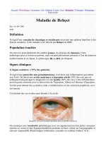
–FIGURE2-3— Typical clinical and laboratory features of acute HBV infection.
Reprinted from Braunwald E, Fauci AS, Kasper DL. Harrison’s Principles of Internal Medicine. 16th ed. New York: McGraw-Hill,
2005, p. 1825, Figure 285-4.
0
ALT
12345
HBsAg
HBeAg Anti-HBe
HBV DNA
6 12 24 36 48 60 120
Months after exposure
Anti-HBc
IgM anti-HBc
–FIGURE2-4— Typical clinical and laboratory features of chronic HBV infection.
Reprinted from Braunwald E, Fauci AS, Kasper DL. Harrison’s Principles of Internal Medicine. 16th ed. New York: McGraw-Hill,
2005, p. 1825, Figure 285-5.
52
CHAPTER 2 / ABDOMINAL AND GASTROINTESTINAL DISORDERS
Diagnosis: IgM and IgG anti-HDV are the serologic markers of HDV infection. Due to the dependence
of HDV on HBV, the diagnosis of HDV infection requires the presence of HB
s
Ag.
HEPATITIS E
Etiology: Hepatitis E virus (HEV) is an RNA hepatitis virus that is waterborne or enterically transmitted.
Clinical Presentation: The incubation period for Hepatitis E is 15–60 days. Patients present with similar
signs and symptoms to other forms of acute viral hepatitis, though this does not progress to a chronic disease.
Fulminant hepatitis can occur rarely and is more frequent in pregnant women, particularly those in their
third trimester.
Diagnosis: Abnormal LFTs are seen with the initial symptoms, and these tests return to normal levels
within one to six weeks after illness develops. There is no serologic marker for routine testing of HEV at this
time.
Hepatorenal Failure
Definition: Acute renal failure in a patient with liver cirrhosis, alcoholic hepatitis, or liver tumors is termed
hepatorenal failure.
Etiology: As liver function worsens, splanchnic vasodilatation occurs and there is a fall in renal perfusion.
This is the end stage complication of the decrease in renal perfusion caused by severe hepatic disease.
Clinical Presentation: Oliguria, low sodium excretion, and increase in plasmacreatinine can all indicate
hepatorenal failure. The emergency physician should beware thaturineoutput may not decrease significantly
until just a few days before the patient’s rapid decompensation and death.
Diagnosis: The diagnosis is made when the glomerular filtration rate and the urine sodium secretion
decrease, azotemia worsens, and renal failure develops.
Treatment: The treatment of the liver disease and improvement in hepatic function (transplantation or
resolution of primary liver disease) is the primary goal of treatment. Transjugular intrahepatic portosystemic
shunt (Tips) placement can aid in the treatment of the liver disease. Hemodialysis may be needed to care
for these patients.
Hepatic Encephalopathy
Etiology: Hepatic encephalopathy occurs in acute liver failure or chronic liver disease and is a response
to cerebral edema in acute disease or to the build up of metabolic waste in chronic disease.
Clinical Presentation: Stages of hepatitic encephalopathy progress from apathy to coma. Patients may
present with lethargy, drowsiness, asterixis (a hand flap when patients hold their hands up and extend at the
wrist), and stupor with hyperreflexia.
Diagnosis: Ammonia levels are typically elevated but are often inaccurate and therefore cannot be relied
upon solely. Patients with this presentation are prone to falls. Therefore a head CT and laboratory studies
should be ordered to identify other etiologies of their encephalopathic presentation.
DISORDERS OF THE LIVER, GALL BLADDER, AND PANCREAS
53
Treatment: Lactulose is the mainstay of treatment and should be given until soft stools are produced.
Lactulose reduces ammoniagenic substrates by lowering the colonic pH to cause the formation of an am-
monium ion, NH
4
+
, from ammonia, NH
3
. Since NH
4
+
is not absorbed in the colon, this effectively lowers
the serum ammonia concentration. Neomycin is an alternative treatment and has been shown to decrease
the amount of intestinal bacteria thereby decreasing protein degradation. Side effects of neomycin include
nephrotoxicity and ototoxicity.
Ascites and Spontaneous Bacterial Peritonitis
Etiology: Ascites occurs secondary to portal hypertension and hypoalbuminemia. Spontaneous bacterial
peritonitis (SBP) is the most frequent complication of cirrhotic ascites.
Clinical Presentation: Patients with ascites present with a distended abdomen, and patients may com-
plain of abdominal pain, shortness of breath, orthopnea, or fatigue. Physical findings include a fluid wave
and hepatomegaly. Patients with SBP present with fever, abdominal pain, and diffuse tenderness. They may
also present with only worsening encephalopathy.
Diagnosis: The physical examination and bedside ultrasound can confirm the presence of fluid. The
diagnosis of SBP requires paracentesis to evaluate the composition of the ascitic fluid. If SBP is suspected,
the fluid should be sent for a cell count, gram stain and culture. The diagnosis of SBP is confirmed if the
ascitic fluid has WBC >1000/mm
3
with PMN >250/mm
3
.
Treatment: Ascites can be managed conservatively or a therapeutic paracentesis may be used for
symptomatic relief. SBP treatment requires broad-spectrum antibiotics (cefotaxime or either ticarcillin-
clavulanate, piperacillin-tazobactam, or ampicillin-sublactam) and hospitalization. In peritoneal dialysis
patients, SBP is typically secondary to skin flora and may be treated with vancomycin infused intraabdomi-
nally with the patient’s dialysate.
Cholecystitis and Biliary Colic
Definition:
r
Biliary colic: Contractions of the gallbladder against an obstructed duct or gallbladder infindibulum
causing abdominal pain
r
Acute cholecystitis: Acute inflammation of the gallbladder
r
Ascending cholangitis: Fulminant infection of the bile duct extending into the liver with secondary
bacteremia and sepsis
Etiology: Gallstones are comprised of cholesterol (70%), pigment (20%), or a mixture of the two (10%). A
variety of conditions and diseases predispose to the formation of gallstones. The presentation of gallbladder
disease is a continuum from biliary colic to ascending cholangitis. The most common bacterial pathogens
in acute cholecystitis and ascending cholangitis are Escherichia coli, Klebsiella, Enterococcus, Bacteroides,
and Clostridium.
54
CHAPTER 2 / ABDOMINAL AND GASTROINTESTINAL DISORDERS
TABLE 2-4 RISK FACTORS F OR GALLSTONES
Female sex
Multiparity
Pregnancy
Obesity
Drastic weight loss
Fasting
Total parenteral nutrition
Sickle cell anemia
Chronic hemolytic anemias
Liver disease
Medications:
Octreotide
Estrogen
Progesterone
Clofibrate
Clinical Presentation:
Biliary colic:
r
Right upper quadrant or epigastric pain which often radiates to the shoulder or back is the classic
presentation for bilary colic.
r
Pain from biliary colic is generally self-limited, lasting anywhere from 2 to 6 hours and often occurring in
the evening or early morning hours. Up to one-third of patients will have no association between meals
and biliary symptoms.
Acute cholecystitis:
r
Pain is similar to biliary colic but persists beyond 6 hours and is often accompanied by fever, nausea,
vomiting, and anorexia. Murphy sign—worsening pain on palpation of the right upper quadrant is 97%
sensitive for acute cholecystitis. Ten to fifteen percent of patients with gallstones will develop pancreatitis
as a complication.
r
Acalculous cholecystitis occurs in 5–10% of patients and generally has a more fulminant course. It is
usually seen in patients with comorbid illness such as diabetes mellitus, burns, multiple trauma, or
sepsis.
Cholangitis:
r
The classically described triad of jaundice, fever, and right upper quadrant pain (Charcot triad) only
occurs in 50–75% of patients with acute cholangitis.
DISORDERS OF THE LIVER, GALL BLADDER, AND PANCREAS
55
r
Severe cases will cause mental confusion and shock in addition (Reynold pentad) and are associated with
significant morbidity and mortality.
r
Immunocompromised or elderly patients may only present with hypotension.
Diagnosis:
Biliary colic: WBC, LFTs, alkaline phosphatase, and serum bilirubin are usually normal. Diagnosis re-
lies on clinical presentation combined with ultrasound. Ultrasound will typically show stores with their
accompanying sonographic shadows.
Acute cholecystitis: Leukocytosis and elevated LFTs with an obstructive picture (elevated alkaline phospho-
tase and bilirubin) are usually present. Serum lipase should also be checked to assess for complication
of pancreatitis. Ultrasound is the definitive diagnostic test (sensitivity 94%, specificity 75%). Hallmark
ultrasound findings are pericholecystic fluid, gallbladder wall thickening (>5 mm), and a sonographic
Murphy sign. Common bile duct distention beyond 3 mm is suggestive of common bile duct obstruction.
CT scan is only 50% sensitive.
Treatment:
Biliary colic: Symptomatic management with antiemetics, fluid replacement, and analgesia is routinely
needed for biliary colic. Surgical referral on an outpatient basis is usually sufficient, unless pain cannot
be controlled in the ED.
Acute cholecystitis: Fluid replacement, bowel rest, analgesia, and antiemetics are indicated in patients with
acute cholecystitis. Antibiotic coverage should be provided as well as emergent surgical referral. If a
common bile duct obstruction is suspected based on ultrasound findings then ERCP or cholangiogram
is indicated.
Cholangitis: Surgical consult, broad-spectrum antibiotics, and ICU admission are all indicated in the treat-
ment of cholangitis.
Pancreatitis
Definition: Pancreatitis is the inflammation of the pancreas.
Etiology: Ninety percent of cases of pancreatitis in the United States are due to cholelithiasis or alcohol
abuse. Pancreatitis may also be caused by drugs, infection, or metabolic disorders such as hypertriglyc-
eridemia.
Clinical Presentation: Midline epigastric pain radiating to the back or flank is the classic presenting
complaint. Pain is often relieved by leaning forward and made worse by a supine position. Fever, vomiting,
and signs of hypovolemia may be present. Cullen sign (bluish discoloration of the periumbilical region) and
Grey Turner sign (bluish discoloration over the flanks) are both signs of retroperitoneal hemorrhage which
can complicate pancreatitis. ARDS and shock are potential complications.
Diagnosis: Elevatedserum amylase or lipaselevelsare an indication of pancreatitis.Amylase is a nonspecific
test that rises sooner but has a shorter half-life and returns to normal levels in 3–4 days. Lipase is more accurate
and generally remains elevated for a longer period of time. A CT scan may be useful in determining the
severity of the disease and the presence of a necrosis or a pancreatic abscess or pseudocyst. A right upper
56 CHAPTER 2 / ABDOMINAL AND GASTROINTESTINAL DISORDERS
TABLE 2-5 CAUSES OF PANCREATITIS
TOXIC
∗
OBSTRUCTION METABOLIC IN FECTIOUS OTHER
Ethanol Gallstones Hemochromatosis Viral: Cystic fibrosis
Methanol Tumor Hypercalcemia Adenovirus DKA
Acetaminophen Divisum Hyperlipidemia Coxsackie Idiopathic
Amiodarone Post ERCP Uremia CMV Post-op
Amlodipine EBV Pregnancy
Antibiotics: Echovirus
Metronidazole Hepatitis viruses
Macrolides HIV
Rifampin Rubella
TMP/SMZ Varicella
Antiretrovirals Bacterial:
Glucocorticoids Campylobacter
Statins Legionella
Thiazides Mycoplasma
Mycobacteria
Fungal:
Asperigillus
Cryptococcus
Cryptosporidium
∗
These are only the most common drugs known to cause pancreatitis. Many others have been implicated in case reports.
quadrant ultrasound should be ordered to rule out cholelithiasis as a cause of pancreatitis. The Ranson
criteria as seen in Table 2-6 is useful to predict mortality but has limited value in the ED setting.
Treatment: Supportive care should be provided with analgesia, bowel rest (NPO status), intravenous
hydration, and electrolyte repletion. Some patients may need antiemetics. Antibiotics are not routinely
indicated but should be considered in patients with suspected pancreatic necrosis. Antibiotic coverage should
include adequate antimicrobial activity against Enterococcus, gram-negative, and anaerobic organisms.
DISORDERS OF THE STOMACH
57
TABLE 2-6 RANSON CRITERIA
SPECIF IC RANSON FACTORS
ON ADMISSION 48 h AFTER ADMISSION
Glucose >200 mg/dL Drop in calcium below 8 mg/dL
Age >55 Decrease in arterial PO
2
below 60 mm Hg
LDH >350 IU/L Drop in hematocrit of >10%orHct< 30%
AST >250 Increase in BUN over 5 mg/dL
WBC >16,000/µL Base deficit over 4 meq/L
MORTALITY ASSOCIATED WITH RANSON CRITERIA
RANSON FACTORS MORTALITY DEAD OR IN ICU FOR >7D
0–2 0.9% 3.7%
3–4 16% 40%
5–6 40% 93%
7–8 100% 100%
DISORDERS OF THE STOMACH
Peptic Ulcer Disease and Gastritis
Definition:
Peptic ulcer disease: Peptic ulcer disease (PUD) is a chronic disease with recurrent ulcerations of the stomach
and proximal duodenum. The lifetime prevalence of PUD is 10% in adult Americans.
Gastritis: Acute or chronic inflammation of the mucosal lining of the stomach.
Etiology:
PUD: Helicobacter pylori infection is present in 95% of duodenal ulcers and 80% of gastric ulcers. NSAIDs
predispose to these conditions by inhibiting the production of prostaglandin and thereby decreasing
bicarbonate and mucous production. Cigarette smoking is also a risk factor for PUD.
Gastritis: Acute gastritis may be secondary to ischemia in the setting of acute illnesses such as shock, severe
burns, or trauma. Chronic gastritis is usually caused by an H. pylori infection.
Clinical Presentation: The typical patient complaint is burning epigastric pain often relieved by inges-
tion of food, milk, or antacids. Physical examination may be normal or notable for epigastric tenderness.
Complications of PUD include vascular ulceration resulting in GI bleed, perforation, and gastric outlet
obstruction secondary to scarring and edema.
58
CHAPTER 2 / ABDOMINAL AND GASTROINTESTINAL DISORDERS
Diagnosis: In the emergency department, this is a diagnosis of exclusion where other emergent causes of
epigastric pain should be excluded. The definitive diagnosis is by endoscopy. The emergency physician can
consider testing for H. pylori infection. If patient has peritoneal signs, an upright CXR may show free air
under the diaphragm in 60% of patients with anterior perforations. In posterior duodenal perforations, no
free air is seen since the posterior duodenum is retroperitoneal.
Gastric outlet obstruction may develop after an ulcer heals causing a scar that blocks the gastric outlet.
Signs and symptoms include abdominal pain, vomiting, and weight loss. Diagnosis can be made by plain
films that demonstrate a dilated stomach with an air–fluid level.
Treatment: The patient should be instructed to discontinue use of alcohol, tobacco, and NSAIDs.
Pharmacologic treatment is indicated with a H2 blocker or proton pump inhibitor. Blood transfusion is
needed in cases of PUD perforation with hemorrhage. Consult GI or general surgery as indicated for
complications.
Gastrointestinal Bleeding
Definition : Upper GI bleeding is defined as bleeding that originates from sites proximal to the ligament
of Treitz. Lower GI bleeding begins distal to this.
Etiology:
Upper GI bleeds: Peptic ulcer disease (PUD) in the most common cause of upper GI bleeding and account
for 60% of cases. Other causes are gastritis and esophagitis (15%), esophageal and gastric varices (6%),
Mallory-Weiss tears, arteriovenous malformations, and epistaxis.
Lower GI bleeds: An upper GI source is the most common cause of lower GI bleeding. Hemorrhoids are the
most common cause of true lower GI bleeds followed in frequency by diverticulosis and angiodysplasia.
Other causes are malignancy, polyps, aortoenteric fistula, arteriovenous malformations, inflammatory
bowel disease, and infectious colitis.
Clinical Presentation: Severe GI bleeding presents with signs of shock such as hypotension and tachy-
cardia. A history of weight loss is suggestive of malignancy. The presence of alcoholic liver disease and
hematemesis should lead one to suspect bleeding esophageal or gastric varices. Labs may show microcytic
anemia that if the bleeding is chronic. BUN may be elevated secondary to hemoglobin breakdown.
Diagnosis: The patient may be lavaged with crystalloid via nasogastric tube to confirm an upper GI source
and help determine if the bleeding is active. Endoscopy and colonoscopy may be both diagnostic and
therapeutic. Other diagnostic tools are angiography and tagged RBC scan (scintigraphy).
Treatment: Airway protection for patients with active GI bleeding should be considered. Patients may need
resuscitation with crystalloid and RBC transfusion. Coagulopathies should be corrected. NG tube placement
is helpful in patients with nausea and vomiting. Endoscopy with sclerotherapy or banding may be successful
for treatment of esophageal or gastric sources of bleeding. Sclerotherapy is also utilized via colonoscopy to
treat some lower GI bleeds. Recommended drug therapy includes high-dose IV proton pump inhibitor for
active peptic ulcer bleeding and IV somatastatin or octreotide for bleeding varices.
DISORDERS OF THE SMALL AND LARGE BOWEL
59
DISORDERS OF THE SMALL AND LARGE BOWEL
Aortoenteric Fistulas
Definition: An aortoenteric fistula is an abnormal connection between the aorta and the bowel lumen.
Etiology: This fistula typically involves the duodenum and occurs more often in patients with a history of
aortic graft placement.
Clinical Presentation: Massive hemorrhaging is the most common presenting sign. Patients may present
with hematemesis, melena, or hematochezia as well. Occasionally an episode of mild bleeding is followed
by a life-threatening hemorrhage.
Diagnosis: An aortoenteric fistula is diagnosed by a high index of suspicion with the right clinical presenta-
tion. Endoscopy is the procedure of choice and excludes other etiologies for an upper GI bleed. Abdominal
CT and aortography can be used to confirm the diagnosis, as can exploratory laparotomy.
Treatment: In the emergency department, supportive care should be provided. This may include blood
transfusion, broad-spectrum antibiotics, and management of shock. Emergent surgery is clearly the treatment
of choice.
Intestinal Obstruction
Definition: An intestinal obstruction is a mechanical obstruction of the intestinal lumen or adynamic
ileus causing inability of bowel contents to pass through the intestines.
Etiology: A mechanical obstruction can be caused by extrinsic or intrinsic compression on the bowel or
by an intraluminal processes.
Clinical Presentation: A mechanical small bowel obstruction (SBO) usually presents with crampy, in-
termittent pain. Patients with an adynamic ileus present with more constant and less severe pain. Either
type of bowel obstruction is often accompanied by vomiting. Emesis is bilious with a proximal obstruction
and feculent with a distal obstruction. Complete bowel obstruction is accompanied by constipation and
obstipation while partial bowel obstruction may present with the continued ability to pass stool. Exami-
nation findings vary with the type, location, and duration of the process. Mechanical obstruction usually
produces high-pitched bowel sounds and a distended, tympanic abdomen. Adynamic ileus frequently results
in hypoactive bowel sounds.
Diagnosis: Plain radiographic findings seen with a complete SBO include plicae circulares (transverse
linear densities that extend completely across the bowel lumen) and air–fluid levels in the small bowel.
Large bowel obstruction (LBO) will have distended loops of colon on the plain film. CT scan is particularly
useful to distinguish partial from complete obstruction and mechanical SBO from adynamic ileus. Barium
enema may diagnose the site of a LBO more accurately then plain films.
Treatment: The treatment involves reducing the contents in the GI tract by keeping the patient NPO and
placing an NG tube. Intravenous fluids should be infused and broad-spectrum parental antibiotics given to
cover anaerobes, gram-negatives, and Enterococcus if indicated. Emergent surgical consultation should be
obtained if mechanical obstruction is suspected.
60 CHAPTER 2 / ABDOMINAL AND GASTROINTESTINAL DISORDERS
Inflammatory Bowel Disease
TABLE 2-7 FEATURES OF ULCERATIVE COLITIS VERSUS CROHN DISEASE
ULCERATIVE COLITIS CROHN DISEASE
Definition
r
Chronic inflammatory disease of the
colon
r
Involves only mucosa and submucosa
r
Inflammation progressively more
severe from proximal to distal colon
r
Rectum involved nearly 100% of time
r
Chronic granulomatous inflammation
anywhere in GI tract
r
All layers of bowel wall involved
r
Discontinuous, “skip areas” of
inflammation are common
r
Rectal sparing common
Etiology Multifactorial Multifactorial
Clinical
Presentation
r
Peak incidence in teens and 20s
r
Bloody diarrhea most common
presentation
r
Extraintestinal manifestations such as
liver disease, arthritis, uveitis
r
10–30% increased risk of colon
cancer
r
Bimodal age of onset, 15–22 years and
55–60 years
r
Chronic abdominal pain, fever, and
diarrhea
r
25–30% of Crohn patients have
extraintestinal symptoms
r
Complications frequent including
thromboembolic disease, toxic
megacolon, obstruction, malignancy
(risk increased three fold), abscess, and
fistula formation
Diagnosis Colonoscopy required for definitive
diagnosis
r
Upper GI series: can show ileal
involvement, segmental narrowing of
small intestine, destruction of normal
mucosal pattern, fistulas
r
Colonoscopy: allows biopsy to
determine extent of bowel wall
involvement
r
CT: useful in acute flares, identifying
abscesses, fistulas, obstruction, or toxic
megacolon
Toxic Megacolon
r
Occurs when inflammation progresses through the wall of the colon and results in
atony and distension. Patients are generally toxic appearing and have peritoneal signs
r
Peritoneal signs may be masked in patients on glucocorticoids
r
KUB demonstrates a dilated colon of 6 cm or greater
r
“Thumb printing” of the bowel wall may be recognized and represents bowel-wall
edema
r
Treatment is immediate decompression with nasogastric tube, hydration,
broad-spectrum antibiotics, and emergent surgical consultation
DISORDERS OF THE SMALL AND LARGE BOWEL 61
TABLE 2-7 FEATURES OF ULCERATIVE COLITIS VERSUS CROHN DISEASE (CONTINUED)
Toxic Megacolon
(continued)
Treatment
r
Identify and treat complications
r
Hydrate with crystalloid
r
Broad-spectrum antibiotics for fulminate colitis (cover enteric flora with
metronidazole and ciprofloxacin or clindamycin and ampicillin)
r
Glucocorticoids
r
Severe exacerbations require complete bowel rest and parenteral nutrition
r
Maintenance therapy with sulfasalazine or a 5-aminosalicylic derivative (Asacol,
Pentasa) is often effective in preventing flares
r
Total colectomy is curative for ulcerative colitis
Acute Appendicitis
Definition: Inflammation of the appendix.
Etiology: Acute appendicitis begins with obstruction of the lumen followed by continued mucous produc-
tion from the glands of the appendix. Subsequently, there is increased intraluminal pressure, which leads to
vascular compromise and, ultimately, necrosis. If untreated, this will result in perforation of the appendix,
abscess formation, and peritonitis.
Clinical Presentation: Classic symptoms of appendicitis are early periumbilical or epigastric pain (the
visceral innervation of the appendix is at the T-10 level), vomiting, and anorexia. As inflammation of the
appendix progresses, there is activation of the somatic fibers and localization of pain to the right lower
quadrant and tenderness at McBurney point (just below the middle of the line connecting the umbilicus
with the anterior superior iliac spine). Beware of anatomic variations of the appendix: only one-half to
two-third of patients have classic symptomatology. Some of these variants include:
r
With inflammation of a retrocecal appendix (present in approximately 25% of the population) there may
be localization of pain to the right flank.
TABLE 2-8 COMMON CAUSES OF BOWEL OBSTRUCTION
DUODENU M SMALL BOWEL LARGE BOWEL
Foreign Body Adhesions Tumor
Stenosis Hernia Fecal impaction
Stricture Intussusception Volvulus
Superior mesenteric artery syndrome Stricture Intussusception
Lymphoma Pseudo-obstruction
62 CHAPTER 2 / ABDOMINAL AND GASTROINTESTINAL DISORDERS
r
Men with inflammation of a retroileal appendix may have testicular pain.
r
Inflammation of a pelvic appendix may cause suprapubic pain and dysuria.
r
Pregnant patients may present with right upper quadrant pain secondary to caudal displacement of the
appendix.
r
A high index of suspicion should be maintained in young children (<5 years), the elderly, and AIDS
patients who may all present with atypical symptoms.
Diagnosis:
Physical examination in a patient with suspected appendicitis includes several special examination maneu-
vers:
r
Rovsing sign: Tenderness of the right lower quadrant or palpation of the left lower quadrant
r
Psoas sign: Increase in pain when the right leg is passively flexed while the patient is in the left lateral
decubitus position
r
Obturator sign: Increase in pain when the right hip is passively internally rotated while the patient is
supine with a flexed right hip and knee
Leukocytosis (30% of patients with appendicitis had a normal WBC count with over 90% of these patients
having a concomitant left shift). 24–95% of plain radiographs in patients with appendicitis are abnormal.
Abnormal findings include an appendicolith, blurring of the psoas muscle margin, appendiceal gas, and
free air. The CT scan is 96% sensitive in identifying appendicitis compared to ultrasound which
has a 76–95% sensitivity. Findings on ultrasound may be limited by body habitus or the technician’s
skill.
Treatment: The primary treatment for acute appendicitis is emergent surgical consultation for appendec-
tomy. The patient should be kept NPO and given broad-spectrum antibiotics with coverage of anaerobes,
grand negatives, and Enterococcus.
Pseudomembranous Enterocolitis
Definition: Inflammation of the bowel characterized by the development of yellowish plaques overlying
inflamed bowel.
Etiology: Clostridium difficile, an anaerobic bacterium, causes pseudomembranous colitis. Risk factors for
infection include recent broad-spectrum antibiotic use, prolonged hospitalization, advanced age, immuno-
compromised status, and recent bowel surgery.
Clinical Presentation: Patients generally present with complaints of crampy abdominal pain, diarrhea,
and fever. The diarrhea may be watery, mucoid, or bloody. Complications include dehydration, electrolyte
abnormalities, toxic megacolon, and perforation.
Diagnosis: The diagnosis is confirmed by identifying C. difficile or its toxin in the stool. Colonoscopy will
show yellow membranous plaques within the bowel lumen.
Treatment: The mainstay of treatmentisto discontinue the broad-spectrum antibiotics thathavecaused the
illness. The patient’s dehydration and electrolyte abnormalities should be treated appropriately. C. difficile
DISORDERS OF THE SMALL AND LARGE BOWEL 63
infection will generally respond to either metronidazole 250 mg PO qid or vancomycin 250 mg PO qid.
Antidiarrheal medications should be avoided.
Viral and Bacterial Diarrhea
Etiology: Eighty percent of cases of acute infectious diarrhea are viral compared with 20% of caused by
bacterial infections. The most common viral causes are rotavirus and Norwalk. Rotavirus occurs in children
6–24 months of age and peaks in the winter as does Norwalk, which infects older children and adults. The
most common bacterial causes of diarrhea are campylobacter, salmonella, and shigella which are invasive
bacteria that affect the large bowel often causing blood and mucous in the stool. Staphylococcal aureus,
Vibrio cholera, Clostridium perfringens, Bacillus, ciguatera fish poisoning, and scromboid fish poisoning are
enterotoxin-producing bacteria that change water and electrolyte transport in the small bowel causing watery
diarrhea.
Clinical Presentation: Symptoms depend on the infectious agent causing the diarrhea and may include
constitutional signs and symptoms associated with dehydration as well as fever, chills, and abdominal pain.
Diagnosis: A wet mount of the stool can be diagnostic for fecal leukocytes which are present in invasive
bacterial diarrhea, pseudomembranous colitis, and inflammatory bowel disease. Other laboratory studies
that may distinguish between causes of diarrhea include a stool culture, stool samples for ova and parasites,
or a C. difficile toxin assay.
Treatment: Supportive care, including fluid hydration as well as antibiotics (fluoroquinolones), is used
for invasive bacterial diarrheas. Antimotility agents should be used with caution in patients not prescribed
antibiotics due to possible delay in clearing the infectious organism.
Diverticulitis
Definition: Diverticulitis is the acute inflammation of the wall of a diverticulum with or without microp-
erforation.
Etiology: Colonic diverticular disease increases with advancing age and occurs due to a combination of
weakening of the muscular layer of bowel wall and increased intraluminal pressure. This results in herniation
of mucosa and submucosa through the muscular layers of the bowel wall. Diverticuli occur most frequently
in the sigmoid colon. Inspissation of undigested food at the neck of a diverticula causes increased pressure
within the diverticulum from mucous production, overgrowth of bowel flora, and ultimately diverticulitis.
Microabscesses may develop but are usually walled off by adjacent loops of bowel or mesentery.
Clinical Presentation: The most common symptom for diverticulitis is left lower quadrant pain and
tenderness on palpation. Diarrhea or constipation may also occur, and fever is frequently present.
Diagnosis: The diagnosis may be based on clinical history. Dual contrast CT scan is the confirmatory test
in the ED and will also reveal complications such as perforation, abscess, and fistula.
Treatment: The emergency department treatment consists of IV hydration, bowel rest, and broad-spectrum
antibiotics with coverage of colon flora. Hospitalization and surgical consultation are warranted for patients
with systemic symptoms, toxic appearance, abscess, or peritoneal signs.
64 CHAPTER 2 / ABDOMINAL AND GASTROINTESTINAL DISORDERS
Volvulus
Definition: A volvulus is a closed loop obstruction of the large bowel leading to bowel obstruction and
possibly infarction.
Etiology: Sigmoid volvulus occurs more commonly than cecal volvulus. Sigmoid volvulus affects patients
with a history of chronic constipation, particularly the elderly, patients with comorbid illnesses, and psychi-
atric illnesses. In children, sigmoid volvulus can be the presenting symptom of Hirschsprung disease. Cecal
volvulus is caused by the anomalous fixation of the right colon, more commonly affects younger patients
than sigmoid volvulus.
Clinical Presentation: Patients can present with crampy abdominal pain, nausea, vomiting, diffuse
abdominal tenderness with distention, and tympany.
Diagnosis: X-rays of the abdomen, which can be helpful in making the diagnosis, show a single dilated
loop of colon. The sigmoid volvulus is typically seen in the left side of the abdomen while a cecal volvulus
is described as having a coffee bean shape usually seen in the upper abdomen.
Treatment: Supportive care, NG tube decompression, and administration of broad-spectrum antibiotics
are the primary goals of emergency department care. The patient should be watched for signs of bowel
infarct including peritonitis and sepsis. Sigmoid volvulus can be reduced using a rectal tube. Recurrence is
common so reduction should be followed by surgery. A cecal volvulus requires early surgery.
ANORECTAL DISORDERS
Hemorrhoids
Definition: Hemorrhoids are engorgement and dilation of the hemorrhoidal plexus veins. Internal hemor-
rhoids are located proximal to the dentate line and drain into the portal venous system. External hemorrhoids
are distal to the dentate line and drain to the iliac veins.
Etiology: Hemorrhoids result from weakened connective tissue of the vessels. This may result from any-
thing that causes increased venous pressure in the rectum such as pregnancy or portal hypertension. Con-
stipation is also risk factor.
Clinical Presentation: The patient with internal hemorrhoids generally complains of painless bright red
blood with bowel movements. Internal hemorrhoids that prolapse and become thrombosed may result in
local tissue ischemia and become very painful. Patients with external hemorrhoids present with complaints
of itching and pain.
Diagnosis: The emergency department diagnosis of hemorrhoids is generally done by visual and digital
rectal examination. Anoscopy may be necessary to visualize internal hemorrhoids.
Treatment: Sitz baths and topical analgesics are the mainstay of therapy for hemorrhoids. Steroids creams,
stool softeners, or high-fiber diet can also provide some relief. Surgical referral is indicated for incarcerated
or strangulated internal hemorrhoids.
ANORECTAL DISORDERS 65
Cryptitis
Definition: Inflammation of the anal crypts.
Etiology: Tissue breakdown resulting in cryptitis can be caused by trauma from foreign bodies, chronic
diarrhea, or passage of hard stool. Cryptitis may progress to fissure or abscess.
Clinical Presentation: The most common presenting symptoms are anal pain, spasm, and itching with-
out rectal bleeding.
Diagnosis: The diagnosis is made by clinical symptoms and palpation of tender and edematous crypts on
physical examination.
Treatment: Treatment of cryptitis includes use of stool softeners and eating a high-fiber diet. If the
symptoms are severe, surgical referral is indicated.
Anorectal Abscesses
Etiology: An anorectal abscess begins with obstruction of an anal gland and cryptitis. Most commonly, the
infection is limited to the superficial perianal area though it may involve the deeper spaces (intersphincteric,
ischiorectal, postanal, supralevator, and perirectal spaces). Theseabscesses are more commonin patients with
Crohn disease, immunocompromised patients, and those with concomitant infections such as gonococcal
prostatitis and other STDs. The most common age range is young to middle-aged males.
Clinical Presentation: A perianal abscess is located on the anal verge at the posterior midline. The
examination will note a discrete, superficial, and tender mass that is often fluctuant. Perirectal abscesses
typically present with a fever, anorexia, and pain on rectal examination. Ischiorectal abscess is the most
common deep-space infection and presents with a tender, fluctuant mass over the medial buttock. Other
deep-space infections may be noticeable only on rectal examination as an exquisitely tender and indurated
mass.
Diagnosis: Endorectal ultrasound, MRI, or CT scan will help differentiate a perianal from a deeper perirec-
tal abscesses.
Treatment: Incision, drainage, and gauze packing of a perianal abscess can be done in the emergency
department. If there is evidence of cellulitis, the patient is immunocompromised state, or has valvular
heart disease, oral antibiotics are indicated. In contrast, with a perirectal abscess, parental broad-spectrum
antibiotic treatment should be given. Admission and emergent surgical referral for operative irrigation and
debridement is necessary.
Fistula In Ano
Definition: Fistulo in ano is an abnormal, epithelial-lined tract that connects an anal gland to the skin.
Etiology: This is most commonly seen as a complication of perianal or ischiorectal abscesses. A fistula in
ano may also result from Crohn disease, ulcerative colitis, cancer, or an STD.
66 CHAPTER 2 / ABDOMINAL AND GASTROINTESTINAL DISORDERS
TABLE 2-9 DIFFERENTIAL FOR ACUTE ABDOMINAL PAIN
DISEASE ETIOLOGY CLINICAL PRESENTATION DIAGNOSIS
Perforated peptic ulcer Peptic ulcers may be
secondary to H. pylori
infection, NSAID use, or
other mediators
Abrupt onset of severe
epigastric pain
Free air on abdominal
x-ray series
Biliary tract disease Common in patients >50
yrs, majority lack fever
RUQ pain with nausea,
vomiting, fever, Murphy
sign
Clinical presentation,
RUQ ultrasound
Pancreatitis 80% of cases secondary
to gallstones or alcohol
abuse
Pain and tenderness in
the upper half of the
abdomen
Elevated serum amylase
and lipase (lipase more
sensitive)
Bowel obstruction May be small or large
bowel obstructions, can
be caused by sigmoid or
cecal volvulus
Colicky abdominal pain
with nausea, vomiting,
abdominal distention,
and high-pitched bowel
sounds
KUB showing air fluid
levels, CT scan in the
case of a nondiagnostic
KUB
Renal colic Obstructing renal stone Colicky abdominal pain
often with radiation to the
groin or costovertebral
angle, may be associated
with nausea and vomiting
Blood in urine (up to
15% will not have
hematuria), helical
noncontrast abdominal
CT
Appendicitis Obstruction of the
appendix from food,
adhesions, or lymph
nodes
Abdominal pain radiating
periumbilical to RLQ,
anorexia, nausea,
vomiting, fever
History and physical
exam and if necessary
imaging studies (CT scan
vs. US)
Diverticular disease Weakening of the
muscular layer of bowel
wall secondary to
increased intraluminal
pressure
When infected, fever and
abdominal pain (though
LLQ pain present in only
25% of patients)
Clinical history, CT
abdomen
Hernias Intraabdominal-wall
defect with protruding
bowel; most are inguinal
Abdominal pain with a
bulge on physical
examination
Physical examination, CT
abdomen
Mesenteric ischemia Embolic disease is abrupt
in onset, nonocclusive
disease has a more
indolent course
Pain out of proportion
with examination, may
have nausea, vomiting,
blood in stool
High index of suspicion,
elevated lactate,
arteriography, CT
abdomen
ANORECTAL DISORDERS 67
TREATM ENT COMPLICATIONS COMMENTS
Broad-spectrum
antibiotics, surgical
consult with likely surgical
intervention
Peritonitis, sepsis Patients may not have prior ulcer
history, elderly patients may have less
peritoneal findings
Broad-spectrum
antibiotics, surgical
consult
Cholecystitis with sepsis, ascending
cholangitis
Cholangitis: Charcot triad: jaundice,
fever, RUQ pain.
Reynold pentad adds altered mental
status and shock
Analgesics, antiemetics,
IVFs, bowel rest
Peripancreatic fluid collections,
complications from gallstones that
may be causing pancreatitis
Ranson Criteria predicts morbidity and
mortality (Table 2-6)
NG tube, IVFs, analgesics,
surgical repair
Ischemic bowel from prolonged
obstruction
Sigmoid volvulus: typically affects
those with history of constipation, the
elderly or patients with neuromuscular
or psychiatric illnesses
Supportive care,
analgesics, urology
consult for stones >
5mm in diameter
Obstructive pyelonephritis 90% of stones are radioopaque and
can be seen on KUB but CT provides
vital information regarding amount of
obstruction caused by the stone.
Surgery, broad spectrum
antibiotics
Peritonitis, sepsis Children may present with atypical
symptoms such as lethargy and the
elderly may have subtle
signs/symptoms Appendicitis is the
most common general surgical
complication of pregnancy
Antibiotics, supportive
care
Perforation, abscess, fistula formation Elderly patients are at risk for
perforation of the colon that is not
seen in younger patients with
diverticulitis
Manual reduction,
surgery if incarcerated or
strangulated
Bowel ischemia if strangulated Indirect inguinal hernia is the most
common in both men and women,
femoral hernias are more common in
women than men
Supportive care,
emergent surgery vs.
interventional radiology
Sepsis and peritonitis The small bowel will infarct within 2 to
3 h of ischemia
68 CHAPTER 2 / ABDOMINAL AND GASTROINTESTINAL DISORDERS
Clinical Presentation: Physical examination demonstrates an open tract that produces a bloody, foul
smelling discharge. Fistulas frequently become blocked and result in perianal or perirectal abscess formation.
Diagnosis: Physical examination is generally diagnostic, though endorectal ultrasound or MRI may aid in
the diagnosis.
Treatment: These patients should be referred for surgical excision.
Proctitis
Definition: Proctitis is a viral or bacterial infection of the prostate gland.
Etiology: Proctitis typically occurs as the result of an STD such as gonococcus, syphilis, chlamydia, or
lymphogranuloma venereum. It is most commonly seen in men who have unprotected anal intercourse.
Clinical Presentation: The patients present with itching, pain, and rectal discharge.
Diagnosis: Diagnosis is made by anoscopy and a gram stain of the rectal discharge.
Treatment: All patients with proctitis should undergo a screening examination for other STDs. Treatment
should include an antibiotic appropriate to the offending organism.
FURTHER READING
Braunwald E, Fauci AS, Kasper DL. Harrison’s Principles of Internal Medicine. 15th ed. New York: McGraw-Hill, 2001.
Meyer GK, DeLaMora PA. Last Minute Pediatrics: A Concise Review for the Specialty Boards. New York: McGraw-Hill,
2004.
Tintinalli JE, Kelen GD, Stapczynski S. Emergency Medicine: A Comprehensive Study Guide. New York: McGraw-Hill,
2004.
Marx JA, Hockberger RS, Walls RM. Rosen’s Emergency Medicine: Concepts and Clinical Practice. St. Louis, MO:
Mosby, 2002.
Ferzoco LB, et al. Acute Diverticulitis. NEJM 1998;338:1521–1526.
Ranson JH, et al. Prognostic Signs and the Role of Operative Management in Acute Pancreatitis. Surg Gynec Obstet
1974;139:69–81.
CHAPTER 3
CARDIOVASCULAR
DISORDERS
DISORDERS OF CIRCULATION
Arterial
ABDOMINAL AORTIC ANEURYSM
Definition: An abdominal aortic aneurysm (AAA) is a localized dilation of all three layers of the wall of the
abdominal aorta. AAAs usually develop in the infrarenal portion of the abdominal aorta. The normal size of
the infrarenal aorta is ≤2 cm diameter. An AAA is diagnosed when the diameter of the infrarenal aorta is ≥3
cm.
Etiology: AAAs form as a result of loss of elastin and collagen from the aortic wall due to genetic, trau-
matic, infectious, and usually degenerative reasons. The primary risk factor for the development of AAAs is
advanced age. The average age at diagnosis is 65–70 years. Other significant risk factors include male gender,
atherosclerotic disease, and immediate family history of AAA.
Clinical Presentation: The classic symptom of a ruptured AAA is sudden onset of severe abdominal
and/or back pain—more often left back/flank. The pain often radiates to the groin, simulating renal colic,
which is the most common misdiagnosis of this condition. The patient may experience neurologic symptoms
including syncope ora femoral neuropathydue to aorticorhematoma compressionona peripheral nerveroot.
The vital signs usually demonstrate tachycardia, although intra-abdominal blood can induce a vagal response
and produce a relative bradycardia. Hypotension is classic, though unreliable; because the abdominal aorta is
a retroperitoneal structure, the initial rupture may tamponade in the confined space of the retroperitoneum
and allow the patient to temporarily stabilize their blood pressure through compensatory increases in vascular
resistance. Physical findings may include a palpable pulsatile abdominal mass, abdominal aortic or femoral
artery bruits, or signs of distal embolization or distal ischemia. None of the physical findings are reliable
enough to exclude the diagnosis.
Patients with a prior history of AAA repair may rarely develop an aortoenteric fistula, producing massive
gastrointestinal (GI) bleeding. The diagnosis should be immediately suspected in any patient with hematem-
sis, melena, or hematochezia who has had a prior AAA repair.
Diagnosis: The diagnosis is initially suggested by the clinical presentation. In a patient presenting with
the classic triad of abdominal/back pain, hypotension, and a pulsatile abdominal mass; or in a patient with
a history of a known AAA who presents with abdominal pain/back pain and hypotension, no further diag-
nostic interventions are needed prior to surgery. Most patients, however, do not present with such a classic
69
Copyright © 2007 by The McGraw-Hill Companies, Inc. Click here for terms of use.
70 CHAPTER 3 / CARDIOVASCULAR DISORDERS
presentation and require imaging studies. Computerized tomography (CT) of the abdomen with intravenous
(IV) contrast is considered the gold standard for diagnosis of ruptured or leaking AAA. Aneurysmal dilatation
can be diagnosed with CT even without IV contrast, although the presence of rupture cannot. Hemody-
namically unstable patients should not be sent for CT. Bedside ultrasonography (US) is an outstanding
tool to diagnose the presence of an AAA (Figure 3-1), although US is not sensitive for detecting rupture.
Nevertheless, US confirmation of the presence of an AAA in the patient with severe abdominal/back pain
and hypotension should be enough information to prompt immediate surgical consultation and exploration.
Plain radiographs may be helpful in demonstrating aortic calcifications, but they are neither sensitive nor
specific enough to be routinely recommended unless searching for an alternate diagnosis.
– FIGURE 3-1 — Ultrasound image of abdominal aortic aneurysm (cross-sectional view).
Treatment: When the diagnosis of a ruptured AAA is suspected, bilateral large bore IV lines should be
placed and blood sent immediately for routine labs as well as type and crossmatch. A vascular surgeon should
be consulted early based on strong suspicion of this diagnosis; consultation in these cases should not await
definitive diagnostic studies. Any delay in operative intervention is associated with a significant increase in
mortality.
DISORDERS OF CIRCULATION 71
THORACIC AORTIC DISSECTION
Definition: Thoracic aortic dissection (TAD) refers to a longitudinal cleavage of the wall of the aorta
through a tear in the intima. Blood “dissects” through this defect into the media of the aorta, creating a false
lumen within the media.
Etiology: The initial defect in the intima is usually caused by the stress of pulsatile blood flow in a
patient with chronic severe hypertension (the most common risk factor). Weakening of the aortic wall
can also be caused by connective tissue disorders, congenital heart disease (e.g., Marfan’s disease, Ehlers-
Danlos syndrome), pregnancy, and syphilis. Aortic dissections can also be caused iatrogenically after aortic
catheterization or surgery. Other commonly reported risk factors include bicuspid aortic valves, coarctation
of the aorta, and trauma. TADs usually occur in the fifth through seventh decades of life.
Clinical Presentation: The high-pressure pulsatile flow of blood can cause the dissection to progress,
leading to the classic presentation of sudden tearing, sharp pain that is maximal at onset, often radiating
to the mid-scapular region of the back. The dissection can also cause sharp pain radiating to jaw, neck,
shoulder, arm, low back, or abdomen. If the dissection involves the carotid or vertebral artery, the patient
may present with stroke symptoms or paraplegia, respectively. If the dissection descends to the iliac ar-
teries, the patient may develop lower-extremity pulse deficits and ischemic pain. The dissection may also
progress proximally toward the heart and disrupt the aortic valve (new diastolic murmur due to aortic re-
gurgitation), occlude a coronary artery (causing MI), or dissect into the pericardium (leading to cardiac
tamponade and rapid cardiovascular collapse). Overall, the clinical presentation will very dependant on
the location of the dissection and its propagation. The vital signs are usually notable for tachycardia. Hy-
pertension is common although patients may be normotensive or hypotensive at the time of the initial
presentation.
Diagnosis: The diagnosis of TAD is based on radiographic or echocardiographic imaging. Chest radio-
graphy is abnormal in more than 80% of cases, with findings such as widened mediastinum (>8cm),
separation of intimal calcification at the aortic arch more than 5 mm, pleural effusion, apical capping,
or rightward deviation of the trachea, bronchus, or esopahagus. CT of the aorta is commonly used to
diagnose TAD (Figure 3-2). It is relatively fast and easily available in most centers. The sensitivity and
specificity of CT in this disorder is 85–95%. Angiography is still considered the gold-standard imaging
test. It provides greater information regarding the aortic anatomy and extent of the dissection, which
assists the surgeons in their approach. Transesophageal echocardiography (TEE) is an outstanding al-
ternative imaging modality, especially in the patient who cannot tolerate IV contrast or is too unsta-
ble to leave the ED for radiography. TEE can be performed at the bedside, and in experienced hands
can provide diagnostic accuracy >95%. However, TEE is far less available in most EDs compared to
other imaging modalities. Magnetic resonance imaging (MRI) is also an outstanding modality for eval-
uation the aorta and branch vessels, but its use is impractical in patients who are actively or potentially
unstable.
Treatment: Immediate thoracic surgeon consultation is paramount. While awaiting surgical consultation,
medical management should begin at once. The initial management of all TADs is focused on reducing the
stress of pulsatile flow of blood in the aorta. IV beta blockers should be used to reduce the heart rate (HR)
to a goal of 50–60 s. IV esmolol is an ideal agent for this purpose, as it can be titrated based on the patient’s
condition. Calcium channel blockers can be used in patients who cannot tolerate beta blockers. Once the
goal HR has been achieved, IV antihypertensives should be added to further reduce the SBP to a goal of
100–110 mmHg. Easily titrateable antihypertensives (e.g., nitroprusside) are ideal because these patients
72
CHAPTER 3 / CARDIOVASCULAR DISORDERS
– FIGURE 3-2
— Computerized tomography with IV contrast demonstrating TAD (arrow indicates the false lumen).
can have very labile blood pressures. Some authors suggest single-drug therapy with the combination alpha-
and beta-blocker IV labetalol for both HR and SBP management, although one should be aware that this
medication has significantly more beta-blocking activity than alpha-blocking activity, and often additional
antihypertensive medications will be required.
Following this initial medical management, surgical evaluation is critical. The decision to perform
operative repair of a TAD is primarily based on the classification of dissection. There are two different
classification systems used for describing TADs, the older DeBakey classification and the newer Stanford
classification. Both classifications utilize the location of the dissection in relation to the left subclavian artery.
The DeBakey classification divides TADs into three groups: Type I involves both the ascending and the
descending aorta; Type II involves only the ascending aorta; and Type III involves only the descending
aorta. The Stanford classification divides TADs into only two groups: Type A includes any dissection that
involves the ascending aorta (includes DeBakey Types I and II); and Type B involves isolated descending
dissections (Figure 3-3). The Stanford classification is generally more relevant to the decision-making process
of the thoracic surgeons—Stanford Type A dissections almost always require operative intervention, whereas
Stanford Type B dissections usually are managed only medically with HR and BP control. Stanford Type B
dissections, however, may require surgery if the patient develops occlusion of a major vessel producing acute
end-organ ischemia (e.g., occluded superior mesenteric artery producing mesenteric ischemia, occluded
renal artery producing renal failure, occluded iliac artery producing ischemic leg, etc.).
ARTERIAL THROMBOEMBOLISM
Definition: Arterial thromboembolism refers to arterial occlusive disease due to either thrombosis or
embolism.
Etiology: Arterial thrombosis generally occurs in patients with risk factors for atherosclerotic disease,
especially diabetes mellitus and cigarette smoking. Arterial embolism tends to occur in patients with atrial
fibrillation or cardiomyopathies. The majority of emboli originate in the heart and primarily affect the lower
DISORDERS OF CIRCULATION
73
–FIGURE3-3— DeBakey and Stanford Classifications for TAD. Illustration by Ben Lawner, D.O.
extremities, although 5–10% of emboli lodge in the visceral circulation and cause mesenteric ischemia, renal
ischemia, or ischemia to other organs. Distal lower extremity emboli can also originate in the abdominal
aorta.
Clinical Presentation: Patients with acute extremity ischemia generally present with one or more of the
“six P’s of ischemia”: pain, pallor, pulse deficit, paresthesias, paresis, and poikilothermia (or polar; cold).
Pain is usually the first symptom that develops and often is described as “pain out of proportion to physical
findings.” Whereas arterial emboli cause an abrupt onset of symptoms, arterial thromboses present with a
history of claudication and signs of chronic ischemia: patients with mesenteric artery thrombosis typically
describe many months of increasing intestinal angina; and patients with lower-extremity thrombosis usually
have loss of distal hair, shiny skin, thickened nails, and poor capillary refill and pulses on the opposite
extremity.
Diagnosis: The diagnosis of arterial thromboembolism in the lower extremities can be confirmed with
Duplex US, which has a sensitivity approaching 85%. Abdominal CT with oral and IV contrast is often
employed for the diagnosis of mesenteric ischemia, although if this diagnosis is strongly suspected, every
effort should be made to obtain angiography, which can often be used therapeutically as well (see Chapter 2
for more information on vascular insufficiency/mesenteric ischemia). The gold-standard diagnostic study for
all forms of arterial occlusive disease is angiography.
Treatment: When the diagnosis of arterial occlusion in the lower extremities is strongly suspected, un-
fractionated heparin should be initiated. A vascular surgeon should be consulted and lower extremity
angiography ordered. If mesenteric ischemia is strongly suspected, a general surgeon and interven-
tional radiologist should be consulted immediately. Options for definitive treatment of lower extremity