Textbook of Traumatic Brain Injury - part 6 docx
Bạn đang xem bản rút gọn của tài liệu. Xem và tải ngay bản đầy đủ của tài liệu tại đây (1019.76 KB, 62 trang )
376 TEXTBOOK OF TRAUMATIC BRAIN INJURY
complaints and diagnosed posttraumatic narcolepsy using
formal sleep studies such as the polysomnogram (PSG)
and MSLT.
We recommend that clinical diagnosis of narcolepsy
should always be accompanied by formal sleep studies and
HLA typing. However, even if a patient is confirmed to
have the appropriate HLA haplotype, the question always
exists whether TBI was the causative factor or a precipi-
tating event.
Post-TBI hypersomnia is an understudied area. The
prevalence, varieties, associated psychiatric disturbances,
and effect on rehabilitation and physical, cognitive, and
social level of functioning are yet to be identified. Such
identification is important because effective management
of treatable disorders can have far-reaching results for the
rehabilitative process.
Sleep-wake cycle disturbances. Sleep-wake cycle distur-
bance, or circadian rhythm sleep disorder, is defined as
inability to go to sleep or stay awake at a desired clock
time. Both the duration and pattern of sleep are normal
when patients with this disorder do fall asleep (Kryger et
al. 2000). There are several varieties of sleep-wake cycle
disturbances, including the delayed, advanced, and disor-
ganized types. The pathogenesis remains unclear,
although dysfunction of the suprachiasmatic nucleus has
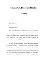
FIGURE 20–3. Epworth Sleepiness Scale.
Source. From Johns MW: “A New Method for Measuring Daytime Sleepiness: The Epworth Sleepiness Scale.” Sleep 14:540–545,
1991. Revised 1997. Used with permission of M.W. Johns. Copyright M.W. Johns 1991–1997.
Fatigue and Sleep Problems 377
been postulated (Okawa et al. 1987). Other factors often
associated with this disorder in the general population
include shift work and travel through different time zones
(Patten and Lauderdale 1992). There is little literature
available on the prevalence of this disorder in the TBI
population.
Schreiber et al. (1998) described circadian rhythm and
sleep-wake cycle abnormalities in all 15 individuals evalu-
ated after mild TBI using actigraphy (described in the sec-
tion Evaluation of Fatigue and Sleep Disturbances in TBI)
and PSG recordings. None had past history of neurological
illness, psychiatric history, or sleep apnea syndrome. More
than one-half of the patients were diagnosed with delayed-
phase type and the rest disorganized-type sleep-wake cy-
cle disturbance.
Quinto et al. (2000) described the case of a 48-year-
old man who presented with sleep-onset insomnia after a
severe closed head injury. His complaints included diffi-
culty in initiating sleep, being able to finally fall asleep
around 3:00–5:00
A.M., and waking up around noon. His
attempts to wake up earlier resulted in poor functioning.
Before the injury, he was reportedly high functioning and
denied problems with sleep. A diagnosis of delayed sleep
phase syndrome was confirmed by sleep logs and actigra-
phy. Patten and Lauderdale (1992) also reported delayed
sleep phase disorder in a 13-year-old boy after mild closed
head injury.
Complaints of sleep disturbance in TBI patients are
common, and therefore awareness and diagnosis of this
disorder are important; some patients may respond to
simple therapies such as adjusting the time of sleep (de-
scribed in the section Chronotherapy) or exposure to
bright light (described in the section Phototherapy).
Parasomnias. Parasomnias are undesirable motor or
behavioral events that occur during sleep that can result
in physical injuries to the patient and mental agony to the
caregivers (Mahowald and Mahowald 1996). Sleepwalk-
ing, sleep terrors, REM sleep behavior disorders, and
nocturnal seizures are some of the varieties of parasom-
nias. Other than occasional case studies (Drake 1986),
there is no literature available on the prevalence and clin-
ical presentation of this condition after TBI.
Evaluation of Fatigue and Sleep
Disturbances in TBI
Evaluation of a brain-injured individual with fatigue or sleep
disturbances should be complete and comprehensive (Table
20–3). It is important to differentiate between fatigue and
sleep disturbance if possible and determine if these symp-
toms are occurring in isolation or are secondary to other
neuropsychiatric disturbances such as mood disorder, anxi-
ety disorder, substance abuse, chronic pain, or dizziness.
Patients with cognitive deficits, especially pertaining to
attention and concentration, often complain of fatigue.
Medical illnesses such as idiopathic sleep disorders, chronic
viral illness, malignancies, and medication side effects should
always be ruled out. The key elements include obtaining a
detailed history from the patient and collateral information
from family members with the patient’s consent, reviewing
old medical records, and performing medical, neurological,
and psychiatric examinations.
If the sleep disturbance is not considered to be secon-
dary to another clinical syndrome, sleep studies should be
performed. These studies not only help in identifying the
type of sleep disturbance but also may be helpful in differ-
entiating fatigue (normal sleep studies) from sleep distur-
bances. The most commonly used objective tests include
the PSG and the MSLT (described in the section Multiple
Sleep Latency Test). Actigraphy is a recently developed
TABLE 20–3. Evaluation of fatigue and sleep
disturbances in traumatic brain injury
Detailed history from patient and collateral informants
Key questions:
Level of physical and mental functioning pre- and postinjury
Sleep pattern and duration pre- and postinjury
Type and severity of brain injury
Various treatments received since injury
Alcohol and substance abuse history
Medical history, including chronic pain, dizziness
Current medications and dosages
Past psychiatric history
Duration and description of current problems
Neuropsychiatric evaluation
Includes physical, neurological, and mental status
examination
Neuropsychological tests in subjects with cognitive deficits
Laboratory tests
Blood count, comprehensive metabolic panel, vitamin B
12
and folate levels, thyroid function test, and erythrocyte
sedimentation rate
Brain scans
Computed tomography and/or magnetic resonance imaging
Specific sleep studies
Polysomnography
Multiple sleep latency test
378 TEXTBOOK OF TRAUMATIC BRAIN INJURY
measure to obtain objective data regarding activity during
sleep and wakeful state and helps supplement the subjec-
tive sleep log. An actigraph is a small device worn around
the wrist or ankle that quantifies and records movements
and thus detects activity during wakefulness and sleep.
Detailed information on these tests can be found in com-
prehensive texts on sleep disorders (Kryger et al. 2000).
Polysomnography
The PSG is the standard tool for measurement of sleep dis-
turbances and includes assessment of breathing, respira-
tory muscle effort, muscle tone, REM sleep, and the four
stages of NREM sleep (Castriotta and Lai 2001). Standard
electrophysiologic recording systems are used in polysom-
nography. Polysomnography includes at least one channel
of electroencephalography, electrocardiography, submen-
tal and anterior tibialis electromyography, and continuous
monitoring of eye movements. If clinically indicated, mul-
tiple respiratory parameters are monitored to evaluate
breathing problems during sleep, extensive electroenceph-
alography is monitored for parasomnias, esophageal pH is
monitored for gastroesophageal reflux, and penile tumes-
cence is monitored for erectile functions. An all-night PSG
will help to accurately quantify sleep and its different
stages. In addition, other abnormalities such as disruption
of sleep architecture, motor activity, or any other abnor-
mality associated with sleep and cardiopulmonary irregu-
larities can also be determined (Mahowald and Mahowald
1996). Polysomnography aids in the diagnosis of sleep dis-
orders such as obstructive sleep apnea, central sleep apnea,
upper airway resistance syndrome, nocturnal seizures, and
periodic limb movements.
Multiple Sleep Latency Test
The MSLT is a well-validated measure of physiological
sleep and provides objective measurement of daytime
sleepiness. It is a useful tool to quantify daytime sleepiness
and differentiate pathological sleep abnormalities from
subjective complaints of sleepiness and fatigue (Mahowald
and Mahowald 1996). It consists of four or five 20-minute
naps at two hourly intervals and quantifies sleepiness by
measuring how quickly one falls asleep during the day and
also identifies abnormal occurrence of REM during the
nap. A mean sleep latency of 5 minutes or less indicates
abnormality. The diagnosis of narcolepsy is based on an
MSLT score of less than 5 minutes, with REM sleep during
at least two of the naps. Posttraumatic hypersomnia is diag-
nosed on the basis of a history of trauma, exclusion of other
sleep disorders, excessive daytime sleepiness, MSLT of less
than 10 minutes without sleep-onset REM periods, and a
relatively normal PSG (Castriotta and Lai 2001).
Treatment
Treatment of fatigue and sleep disturbances includes phar-
macological and nonpharmacological measures. Knowl-
edge regarding pharmacotherapy in brain-injured patients
is derived mainly from our experience in taking care of
patients with primary psychiatric disorders and from case
reports or small case series. Pharmacological interventions
should target the observable symptom and any other coex-
isting psychiatric disorder, if present. If fatigue or sleep dis-
turbance, or both, is secondary to any other psychiatric or
medical disorder, the underlying disease should be treated.
Because individuals with TBI may be sensitive to medica-
tions, it is important to start at the lowest dose and gradu-
ally increase, if necessary. Although there is overlap both
pharmacologically and nonpharmacologically between
fatigue and sleep disorders, we describe each of them sepa-
rately (Tables 20–4 through 20–6).
TABLE 20–4. Management of fatigue
Pharmacological measures
Psychostimulants
Dopamine agonists
Amantadine
Modafanil
Nonpharmacological measures
Balanced diet and lifestyle
Sleep hygiene
Regular exercise
Psychotherapy
Always treat underlying medical and psychiatric disorders
TABLE 20–5. Sleep hygiene
Keep a regular sleep schedule of going to bed and awakening
around the same time every day, including holidays and
weekends.
Avoid lengthy naps during the day.
If unable to fall asleep within 10 minutes of lying in bed, get up
and stay awake.
Avoid coffee, sodas, alcohol, and strenuous exercise late in the
day, as they may be too stimulating and delay sleep.
Avoid bright lights and loud noise in the bedroom, especially
before bedtime.
Maintain a sleep log, noting duration and quality of sleep.
Fatigue and Sleep Problems 379
Treatment of Fatigue
Pharmacological Measures
There are only a few studies available on the treatment of
fatigue specifically after TBI. Psychostimulants, amanta-
dine, and dopamine agonists have been used to treat
impaired arousal, fatigue, inattention, and hypersomnia
after brain injury (Gualtieri and Evans 1988; Neppe
1988). However, there are no studies available specifically
for the treatment of fatigue in the TBI population.
Psychostimulants. Psychostimulants exert their effect by
augmenting the release of catecholamines into the synapses.
Methylphenidate (10–60 mg/day) and dextroamphetamine
(5–40 mg/day) are the commonly used stimulants. Pemoline
(18.75–75.0 mg/day), which is another stimulant, is less
commonly used because of its potential for hepatotoxicity as
well as its long half-life that prevents rapid clearance from
the body in the event of an adverse reaction (Gualtieri and
Evans 1988). Psychostimulants are usually taken twice a day,
with the second dose taken approximately 6–8 hours before
sleep to prevent initial insomnia. Treatment is usually begun
at the lowest dose and gradually increased if necessary. Pos-
sible side effects include paranoia, dysphoria, agitation, dys-
kinesia, anorexia, and irritability. There is a potential for
abuse, and, hence, patients taking these drugs should be
closely monitored.
The efficacy of psychostimulants in the treatment of
cancer, human immunodeficiency virus infection, and MS
has been studied. In a prospective, open-label pilot study,
methylphenidate was used successfully to treat cancer fa-
tigue in seven of the nine patients (Sarhill et al. 2001). In
another randomized, double-blind, placebo-controlled
trial of psychostimulants such as methylphenidate and
pemoline for the treatment of fatigue associated with hu-
man immunodeficiency virus infection, both of the psy-
chostimulants were found to be equally effective and su-
perior to placebo in decreasing fatigue severity and
improving quality of life (Breitbart et al. 2001). Studies of
MS patients have not favored pemoline over placebo for
the treatment of fatigue (Branas et al. 2000).
Dopaminergic agonists. Carbidopa/levodopa (10/100
mg to 25/100 mg qid) and bromocriptine (2.5–10.0 mg/
day) are both dopamine agonists that have been studied in
small uncontrolled case studies for the treatment of
mood, cognition, and behavior problems in TBI patients
(Dobkin and Hanlon 1993; Lal et al. 1988). Bruno et al.
(1996), in a study of five postpolio patients with history of
moderate to severe fatigue, noted significant improve-
ment in fatigue and cognitive tests of attention and infor-
mation processing in three patients when treated with
bromocriptine up to a maximum of 12.5 mg/day.
Amantadine. Amantadine was first used in the treat-
ment of influenza in the 1960s and was later found to have
antiparkinsonian effects. It enhances release of dopamine,
inhibits reuptake, and increases dopamine activity at the
postsynaptic receptors (Nickels et al. 1994). Case reports
have found amantadine to be useful in the treatment of
mutism, apathy, inattention, and impulsivity. The usual
doses are 100–400 mg/day. Confusion, hallucinations,
pedal edema, and hypotension are common side effects.
Krupp et al. (1995) conducted a double-blind, randomized
parallel trial of amantadine, pemoline, and placebo in 93
patients with MS who complained of fatigue. Amantadine-
treated patients improved significantly (both by verbal
report and on the MS-specific Fatigue Severity Scale)
compared with pemoline and placebo. The benefit was
not due to changes in sleep, depression, or physical dis-
ability. Studies on the efficacy of amantadine for the treat-
ment of fatigue in TBI patients are warranted.
Modafinil. Modafinil is a new agent with unclear mech-
anism of action but appears to activate the brain in a pat-
tern different from that of the classic psychostimulants
(Elovic 2000). Lin et al. (1996), in studies of cats given
equivalent doses of modafinil, amphetamines, and meth-
ylphenidate, noted that although the latter two drugs
brought about widespread increase in activation of the
cerebral cortex and dopamine-rich areas such as the stri-
atum and mediofrontal cortex, modafinil was associated
with activity in the anterior hypothalamus, hippocampus,
and amygdala. Modafinil’s effect was supposed to be
more selective on the pathways that regulate sleep. With
TABLE 20–6. Management of sleep disturbances
Pharmacological measures
Benzodiazepine sedative-hypnotics
Nonbenzodiazepine sedative-hypnotics
Modafinil
Melatonin
Nonpharmacological measures
Balanced diet and lifestyle
Sleep hygiene
Phototherapy
Chronotherapy
Psychotherapy
Always treat underlying medical and psychiatric disorders
380 TEXTBOOK OF TRAUMATIC BRAIN INJURY
regards to the neurotransmitter activity, modafinil has
been shown to inhibit γ-aminobutyric acid levels and
increase glutamate levels (Ferraro et al. 1999). It has been
found to have little activity on the catecholamine system,
cortisol, melatonin, and growth hormone (Brun et al.
1998; Elovic 2000). The addictive potential of modafinil
is much less than the classic stimulants.
Currently, there are no specific data on the use of
modafinil for the treatment of fatigue in TBI patients.
Teitelman (2001) conducted an open-label study in 10 in-
dividuals with closed head injury who complained of ex-
cessive daytime sleepiness and in two individuals with
somnolence secondary to sedating psychiatric drugs.
Modafinil was well tolerated at a dose of 100–400 mg
given once a day. All patients reported improvement in
daytime sleepiness. No adverse effects were encountered.
Modafinil has been studied for the treatment of fa-
tigue in MS. Rammohan (2002) conducted a single-blind
Phase II study in MS patients and found that modafinil ef-
fectively treated fatigue. Similar results were found by
Zifko et al. (2002) in an open-label study of modafinil and
fatigue in MS patients. Side effects were minimal in both
studies.
Nonpharmacological Measures
Education. Patient and family members should be edu-
cated about the frequent occurrence of fatigue in TBI as
an isolated problem or secondary to other psychiatric dis-
turbances, or both. Often, it enhances the patient’s self-
esteem to be told that the “feeling of tiredness” is not a
sign of laziness but a symptom of the brain injury.
Diet and lifestyle. Good nutrition and a balance between
regular exercise and adequate rest are important measures
to combat fatigue. Patients should be encouraged to have
three well-balanced meals a day. Regular exercise is
important because it prevents deconditioning and pro-
motes normalization of physical efficiency and perfor-
mance, both physically and mentally. The exercise proto-
col should be individualized because too much or too
little exercise can be detrimental. In addition, adequate
rest is also important, and patients should be encouraged
to practice good sleep hygiene measures (see Table 20–5).
Lezak (1978) has suggested that individuals who have dif-
ficulty with fatigue should be encouraged to perform
most important activities in the morning or at a time
when they feel best.
Psychotherapy and behavioral therapy. Cognitive-behav-
ioral therapy has been found to be useful in patients with
chronic fatigue syndrome (Prins et al. 2001). In a large
multicenter randomized, controlled trial, cognitive-behav-
ioral therapy was found to be significantly more effective
than control conditions both for fatigue improvement and
functional performance. Studies of this approach are lack-
ing for the treatment of fatigue after brain injury.
Treatment of Sleep Disturbances
The general guidelines for the management of sleep dis-
turbances are similar to those for fatigue. Establishing a
diagnosis is crucial. Recognition and treatment of other
coexisting psychiatric and medical disorders are impor-
tant because they could be contributing to or exacerbating
the sleep disturbance. Management includes pharmaco-
logical interventions and an array of nonpharmacological
measures such as sleep hygiene techniques, phototherapy,
chronotherapy, and psychotherapy.
Pharmacological Measures
Even though sleep disturbances are commonly seen in
TBI patients, there are only a few drug trial studies avail-
able in the TBI literature. Medications are mentioned
here based on our knowledge of treatment of primary
psychiatric disorders and sleep disturbances in the general
population.
Benzodiazepine sedative-hypnotics. The mechanism of
action of benzodiazepines in the treatment of insomnia is
unclear, although there is subjective and objective evi-
dence of improvement in sleep (Chokroverty 2000).
However, animal studies reveal impairment of neuronal
recovery with the administration of benzodiazepines after
laboratory-induced brain injury (Schallert et al. 1986;
Simantov 1990). Similarly, studies in humans have
shown poorer sensorimotor functioning in stroke
patients who received benzodiazepines compared with
those who did not (Goldstein and Davies 1990). There-
fore, benzodiazepines should be used with caution in
individuals with brain injury because they theoretically
may impair neuronal recovery. Benzodiazepines com-
monly used as hypnotics include lorazepam (0.5–2.0 mg
at bedtime), temazepam (7.5–30.0 mg at bedtime), and
clonazepam (0.25–2.0 mg at bedtime). The main indica-
tion is for the treatment of transient insomnia or insom-
nia of short duration. Benzodiazepines should not be used
for more than a few days to a couple of weeks because of
the risk of dependence.
Nonbenzodiazepine sedative-hypnotics. Zolpidem (5–
10 mg at bedtime) and zaleplon (5–10 mg at bedtime) are
two nonbenzodiazepines also used in the treatment of
transient insomnia. They are structurally different from
the benzodiazepines but act on the benzodiazepine recep-
Fatigue and Sleep Problems 381
tor complex with more selectivity to the type 1 receptors
that are involved in the mediation of sleep (Damgen and
Luddens 1999; Wagner et al. 1998). Because of nonben-
zodiazepines’ selectivity, they are less likely to produce
cognitive side effects. They also have short half-lives and
are less likely to cause daytime drowsiness. Common side
effects include anxiety, nausea, and dysphoric reactions,
although rebound insomnia and anterograde amnesia
have also been reported.
In a randomized, placebo-controlled, double-blind
study comparing a 10-mg dose of zolpidem with a 10-mg
dose of zaleplon given 5, 4, 3, and 2 hours before awaken-
ing in the morning to 36 healthy subjects, zaleplon was
found to be free of hypnotic or sedative effects when ad-
ministered as late as 2 hours before awakening (Danjou et
al. 1999). Zaleplon was found to be indistinguishable
from placebo in terms of subjective and objective assess-
ment of memory and even adverse reactions. Zolpidem,
in contrast, produced results different from that of pla-
cebo. Memory problems (immediate and delayed recall)
were detected up to 5 hours after nocturnal administra-
tion. The differences between the two drugs are more
likely to be due to their pharmacokinetic profiles than to
their pharmacology (Danjou et al. 1999). Vermeeren et
al. (2002), in their study of 30 healthy volunteers, demon-
strated that zaleplon, 10–20 mg, could be taken at bed-
time or even later (up to 5 hours before driving) with no
serious risk of impairment. No studies are currently avail-
able on the use of zaleplon or zolpidem in TBI subjects.
Modafinil. Modafinil has been found to be both safe and
efficacious in the treatment of narcolepsy at a dosage of
200–400 mg/day. However, in patients with liver dysfunc-
tion, one-half of the recommended dose should be pro-
vided because there is a rare chance it can cause liver tox-
icity (Elovic 2000). Beusterien et al. (1999) performed a
double-blind, placebo-controlled study and looked at
quality-of-life issues in patients with narcolepsy. The
treatment group reported improvement in energy level
and in overall social functioning, increased productivity,
and improved psychological well-being. Headache was
the only common side effect in clinically therapeutic
doses of 200–400 mg/day. Although modafinil appears to
be useful in the treatment of hypersomnia, controlled
studies need to be conducted to determine efficacy and
side effects after brain injury in individuals with compli-
cated and uncomplicated sleep disorders.
Melatonin. Melatonin is a hormone secreted by the
pineal gland. It is a metabolite of serotonin. Darkness
augments the production of melatonin, and light sup-
presses its secretion. It plays an important role in main-
taining the body’s biological rhythm and synchronizing
the sleep-wake cycle with the environment. The supra-
chiasmatic nucleus, which mediates the circadian rhythm,
has several melatonin receptors, suggesting the impor-
tance of melatonin in maintaining the body’s internal
clock (Reppet et al. 1988). Studies in the general popula-
tion have shown that exogenous melatonin may be useful
in improving duration and quality of sleep and altering
the biological rhythm (Lewy et al. 1992).
Information on this drug is limited. Although some
people report improvement in sleep while taking a dose of
1.5 mg, the actual therapeutic dose is unknown. Its man-
ufacture is not regulated by government agencies. Be-
cause of its vascular constriction property, melatonin
should be avoided in patients with atherosclerosis, heart
disease, and stroke. Drowsiness is a common side effect of
melatonin.
Herbal supplements. Herbs and natural remedies have
been widely used to treat numerous ailments, including
sleep disturbances (Tariq 2004). A number of these natu-
ral remedies have been purported to be effective in the
treatment of insomnia. However, there is a paucity of
studies in this area (Sateia et al. 2004).
Valerian is one of the traditional herbal sleep remedies
that has been studied. Ziegler et al. (2002) conducted a
randomized, double-blind, comparative clinical study in
which insomnia patients (ages 18–65 years) took either
600 mg/day valerian extract LI 156 or 10 mg/day ox-
azepam for 6 weeks. The results found that valerian was
as safe and efficacious as oxazepam. However, Glass et al.
(2003) conducted a placebo-controlled, double-blind,
crossover study comparing single doses of temazepam (15
mg and 30 mg), diphenhydramine (50 mg and 75 mg), and
valerian (400 mg and 800 mg) in 14 healthy elderly volun-
teers (mean age, 71.6 years; range, 65–89 years). Valerian
was comparable to placebo in measures of both sedation
and psychomotor performance.
Nonpharmacological Measures
Diet and lifestyle. Diet, rest, exercise, and sleep hygiene
programs, as mentioned in the section Treatment of Fatigue,
should be recommended to patients with sleep disturbance.
Patients and their families should also be educated about
their symptoms and the treatment options available.
Phototherapy. Circadian rhythm disorders may respond
to phototherapy. The actual mechanism of action is
unknown, but exposure to bright light at strategic times of
the sleep-wake cycle produces a shift of the underlying bio-
logical rhythm (Mahowald and Mahowald 1996). The tim-
385
21
Headaches
Thomas N. Ward, M.D.
Morris Levin, M.D.
POSTTRAUMATIC HEADACHE (PTH) affects mil-
lions of people annually. It is the most common present-
ing complaint of postconcussion syndrome (see Chapter
15, Mild Brain Injury and the Postconcussion Syndrome).
PTH is defined as a new headache beginning after brain
injury. Headache associated with brain or neck injury usu-
ally is short-lived; when it persists for months to years af-
ter the event, it is termed chronic. Awareness of this phe-
nomenon allows proper evaluation, diagnosis, treatment,
and ascertainment of prognosis.
Prevalence
Estimates of PTH after injury to the brain or neck vary
from 30% to 90% (Gfeller et al. 1994; Rimel et al. 1981).
However, definitions are inconsistent, making compari-
sons of reports problematic. For example, the current
International Headache Society (IHS) criteria for PTH
do not recognize late-onset headaches (headaches begin-
ning more than 7 days after the injury or after regaining
consciousness therefrom) (International Headache Soci-
ety 2004). However, such headaches are described. Brain
injury may also occur as part of “whiplash” injuries. Just
as headache is the most frequent symptom of postconcus-
sion syndrome, occurring in up to 90% of patients, more
than 90% of patients evaluated medically after whiplash
events complain of headaches (Machado et al. 1988). Pre-
cise numbers are elusive because most whiplash events are
not reported. Given the common co-occurrence of brain
injury and whiplash, an estimate of 4 million cases of
PTH annually in the United States is conservative.
PTH seems to occur more frequently in milder brain
injuries. There appears to be no clear relationship be-
tween the severity or duration of PTH and gender, age,
intelligence, occupation, or conditions under which the
injury occurred (Guttman 1943).
Definitions
The IHS criteria defines acute PTH as beginning within
7 days of the trauma (or of awakening therefrom) and
resolving within 3 months. Chronic PTH is defined as
persisting beyond 3 months (International Headache
Society 2004). In that the majority of PTH resolves
within 6 months, it has been proposed that persistence
beyond 6 months is a more practical definition of chronic
PTH (Packard and Ham 1993). The IHS criteria addi-
tionally specify two subtypes of acute PTH. First is acute
PTH with significant head trauma (having at least one of
the following: loss of consciousness; posttraumatic amne-
sia lasting longer than 10 minutes; and at least two abnor-
malities among the clinical neurological examination,
including skull X ray, neuroimaging, evoked potentials,
and cerebrospinal fluid [CSF], vestibular function, and
neuropsychological tests). Acute PTH after minor head
trauma and no confirmatory signs is the other subtype.
Whiplash injuries refer to flexion-extension and lat-
eral motions of the neck related to acceleration-deceleration
injuries. Because these movements also affect the head
and brain, it is not surprising that both are injured con-
comitantly and that there is great overlap between post-
concussion syndrome and whiplash syndrome.
Pathophysiological Changes
The mechanism(s) of PTH are not fully understood.
Most cases of PTH clinically resemble tension-type
386 TEXTBOOK OF TRAUMATIC BRAIN INJURY
headache (TTH) (Table 21–1), which also is poorly
understood. The spinal trigeminal nucleus caudalis is
thought to be a point of physiological and anatomical
convergence relevant to the genesis of headache. It
receives input from the distribution of the trigeminal
nerve as well as upper cervical segments. This arrange-
ment explains how neck pain might be referred to the
head and vice versa.
It has been speculated that PTH may be due to “central
sensitization.” It is suggested that persistent peripheral in-
put through the spinal trigeminal nucleus caudalis results
in permanently altered function of second- and third-order
neurons along the pain pathway in the spinal trigeminal
nucleus and thalamus (Post and Silberstein 1994). If cor-
rect, this concept might explain how persistent musculo-
skeletal injuries could generate chronic PTH.
During head injury or whiplash, shear forces affect the
brain. Asynchronous movements occur between the con-
tents of the posterior fossa (i.e., brainstem and cerebel-
lum) and the cerebral hemispheres. Direct impact is un-
necessary (Gennarelli 1993). Acceleration-deceleration
and/or rotational forces can result in stretching, compres-
sion, even anatomical disruption of axons (diffuse axonal
injury). These pathological changes most often occur in
the internal capsule, corpus callosum, fornices, dorsolat-
eral midbrain, and pons (Blumbergs et al. 1989). Axons
traversing the upper brainstem seem to be particularly at
risk for axonal injury in this setting. The area encompass-
ing the periaqueductal gray/dorsal raphe nucleus is in this
region and has been implicated in headache (migraine)
activity. Also in the midbrain/upper pons is the ascending
reticular activating system. Damage to the ascending re-
ticular activating system might explain the sleep-wake
disturbances and attentional and concentration problems
frequently described in postconcussion syndrome.
Severe brain injury may result in ischemic brain dam-
age, but even with lesser degrees of insult posttraumatic
vasospasm or abnormal cerebrovascular autoregulation
may occur (Junger et al. 1997; Zubkov et al. 1999). Ab-
normalities demonstrated on cerebral blood flow studies
and single-photon emission computed tomography
(SPECT) have been reported to persist up to 3 years after
the trauma (Taylor and Bell 1996). Similarly, positron
emission tomography (PET) studies may be abnormal.
However, PTH patients generally have not had such
studies before their injuries, and SPECT and PET stud-
ies are also abnormal during headache.
Packard and Ham (1997) have noted similarities in
neurochemical changes between experimental brain in-
jury and migraine. These include increased extracellular
potassium; increased intracellular sodium, calcium, and
chloride; increased release of excitatory amino acids
(glutamate); decreased intracellular and total brain mag-
nesium; and possible changes in nitric oxide.
There seems to be an inverse relation between the se-
verity of the brain injury or whiplash and the severity of
postconcussion syndrome. Perhaps dysfunction or dam-
age to brain systems allows the genesis of headache,
whereas more severe injury (destruction) does not (Pack-
ard and Ham 1997).
Assessment
The evaluation of acute posttraumatic headache usually
transpires in the emergency department setting. A thor-
ough history and general physical and neurological exam-
inations need to be performed expeditiously to rule out
potentially life-threatening conditions (Table 21–2)
(Ward et al. 2001). Cervical spine injury should be con-
sidered and evaluated and treated as part of the initial
examination. Patients requiring immediate treatment or
in whom a period of observation is deemed prudent are
hospitalized. Otherwise, patients may be sent home with
supervision and instructions regarding under what cir-
cumstances to return for reevaluation. Arrangements for
appropriate follow-up appointments should be made.
When patients are evaluated for chronic PTH, the
strategy is somewhat different. The possible causes of
chronic PTH are slightly different from the acute situa-
tion (Table 21–3). Trauma can trigger the development of
TABLE 21–1. International Headache Society
criteria for episodic tension-type headache
A. At least 10 previous episodes occurring <15/month, fulfilling
criteria B through D
B. Headache lasting from 30 minutes to 7 days
C. At least two of the following pain characteristics:
1. Bilateral location
2. Pressing/tightening (nonpulsating) quality
3. Mild or moderate intensity
4. Not aggravated by routine physical activity such as
walking or climbing stairs
D. Both of the following:
1. No nausea and vomiting (anorexia may occur)
2. No more than one of photophobia or phonophobia
Source. Reprinted from Headache Classification Subcommittee of the
International Headache Society: “The International Classification of
Headache Disorders: Second Edition.” Cephalalgia 24 (suppl 1):9–160,
2004. Used with permission.
Headaches 387
headaches that mimic primary headaches, but obvious
structural etiologies still should be considered. One needs
to ensure that nothing was overlooked during the initial
evaluation and that a new problem has not declared itself,
and to remember that some patients have more than one
type of headache.
The patient should be examined again, without pre-
conceptions. It is not sufficient simply to rely on prior
normal neuroimaging and other evaluations. An adequate
assessment includes a neurological examination (with
mental status examination) and attention to the head and
neck. Any abnormality should prompt consideration of
further investigation.
The cranial examination should include inspection for
local residua of trauma. Posttraumatic temporomandibular
joint syndrome may be a source of discomfort as well as a
headache trigger. Typically, there are clicking and popping
of the joint, pain with use, and restriction of jaw opening.
One may appreciate associated masseter muscle spasm.
The head should be inspected and palpated for the possible
presence of painful scars and neuromas. The finding of ot-
orrhea or rhinorrhea suggests a CSF leak, which could
cause orthostatic headache (CSF hypotension) or predis-
pose the patient to acquiring meningitis. A Tinel’s sign
over the occipital nerve may suggest occipital neuralgia.
However, if there is a persistent side-locked headache with
decreased sensation in the ipsilateral C2 or C3 dermatome,
the possibility of an upper cervical root entrapment should
be considered (Pikus and Phillips 1996).
An abnormality on the examination, or even a worri-
some history (worsening headache pattern), should
prompt further testing. Otherwise, the patient’s descrip-
tion of the head pain should allow a diagnosis to be as-
signed. Though PTH may mimic the primary headaches
described by the IHS, posttraumatic neuralgia may also
occur. For example, injury or fracture to the styloid pro-
cess may cause Eagle’s syndrome, which is essentially a
symptomatic form of glossopharyngeal neuralgia (Young
et al. 2001). Paroxysms of pain occur in the oropharynx or
radiate toward the ear. The diagnosis requires a careful
description of the head pain(s).
In our experience, the most likely causes of symptomatic,
chronic PTH are chronic subdural hematoma, late-onset
hydrocephalus, upper cervical root entrapment, unsuspected
vascular dissection, and cerebral vein or venous sinus throm-
bosis. It is important to remember that increased intracranial
pressure may occur (with or without hydrocephalus) and
papilledema need not always be present (Mathew et al.
1996). Last, it has been reported that PTH may be perpetu-
ated by overuse of symptomatic medications, so-called anal-
gesic rebound headache (Warner and Fenichel 1996). In this
situation, symptomatic pain medications used daily or nearly
daily actually lead to a worsening of the headache pattern.
Getting the patient out of this pattern may lead to dramatic
improvement.
If the history or examination, or both, suggests the
need for further testing, test selection for chronic PTH
is somewhat different from that in the emergency de-
partment. Although brain computed tomography scan-
ning is often preferred in the acute setting because it is
usually more readily available and detects acute hemor-
rhage well, magnetic resonance imaging, angiography,
or venography is usually desired to search for diffuse ax-
TABLE 21–2. Secondary (“threatening”) causes
of acute posttraumatic headache
Condition Useful tests
Epidural hematoma CT scan
Subdural hematoma CT scan
Vascular dissection Magnetic resonance angiography,
angiography
Subarachnoid hemorrhage CT scan, lumbar puncture,
angiography
Intracerebral hematoma CT scan
Cerebral venous sinus
thrombosis
Magnetic resonance venography,
angiography
Ischemic stroke Magnetic resonance imaging, CT
scan
Cervical spine fracture X ray, CT scan
Note. CT=computed tomography.
TABLE 21–3. Causes and triggers of chronic
posttraumatic headache
Whiplash or cervical spine injury
Upper cervical root entrapment
Temporomandibular joint injury
Dysautonomic cephalgia
Vascular dissection (carotid, vertebral arteries)
Subdural hematoma (rarely, epidural hematoma)
Neuromas
Neuralgias (e.g., Eagle’s syndrome)
CSF hypotension (CSF leak)
Intracranial hypertension or hydrocephalus
Venous sinus thrombosis, cerebral vein thrombosis
Posttraumatic seizures
Note. CSF=cerebrospinal fluid.
388 TEXTBOOK OF TRAUMATIC BRAIN INJURY
onal injury, subdural hematoma, vascular dissection, hy-
drocephalus, or venous sinus thrombosis. After mass le-
sion has been ruled out, lumbar puncture may be
performed if increased or decreased (by CSF leak) intra-
cranial pressure is being considered. Further tests, such
as bloodwork, are selected in accordance with diagnostic
possibilities suggested by the history and examination. If
upper cervical root entrapment is suspected on clinical
grounds, a deep computed tomography–guided root
block may be diagnostic.
Electroencephalography (EEG) is frequently abnor-
mal in patients with PTH; however, the findings are not
specific. If seizures are a diagnostic possibility, then EEG
is appropriate. Many other tests are often abnormal in
PTH. These include evoked potentials, quantitative EEG
(brain mapping), SPECT, and PET. Again, the findings
are generally not specific for brain injury and are not di-
rectly useful for patient management. For example, the
American Academy of Neurology (1996) labels the use of
SPECT in the evaluation of PTH “investigational.” Al-
though of interest in a research setting, these investiga-
tions should not be routinely performed.
Many patients with PTH have other symptoms of
postconcussion syndrome (Table 21–4). If vertigo is a
prominent symptom, ear, nose, and throat referral, in-
cluding electronystagmography, may document dysfunc-
tion of the vestibular apparatus. If psychiatric or cognitive
complaints, or both, are found, psychiatric consultation
and/or neuropsychological testing may be invaluable. If
sleep dysfunction is evident, evaluation by a sleep special-
ist, and possibly polysomnography, might be helpful.
Natural History
Approximately 80% of patients with PTH improve by the
end of the first year. Studies show that 1 year after mild
traumatic brain injury, 8%–35% of patients had persis-
tent headache (Dencker and Lofving 1958; Rutherford et
al. 1978). However, after the passage of another 3 years,
20%–24% still had headache. Therefore, Packard (1994)
suggests that if reasonable therapeutic maneuvers have
been attempted, PTH is likely to be permanent if it lasts
longer than 12 months, or longer than 6 months with a
lack of further improvement for 3 months.
Much has been made of the potential confounding ef-
fects of litigation and financial compensation on resolu-
tion of PTH. Financial settlement does not seem to pre-
dict persistence or resolution of symptoms in most cases.
Although malingering occasionally occurs, probably
fewer than 10% of patients are thought to be manipulat-
ing the situation for financial reasons (Gutkelch 1980).
Complications
It is difficult to discuss complications of PTH without
including those of postconcussion syndrome (see Table
21–4). In approximately one-fifth of patients, the head-
aches fail to resolve. Beyond the head pain itself, the cog-
nitive and psychiatric problems occurring as part of post-
concussion syndrome lead to significant disability. These
symptoms may actually become more prominent clini-
cally as the headaches improve (Packard 1994).
Many of the complications of PTH are related to drug
therapy. Overuse of narcotics can lead to dependence, and
overuse of other analgesics has led to untold numbers of
cases of renal failure, hepatic damage, and gastrointestinal
bleeding.
Treatment
The approach to the patient with PTH must be individu-
alized. Although the type(s) of headache must be diag-
nosed, all of the patient’s symptoms must be inventoried
to select the appropriate treatments. Comorbid and coex-
istent conditions impose therapeutic limitations but may
also suggest therapeutic opportunities (Table 21–5).
Many associated symptoms may be quite disabling in
their own right, such as vestibular symptoms, cognitive
TABLE 21–4. Symptoms of postconcussion
syndrome
Headaches
Psychiatric symptoms
Anxiety
Depression
Irritability
Mania
Difficulty concentrating
Sleep disturbances
Seizures
Dystonia
Tremor
Vertigo, tinnitus, hearing loss
Blurred vision, double vision
Anosmia
Neuralgia
Temporomandibular joint dysfunction
Headaches 389
dysfunction, and mood changes, and failure to recognize
them may impair compliance and delay recovery.
For headaches due to an obvious underlying etiology,
treatment is directed against the underlying condition.
This is particularly true for headache in the acute post-
traumatic period. Many cases of chronic PTH mimic pri-
mary headache (e.g., migraine and TTH), and in these
cases treatment is directed at that type of headache.
Options include nonpharmacological measures such as
physical therapy, cognitive-behavioral therapy, and bio-
feedback. Pharmacological measures include acute medi-
cations for specific episodes and preventive drugs to at-
tempt to lessen the frequency, duration, and severity of
the headaches (Ward 2000).
An essential first step in the treatment of PTH is to
educate the patient about the diagnosis and integrate his
or her participation into the headache plan. The patient’s
condition should be clearly explained and the natural his-
tory of likely substantial clinical improvement empha-
sized. Patient preferences regarding therapy should be
considered to enhance compliance. Limits on acute med-
ication intake should be set to avoid causing analgesic re-
bound and inadvertently prolonging the clinical course.
The patient’s progress should be monitored regularly and
any new problems or setbacks dealt with promptly. The
use of headache calendars or diaries is very important. Pa-
tients must understand that optimal treatment is often a
team effort, with various consultants involved for the
management of specific problems as they are identified.
In general, nonpharmacological measures are nearly
always indicated. These treatments may enhance compli-
ance, help identify problems, and may reduce the need for
medication. Lifestyle adjustments such as sleep regula-
tion, avoidance of trigger activities, discontinuation of
nicotine and alcohol, and regular appropriate exercise
should be encouraged. Relaxation techniques, includ-
ing thermal and myographic biofeedback, imagery, and
hypnotherapy, have proven helpful for many patients.
Cognitive-behavioral programs can also be highly effec-
tive but are clearly limited in patients with significant
cognitive impairment. Individual (as well as family or
group) psychotherapy can address associated posttrau-
matic mood and behavioral changes, but can also provide
effective pain-coping strategies. Massage, mobilization
techniques, and myofascial release can be effective in
management of PTH, particularly in patients in whom
cervicogenic headache seems significant. Transcutaneous
electrical nerve stimulation and acupuncture may be
helpful in some patients as well.
Acute symptomatic treatment of PTH pain is best
treated with nonaddictive medication. Specific choices, in-
cluding nonsteroidal anti-inflammatory drugs (NSAIDs),
muscle relaxants, and others, are discussed below. Pro-
phylactic pharmacological therapy for PTH should be
considered when acute medications are ineffective, re-
quired frequently, or are not well tolerated. Doses should
be low initially and advanced as necessary and as toler-
ated. Adverse-effect profiles should be tailored to the in-
dividual and carefully explained. Multiple symptoms
should be targeted with the minimum of medications
(e.g., the choice of tricyclic antidepressants for patients
with concomitant depression and pain). Daily preventive
medications should be challenged for effectiveness and
discontinued when possible. The United States Head-
ache Consortium has published evidence-based treat-
ment guidelines that may be downloaded from the Inter-
net (). These guidelines address both
nonpharmacological and pharmacological options.
For TTHs that are intermittent, NSAIDs, including
cyclooxygenase-2 inhibitors, can be useful. These may in-
clude over-the-counter or prescription drugs. Acetamin-
ophen is also useful. Muscle relaxants may be used if there
is significant neck discomfort. Frequent headaches may
require prophylaxis, and amitriptyline or other tricyclic
antidepressants in relatively small doses given at bedtime
may be of great use.
TABLE 21–5. Therapeutic opportunities and
constraints in posttraumatic headache
Comorbid or coex-
istent conditions Possibly useful
Relatively
contraindicated
Raynaud’s
phenomenon
Calcium channel
agents
β-Blockers
Epilepsy Sodium valproate,
gabapentin,
topiramate
Tricyclic
antidepressants
Mitral valve prolapse β-Blockers —
Depression Tricyclic
antidepressants,
MAOIs
β-Blockers
Bipolar disorder Sodium valproate Tricyclic
antidepressants,
MAOIs
Hypertension β-Blockers,
calcium channel
drugs
—
Asthma Leukotriene
inhibitors
(montelukast,
zafirlukast)
β-Blockers
Note. MAOIs=monoamine oxidase inhibitors.
This page intentionally left blank
393
22
Balance Problems
and Dizziness
Edwin F. Richter III, M.D.
DIZZINESS AND IMPAIRED balance are among the
known consequences of traumatic brain injury (TBI).
Dizziness may include sensations of unsteadiness, nausea,
light-headedness, or other vague symptoms. Vertigo is a
more specific sensation of the environment spinning
around the patient. Because this is a more distinct phe-
nomenon, some clinicians stress the term true vertigo in
their assessments. Although the distinctions between ver-
tigo and other forms of dizziness are of some importance,
one should not conclude from the popular use of the term
true vertigo that other complaints of dizziness are either
false or unimportant.
Dizziness is a subjective symptom. It may be experi-
enced at rest or when in motion. Objective examination
findings may be associated with conditions known to
cause dizziness. Even when such findings are present, pa-
tients express various levels of distress.
Impaired balance is an objective sign. Ability to main-
tain body position can be measured. Visual observation
and other tests provide objective assessments of dysequi-
librium. There may still be substantial differences in how
individuals report their complaints for a given degree of
impairment. Prior activity levels and current comorbidi-
ties influence perceptions of disability. Some patients with
visible stigmata of recurrent falls, such as ecchymoses,
may verbalize less distress than others who perceive
themselves at risk for falls.
Various factors contribute to difficulty maintaining
balance after TBI. Some are relatively easy to detect and
understand. Patients with motor deficits may demon-
strate difficulty controlling body position. Somatosensory
deficits also cause balance deficits, especially if proprio-
ception and kinesthesia are impaired. Cerebellar lesions
may be associated with significant ataxia.
Vestibular deficits may cause functional impairments
after head trauma. Gait may become less stable. Stabiliz-
ing gaze during head motions may become more difficult.
Balance deficits may be subtle. Some patients appear
to ambulate normally under ordinary conditions but
struggle with uneven terrain or moving surfaces. Envi-
ronmental factors may trigger balance problems. A mis-
match between subjective complaints and conventional
examination findings may pose a management challenge.
Prevalence
The incidence of dizziness and balance problems after TBI
varies with several factors. Dysfunction of the vestibular
system can occur in approximately one-half of cases with
skull fractures. If a temporal bone fracture is involved, inci-
dence has been reported as great as 87%–100% (Toglia
1976; Tuohima 1978). Transverse fractures of the temporal
bone are more likely to cause anatomical damage to the
vestibular system. Unilateral injuries may include acute
spontaneous nystagmus, provoked vertigo, and impaired
balance. (Provoked vertigo is a spinning sensation elicited
by various combinations of head turning, sudden eye
movements, or other challenging stimuli.) Bilateral injuries
may feature oscillopsia (to-and-fro eye motions) and pro-
found balance disorders (Herdman 1990). Longitudinal
temporal fractures more often cause anatomical injury to
the middle ear, with prominent conductive hearing loss,
but vestibular dysfunction may also be seen.
The overall incidence of balance problems or dizzi-
ness, or both, after TBI is difficult to determine accu-
rately. Reports of vestibular symptoms ranging from 30%
to 60% have been reported in various studies of TBI pop-
394 TEXTBOOK OF TRAUMATIC BRAIN INJURY
ulations (Gibson 1984; Griffiths 1979; Healy 1982).
Given varying access to services in populations at risk for
brain injury and the potential for underreporting of mild
TBI, a precise estimate may not be possible.
Physiology
To understand posttraumatic vestibulopathy, one must
consider the structure of the vestibular apparatus (Hain
and Hillman 2000; Shumway-Cook 2001). The periph-
eral sensory receptors are located within the membranous
labyrinth of the inner ear. The structures include the
semicircular canals, the utricle, and the saccule. These
receptors and the vestibular fibers of cranial nerve VIII
constitute the peripheral component of the vestibular sys-
tem. Information from this system passes through the
vestibular nuclei to ascending and descending tracts. The
vestibular nuclei and the structures to which they connect
constitute the central vestibular system.
Within each inner ear, the three semicircular canals
are each oriented in a different plane. Each canal is paired
with a symmetrical counterpart in the opposite ear. Each
canal is filled with endolymphatic fluid and surrounded
with perilymphatic fluid. If the head rotates in the plane
of a canal, the endolymphatic fluid tends to stay at rest
within the canal. Because the canal itself moves with the
head, there is a relative motion of the fluid in the canal.
At the end of each canal is an enlarged area called the
ampulla. Within each ampulla lie upward projections
called cupula. They are deformed by motion of the canal
because the endolymphatic fluid surrounding them does
not initially move. The cupula contain projections from
the hair cells. These tufts bend with the cupula during ro-
tation within the plane of their canal.
The hair cells are connected to the vestibular nuclei via
bipolar neurons. At rest, these neurons fire at a fixed rate.
The firing frequency of these neurons changes with bend-
ing of the hair cells, increasing or decreasing depending on
the direction of motion. Because the canals are paired, an-
gular acceleration within the plane of a pair of canals results
in activation of the receptors on both sides.
Hair cells within the vertical saccule and horizontal
utricle project into masses called otoliths. These contain
crystals called otoconia. Linear acceleration or lateral tilt-
ing of the head causes motion of the otoliths and bending
of the hair cells. The presence of paired structures on op-
posite sides of the head allows concurrent input of data.
Redundancy may allow for compensation for unilateral
injuries.
Information from the hair cells travels along the ves-
tibular nerve to the vestibular nuclei, located at the junc-
tion of the pons and medulla. There are also connections
to the cerebellum, reticular formation, thalamus, and ce-
rebral cortex. Proprioceptive, visual, and auditory infor-
mation is also processed by the vestibular nuclei.
Information from the vestibular system drives the ves-
tibuloocular reflex (VOR). This reflex rotates the eyes in
the direction opposite to the direction of head rotation. A
rapid resetting motion follows this eye rotation. This is
called nystagmus. This system relies on the horizontal ca-
nals in particular to detect the direction and rate of accel-
eration of movement. Normally, each canal should gener-
ate signals of equal magnitude. (Unilateral injury may
cause conflicting data to be presented to the central ner-
vous system.)
Vestibular input also drives the vestibulospinal reflex.
Rapid acceleration of head motion may excite the vestib-
ulospinal tract, which activates antigravity muscles.
Reflex activation of cervical muscles to oppose de-
tected motion also occurs. Vestibulocollic reflex head
movement counters perceived head motion detected by
the vestibular system.
The vestibular nuclei directly activate the reflexes, but
the cerebellum plays a critical role in the central vestibular
system. It regulates the sensitivity of the reflexes and prob-
ably plays a critical role in compensating for disorders.
Cortical interaction with the vestibular system is far
from fully understood. Parietal processing of vestibular
information occurs, but the exact process is not known. It
is clear that the brain must somehow coordinate visual,
vestibular, and proprioceptive information to facilitate
gaze stability and postural stability.
Because multiple sites within the brain may be associ-
ated with modifying and perceiving input from the visual
and vestibular systems, dysfunction may occur after even
mild TBI. The sensory organs themselves may be either
injured or intact in this scenario. If intact, they might be
sending correct data that are not accurately processed. If
sensory organs are injured, there might not be adequate
ability to compensate in the central nervous system. Any
resulting perceptions of dizziness or dysequilibrium
would not help problems of irritability or distractibility.
Diagnostic Procedures
History
As with most clinical disorders, careful attention to the
history is the most critical aspect of the diagnostic pro-
cess. Many patients do not have a precise vocabulary for
matters relating to dizziness and dysequilibrium (Table
22–1). Vague references to being “light-headed” or
Balance Problems and Dizziness 395
“floating” may be the first clues to the existence of a sig-
nificant deficit. Other patients may have heard terms such
as vertigo or vestibular disorder without accurately under-
standing them, and may then use them while relating
their history.
Patients should be asked about the presence or ab-
sence of spinning sensations (vertigo), feeling off balance,
vision problems, difficulty reading, hearing problems, or
tendencies to veer to one side while walking. Exacerbat-
ing conditions should be noted if any of these problems
are reported.
Patients should be asked about past history of inner
ear disorders. Any premorbid visual or hearing impair-
ment should be noted.
Academic and vocational history is sometimes used to
infer levels of cognitive function before brain injury.
Some patients may be able to recall their scores on the
Scholastic Aptitude Test or their grades in school. A clini-
cian may consider such information when neuropsycho-
logical testing reveals evidence of cognitive impairments.
Few patients have had comparable formal balance testing
before presenting with their complaints. One can some-
times infer from vocational or avocational histories how
certain individuals previously functioned. A valid history
of high-level athletic performance, prolonged work at el-
evated heights, or extensive exposure to extreme motion
without prior difficulty can indicate good underlying ves-
tibular system functioning. Individuals who always
tended to develop motion sickness riding in conventional
vehicles may have been living with less resilient vestibular
systems. One may obtain a hint of past function by asking
about prior experiences traveling by airplane or boat, past
participation in relevant recreational sports, or even
amusement park experiences.
In addition to eliciting a current list of symptoms, it is
useful to inquire about performance of common func-
tional tasks. During reading, the eyes scan across pages in
a manner that may challenge the compromised vestibular
system. Shopping in a grocery store is potentially quite
difficult. This activity requires scanning across both sides
of an aisle, processing extensive visual information, while
moving through the environment and avoiding both sta-
tionary and moving obstacles. The colorful packaging
and ambient noise provide additional sensory stimuli.
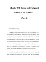
Standard batteries have been developed. The Dizziness
Handicap Inventory is a 25-item questionnaire with phys-
ical, emotional, and functional sets of questions (Jacobson
and Newman 1990) (Figure 22–1). Correlation with bal-
ance platform testing has been shown (Robertson and Ire-
land 1995). A short form has recently been developed
(Tesio et al. 1999). This 13-item version appears promising
but has not been tested as widely as the original.
A detailed medication history should be taken, includ-
ing any over-the-counter medications, vitamins, or herbal
supplements. There is a trap to be avoided when review-
ing medications of the patient with dizziness, because nu-
merous medications are known to include dizziness as a
potential side effect. One must always look carefully at the
temporal relationship between the onset of dizziness and
the initiation of any drug suspected of either causing or
exacerbating the condition (Table 22–2). Stimulants, ben-
zodiazepines, tricyclic antidepressants, tetracyclics,
monoamine oxidase inhibitors, selective serotonin reup-
take inhibitors, neuroleptics, anticonvulsants, selective
serotonin agonists, and cholinesterase inhibitors are
among the classes of drugs with multiple members re-
ported to cause dizziness. There are also many medica-
tions that patients might be taking for conditions unre-
lated to brain injury that could cause dizziness.
Certain anticonvulsants, such as phenytoin, may cause
nystagmus in the absence of any noxious symptoms. This
is not so much an adverse reaction as a potential con-
founding factor for the physical examination.
Physical Examination
Observation of the patient begins before the formal parts
of the physical examination. Grooming and attire may
reflect how well an individual performs his or her morning
routine of activities of daily living. Signs of recent minor
injuries might indicate balance or coordination problems.
Ambulatory patients may be observed walking
through a waiting area or within the examination room.
One may note greater difficulty maneuvering through a
busy environment than in a quiet area without distrac-
tions or hazards. Some patients with vestibular dysfunc-
tion after brain injury are very sensitive to visual or audi-
tory distractions. (If a patient demonstrates much more
TABLE 22–1. Common somatic complaints
associated with dysequilibrium after traumatic
brain injury
Dizziness (“shaky,” “light-headed,” many other vague
synonyms)
Vertigo (environment spins)
Imbalance (+/–falls), veering
Visual blurring and fatigue, difficulty reading (+/–headache)
Tinnitus (ringing or buzzing sensation in ears)
Difficulty distinguishing speech from background noise
Difficulty hearing
Sensitivity to noise
396 TEXTBOOK OF TRAUMATIC BRAIN INJURY
difficulty with ambulation when formally asked to dem-
onstrate walking than at other times, one may be con-
cerned about an attempt at simulating pathology.)
Visual acuity screening is appropriate, but many visual
impairments may be missed by use of an eye chart alone. A
visual field cut, for example, might spare central vision, but
loss of a peripheral visual field could create significant safety
problems. Extraocular movements and pupillary responsive-
ness should be assessed. These evaluations may yield signs of
cranial nerve injury. (Impaired eye movement may hinder
efforts at teaching compensatory strategies. A therapist seek-
ing to teach a patient how to compensate for a field cut ben-
efits from knowing how the eyes move during scanning.)
There are other components of the visual system ex-
amination that are of special interest when assessing pa-
tients with suspected vestibular disorders. Nystagmus de-
scribes involuntary rhythmic movements of the eye, with
a rapid saccadic component followed by a slow return to
the opposite direction. Spontaneous nystagmus is most
often seen in acute settings. Gaze-induced nystagmus,
noted during testing of smooth pursuit, is more common
in subacute and chronic cases. A deviation of approxi-
FIGURE 22–1. Dizziness Handicap Inventory items.
Source. Reprinted from Jacobson GP, Newman CW: “The Development of the Dizziness Handicap Inventory.” Archives of Otolaryn-
gology—Head and Neck Surgery 116:424–427, 1990. Used with permission.
(E=emotional, F=functional, P=physical)
"Yes" 4 points, "Sometimes" 2 points, "No" 0 points.
P1. Does looking up increase your problem?
E2. Because of your problem do you feel frustrated?
F3. Because of your problem do you restrict your travel for
business or recreation?
P4. Does walking down the aisle of a supermarket increase
your problem?
F5. Because of your problems do you have difficulty getting
into or out of bed?
F6. Does your problem significantly restrict your participation in
social activities such as going out to dinner, movies,
dancing, or parties?
F7. Because of your problems do you have more difficulty
reading?
P8. Does performing more ambitious activities like sports,
dancing, and household chores such as sweeping or
putting away dishes increase your problem?
E9. Because of your problem are you afraid to leave your home
without having someone accompany you?
E10. Because of your problem have you been embarrassed in
front of others?
P11. Do quick movements of your head increase your problem?
F12. Because of your problem do you avoid heights?
P13. Does turning over in bed increase your problem?
F14. Because of your problem is it difficult for you to do
strenuous housework or yard work?
E15. Because of your problem are you afraid people may think
you are intoxicated?
F16. Because of your problem is it difficult for you to go for a
walk by yourself?
P17. Does walking down a sidewalk increase your problem?
E18. Because of your problem is it difficult for you to
concentrate?
F19. Because of your problem is it difficult for you to walk
around your house in the dark?
E20. Because of your problem are you afraid to stay home
alone?
E21. Because of your problem do you feel handicapped?
E22. Has your problem placed stress on your relationships with
members of your family or friends?
E23. Because of your problem are you depressed?
F24. Does your problem interfere with your job or household
responsibilities?
P25. Does bending over increase your problem?
Balance Problems and Dizziness 397
mately 30 degrees is appropriate to test for this finding. At
the extremes of eye movement, endpoint nystagmus may
be seen in healthy individuals.
Other clinical visual tests include checking saccades
(quick movements between targets), tracking a target
while the head moves with it (vestibuloocular cancella-
tion), and fixating on a target while the head is moved
horizontally or vertically (vestibuloocular reflex; VOR).
(Detailed reviews of vision tests and related issues are pro-
vided in Chapter 23, Vision Problems.) Clinicians who do
not specialize in visual disorders may still incorporate
brief screening in their own examination to guide a deci-
sion on referral to an appropriate eye specialist. Because
many rehabilitation therapies present visual information
to patients, visual impairments may impede progress.
Brief auditory screening can similarly be done in a
bedside or office setting. Ability to hear a tuning fork vi-
brating at 512 Hz is one of the simplest parameters to test.
Functional observation of how well a patient responds to
auditory stimuli may also be useful. Audiometric testing is
safe and painless but does require some basic ability to at-
tend to a task and follow directions. Patients who are un-
likely to do so may be referred instead for auditory evoked
potentials. Auditory pathology may be present indepen-
dent of vestibular pathology. Hearing problems may inter-
fere with a patient’s ability to process verbal instructions.
There are data suggesting that impaired auditory sensory
gating may produce attention and memory impairments
(Arciniegas et al. 2000) after brain injury. One should look
closely at auditory pathways in balance and dizziness eval-
uations given the close proximity of the systems.
Olfactory screening is rarely if ever performed by
most clinicians (on the basis of personal observation after
reviewing many hospital and office charts). The Univer-
sity of Pennsylvania Smell Identification Test (Doty et al.
1984) is a commercially available (Sensoronics, Haddon
Heights, NJ) standardized test. Brain injury specialists
are well aware of the risk of injury to olfactory nerves tra-
versing the cribriform plate in frontal injuries. This can
cause hyposmia or anosmia. (A number of patients at our
center have complained of somewhat disabling hyper-
acute olfactory function. There is no obvious mechanism
by which brain injury would improve function of the
nose, but these patients are easily distracted by odors in
their environment.)
Somatosensory testing is undoubtedly critical when
evaluating any patient with balance issues. Pinprick and
light touch are most often documented in standard neu-
rological examinations. Assessments of proprioception,
kinesthesia, and vibration sense are also indicated in pa-
tients with balance issues.
Ataxia is not anticipated in patients with isolated ves-
tibular deficits in the absence of cerebellar injury. (Both
are common after TBI.) A patient with a remote history
of head trauma is still at risk of developing a cerebellar or
pontine tumor or stroke, multiple sclerosis, or other new
disorder. Development of a new finding not explained by
the known history would generate a legitimate need for
further investigation.
Musculoskeletal factors should be evaluated carefully.
Strength of postural muscles must be adequate for static
and dynamic balance tasks before more subtle deficits can
be addressed. Chronic problems such as leg-length dis-
crepancies or skeletal deformities may no longer be com-
pensated for adequately if balancing mechanisms sustain
an injury. Patients who sustained musculoskeletal injuries
in addition to brain injuries may have residual impair-
ments limiting mobility. (Vestibular symptoms may not
be noted if a patient is confined to a bed or wheelchair
during acute care.)
Direct examination of balance can be performed in
several ways. Severe deficits can be picked up on observa-
tion of poor sitting or standing balance or a markedly un-
steady gait. Patients with mild to moderate brain injuries
may look normal in this context or their deficits may only
be evident when fatigued or otherwise stressed. (Variabil-
ity that can be logically explained differs conceptually
from “inconsistency,” which raises concerns about efforts
to simulate pathology.)
Romberg testing begins with a patient standing with
feet apart and eyes open. The feet are placed directly to-
gether at the heels and toes. (Some patients need exten-
sive prompting to do so and may “cheat” by moving the
feet apart if not monitored.) If patients can maintain bal-
ance in this condition, then they are instructed to close
their eyes. Ability to maintain balance and extent of sway
are noted over at least 60 seconds if the patient is able to
maintain for that long. The degree of difficulty can be in-
creased by changing the positions of the feet. Standing
with one foot directly in front of the other provides the
sharpened Romberg position. Ability to stand on one leg
TABLE 22–2. Psychiatric and neurologic drug
classes potentially aggravating dizziness
Antidepressants (including tricyclic, monoamine oxidase
inhibitor, and selective serotonin reuptake inhibitor agents)
Benzodiazepines (occasionally used as treatment)
Anticonvulsants
Stimulants
Neuroleptics
Cholinesterase inhibitors
398 TEXTBOOK OF TRAUMATIC BRAIN INJURY
is another test of standing balance, with a somewhat
greater dependence on lower extremity motor power.
Office testing of static balance is usually performed on
a conventional floor. Sensitivity can be increased by add-
ing use of a foam mat. Lighting and background noise
may also affect aspects of performance.
Dynamic testing attempts to simulate some of the
challenges faced in the “real world,” where the body’s
center of gravity moves during functional tasks. The
Fukuda Stepping Test (Fukuda 1959) evaluates ability to
march in place with eyes open and closed. Moving for-
ward more than 50 cm or turning more than 30 degrees is
abnormal.
Functional reach from a standing position is another
readily measured dynamic assessment. It is easily mea-
sured with a measuring tape or ruler, correlates with cen-
ter of pressure testing, and has some ability to predict falls
(Duncan et al. 1992).
The Dynamic Gait Index is a low-tech quantitative
measure using a shoe box, cones, and stairs (Shumway-
Cook 1995). It consists of eight tasks related to gait. Pa-
tients can score up to 3 points on each task. Scores below
19 suggest an increased fall risk in elderly patients.
The Berg Balance Scale (Berg 1989; Thorbahn and
Newton 1996) is a 14-item test of various balancing tasks.
Up to 4 points are awarded on each task, for a maximum
total of 56. Scores below 36 correlate with very significant
fall risks in elderly patients. Although published studies
have primarily looked at predicting falls in geriatric pop-
ulations, it is reasonable to use this scale for evaluation of
patients with TBIs.
For patients with TBI, it has been suggested that tests
of balance should be combined with performance of cog-
nitive tasks (Shumway-Cook 2000). This would reflect
the reality that in normal life people do not concentrate
on how they are maintaining their equilibrium while they
move through their environment. A patient with mar-
ginal balance might be able to compensate when concen-
trating on a specific balancing task in a clinical setting.
This does not necessarily mean that he or she could re-
peat the performance while multitasking in a community
setting. One could observe performance while engaging a
patient in conversation as a simple application of this con-
cept. Therapists may take patients on community excur-
sions such as a trip to a store.
Physical examinations should also include evaluation
for medical disorders that might contribute to gait or bal-
ance disorders. Problems such as orthostatic hypotension
should be addressed appropriately.
When evaluating older patients after brain injury, one
may consider vascular pathology. Vertebrobasilar disease
may mimic vestibular dysfunction. Screening for verte-
brobasilar insufficiency carries potential pitfalls. Flow in
the vertebral or basilar artery may be compromised by
atherosclerotic disease or external masses, and when com-
bined with the effects of certain neck positions, patients
may experience dizziness or even syncope. Cervical rota-
tion and extension performed in supine position may
elicit symptoms of benign positional vertigo. Testing in a
seated position avoids this potential confounding factor
(Clendaniel 2000). Table 22–3 highlights points to cover
during a physical examination.
Laboratory Tests
The diagnostic workup after head trauma routinely
includes imaging by at least computed tomography scan-
ning, and often may include magnetic resonance imaging
(MRI). In patients with dizziness and balance problems,
one might consider the value of MRI in evaluating the
posterior fossa (Halmagyi and Cremer 2000). This helps
exclude subtle infarctions, tumors, and demyelinating dis-
orders. (One might therefore pursue such testing when
the correlation between onset of dizziness and TBI is not
clear.) Negative studies do not exclude either central or
peripheral forms of vestibular dysfunction. Patients who
cannot undergo MRI might benefit from computed
tomography scanning, with particular attention to the
posterior fossa.
Electronystagmography (ENG) is an electrodiagnos-
tic test of eye movements. It relies on differences of po-
tential between the cornea and the retina, which allow
surface electrodes to detect eye rotation. Data can be re-
corded graphically and electronically. ENG is notably less
sensitive than direct inspection by an examiner and is not
able to quantify vertical movements because of the con-
founding effects of blinking (Honrubia 2000). Despite
those limitations, ENG does allow reliable objective mea-
TABLE 22–3. Points to cover during physical
examination after brain injury
Observation
Olfactory (optional)
Eyes: acuity, tracking, saccades, nystagmus
Ears: hearing screen (otoscopic examination and/or ear, nose,
and throat referral if abnormal)
Sensation: sharp, light touch, proprioception, vibration
Motor: power, coordination
Balance: sitting, sit-to-stand transfer, standing (eyes open or
closed, feet apart or together or in tandem stance, or on one leg)
Gait: walking, tandem walking, turning
Balance Problems and Dizziness 399
surement of horizontal rotation. It can be combined with
various provocative maneuvers to record physiological
data.
One can elicit the VOR with caloric stimulation. Ca-
loric testing requires irrigating the external auditory ca-
nals with water at 7˚C higher or lower than body temper-
ature. The patient is positioned supine with the head
tilted back 60 degrees from the upright position. The re-
sulting temperature gradients in the horizontal canals
create currents within the endolymphatic fluid, triggering
deformation of hair cells. With warm water, there is a
slow deviation away from the site of irrigation followed by
nystagmus toward that side. (The response is named by
convention on the basis of the direction of the nystag-
mus.) Cold water elicits the opposite response. (Thus, the
mnemonic COWS refers to the principle of cold opposite,
warm same in this situation.)
There are limitations to this test. Anatomical varia-
tions may alter the process of heat transfer. Fixation al-
lows some individuals to suppress nystagmus to varying
degrees. Quantitative analysis can be performed with use
of ENG. One can compare the maximum slow compo-
nent velocity of nystagmus between left ear and right ear
stimulation responses or measure the ability to suppress
with fixation. There are many procedural variables to
consider (Honrubia 2000). The test does have some abil-
ity to localize lesions. Unilateral response would indicate
contralateral peripheral dysfunction. Bilateral normal re-
sponses would not rule out some central pathology.
Rotatory (Barany) chair testing can be performed in a
simple manner by rapidly rotating a chair, with the back-
rest tilted back 60 degrees. One can then observe the du-
ration of resulting nystagmus or record the severity of
subjective complaints. More sophisticated testing uses
ENG and automated programs of rotation (Honrubia
2000).
Quantitative balance testing can be performed in sev-
eral ways. Force platforms can record the perturbations of
the center of gravity in varying conditions. Removing vi-
sual input or providing visual inputs that contrast with ac-
tual conditions can pose added challenges.
One might seek information about how postural mus-
cles respond to environmental challenges. Dynamic pos-
turography can include electromyographic measurement
of lower extremity muscle responses on a moving plat-
form. Patients may rely on varying strategies to maintain
balance, including use of motions about the ankle or hip.
Muscles stabilizing the ankle respond to perturbations of
smaller amplitude or velocity. Hip muscles are recruited
in more severe challenges. The most severe perturbations
require moving the feet (Pai and Patton 1997). Patients
who lose their balance during testing before initiating
typical strategies may be given exercises to address defi-
cits in involved muscles or may be trained to recruit these
muscles sooner with biofeedback.
Attention has been paid to indicators of psychogenic
balance disorders (Goebel et al. 1997). Worse perfor-
mances on easier conditions, unusually large variability
within trials of the same test, and a regular frequency of
sway all raise concerns. Krempl and Dobie (1998) re-
ported that dynamic posturography was effective in dis-
tinguishing between malingering and best-effort perfor-
mance in healthy subjects. Table 22–4 provides a
summary of laboratory testing.
TABLE 22–4. Laboratory test summary
Test Purpose Indication
Magnetic resonance imaging/
computed tomography
Shows anatomy To localize visible lesions; may lead to surgery
ENG Records eye motion To record/localize signs of oculomotor pathology; may guide therapy or
document change on retesting
Caloric stimulation Tests VOR To provoke involuntary response, measurable with ENG (see above), not
dependent on effort
Rotatory chair Tests VOR To provoke involuntary response, measurable with ENG (see above), not
dependent on effort
Posturography: force plates Tests balance To record signs of balance pathology or potential simulation; may guide
therapy or allow documentation of change on retesting
Posturography: surface
electromyography
Tests balance To add information on motor strategies to platform tests (see above)
Note. ENG=electronystagmography; VOR=vestibuloocular reflex.
400 TEXTBOOK OF TRAUMATIC BRAIN INJURY
Peripheral Vestibular Dysfunction
Benign Positional Paroxysmal Vertigo
The most commonly attributed cause of vertigo after TBI
is benign positional paroxysmal vertigo (BPPV). It is also
the most common cause of vertigo seen in outpatient pop-
ulations in general. Vertigo and dysequilibrium are elicited
by common motions or positions. The proposed etiology
is a disturbance of semicircular canal function caused by
debris from the otolithic organs. Provocative maneuvers
can be used to elicit vertigo and nystagmus. The Hallpike-
Dix (also referenced as Dix-Hallpike) maneuver (Dix and
Hallpike 1952) involves rotating the head 45 degrees and
quickly lying down with the head hanging 30 degrees
below horizontal. Within 30 seconds, this maneuver will
elicit nystagmus if the affected side is inferior.
Single-treatment interventions for BPPV have been
developed on the basis of the underlying problem of debris
that was displaced from otolithic organs into the canals
(Epley 1992; Herdman et al. 1993). Simply put, these inter-
ventions all involve maneuvering the head to facilitate flow
of the debris out of the canals. Habituation regimens teach
patients to repeatedly position themselves several times a
day in provoking positions (Brandt and Daroff 1980).
Developers of all of these techniques have reported
high success rates. Although most reports lacked control
groups, it does appear that the rapid remission of symp-
toms can often be attributed to the intervention. (A
much-delayed response might reflect a spontaneous re-
covery.) One problem is that patients must tolerate the
transient induction of symptoms that these procedures
require. They must also comply with instructions regard-
ing positioning over a 2- to 5-day period. Use of a cervical
collar may be indicated during this period.
Patients who sustained TBIs may have cervical path-
ology. Cervicalgia in the absence of demonstrated ortho-
pedic or neurological cervical pathology would not for-
mally contraindicate these maneuvers, but patient
response might be problematic.
Perilymphatic Fistula
Trauma to the round or oval windows may lead to a peri-
lymphatic fistula, with communication between the mid-
dle and inner ears. A popping sensation may be noted at
the time of onset. Symptoms include vertigo, tinnitus,
and hearing loss. Valsalva maneuvers may exacerbate the
symptoms.
Diagnosing this condition may be difficult because
usually no single test is definitive. Application of pressure
over the tympanic membrane may induce vertigo (Hen-
nebert’s sign) or nystagmus. Concurrent use of computer-
ized balance platform testing allows quantitative mea-
surement of increased sway during this maneuver. (This
form of posturography uses force plates under the feet to
detect displacement of the center of gravity.) Audiometric
testing may show significant hearing loss, especially at
higher frequencies. ENG may show dysfunction in the af-
fected ear.
Bed rest with the head elevated may be of some help.
Avoidance of constipation or other causes of straining is
advisable. Persistent symptoms may be managed surgi-
cally, with exploration and repair of defects of the win-
dows. Differing opinions about the success rate of sur-
gical interventions have been offered (Fetter 2000;
Fitzgerald 1995). It is reasonable to suppose that a num-
ber of patients with chronic dizziness have undiagnosed
perilymphatic fistulas, but identifying this subset of pa-
tients can be difficult.
Ménière’s Disease
Classically, Ménière’s disease is regarded as an idiopathic
disorder that typically begins in middle age. It begins with
potentially severe bouts of vertigo accompanied by a
sense of fullness in the affected ear, episodic hearing
reduction, and tinnitus. The hearing loss does not always
remit after each episode.
A syndrome such as Ménière’s can be seen after head
trauma (Healy 1982). Bleeding into the membranous lab-
yrinth or altered bony anatomy after temporal fracture
are two possible mechanisms.
The disorder is associated with endolymphatic hy-
drops (excessive accumulation of fluid). This is usually at-
tributed to malabsorption of endolymph. Restriction of
sodium, caffeine, nicotine, and alcohol intake has been
recommended traditionally, whereas diuretics and fluid
restrictions are also sometimes added. There is a lack of
strong data to support these interventions. The relapsing
and remitting nature of the disorder would make further
investigation difficult.
The effectiveness of endolymphatic sac surgery is
controversial, but such procedures are not expected to
harm any existing function of the vestibular and auditory
systems. Labyrinthectomy and vestibular nerve resections
are both effective at stopping vertigo (Mattox 2000), but
the latter is preferred if preserving hearing is a goal.
Central Vestibular Dysfunction
Although the reflex circuits from the vestibular sensory
organs to oculomotor, cervical, and postural muscles are
Balance Problems and Dizziness 401
the best-identified pathways, it is clear that data must
also flow to other areas within the central nervous sys-
tem. By convention, pathology involving this network is
referred to as central vestibular dysfunction even if the sen-
sory end organs are intact. The central vestibular system
may be defined as the vestibular nuclei and their connec-
tions to other parts of the brain and spinal cord. A subset
of brain-injured patients presents with complaints of
dizziness and imbalance related to central dysfunction.
It is to some extent a diagnosis of exclusion because
imaging of the vestibular apparatus or testing of the
reflex arcs (e.g., caloric stimulation) can help to uncover
peripheral lesions. Patients who fit a profile of vestibular
dysfunction after brain injury but who do not have evi-
dence of a peripheral lesion or other etiologies are
included in the central category.
An important role for the cerebellum in the vestibular
system has been accepted. The cerebellar flocculus, in
particular, seems to play a critical role in VOR adapta-
tions. There is reason to believe that some forms of learn-
ing and adaptation take place in areas of the cerebellum
and the brainstem (du Lac et al. 1995). Trauma affecting
the cerebellum may therefore affect subjective sensations
of dizziness or objective signs of balance problems even if
gross ataxia is not present.
Brandt and Dieterich (1994, 1995) have made exten-
sive reviews of central vestibular syndromes. Sites from
the brainstem to the thalamus to sensory cortex have been
implicated (including an area of the parietoinsular cortex
in monkeys). Reviews of cases of individuals with well-
circumscribed lesions are, of course, critical to the current
understanding of brain pathology. Functional MRI stud-
ies are adding new dimensions to that knowledge. Opto-
kinetic stimulation has been noted to activate vestibular
cortex on functional MRI (Dietrich et al. 1998).
Pharmacological Management
Medications for dizziness and vertigo may be referred to
as vestibular sedatives (Table 22–5). They tend to have gen-
erally sedating properties. Their exact mode of action for
dizziness reduction is not known. Meclizine, which has
antihistaminic and anticholinergic properties, is a com-
mon choice. Promethazine and prochlorperazine also
have properties of phenothiazines. Transdermal scopol-
amine is another anticholinergic option.
There are general precautions about use of vestib-
ular sedatives in patients with asthma, glaucoma, or
prostatic hypertrophy. More specifically, there is little
basis for prolonged use of these medications for chronic
dizziness (Zee 1985). They may be quite helpful for
acute motion sickness or other acute disorders but have
not been shown effective in chronic deficits after brain
injury. Vestibular sedatives might actually slow the pro-
cess of adaptation after injury. Sedating effects may neg-
atively affect arousal. The potential for drug interactions
in patients taking other medications should also be con-
sidered. Polypharmacy also poses additional problems
for cognitively impaired patients who have difficulty
keeping track of medications.
Benzodiazepines and other sedating drugs are some-
times prescribed for patients with dizziness. These may
address associated anxiety but are not known to be of di-
rect benefit. Prolonged use in patients with brain injury
should be approached with great caution.
TABLE 22–5. Medications for dizziness and vertigo
Medication
Dosage
(typical ranges) Precautions (common) Reactions (partial list)
Meclizine (Antivert) 12.5–25.0 mg, bid–tid Bladder obstruction, asthma,
glaucoma
Sedation, confusion, dry mouth (common),
ototoxicity, tachycardia hallucinations
(serious)
Prochlorperazine
(Compazine)
5–10 mg, tid–qid Bladder obstruction, asthma,
glaucoma, bone marrow
depression, epilepsy, many others
Sedation, confusion, dry mouth (common),
hematologic, hepatic, neuroleptic
malignant syndrome (serious)
Promethazine
(Phenergan)
12.5–25.0 mg, qid Bladder obstruction, asthma,
glaucoma, epilepsy, liver
dysfunction
Sedation, confusion, dry mouth,
tachycardia (common) hematologic,
respiratory depression, bradycardia
(serious)
Scopolamine
(Transderm Scop)
1.5-mg patch, apply
4 hours before travel,
lasts 72 hours
Bladder or intestinal obstruction,
asthma, glaucoma, epilepsy, liver
or kidney dysfunction
Sedation, confusion, dry mouth,
respiratory depression, bronchospasm
402 TEXTBOOK OF TRAUMATIC BRAIN INJURY
Vestibular Rehabilitation
Techniques of therapy have been developed for patients
with various vestibular disorders. These have been used in
brain-injury populations, although it is widely under-
stood that patients with multiple areas of dysfunction face
special challenges.
Vertiginous symptoms are addressed with habituation
exercises (Brandt and Daroff 1980). Repetition of move-
ments that provoke vertigo eventually reduces symptoms.
Behavioral or cognitive problems are known to increase
the difficulty in applying this approach to brain-injured
patients (Shumway-Cook 2000).
Gaze stabilization exercises are used to improve the
efficiency of vestibuloocular coordination. These exer-
cises are initially performed with the head still and later
are performed during movement.
Balance retraining may stress challenging vestibular
function by minimizing availability of other sensory in-
puts. For patients who cannot progress with this ap-
proach, efforts at optimizing their use of visual or propri-
oceptive strategies for balance may be proposed.
To whatever extent normal function cannot be re-
stored, adaptive techniques can be taught. Patients may
need to modify how they perform routines for dressing
and grooming. A shower bench may be needed if they
cannot balance safely with eyes shut. These interventions
may require collaboration between physical and occupa-
tional therapists. If patients or family members resist such
recommendations, then psychologists or social workers
on the rehabilitation team will need to understand the un-
derlying rationale to intervene effectively.
Our center uses a separate team of physical therapists
for vestibular therapy. Given the known emotional chal-
lenges of vestibular disorders, a pathway has been estab-
lished to facilitate referral of patients without brain injury
to psychologists with expertise in treating this population.
For patients with brain injury, particularly mild TBI, we
have found that an interdisciplinary team can provide a
closer level of coordination and communication. Occupa-
tional therapists, speech pathologists, and neuropsycholo-
gists may need to modify their approaches to accommodate
patients with limited tolerance of visual or auditory stimuli.
Social workers and vocational counselors should under-
stand these issues as they advise families or employers.
It is important for clinicians and patients to under-
stand that aspects of a vestibular therapy program may
make the patient feel worse acutely. The potential for fa-
cilitating habituation should be explained. As patients
practice fixing gaze on a target while turning the head as
quickly as possible or walking through a hallway while
turning to look at targets on the walls, dizziness may be
elicited. With further practice, however, the central ves-
tibular system may adapt and no longer perceive discom-
fort. As patients practice maintaining balance on soft
foam pads or moving platforms, their bodies may become
more efficient at maintaining their center of gravity in a
stable position.
Extra emotional support might be needed in the early
stages of a program. As time passes, reviewing measurable
clinical progress is a reasonable strategy to counteract dis-
couragement over any persistent symptoms. One can re-
view clinical measures such as the rate at which patients
can turn their heads from side to side while keeping their
eyes fixed on a target. The length of time that balance is
maintained during Romberg testing is another easily
measured parameter. Functional performance in daily life
can also be reviewed, such as the length of time spent out
of bed or distance ambulated daily.
Once progress is made, the reinforcement of compli-
ance with home exercises may be necessary. If a plateau is
reached after a prolonged course of therapy, counseling
should focus on the need to move on with life rather than
hope for a dramatic improvement with more of the same
treatment.
Emotional Factors
Dizziness and nausea are noxious stimuli. Impaired bal-
ance carries a risk of injury that is readily understood by
most patients. These problems can therefore have an
adverse emotional effect on patients. There is also con-
cern that expressions of vestibular symptoms might
reflect a primary psychiatric disorder or pursuit of secon-
dary gain.
Patients with dizziness have a significant risk of psy-
chiatric dysfunction. Rates as high as 50% have been cited
for either panic disorder or depression in patients with
vestibular hypofunction (Eagger et al. 1992). (The subset
of dizzy patients who present after head trauma was not
studied separately.) Anxiety and dizziness overlap more
than would be predicted by chance and carry a worse
prognosis for resolution of dizziness and greater degree of
reported handicap, but this does not mean that vestibular
symptoms should be readily dismissed as not having a
physiological basis. Jacob and Furman (2001) proposed a
linkage via overlapping circuits, including the parabra-
chial nucleus network. A better understanding of the neu-
rophysiology underlying anxiety and dizziness may re-
duce the temptation to dismiss “psychogenic dizziness” as
strictly an emotional disorder.
405
23
Vision Problems
Neera Kapoor, O.D., M.S.
Kenneth J. Ciuffreda, O.D., Ph.D.
VISION IS ONE of the primary sensory modalities in-
volved in tasks such as stance, gait, reading, and other ba-
sic activities of daily living (ADLs). Furthermore, ade-
quate vision is a requisite for evaluation and treatment
performed during most types of rehabilitation, such as
optometric, ophthalmological, neuropsychological, phys-
ical, vestibular, occupational, and speech and language
therapies. Nonetheless, diagnosis and management of
functional vision deficits have been frequently overlooked
in textbooks and teaching curricula used by many rehabil-
itation professionals (Wainapel 1995). The recent in-
creasing interest in functional vision and its integrative ef-
fect on rehabilitation in patients with traumatic brain
injury (TBI) (Altner et al. 1980; Fisher 1987; Tinette et al.
1995; Wainapel et al. 1989) serves as the impetus for this
chapter.
In this chapter, we discuss the prevalence and patho-
physiology of vision problems and provide an overview of
functional vision anomalies in patients with TBI. A glos-
sary of ophthalmic terms used in the following text is
found in the appendix at the end of the chapter.
Prevalence of Vision Problems in TBI
Vision problems have been reported in TBI patients with
varying prevalence, depending on the source used and
diagnostic criteria adopted (Al-Qurainy 1995; Baker and
Epstein 1991; Gianutsos et al. 1988; Hellerstein et al.
1995; Lepore 1995; Sabates et al. 1991; Schlageter et al.
1993; Suchoff and Gianutsos 2000; Suchoff et al. 1999,
2000; Suter 1995; Zost 1995) (Table 23–1). The most
common problems adversely affecting visual function
directly are versional and vergence oculomotor anoma-
lies, accommodative dysfunctions, dry eye, cataracts, and
visual field defects. Other vision problems affecting func-
tion more indirectly include orbital fractures, lid anoma-
lies, blepharitis, blepharoconjunctivitis, pupillary anoma-
lies, optic nerve anomalies, and retinal defects (Suchoff et
al. 1999).
Pathophysiology
The pathophysiology for all vision deficits in TBI has not
been reported in the literature in detail, but it is more evi-
dent for some deficits than for others. Oculomotor defi-
cits (Table 23–2) resulting in diplopia, loss of place while
reading, nystagmus, and oscillopsia may occur because of
sheared or severed cranial nerves (CNs) (i.e., CN III, CN
IV, CN VI), mechanical restriction of an extraocular mus-
cle, or damage at the level of the neuromuscular junction
(Baker and Epstein 1991). Accommodative deficits result-
ing in blurred vision may occur as a result of damage to
the oculomotor nerve (i.e., CN III), more central neuro-
logical anomalies, or a side effect of medications (Ciuf-
freda 1991; Cooper 1998; Suchoff et al. 2000).
With respect to ocular pathology, dry eye resulting in
intermittent blurred vision and a gritty sensation is quite
common in the TBI population. It is typically an ocular
side effect of antidepressants, antihypertensives, and oral
contraceptives (Bartlett and Jaanus 1995; Jaanus and Bart-
lett 1984). Blepharitis and blepharoconjunctivitis are also
frequently found and typically occur because of poor lid
hygiene (Catania 1988). Pupillary anomalies may result
from damage along the pupillary pathway in association
with a CN III palsy, asymmetrical optic nerve disease or
anomaly, the presence of a space-occupying lesion, or dis-
rupted autonomic innervation. Visual field defects such as
noncongruous hemianopias and quadrantanopias may oc-
406 TEXTBOOK OF TRAUMATIC BRAIN INJURY
cur with TBI depending on the nature and severity of the
injury, but they are more typically associated with stroke.
Clinical experience has demonstrated that TBI patients
present with scattered visual field defects and no evidence
of hemifield lateralization, as described in the section Vi-
sual Field Deficits. The etiology of this scattered visual
field defect remains poorly understood.
There are other ocular sequelae that may occur with
blunt trauma to the periorbital region but are not common
in TBI. These sequelae are orbital fracture, lid anomaly,
corneal abrasion, lens dislocation, angle recession, trau-
matic glaucoma, traumatic cataract, traumatic uveitis, and
retinal or vitreal detachment (Vogel 1992). The patho-
physiology of these conditions is not addressed further be-
cause it is beyond the scope and aim of this chapter.
However, in the TBI population, there is an increased
frequency of some of the above conditions when compared
with the non-brain-injured population (Suchoff et al. 1999;
Vogel 1992), which may result in reduced visual acuity, re-
duced contrast sensitivity, and/or visual field defects. Or-
bital fractures and lid anomalies secondary to blunt and se-
vere head trauma require immediate medical intervention
because of the concern of additional inflammation or infec-
tion (e.g., orbital cellulitis). Inflammation, infection, shear-
ing, or compression may occur at any point along the optic
radiations in the primary visual pathway between the oc-
cipital cortex and retina as a result of trauma. Retinal de-
fects and tears occur often with severe blunt trauma. Reti-
nal vascular insufficiencies, which are often associated with
hypertension and diabetes, are also possible sequelae. Such
TABLE 23–1. Percentage of visual and ocular conditions in acquired brain-injured (ABI) sample with
comparative values for a random adult population
Ocular/visual condition
Occurrence in
an ABI
sample (%)
Occurrence in a
random adult
population (%)
Occurrence in an
ABI/random adult
occurrence
Exophoric deviations 41.9 2.1 19.9
Esophoric deviations 1.6 1.3 1.3
Vertical deviations 9.7 1.6 6.1
Oculomotor dysfunctions 39.7 NA NA
Accommodative dysfunctions 9.6 NA NA
External eye pathologies: dry eye/blepharitis/keratitis/
pterygium/corneal degeneration
22.6 11.2 2.0
Lid defects: ptosis/dermatochalasis/blepharochalasis 4.8 2.1 2.3
Aphakia/pseudophakia/cataracts 24.1 12.3 2.0
Optic nerve cupping/optic atrophy/glaucoma suspect/
glaucoma
19.4 8 2.4
Color vision defect 0 8.3 (male);
0.5 (female)
0
Contrast sensitivity defect 0 NA NA
Posterior pole anomalies: retinopathies (including diabetic
retinopathy, hypertensive retinopathy, and maculopathy)
9.7 1.5 6.5
Retinal defects/detachments 1.6 0.1 20.0
Peripheral retinal degenerations/vitreoretinal
degenerations
9.7 2.6 3.7
Blindness/enucleation 6.5 1.6 4.1
Pupillary anomaly 1.6 1 1.6
Visual field defects 32.5 NA NA
Note. NA=normative data for a random adult population not available.
Source. Adapted from Suchoff IB, Kapoor N, Waxman R, et al: “The Occurrence of Ocular and Visual Dysfunctions in an Acquired Brain-Injured
Patient Sample.” Journal of the American Optometric Association 70:301–309, 1999. Used with permission.