Atlas of the Diabetic Foot - part 8 ppt
Bạn đang xem bản rút gọn của tài liệu. Xem và tải ngay bản đầy đủ của tài liệu tại đây (860.47 KB, 22 trang )
160 Atlas of the Diabetic Foot
and subungual debris develop. In proximal
subungual fungal infection, the second
commonest form, Trichophyton rubrum
accumulates hyperkeratotic debris under
the nail plate and loosens the nail,
eventually separating it from its bed.
This fungus infects the underlying matrix
and nail plate leaving the nail surface
intact. Leuconychia mycotica, caused by
Trichophyton metagrophytes, infects the
nail superficially. The nail surface becomes
dry, soft and friable but the nail remains
attached to its bed. In addition to these
fungi Epidermophyton floccosum may also
be isolated from infected areas.
Itraconazole and fluconazole are also
effective in the treatment of chronic ony-
chomycosis.
Keywords: Onychomycosis; distal subun-
gual onychomycosis; proximal subungual
fungal infection; leuconychia mycotica; Tri-
chophyton metagrophytes, Trichophyton
rubrum or Epidermophyton floccosum;ter-
binafine; itraconazole; fl uconazole
FUNGAL INFECTION WITH
MULTIMICROBIAL
COLONIZATION
Superficial ulcers of 10 days’ duration on
the facing sides of the left first and sec-
ond toe of a 70-year-old type 2 dia-
betic lady with diabetic neuropathy, before
debridement are shown in Figures 8.8 and
8.9. Note soaking of the skin. An X-
ray excluded osteomyelitis. Staphylococcus
coagulase-negative, Pseudomonas aerugi-
nosa and enterobacteriaceae were recov-
ered after swab cultures in addition to Can-
dida albicans. She was treated successfully
with itraconazole for 5 weeks. The patient
used a clear gauze in order to keep her
toes apart, together with local hygiene pro-
cedures twice daily. Weekly debridement
was carried out and no antimicrobial agent
was needed.
Keywords: Fungal infection
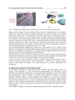
Figure 8.8 Neuro-ischemic ulcers facing each other on the first and second toe with fungal
infection and soaked skin in addition to claw toes. Foot shown from the plantar aspect
Infections 161
Figure 8.9 Neuro-ischemic ulcers facing each other on first and second toe, with fungal infection.
Front aspect of Figure 8.8
DEEP TISSUE INFECTION
AFTER
INTERPHALANGEAL
MYCOSIS
A 60-year-old female patient with type 2
diabetes diagnosed at the age of 47 years
and treated with sulfonylurea and met-
formin and with poor glycemic control, was
referred to the diabetic foot outpatient clinic
because of a severe foot infection.
The patient had known mycosis between
the fourth and the fifth toes of her right
foot. Three days before her visit she noticed
redness and mild pain on the dorsum of
her toes. Her family doctor gave her cefa-
clor, but she became febrile and her foot
became swollen, red and painful. No trauma
was reported.
On examination, her foot was red, warm
and edematous with pustules on its dorsum
(Figure 8.10). The peripheral arteries were
normal on palpation and peripheral neu-
ropathy was present. Pathogen entry was
probably via the area of the mycosis.
The patient was admitted to the hospital
and treated with intravenous ciprofloxacin
and clindamycin. No osteomyelitis was
found on repeated radiographs. Extensive
surgical debridement was carried out. Deep
tissue cultures revealed Staphylococcus au-
reus, Escherichia coli and anaerobes. The
patient was discharged in fair condition
after a stay of 1 month.
Keywords: Mycosis; deep tissue infection
DEEP TISSUE INFECTION
A 50-year-old type 1 male diabetic patient
with known diabetes since the age of
25 years was referred to the outpatient dia-
betic foot clinic for a large infected neuro-
ischemic ulcer.
The patient suffered from retinopa-
thy — treated with laser — established dia-
betic nephropathy, hypertension — treated
with enalapril and furosemide — and severe
neuropathy.
162 Atlas of the Diabetic Foot
Figure 8.10 Deep tissue infection of the foot following web space mycosis. Redness and edema
of the whole foot with pustules on t he dorsum can be seen along with claw toes
Six months before visiting a surgeon, the
patient had noticed a painless superficial
ulcer caused by a new pair of shoes.
Hoping it would subside quickly, he did not
seek a doctor’s advice and continued his
daily activities although the ulcer became
larger with surrounding erythema and
eventually became purulent and odorous.
Fever developed. A deep tissue c ulture
revealed Staphylococcus aureus, Klebsiella
spp. and anaerobes. Surgical debridement
was carried out, and amoxicillin–clavulanic
acid treatment was initiated. After 1 month
of stabilization, with dressings being
changed daily, the patient noticed increased
purulent discharge and an intense foul odor.
On examination at the diabetic foot
clinic, the patient was febrile and weak.
He had complete loss of sensation. Periph-
eral pulses were palpable. Gross ankle and
forefoot edema was noted and the short
extensor of the toes and anterior tibial ten-
dons was exposed (Figure 8.11). The com-
mon tendon sheath and subcutaneous tis-
sue were necrosed. An acrid odor emanated
from the foot even before the bandages
were removed. A seropurulent discharge
was being emitted from deeper structures.
The patient was referred back to his sur-
geon; admission to the hospital and intra-
venous antibiotics together with extensive
debridement followed, and due to abiding
Infections 163
Figure 8.11 Deep tissue infection of the foot with gross ankle and forefoot edema. The short
extensor of the toes and the anterior tibial tendons are exposed, while the common tendon sheath
and subcutaneous tissue are necrosed
septic fever and the critical condition of
the patient, a below-knee amputation was
undertaken 2 days later.
Keywords: Deep tissue infection; amputa-
tion
DEEP TISSUE INFECTION
OF A CHARCOT FOOT
WITH A NEUROPATHIC
ULCER
A 65-year-old female patient with type 2
diabetes mellitus since the age of 40 years
attended the diabetic foot clinic because of
a large ulcer of the sole of her left foot.
She was being treated with insulin result-
ing in acceptable diabetes control (HbA
1c
:
7.28%). She had a history of hypothy-
roidism as well as a history of ulcers under
her right foot at the age of 63 years, which
had healed completely.
The present ulcer had developed after
a minor trauma to the sole of her foot
while walking barefoot during the summer.
It evolved within a month together with a
fast progressing gross deformity of the foot.
The patient complained of mild discomfort
but no pain, so she kept on using both
feet without any means of reducing the
pressure on her ulcerated foot. S he was
treated with amoxicillin–clavulanic acid
and clindamycin for 20 days.
On examination, her left foot was swol-
len, with midfoot collapse; it was warm
(2.5
◦
C temperature difference to the con-
tralateral foot), and crepitus was heard on
passive movement. A large neuropathic
164 Atlas of the Diabetic Foot
Figure 8.12 Neuro-osteoarthropathy. A large
neuropathic non-infected ulcer surrounded by
callus occupies the midsole
non-infected ulcer of size 8 × 7 × 0.4cm
occupied the midsole surrounded by cal-
lus (Figure 8.12). A small, full-thickness
neuropathic ulcer was present within an
area of callus formation over the right
first metatarsal head (Figure 8.13). The skin
on both her feet was dry and the periph-
eral pulses were palpable. The vibration
perception threshold was 20 V in both
feet. Monofilament sensation was absent,
as were sensations of light touch, pain and
temperature perception.
Debridement was carried out; an X-ray
showed disruption of the tarsometatarsal
joint (Lisfranc’s joint), bone absorption of
the first and second cuneiforms and dislo-
cation of the cuboid bone (Figure 8.14). A
diagnosis of acute neuro-osteoarthropathy
Figure 8.13 Right foot of the patient whose
left foot is shown in Figure 8.12. A small,
full-thickness neuropathic ulcer within an area
of callus is present over the right first metatarsal
head
was made and a single dose of 90 mg of
pamidronate was administered. The pres-
ence of ulcers prevented the use of a
total-contact cast since daily changes of
dressings were needed. The patient was
instructed to refrain from walking and
to visit the diabetic foot clinic on a
weekly basis. After 1 month the mid-
sole ulcer was smaller compared to its
initial size (Figure 8.15) and showed no
signs of infection. The ulcer under her
right sole healed. There was no differ-
ence in the temperature between the two
feet.
After an absence of 3 weeks the patient
visited the clinic with acute foot infec-
tion and fever. The midsole ulcer was
Infections 165
Figure 8.14 Plain radiographs showing neuro-osteoarthropathy in the left foot of the patient whose
feet are illustrated in Figures 8.12 and 8.13. Disruption of the tarsometatarsal joint (Lisfranc’s joint),
resorption of the first and second cuneiforms and midfoot collapse can be seen
Figure 8.15 Left neuro-osteoarthropathic
foot of the patient whose feet are shown in
Figures 8.12–8.14. Progress of the plantar
neuropathic ulcer after 1 month of chiropody
treatment. Healthy granulated tissue covers
the bed of the ulcer
166 Atlas of the Diabetic Foot
Figure 8.16 Left neuro-osteoarthropathic foot of the patient whose feet are shown in
Figures 8.12–8.15 , 3 weeks after the photograph shown in Figure 8 .15 was taken. Signs of infection
(cellulitis, blisters and edema) are present
much smaller (Figure 8.16), surrounded by
cellulitis, and a new infected ulcer was
present on the lateral aspect of the hind-
foot (Figure 8.17). The patient insisted that
she had complied with the instructions,
except for the last week, when she felt
confident that the ulcer had healed. She
was admitted to the hospital and under-
went extensive surgical debridement. Intra-
venous antibiotics (ciprofloxacin, penicillin
and clindamycin) were administered but the
high fever persisted despite treatment; the
infection spread to the lower tibia and the
patient became septic. On the 10th day of
hospitalization, the critical condition of the
patient necessitated a below-knee amputa-
tion. She was discharged in good clinical
condition after 1 week.
Keywords: Deep tissue infection; acute
neuro-osteoarthropathy; neuropathic ulcer;
below-knee amputation
Infections 167
Figure 8.17 Lateral aspect of the foot shown in Figure 8.16. Infection has spread to the whole
foot and the lower tibia. The superficial ulcer on the lateral aspect of the hindfoot may have been
caused by rupture of a blister
OSTEOMYELITIS
A 69-year-old female patient with type 2
diabetes diagnosed at the age of 54 years
and treated with sulfonylurea, was referred
to the outpatient diabetic foot clinic for
an infection of her right second toe. She
had background diabetic retinopathy a nd
hypertension. She complained of numbness
and a sensation of pins and needles in her
feet at night.
On examination, she had findings of
severe neuropathy (no feeling of light
touch, pain, temperature, vibration or a 5.08
monofilament; Achilles tendon reflexes
were absent; the vibration perception thres-
hold was >50 V in both feet). Peripheral
pulses were weak and the ankle brachial
index was 0.7. Dry skin and nail dystro-
phies were present. A superficial ulcer with
a sloughy base was seen on the dorsum of
her right second toe which was red, swollen
and painful, having a sausage-like appear-
ance (Figure 8.18). She did not mention any
trauma, but inspection of her shoes revealed
a prominent seam inside the toe box of her
right shoe.
The sausage-like appearance of a toe
usually denotes osteomyelitis. Bone infec-
tion was confirmed on X-ray, showing
osteolysis of the first and second pha-
langes. Staphylococcus aureus and Kleb-
siella pneumoniae were cultured from the
base of the ulcer. The patient was treated
with cotrimoxazole and clindamycin for
2 months. She was also referred to the
Vascular Surgery Department for a per-
cutaneous transluminal angioplasty of her
right popliteal artery. After 2 months the
ulcer was still active and the patient had
local extension of osteomyelitis despite
the restoration of the circulation in the
periphery. She eventually had her second
168 Atlas of the Diabetic Foot
Figure 8.18 Sausage-like toe deformity usu-
ally denotes underlying osteomyelitis
ray amputated. A bone culture revealed
the presence of Staphylococcus aureus.
She continued with cotrimoxazole for two
more weeks.
Keywords: Osteomyelitis; painful–pain-
less feet
OSTEOMYELITIS OF THE
HALLUX
A 30-year-old male patient with type 1 dia-
betes diagnosed at the age of 11 years was
admitted because of infected foot ulcers on
his right hallux. He had a mild fever and
a history of proliferative diabetic retinopa-
thy and microalbuminuria. Diabetes con-
trol was poor (HBA
1c
: 9.5%). He reported
a trauma to his left foot 2 months earlier
when an object fell on his feet while work-
ing. A superficial ulcer had developed on
the dorsal aspect of his right great toe;
the ulcer had become infected because the
patient felt no pain and therefore did not
seek medical advice.
On examination, pedal pulses were nor-
mal. Severe peripheral neuropathy was
found and the vibration perception thresh-
old was 30 V in both feet. An infected right
hallux with purulent discharge, necrotic tis-
sue at the tip, and cellulitis were observed
(Figure 8.19). A plain radiograph showed
osteomyelitis involving both distal pha-
langes (Figure 8.20).
A culture of the pus revealed Pseudo-
mans maltophila, Enterobacter cloacae and
Figure 8.19 Infection of the hallux
with purulent discharge, necrotic tissue
at the tip and cellulitis
Infections 169
Figure 8.20 Osteolysis of the distal
phalanx and condyle of the proximal
phalanx due to osteomyelitis of the
hallux. Plain radiograph of the foot
shown in Figure 8.19
anaerobes, and the patient was treated with
ciprofloxacin and ampicillin–sulbactam for
2 weeks, based on the antibiogram. An
amputation of the right great toe was under-
taken due to persistent osteomyelitis.
Keywords: Hallux; osteomyelitis; amputa-
tion
PHLEGMON
A 62-year-old male diabetic patient with
type 2 diabetes diagnosed at the age of
42 years and treated with sulfonylurea,
biguanide and acarbose and whose diabetes
control was acceptable, visited the outpa-
tient diabetic foot clinic due to infection of
the sole of his right foot. He had hyperten-
sion and coronary heart disease treated with
metoprolol and aspirin. He had no previous
history of foot problems.
On examination, the patient had fever,
severe diabetic neuropathy, and bound-
ing pedal pulses. He had hallux valgus,
claw toes, prominent metatarsal heads, ony-
chodystrophy and dry skin. Callus forma-
tion superimposed on a neuropathic ulcer
over his third metatarsal head was present;
a callus was also noted over his fifth
metatarsal head. A superficial, painless,
170 Atlas of the Diabetic Foot
infected ulcer with purulent discharge was
present under Lisfranc’s joint (Figure 8.21).
This infection progressed to a phlegmon
2 days after a minor shear trauma.
The patient was admitted, and intra-
venous amoxicillin–clavulanate was initi-
ated. A plain radiograph excluded osteo-
myelitis or gas collection within the soft
tissues. Computerized tomography revealed
a phlegmonous subcutaneous mass under
the base of the metatarsals (Figure 8.22).
A sterile probe was used to detect any
sinuses or abscesses, but none was found.
The patient remained bedridden for 1 week
and the infection subsided. He continued
antibiotics for one more week with lim-
ited mobilization and he was released from
hospital in excellent condition. Oral antibi-
otics were continued for two more weeks.
Preventive footwear was prescribed and the
Figure 8.21 Superficial infected ulcer
with purulent discharge under Lisfranc’s
joint. Callus formation is superimposed
on neuropathic ulcer over the third meta-
tarsal head with callus formation over the
fifth metatarsal head. Hallux valgus, claw
toes, prominent metatarsal heads, ony-
chodystrophy and dry skin can be seen
Infections 171
Figure 8.22 Computerized tomography of the feet of the patient whose right foot is shown i n
Figure 8.21. A phlegmonous subcutaneous mass is present under the base of the metatarsals (arrow)
patient continued to visit the outpatient dia-
betic foot clinic on a regular basis.
Computerized tomography is useful in
identifying areas of phlegmon within the
soft tissues. It may provide information
about the exact anatomic location and
extent, so that aspiration or surgical drain-
age can be undertaken. Magnetic resonance
imaging and ultrasound studies are also
helpful in this respect.
Keywords: Neuropathic ulcer; computer-
ized tomography; phlegmon
INFECTED PLANTAR
ULCER WITH
OSTEOMYELITIS
A 50-year-old female diabetic patient with
type 2 diabetes diagnosed at the age of
44 years and treated with sulfonylurea, was
referred to the outpatient diabetic foot
clinic because of a chronic infected ulcer
on her left foot. The patient lived alone
and she was being treated for depression;
she had good diabetes control. A minor
trauma under her left foot was reported to
have occurred 2 years previously. She had
treated the injury with different types of
gauzes and creams, but it failed to heal.
She presented to the clinic with a large,
painless, infected ulcer under her left foot
(Figure 8.23).
On examination, an irregular, soaked,
foul-smelling ulcer with sloughy bed, and
surrounding cellulitis of 3 cm in diameter
was found; body temperature was normal.
Diabetic neuropathy was diagnosed, while
peripheral pulses were normal. Signs
of osteomyelitis (osteolysis of the first
metatarsal head, and the base of proximal
phalanx of the hallux, with periosteal
reaction) were noted on the radiograph
(Figure 8.24). A post-debridement swab
culture from the base of the ulcer revealed
methicillin-resistant Staphylococcus aureus
and Escherichia coli. The patient was
admitted to the hospital. The white blood
cell count was 14,700/mm
3
,anemia(Hb:
9.8 g/dl) characteristic of chronic disease
was found, the erythrocyte sedimenta-
tion rate was 90 mm/h and the level
of C-reactive protein was 70 mg/dl. She
was treated with 600 mg teicoplanin
172 Atlas of the Diabetic Foot
Figure 8.23 A large, irregular, soaked and
infected neuropathic ulcer with sloughy bed
and surrounding cellulitis of 3 cm in diameter
is shown here. A minor trauma reported to
have occurred 2 years earlier was the cause of
this ulcer
intravenously once daily and the ulcer
was debrided and dressed. The cellulitis
progressively subsided, the ulcer became
clear and healthy granulating tissue began
to cover the ulcerated area (Figure 8.25).
The patient was discharged from the
hospital in good clinical condition. She
continued treatment with intramuscular
teicoplanin for three more months and
attended the outpatient diabetic foot clinic
on a weekly basis. Complete offloading
of pressure from the ulcerated area was
achieved by the use of a wheelchair
for most of her activities. Platelet-
derived growth factor-β (becaplermin) was
Figure 8.24 Osteolysis of the first metatarsal
head and the base of proximal phalanx of
the hallux with periosteal reaction due to
osteomyelitis are shown on this plain radiograph
of the foot illustrated in Figure 8.23
applied once daily. The ulcer diminished
progressively (Figure 8.26) and healed in
4 months; no relapse occurred.
All patients with deep or long-standing
ulcers should be evaluated for osteomyeli-
tis. The possibility of an ulcer being com-
plicated by osteomyelitis increases when
the diameter of the ulcer exceeds 2 cm
and the depth is greater than 3 mm; the
possibility of complications becomes even
higher when the white blood cell count, the
erythrocyte sedimentation rate and the C-
reactive protein levels are high.
Treatment of acute osteomyelitis in-
cludes parenteral administration of antibi-
otics for 2 weeks initially, and the continua-
tion of oral treatment for a prolonged period
(at least 6 weeks).
Infections 173
Figure 8.25 Clear ulcer with healthy granulat-
ing tissue after 1 month of appropriate treat-
ment in the patient whose foot is shown in
Figures 8.23 and 8.24
Keywords: Neuropathic ulcer; acute osteo-
myelitis; platelet-derived growth factor-β
(PDGF-β, becaplermin)
NEUROPATHIC ULCER
WITH OSTEOMYELITIS
A 57-year-old obese male patient with type
2 diabetes diagnosed at the age of 40 years
was referred to the outpatient diabetic foot
clinic because of a chronic ulcer under
his right foot. He was being treated with
insulin and metformin with acceptable dia-
betes control ( HBA
1c
:7.8%).Hehadahis-
tory of background retinopathy and cataract
in both eyes. He reported a severe deep
tissue infection 5 years earlier after a burn
sustained under his right foot. At that time
he was hospitalized for about 1 month and
treated with intravenous antibiotics and sur-
gical debridement.
On examination, the patient had severe
diabetic neuropathy with loss of sensation
of pain, light touch, temperature, vibration,
Figure 8.26 Healing of the ulcer affecting
the foot shown in Figures 8.23–8.25.This
photograph was taken 3 months after that
shown in Figure 8.25
174 Atlas of the Diabetic Foot
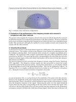
Figure 8.27 Full-thickness neuropathic ulcer post-debridement under a prominent fourth meta-
tarsal head
Figure 8.28 Commercially-available extra depth therapeutic shoe
and 5.07 monofilaments. Achilles tendon
reflexes were absent. The vibration percep-
tion threshold was above 50 V bilaterally,
while the peripheral pulses were normal.
A scar was noted on the dorsum of his
right foot which had an overriding fourth
toe, as a result of past surgical proce-
dures. A full-thickness neuropathic ulcer
was present under his fourth metatarsal
head surrounded by callus (Figure 8.27).
A bony prominence could also be felt
under the ulcerated area. A plain radio-
graph did not show osteomyelitis or neuro-
osteoarthropathy. Debridement of the ulcer
was carried out and extra depth therapeu-
tic shoes with a flat insole were prescribed
(Figure 8.28); a window was made in the
insole in order to offload pressure on the
ulcerated area; the ulcer began to heal well
(Figure 8.29).
Infections 175
Figure 8.29 Healing of the neuropathic ulcer shown in Figure 8.27 pre-debridement
The patient kept himself very active.
He returned to the clinic after 3 weeks
absence with a deeper ulcer involving the
tendons (Figure 8.30). The underlying bone
could not be detected with a sterile metal
probe and a plain radiograph did not show
osteomyelitis. An elevated erythrocyte sed-
imentation rate (74 mm/h) and mild leuko-
cytosis were found, therefore the possi-
bility of osteomyelitis was high. A mag-
netic resonance imaging-T1-weighted sagit-
tal image of the foot was obtained, showing
a phlegmonous mass starting from the skin
and extending to the deeper tissues caus-
ing erosion of the fourth metatarsal head
(Figure 8.31). The patient was hospitalized
so that offloading pressure from the ulcer-
ated area was enforced, and intravenous
antibiotics were administered. Two weeks
later the size of the ulcer had decreased by
almost 50%.
Several methods are used for the diag-
nosis of osteomyelitis. Probe-to-bone tests
(contacting the bone with a sterile metal
probe) have a sensitivity of more than
90% and they are carried out at the bed-
side. Plain radiographs have a sensitiv-
ity of 55%, but when repeated — usually
2 weeks later — the sensitivity is higher,
making this the most cost-effective diag-
nostic procedure. Computerized tomogra-
phy may reveal areas with subtle abnor-
malities such as periosteal reactions, small
cortex erosions and soft tissue abnormal-
ities. Magnetic resonance imaging has a
sensitivity of almost 100% and a speci-
ficity of over 80% and has the poten-
tial to reveal abscesses. Therefore this
is the preferred method for the diagno-
sis of osteomyelitis in many centers in
cases where the plain r adiographs do not
provide sufficient information to make a
conclusive diagnosis. However, the speci-
ficity of MRI decreases in the presence of
neuro-osteoarthropathy, prior bone biopsy,
recent bone fracture or recent surgery.
Magnification radiography is also a very
useful method for the detection of early
osteomyelitis and it is used to follow up
the disease.
Bone scintigraphy imaging is explained
in Figure 8.37.
Keywords: Neuropathic ulcer; magnetic
resonance imaging; MRI; osteomyelitis;
diagnostic methods for osteomyelitis
176 Atlas of the Diabetic Foot
Figure 8.30 The neuropathic ulcer shown in
Figures 8.27 and 8.29 has been aggravated by
the patient’s refusal to reduce activity levels
and poor compliance with measures to offload
pressure from the affected area
OSTEOMYELITIS OF THE
FIRST METATARSAL HEAD
A 74-year-old male patient with type 2
diabetes attended the outpatient diabetic
foot clinic because of a chronic painless
ulcer on the medial aspect of the right first
metatarsal head (Figure 8.32). The ulcer
developed over a bunion deformity, and had
persisted for 10 months.
On examination, the peripheral pulses
were palpable and the patient had severe
peripheral neuropathy. He could not feel
pain, light touch, vibration or 5.07 monofil-
aments. The vibration perception threshold
was above 50 V in both feet. After debride-
ment, the underlying bone could be felt by
means of a sterile probe. A plain radiograph
revealed osteomyelitis of the first metatarsal
head and the proximal phalanx of the right
great toe (Figure 8.33). The patient sus-
tained a first ray amputation.
Chronic osteomyelitis needs surgical re-
moval of the infected bone. However,
recent data suggest that prolonged treatment
with antibiotics (for 1 or 2 years) may erad-
icate chronic osteomyelitis. However, no
consensus on this issue exists at present.
Keywords: Chronic osteomyelitis; first ray
amputation; neuropathic ulcer
CHRONIC NEUROPATHIC
ULCER WITH
OSTEOMYELITIS
A 46-year-old male patient with type 1
diabetes diagnosed at the age of 27 years
was referred to the outpatient diabetes foot
clinic because of a chronic ulcer under his
right fifth metatarsal head. He had accept-
able diabetes control (HBA
1c
:7.7%),pro-
liferative diabetic retinopathy treated with
laser in both eyes, but no nephropathy. He
complained of muscle cramps during the
night and chronic constipation interrupted
by episodes of nocturnal diarrhea. The
patient had a history of painless diabetic
foot ulceration for 3 years under his right
foot after a burn injury. He had attended the
surgery department of a country hospital,
Infections 177
Figure 8.31 MRI image showing osteomyelitis. A magnetic resonance imaging-T1-weighted
sagittal image of the foot illustrated in Figure 8.30 showing a phlegmonous mass (arrow) extending
from the skin into the deeper tissues and causing erosion of the fourth metatarsal head
Figure 8.32 Chronic neuropathic ulcer over a bunion deformity
178 Atlas of the Diabetic Foot
Figure 8.33 Plain radiograph of the foot illustrated in Figure 8.32 showing bone resorption,
periosteal reaction and destruction of metatarsophalangeal joint of the hallux due to osteomyelitis
where he had his foot dressed a nd several
courses of antibiotics were prescribed. The
patient continued to keep himself active,
without any special footwear since he felt
no discomfort or pain.
On examination, severe diabetic neu-
ropathy was found. The peripheral pulses
were palpable and a full-thickness neuro-
pathic ulcer with gross callus formation was
observed under his right fifth metatarsal
head (Figure 8.34). Sharp debridement was
carried out and the underlying bone was
probed with a sterile probe. A plain radio-
graph revealed pseudoarthrosis of a stress
fracture of the upper third of his fifth
metatarsal, bone resorption in the metatar-
sophalangeal joint, and osteolytic lesions in
the fifth metatarsal epiphysis (Figures 8.35
and 8.36). Post-debridement cultures from
the base of the ulcer revealed Staphylococ-
cus aureus, Proteus vulgaris and Entero-
coccus spp. The patient was treated with
amoxicillin–clavulanic acid 625 mg three
times daily for 2 weeks. He was advised to
rest and appropriate footwear and insoles
were prescribed. A fifth ray amputation was
undertaken and antibiotics continued for
two more weeks. A bone culture revealed
Staphylococcus aureus. The wound healed
completely in 2 weeks.
A ray amputation consists of removal of
a toe together with its metatarsal. The unin-
volved half of the fifth metatarsal shaft was
preserved, so that it retained the insertion of
the short peroneal muscle. Ray amputation
results in narrowing of the forefoot, but the
cosmetic and functional result is excellent.
However, the biomechanics of the foot are
disturbed after such an operation and this
leads to the exertion of high pressure under
the metatarsal heads of the adjacent rays.
Keywords: Neuropathic ulcer; osteomyeli-
tis; ray amputation
BONE SCINTIGRAPHY
IMAGING
The scintigraphy findings of a patient with
possible osteomyelitis are discussed below
and the history of this patient is illustrated
in Figures 9.3 to 9.5 in Chapter 9.
A plain radiograph showed findings
compatible with osteomyelitis or neuro-
osteoarthropathy of the second and third
Infections 179
Figure 8.34 Full-thickness chronic neuropa-
thic ulcer with gross callus formation under the
right fifth metatarsal head
metatarsal heads of this female patient. She
was referred for a technetium-99m (
99
Tc)
phosphonate scan. Images obtained dur-
ing the flow phase are shown in the left
upper panel of Figure 8.37; during this
phase a series of 3-s image acquisitions
of the site in question is obtained. They
showed increased radionuclide uptake by
the tarsometatarsal area of her left foot.
A static blood pool image (blood pool
phase) obtained 5 min later is shown in
the right upper panel. A static delayed
image (delayed phase) obtained 3 h later
is shown in the left lower panel. All
images showed increased uptake in the
same areas. A gallium-67 citrate study per-
formed on the same day (right lower panel
of Figure 8.37), showed increased radionu-
clide uptake at the tarsometatarsal area.
Based on the results of bone scintigra-
phy the patient was diagnosed as having
osteomyelitis in the tarsometatarsal area.
99
Tc scintigraphy is useful in cases
of questionable osteomyelitis. It has a
high sensitivity (over 90%) but a low
specificity (33%), particularly in the pres-
ence of neuro-osteoarthropathy. Although
increased radionuclide uptake during the
flow and pool phase is not specific to
the diagnosis of osteomyelitis (it may
mean soft tissue, bone infection or both),
delayed images of the
99
Tc scintigraphy
showed increased blood flow to the bones
only, thus increasing the specificity of the
method in the diagnosis of bone infection.
Patients with neuro-osteoarthropathy have
increased bone blood flow in the absence
of osteomyelitis.
Like
99
Tc scintigraphy, gallium-67 cit-
rate accumulates in both osteomyelitis and
neuro-osteoarthropathy. This is the reason
for its low specificity in the diagnosis of
osteomyelitis in diabetic patients. Indium-
111 white blood cell imaging (
111
In WBCs)
is expensive, time consuming, has poor spa-
tial resolution and does not distinguish soft
tissue from bone infection.
Keywords: Scintigraphy; bone scans; diag-
nosis of osteomyelitis
OSTEOMYELITIS OF THE
HEEL
A 71-year-old female patient with type
2 diabetes was admitted to the hospital
180 Atlas of the Diabetic Foot
Figure 8.35 Anteroposterior plain radiograph
of patient of Figure 8.34. Osteomyelitis. Pseu-
doarthrosis of a stress fracture o f the upper third
of the fifth metatarsal, bone resorption at the
metatarsophalangeal joint, and osteolytic lesions
at the fifth metatarsal epiphysis
because of a severe infection of her right
foot. She had a history of type 2 diabetes
diagnosed at the age of 51 years, diabetic
nephropathy, background diabetic retinopa-
thy — treated with laser — hypertension
and ischemic heart disease. She also had
a history of stroke at the age of 69 years.
A heel ulcer caused after the rupture of a
Figure 8.36 Lateral plain radiograph of foot
shown in Figures 8.34 and 8.35, focused on the
osteomyelitic lesion in the fifth metatarsal. Note
that the phalanx of fifth toe continues over fourth
metatarsal head
blister under her right heel, which devel-
oped after walking in tight new shoes, had
persisted for about 1 year. The ulcer pro-
gressively became deeper and larger. The
patient reported two septic episodes with
infection at the same site, for which she
was hospitalized for prolonged periods.
On examination, her body temperature
was 39.2
◦
C, blood pressure 90/50 mmHg,
heart rate 120 beats/min and weak, and
she was anuric. Her right foot and
the tibia were warm, r ed and swollen.
A large, foul-smelling, neuro-ischemic
ulcer with gross purulent discharge was
seen on the posterior surface of her
right heel (Figure 8.38). The c alcaneus
was exposed.
Infections 181
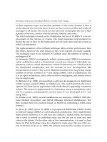
Figure 8.37 Increased radionuclide uptake by the tarsometatarsal bones, possibly due to
osteomyelitis. Technetium-99m (
99
Tc) phosphonates scan: flow phase (left upper p anel); blood
pool phase (right upper panel); delayed phase (left lower panel). Gallium-67 citrate study (right
lower panel)
A plain radiograph showed a large
skin defect on the posterioplantar aspect
of her heel and bone resorption of the
posterior calcaneus (Figure 8.39). Exten-
sive calcinosis of the posterior tib-
ial artery and medial plantar branch
artery was also noted. After surgical
debridement, bone and deep tissue cul-
tures were obtained. Immediate support
with i.v. fluids and antibiotic admin-
istration was commenced (ciprofloxacin
400 mg × 3 and clindamycin 600 mg × 3)
and her situation improved within 12 h.
Tissue cultures revealed Enterococcus
spp., Acinetobacter baumannii,andPro-
teus mirabilis. Based on an antibio-
gram, treatment was changed to ampicil-
lin–sulbactam and continued for 2 weeks.
Disarticulation through the ankle joint
(Syme ankle disarticulation) was not fea-
sible; a healthy heel flap and the heel pad
is a prerequisite for this procedure so that
the end of the stump is capable of bear-
ing the patient’s weight. Two weeks after
her admission the patient sustained a below-
knee amputation.