Atlas of the Diabetic Foot - part 10 pps
Bạn đang xem bản rút gọn của tài liệu. Xem và tải ngay bản đầy đủ của tài liệu tại đây (422.88 KB, 22 trang )
Neuro-Osteoarthropathy. The Charcot Foot 205
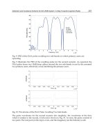
Figure 9.23 Healed ulcer in the patient whose foot is shown in Figures 9.19–9.22. Recurrent
ulceration of the midsole in a patient with midfoot collapse is an indication of osteotomy in the
protruding bones
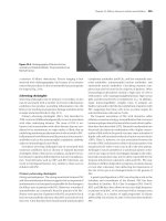
Figure 9.24 Clinical presentation of
acute neuro-osteoarthropathy of the
right ankle which is red, warm and
swollen
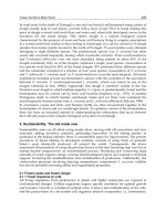
206 Atlas of the Diabetic Foot
of the articular surfaces of the right tibia
and talus. Bone fragments protruded medi-
ally (Figure 9.25). A diagnosis of acute
neuro-osteoarthropathy was made and the
patient was advised to rest, with his right
foot in a total-contact cast. The cast was
changed fortnightly for the first month
and monthly for the next year. After this
time osteoarthritic changes remained only
in the affected joint and no major deformity
was sustained.
Neuro-osteoarthropathy in the ankle is
the third most common pattern of this
Figure 9.25 Plain radiograph of chronic
neuro-osteoarthropathy of the right ankle and
foot as illustrated in Figure 9.24.Thereis
erosion of the articular surfaces of the right
tibia and talus and bone fragments protruding
medially
condition (frequency of 13%) and may
result in severe structural deformity and
instability. An extensive period of immo-
bilization is required in order to prevent
deformities.
Keywords: Acute neuro-osteoarthropathy
NEURO-
OSTEOARTHROPATHY:
SANDERS AND FRYKBERG
PATTERN IV; DOUNIS
TYPE III (a, b, and c)
A 67-year-old patient with type 2 diabetes
diagnosed at the age of 41 years attended
the outpatient orthopedic clinic because of
worsening painful ankle swelling after a
strain in his right ankle 2 weeks previously.
He had severe peripheral neuropathy and
normal feet pulses.
A plain film showed resorption of the
distal parts of the tibial and peroneal
bones and involvement of the ankle joint
(Figure 9.26). Pattern IV neuro-osteoarth-
ropathy was diagnosed and the foot was
placed in a total-contact cast and bed rest
was advised. The patient did not comply
with the advice and continued to be active
while wearing the cast. One month later
extensive resorption and fragmentation of
the talus and resorption of the distal areas of
the tibia and fibula was observed on a sec-
ond radiograph. A bone fragment protruded
posteriorly (Figure 9.27). Six months later
a plain film showed extensive resorp-
tion of the talus, subchondral osteosclero-
sis of the tibia and calcaneus and exten-
sive ligament ossification (the reconstruc-
tive stage of neuro-osteoarthropathy). Bone
fragments protruded laterally (Figure 9.28).
The patient admitted that during this time
he had been active. He had significant
Neuro-Osteoarthropathy. The Charcot Foot 207
Figure 9.26 Plain radiograph showing acute neuro-osteoarthropathy. Resorption of the distal areas
of the tibia and fibula and involvement of the ankle joint are evident
Figure 9.27 Plain radiograph showing pro-
gress of neuro-osteoarthropathy 1 month after
the X-ray shown in Figure 9.25 was taken.
There is extensive resorption and fragmentation
of the talus and resorption of distal areas of the
tibia and fibula and a bone fragment protrudes
posteriorly
instability and varus foot deformity. Even-
tually the patient sustained a below-knee
amputation.
A major problem in this pattern of
neuro-osteoarthropathy is functional insta-
bility and foot deformity. Reconstructive
Figure 9.28 Plain radiograph showing pro-
gress of neuro-osteoarthropathy 6 months after
the X-ray shown in Figure 9.27 was taken.
There is extensive resorption of the talus, sub-
chondral osteosclerosis of the tibia and calca-
neus and extensive ligament ossification. Bone
fragments can be seen to protrude laterally
208 Atlas of the Diabetic Foot
procedures (such as arthrodesis) were not
possible due to extensive bone absorption.
With this type of articular destruction reha-
bilitation will be more successful if the
patient uses a below-knee prosthesis rather
than a patellar-tibial-bearing orthosis.
Keywords: Ankle neuro-osteoarthropathy;
talus resorption; reconstructive stage
NEURO-
OSTEOARTHROPATHY:
SANDERS AND FRYKBERG
PATTERN IV; DOUNIS
TYPE IIIa
A type 2 diabetic female patient with bilat-
eral chronic neuro-osteoarthropathy (in the
reconstructive stage) resulting in marked
bilateral varus foot deformity (Figures 9.29
and 9.30), attended the outpatient orthope-
dic clinic. She was unable to walk with-
out crutches due to significant instability.
On a plain radiograph complete destruc-
tion of the ankle joint and subchondral
osteosclerosis at the distal ends of the tibia
and fibula were seen, together with lat-
eral resorption of the talus. Bone frag-
ments were observed laterally in the ankle
joint as were medial exuberant osteo-
phytes (Figure 9.31). The patient under-
went a realignment arthrodesis of the ankle
joint by lateral ankle incisions and the
ankle joint was fixed with a Huckstep
nail (Figure 9.32). The postoperative results
were excellent (Figure 9.33).
Significant deformity and instability is
the main indication for arthrodesis in
Figure 9.29 Bilateral varus deformity of the feet due to chronic neuro-osteoarthropathy. Signifi-
cant instability resulted in the patient’s inability to walk without crutches
Neuro-Osteoarthropathy. The Charcot Foot 209
Figure 9.30 Lateral view of Figure 9.29
Figure 9.31 Plain radiograph of
neuro-osteoarthropathy of the right
foot of the patient whose feet are
shown in Figures 9.29 and 9.30.There
is complete destruction of the ankle
joint, subchondral osteosclerosis in
the distal areas of the tibia and fibula,
together with lateral resorption of
talus. Bone fragments are seen later-
ally in the ankle joint and exuberant
osteophytes medially
210 Atlas of the Diabetic Foot
Figure 9.32 Plain postoperative radiograph of
the right foot of the patient whose feet are
illustrated in Figures 9.29–9.31. Arthrodesis of
the ankle joint with the use of a Huckstep nail
has been carried out
patients with neuro-osteoarthropathy. In
experienced hands it is possible in almost
80% of cases to achieve the goal of a sta-
ble and shoeable foot after an arthrodesis in
patients with neuro-osteoarthropathy. The
use of modern techniques of internal fix-
ation has significantly improved prognosis
in these patients. The period of immobi-
lization after an arthrodesis in patients with
neuro-osteoarthropathy is prolonged, usu-
ally more than 4 months.
Keywords: Neuro-osteoarthropathy; arthro-
desis; Huckstep nail
Figure 9.33 Postoperative photograph of the
right foot of the patient whose feet are shown in
Figures 9.29–9.32 after successful arthrodesis
of the ankle joint
NEURO-OSTEO-
ARTHROPATHY: SANDERS
AND FRYKBERG PATTERNS
IV AND V; DOUNIS TYPE III
(a, b and c): INVOLVE-
MENT OF THE HINDFOOT
Chronic neuro-osteoarthropathy often leads
to extensive resorption of the hindfoot
(talus and calcaneus), navicular and cuboid
bones (Figure 9.34). The patient whose
Neuro-Osteoarthropathy. The Charcot Foot 211
Figure 9.34 Plain radiograph showing chronic neuro-osteoarthropathy. Extensive resorption of
the hindfoot (talus and calcaneus), navicular and cuboid bones is evident
X-ray is shown in Figure 9.34 is a 45-
year-old female with long-standing type 1
diabetes who developed this complication
after a severe ankle sprain. She suffered
complete loss of sensation in her feet
and symptomatic autonomic neuropathy
(gastroparesis, diabetic diarrhea and ortho-
static hypotension). Gait instability devel-
oped within 8 months, to the point where
the patient was unable to walk with-
out crutches. Although she used a total-
contact cast, bone resorption was rapid and
Figure 9.35 Plain radiograph showing extensive resorption of most of the talus and calcaneus and
of the distal end of the tibia–fibula in a patient with chronic neuro-osteoarthropathy. Osteolysis in
the lower part of the calcaneus is due to osteomyelitis following a perforated ulcer
212 Atlas of the Diabetic Foot
relentless, so that eventually the patient suc-
cumbed to a below-knee amputation.
Keywords: Chronic neuro-osteoarthrop-
athy
NEURO-
OSTEOARTHROPATHY:
SANDERS AND FRYKBERG
PATTERNS IV AND V;
DOUNIS TYPE III (a, b and c)
Figure 9.35 shows extensive resorption of
most of the talus and calcaneus, in addition
to the distal end of the tibia–fibula in
a patient with neuro-osteoarthropathy. The
osteolysis in the lower part of the calca-
neus is due to osteomyelitis. A chronic
Figure 9.36 Chronic neuro-osteoarthropathy.
The osteomyelitis in the heel has been super-
imposed with a deep neuropathic ulcer in
the patient whose X-rays are illustrated in
Figure 9.35
neuropathic heel ulcer is present, caused
by a foreign body (Figure 9.36). Eventually
the patient, who had long-standing diabetes
and severe diabetic neuropathy, sustained a
below-knee amputation.
Keywords: neuro-osteoarthropathy; heel
ulcer; osteomyelitis
BIBLIOGRAPHY
1. Sanders LJ, Frykberg RG. Diabetic neuro-
pathic osteoarthropathy: the Charcot foot.
In Frykberg RG (Ed.), The High Risk Foot
in Diabetes Mellitus. New York: Churchill
Livingstone, 1991.
2. Dounis E. Charcot neuropathic osteoarthrop-
athy of the foot. Acta Orthopaed Hellenica
1997; 48: 281–295.
3. Harris JR, Brand PW. Patterns of disinte-
gration of the tarsus in the anaesthetic foot.
J Bone Joint Surg 1966; 5: 95–97.
4. Lennox WM. Surgical treatment of chronic
deformities of the anaesthetic foot. In
McDowell F, Enna CD (Eds), Surgical
Rehabilitation in Leprosy, and in Other
Peripheral Nerve Disorders. Baltimore:
Williams and Wilkins, 1974; 350–372.
5. Horibe S, Tada K, Nagano J. Neuroarthrop-
athy of the foot in leprosy. J Bone Joint Surg
(Br) 1988; 70-B: 481–485.
6. Brodsky JW, Rouse AM. Exostectomy for
symptomatic bony prominences in diabetic
Charcot foot. Clin Orthop 1993; 296: 21–26.
7. Barjon MC. Les ost
´
eoarthropathies destruc-
trices du pied diab
´
etique. In H
´
erisson C,
Simon L (Eds), Le Pied Diab´etique.Paris:
Masson, 1993; 77–91.
8. Johnson JE. Neuropathic (Charcot) arthrop-
athy of the foot and ankle. AAOS 1995
Instructional course #349. Handoutcover.
9. Eichenholtz SN. Charcot Joints. Spring-
field, IL: Charles C Thomas, 1966.
10. Onvlee GJ. The Charcot foot. A critical
review and an observational study of 60
patients. Thesis, Universiteit van Amster-
dam, 1998.
11. Shaw JE, Boulton AJM. The Charcot foot.
Foot 1995; 5: 65–70.
Appendix 1
ANATOMY OF THE FOOT
Atlas of the Diabetic Foot.
N. Katsilambros, E. Dounis, P. Tsapogas and N. Tentolouris
Copyright © 2003 John Wiley & Sons, Ltd.
ISBN: 0-471-48673-6
Anatomy of the Foot 215
Figure A1 Dorsal aspect of the bones in
the foot
Figure A3 Plain radiograph of the foot shown
in lateral view
Figure A2 Plantar aspects of the bones in
the foot
Appendix 2
MANUFACTURERS OF PREVENTIVE
AND THERAPEUTIC FOOTWEAR
Atlas of the Diabetic Foot.
N. Katsilambros, E. Dounis, P. Tsapogas and N. Tentolouris
Copyright © 2003 John Wiley & Sons, Ltd.
ISBN: 0-471-48673-6
Manufacturers of Preventive and Therapeutic Footwear 219
The therapeutic and preventive footwear and insoles described in this book are products
of various companies including:
Acor Orthopedic, USA
Aircast, Inc., USA
AliMed, Inc., USA
Buratto Advanced Technology, Italy
Darco International, Inc., USA
F. W. Kraemer KG, Germany
Orthopaedic Systems, UK
Index
Note: page numbers in italics refer to figures and tables
abscess
drainage 146
plantar 133
Achilles tendon reflexes 5
Acinetobacter baumannii 181
amputation
arteriography prior to 20
below-knee 129, 140, 147
chronic neuro-osteoarthropathy 212
deep-tissue infection 163, 166
osteomyelitis of heel 181
biomechanics of foot 69
Charcot foot 95
Chopart disarticulation 68–9
foot deformities 31
hallux 109, 110, 169
heel ulcers 98
limited distal 127
prevalence 3
ray 140, 178
fifth 178
first 61–3, 176
fourth 70
risk 3
toe 140, 146
fifth 121, 122
fourth 134
second 203
wet gangrene 143, 146 – 7
angiography 29
digital subtraction 132, 134, 136, 137–8
stenosis location 135, 140, 141
angioplasty
suboptimal 135
see also percutaneous transluminal
angioplasty
anhidrosis 3
ankle
arthrodesis 208, 210
bone fragments 208, 209
disarticulation 181
edema 53, 110, 112
deep-tissue infection 162, 163
neuro-osteoarthropathy 204, 205, 206, 207
osteophytes 208, 209
pressure 8, 20
swelling 204, 205, 206
ankle brachial index
calculation 20
cardiovascular risk 18
foot ulcer classification 26
ischemic ulcers 28
monitoring 18, 20
neuropathic ulcer 28
peripheral vascular disease detection 16
antibiotics 154
bone bioavailability 182
broad-spectrum 154
intravenous 154
osteomyelitis of heel 182–3
resistance 154
antifungal drugs 160
aorta, abdominal, stenosis 138
aorto — femoral bypass graft 127
aorto — popliteal bypass graft 150
Apligraf
see Graftskin
arterial calcification 9, 10
arterial insufficiency, antibiotic therapy 154
arterial stenosis 11
criteria in spectral analysis 13
ultrasonography 12
see also named arteries
arteriography 20
arthrodesis, realignment 208, 210
Atlas of the Diabetic Foot.
N. Katsilambros, E. Dounis, P. Tsapogas and N. Tentolouris
Copyright © 2003 John Wiley & Sons, Ltd.
ISBN: 0-471-48673-6
222 Index
aspirin 16
autolytic debridement 119
becaplermin see platelet-derived growth factor
β (PDGF-β)
bedridden patients 97–8
biosurgery 119, 121
biothesiometer 6
blisters
rupture 146, 167
soft tissue infection 156, 157
blood pressure control 16
bone demineralization 92
bone scintigraphy imaging 175, 178–9, 181
phases 179, 181
bony prominence
osteotomy 197, 199
ulcer 201, 202, 203, 204–5
bunion 48–9
chronic neuropathic ulcer 177
ulcers 49
bunionette formation 43–4, 46
ulceration 46
varus deformity 48
burns
forefoot 98, 99, 100, 101, 102
squamous cell carcinoma in scars 81
toes 98, 99, 100, 101, 102
bursitis 48–9
bypass grafting 127, 201
neuro-ischemic ulcers 113
wet gangrene 147
calcaneocuboid joint
Chopart dislocation 69
collapse 201, 202
neuro-osteoarthropathy 203
calcaneus
bone resorption 183
exposure 180
osteolysis 211, 212
resorption 210, 211, 212
subchondral osteosclerosis 206, 207
calcium pyrophosphate dihydrate (CPPD)
deposition disease 77–8
calf support device 129, 131
callus formation 3
under bone prominence 63 – 4
claw toe 155
hemorrhagic 65–6, 67, 68, 88, 200
metatarsal heads 45, 50–1
neuropathic ulcers 89, 94
prominent 64–5
second toe removal 59, 60, 61, 62
plantar pressure 88
pressure loading 87–8
prevention 67
removal 65
ulcer under 66 – 7
Candida albicans 71
fungal infection with multimicrobial
colonization 160
wound colonization 121
cardiovascular risk factors 16
cast, total-contact 34, 195–7, 198, 200,
206, 211
contraindications 34
cellulitis
deep-tissue infection 166
infected foot ulcer 153
infected plantar ulcer with osteomyelitis
172
neuro-ischemic ulcers 114, 115
non-ulcerated skin 154
treatment 154
wet gangrene 146
wound infections 153
Charcot foot 27
amputation 95
with neuropathic ulcer and deep-tissue
infection 163–4, 165, 166, 167
radiography 192
ulcers 95 – 6
Chopart dislocation, bilateral 68–9
Chopart’s joint 203
claw toe 30, 31, 46–8
bunionette formation 46
callus formation 155
under bone prominence 64
hemorrhagic 65–6
convex triangular foot 51
fungal infection 159
hallux valgus with overriding toes 52–4,
56
with heloma durum 48–9
infection
under callus at tip 155
soft-tissue 156
muscle atrophy 103
neuro-ischemic ulcers 120, 121
neuro-osteoarthropathy 191, 195
neuropathic ulcers of metatarsal heads
93–4
onychodystrophy 155
pes cavus 45
phlegmon 169
plantar arch collapse 200
prominent metatarsal heads 58–9
Index 223
second 99
ulcers 58, 59, 93 – 4, 120, 121
under callus area 66 – 7
wet gangrene 150
cocked-up toes see claw toe
coeliac aortic bifurcation stenosis 134, 137
collagen bundles, hyalinized 80, 81
collagenase 119
collateral circulation 8, 15, 19, 122
development 138, 150
popliteal artery 132, 135
collateral vessel development 142
compliance with medical instructions 3
computed tomography (CT)
osteomyelitis diagnosis 175
phlegmon imaging 171
spiral 14
convex triangular foot 51–2
corns 49–50
see also heloma durum
critical leg ischemia 127
cuboid bone
collapsed 203
fragmentation 199–200
resorption 210, 211
cuneiform bone
fragmentation 201
osteolysis 197
resorption 203
curly toe deformity 48
hemorrhagic callus formation 65–6
debridement 119, 121
depth — ischemia classification 26
Dermagraf
38
dermatofibrosarcoma protruberans 82 – 3
dermis, bioengineered 38
dextranomers 119
diabetes 3
diabetic bullae 157
diabetic neuropathy 3–6
definition 4
painful — painless foot 90
peripheral vascular disease co-existence 7
digital arteries
calcification 201
thrombosis 158
digital subtraction angiography see
angiography, digital subtraction
dorsalis pedis palpation 18
dressings 36, 37–8
ecchymosis 193, 194
eczema, hyperkeratotic 78, 79
edema
ankle 53, 110, 112
deep-tissue infection 162, 163
foot 158
forefoot deep-tissue infection 162, 163
neuro-osteoarthropathy 193
wet gangrene 140
Enterobacter 103, 104
fungal infection with multimicrobial
colonization 160
Enterobacter cloacae 168
Enterococci 156, 178, 181
enzymatic debridement 119
Epidermophyton floccosum 158, 160
Escherichia coli 62, 98
deep-tissue after interphalangeal mycosis
161
infected ulcers 154
osteomyelitis 104, 171
web space infection 158
wet gangrene 146
femoral artery
bruits 18
obstruction 122, 123
stenosis 7, 132, 134, 138
femoral artery, common
atheromatous disease 114, 146
obstruction 200
peak systolic velocity 148, 150
stenosis 17, 148, 150
femoral artery, superficial 19
atheromatous disease 139, 144, 146
atherosclerosis 117
spectral waveform 13 , 16
stenosis 7, 10, 15, 17, 140, 141
collateral vessel development 142
dry gangrene 127
neuro-ischemic ulcers 114
stents 135
wet gangrene 150
stents 135, 141
femoro — popliteal bypass graft 117, 127,
140
neuro-osteoarthropathy 95
femoro — tibial bypass graft 123
fibroblasts 39
fibula
resorption 206, 207, 211, 212
subchondral osteosclerosis 208, 209
flat foot see pes planus
Fontaine clinical staging 7–8
neuro-ischemic ulcers 116
224 Index
foot
anatomy 215
see also forefoot; heel; hindfoot; midfoot;
sole of foot; toe(s)
foot care, patient education 31– 2
foot deformities 3
definition 31
see also pes cavus; pes planovalgus; pes
planus; varus deformity
foot ischemia, hyperbaric oxygen 38
foot pulses
palpation 7
peripheral 28
footwear, preventive 29, 31, 111, 219
commercially-available 120
prescription 117
pressure relief shoes 35 – 6
refusal to wear 116
see also shoes
forefoot
burns 98, 99, 100, 101, 102
edema with deep-tissue infection 162, 163
neuro-ischemic ulcers with osteomyelitis
114, 116–17
foreign objects
neuropathic heel ulcer 212
in shoes 156, 157
fractures
avulsion of metatarsal base 193
see also stress fractures
fungal infection 158
claw toe 159
interphalangeal deep-tissue infection 161,
162
with multimicrobial colonization 160
palmoplantar keratoderma 76 – 7
web space 158
see also onychomycosis
gait instability 211
gallium-67 citrate 179, 181
gangrene 7–8
bedridden patients 97–8
critical limb ischemia 18
diabetic 133
dry
heel 129–30
with ischemic necrosis of skin 129
patient education 143
toes 127, 128, 130, 131, 133–4, 136 –8
infection 127
ischemic ulcers 29
necrosis 127, 128, 139
transcutaneous oxygen pressure 10
wet 127
amputation 143, 146–7
blistering 146
cellulitis 146
Escherichia coli 146
extensive of foot 143–4, 146–7
foot 140, 144
hallux 147, 148, 149–50
leading to mid-tarsal disarticulation
142–3, 145
necrotic tissue debridement 146
sepsis 130, 132–3, 134, 135, 136
Staphylococcus aureus 132, 146
toes 139–40, 144
wound infections 153
Graftskin 38–9
granulocyte colony stimulating factor (G-CSF)
39
hallux
amputation 109, 110, 169
disarticulation 63, 95, 99, 102, 109, 110
hemorrhagic callus 55
infection 103
ingrown nails 146
neuro-ischemic ulcers 107, 108
osteomyelitis 107–9, 110, 117–21
osteomyelitis 168–9, 176, 178
neuro-ischemic ulcers 107–9, 110,
117–21
removal 63
subungual hematoma 110
ulcer under 67, 68
wet gangrene 147, 148, 149–50
hallux valgus 31, 45
after second toe removal 59, 60, 61, 62
bunionette formation 46
callus over prominent metatarsal heads 65
collapsed midfoot 202, 203
hammer toes 57
onychomycosis 54–6, 57
with overriding toe 50–1, 52–4, 56, 57
phlegmon 169
with quintus varus 51–2
hammer toe 31, 47, 58
hallux valgus 57
onychomycosis 54–6, 57
hands, hyperkeratosis of palms 76
heel
cracks 65, 67 – 8
dry gangrene 129 – 3 0
osteomyelitis 179–83
ulcers
debridement 132–3
Index 225
neuro-ischemic 114, 115
neuropathic 96–8, 212
heel protector ring 129, 130
heloma durum 48–9, 50
heloma molle 49–50
hemorrhagic callus formation 66
kissing 50
hindfoot
preservation 69
resorption 210, 211
Huckstep nail 208, 210
Hyaff
39
hydrocolloids 119
hydrogels 119
hydrotherapy 119
hyperhidrosis 158
hyperkeratosis
eczema 78, 79
fifth toe 155
metatarsal heads 95
hypoesthesia
neuropathic ulcers 27
zone 5
iliac artery
atheromatous disease 114, 139, 144
common, stenosis 127
proximal, stenosis 134, 137
stenosis 7, 132, 134, 135, 140
infection
under callus
over fifth toe 155–6
at tip of claw toe 155
deep-tissue 161–3
after interphalangeal mycosis 161, 162
Charcot foot with neuropathic ulcer
163–4, 165, 166, 167
gangrene 127
ulceration 153–4
web space 157 –9
see also abscess; cellulitis; fungal i nfection;
sepsis; soft-tissue infection
insoles, flat 174
insoles, shock-absorbing 31, 32
convex triangular foot 52
custom-molded 32, 33
pes cavus 46
intermittent claudication 7, 16
digital subtraction angiography 138
ischemic ulcers 28
internal fixation 210
interphalangeal joint, distal
calcium pyrophosphate dihydrate deposition
disease 77, 78
plantar flexion 47, 56
interphalangeal joint, proximal
osteomyelitis 109
plantar flexion 47
intrinsic muscles of foot, atrophy 47
joint mobility, limited 3
plantar pressure 33
keratinocytes 38
keratoderma, palmoplantar 75 – 7
Klebsiella, deep-tissue 162
Klebsiella pneumoniae 168
larval therapy 119, 121
latissimus dorsi musculocutaneous flap 81,
82, 83
leuconychia mycotica 160
lichenification 78
ligament ossification 206, 207
limb ischemia, critical 18
lipid level reduction 16
Lisfranc joint see tarsometatarsal j oint
lymphangitis 153
maggot debridement 119, 121
magnetic resonance angiography (MRA) 14,
16
magnetic resonance imaging (MRI)
osteomyelitis diagnosis 175, 177
phlegmon imaging 171
male patients, foot ulcers 3
malleolus, ulcer 43
mallet toe 56, 58
Marjolin’s ulcer 81
mechanical debridement 119
medial arterial calcification 8
Meggitt — Wagner classification of ulcers 25
modification 26
metastases, dermatofibrosarcoma protruberans
83
metatarsal bone
avulsion fracture 193
diaphysis resorption 203, 204
fifth
pencil-like appearance 203, 204
pseudoarthrosis 199
lateral displacement 195, 196
osteomyelitis 191
phlegmon under base 170, 171
subluxation of second 195, 196
metatarsal epiphysis, osteolytic lesions 178
metatarsal heads
broadening 192
226 Index
metatarsal heads (continued )
callus formation 45, 50–1, 53
hallux disarticulation 99
second toe removal 59, 60, 61, 62
cup formation 190
erosion in osteomyelitis 103
fat pad
displacement 47, 65, 90, 91, 103
reduced thickness 91–2
fifth
callosity 132
exostosis 46
neuro-ischemic ulcer with osteomyelitis
121–3
neuro-osteoarthropathy 193–4
neuropathic ulcer 99
osteomyelitis 121–3, 201
pseudoarthrosis in stress fractures 178
ulcer 51
first
neuro-ischemic ulcer with osteomyelitis
113–14
neuropathic ulcer 87–8
osteomyelitis 62, 113–14, 176,
177, 178
fourth
erosion 175, 177
neuropathic ulcer 69–71, 90–2,
174
hyperkeratosis 43, 95
necrosis 61
osteolysis 172, 191
proliferative changes 192
prominent 3, 5, 31, 58 – 9
callus 64–5
neuropathic ulcer 87–90, 93–5
phlegmon 169
plantar pressure 66
soft tissue infection 156
ulcer under callus area 66–7
stress fractures 92
third and hemorrhagic callus 65– 6
ulcer formation 67
metatarsal joint, dislocation of first 197
metatarsophalangeal joint
arthrodesis 61, 62
bone resorption 178
dorsiflexion 47
fourth toe disarticulation 70–1
hallux disarticulation 63, 95
osteoarthritis 134, 139
osteolytic destruction 190
subluxation 190
methicillin-resistant Staphylococcus aureus
(MRSA) 121, 156
infected plantar ulcer with osteomyelitis
171
painful ulcer 200
mid-tarsal disarticulation 127, 129, 130
wet gangrene 142 – 3, 145
midfoot
chronic ulcer 201, 202, 203
collapsed 90, 200–1, 202
midsole, neuro-ischemic ulcers 114, 115
Moh’s micrographic surgery 83
monofilaments 5–6
muscles, small 5
musculocutaneous flap, latissimus dorsi 81,
82, 83
mycosis, interphalangeal 161, 162
nails
deformities 75, 76
ingrown 71, 146
repetitive trauma 75
navicular bone
collapsed 203
resorption 210, 211
naviculocuneiform joint
bone resorption 201, 202
collapse 201, 202
destruction 203, 204
neuro-osteoarthropathy 203
necrobiosis lipoidica 78–80, 81
necrotizing fasciitis 153
nephropathy, diabetic 53
neuro-osteoarthropathy 3, 164, 165, 166
acute 189–91, 193, 197, 206
acute osteomyelitis differential diagnosis
191–3
acute dissolution phase 197
ankle 204, 205, 206, 207
arthrodesis 199
blood supply 201
bone destruction 189
bone/joint destruction anatomic patterns
187–8
calcaneocuboid joint 203
classification 187–9
claw toe 191
clinical presentation 188
cuboid bone fragmentation 199–200
differential diagnosis 189
Dounis classification 187–8
type I 189–91
type II 193–203, 204–5
type III (a, b and c) 206–8, 210–12
Index 227
neuro-osteoarthropathy (continued)
type IIIa 208, 209, 210
type IIIα 204, 205, 206
edema 193
Eichenholtz stage I 197
Eichenholtz stage III 199
femoral — popliteal bypass graft 95
foot deformities 31, 207
functional instability 207
immobilization 199, 206, 210
internal fixation 210
metatarsal heads 193 –4
midfoot ulcers 36
naviculocuneiform joint 203
osteophyte formation 204
radiological findings 188
reconstructive stage 206, 208
Sanders and Frykberg classification 187
pattern I 189–91
pattern II and III 193–203, 204–5
pattern IV 204, 205, 206–8, 209,
210
pattern IV and V 210–12
stages 188
talonavicular joint 203
tarsometatarsal joint 193, 197, 203
neurothesiometer 6
onychocryptosis 71
wet gangrene 150
onychodystrophy 134, 136
claw toe 155
ischemic ulcers 28
peripheral vascular disease 132
phlegmon 169
wet gangrene 146
onychogryposis 75, 76
onychomycosis 54–6, 57, 159–60
callus under bone prominence 64
distal subungual 159–60
neuropathic ulcers of metatarsal heads 94
proximal subungual 160
orthosis, patellar-tibial-bearing 208
osteoarthritis, metatarsophalangeal joint 134,
139
osteomyelitis 95, 96, 167–8
bone destruction 189
bone scintigraphy imaging 179, 181
burns 102
calcaneus osteolysis 211, 212
chronic neuro-osteoarthropathy 211
chronic ulcer 203
diagnosis 175
Escherichia coli 104
hallux 168–9, 176, 178
with neuro-ischemic ulcers 117, 118,
119, 120, 121
heel 179 – 83
infected ulcer 153
plantar 171–3
Klebsiella pneumoniae 168
metatarsal head
fifth 201
first 62, 176, 177, 178
metatarsals 191
neuro-ischemic ulcer
fifth metatarsal head 121–3
first metatarsal 113 – 1 4
forefoot 114, 116–17
hallux 117, 118, 119, 120, 121
neuropathic ulcer complication 102–4,
173–5, 176, 177
chronic 176, 178, 180
pathogens 103, 104
proximal phalanx 63
Pseudomonas aeruginosa 119
radiographic evaluation 154
radiography 192
Staphylococcus aureus 103, 104, 108,
113–14, 116, 168–9
treatment regimens 104
acute infection 172
osteophytes 204
ankle 208, 209
oxygen
hyperbaric 36, 38
transcutaneous pressure (TcPO
2
) 9–10
pain
neuropathic 4
plantar heel spur 43
see also rest pain, ischemic
painful — painless foot 90
Palmaz stent 137
papain 119
papules, violaceous 79
paronychia 71
progression to gangrene 7–8
patellar-tibial-bearing orthosis 208
peak systolic velocity (PSV) ratio 12, 14, 15,
16, 17
wet gangrene 148, 150
pedal arteries
calcification 112
pressure index 10
reconstitution 140
228 Index
percutaneous transluminal angioplasty 135
gangrene 127
neuro-ischemic ulcers 113
popliteal artery 168
wet gangrene 147
peripheral neuropathy 3
claw toes 47
painless ulcer 30
prevalence 4
symptoms 5
tests 5–6
peripheral vascular disease 3, 6 – 14, 15, 16,
17, 18, 19, 20
detection and follow-up 16, 18, 20
gender differences 140
neuropathy co-existence 7
prevalence 6–7
peroneal artery stenosis 140
pes cavus 44–6
callus over prominent metatarsal heads 65
pes planovalgus 51
pes planus 43, 112
bunionette 43–4
phalanges, concentric resorption 189–90
phalangophalangeal joint
hemorrhagic callus 67, 68
subluxation 190
phalanx, proximal, osteomyelitis 63
phlegmon 169–71
photoplethysmography 11
plantar abscesses 133
plantar arch
collapse 200–1
high 45
plantar arteries, reconstitution 140
plantar branch artery, medial 181, 183
plantar cracks 30
plantar heel spur pain 43
plantar ligament preservation 90
plantar pressure
callus formation 88
convex triangular foot 52
fat pad thickness reduction 91 – 2
high 3
in-shoe measurement 52, 54, 91, 92
limited joint mobility 33
offloading 33–6
prominent metatarsal heads 66
redistribution 32
reduction 31, 32
ulcer under callus area 67
platelet-derived growth factor β (PDGF-β)
38, 172
plethysmography, segmental 11
ischemic ulcers 29
popliteal artery
atheromatous disease 146
collateral supply 132, 135
obstruction 122, 123
percutaneous transluminal angioplasty 168
stenosis 114, 127
popliteal — peripheral bypass 117, 150
pre-ulcer formation 88
pressure loading 87–8
pressure offloading 103, 107, 114
heel 131
neuropathic ulcer 174
pressure perception 5–6
pressure ulcers 129
gangrenous 132, 133
probe-to-bone tests 175
prosthesis, below-knee 208
Proteus infection 153
Proteus mirabilis 181
Proteus vulgaris 178
pseudo-gout see calcium pyrophosphate
dihydrate (CPPD) deposition disease
Pseudomonas 153
infected ulcers 154
Pseudomonas aeruginosa 104
fungal infection with multimicrobial
colonization 160
osteomyelitis 119
wet gangrene 132
wound colonization 121
Pseudomonas maltophila 168
pulsed Doppler imaging 12
quality of life 3
quintus varus 51–2
radiography
Charcot foot 192
magnification 175
neuro-osteoarthropathy 188
osteomyelitis 154, 175, 192
Regranex
see platelet-derived growth factor
β (PDGF-β)
rest pain, ischemic 7
persistent recurring 127
toe pressure 9
transcutaneous oxygen pressure 10
revascularization procedures 114
heel gangrene 130
rocker bottom deformity 90
scars, squamous cell carcinoma formation 81
scintigraphy see bone scintigraphy imaging
Scotch-cast boot 35
Index 229
segmental pressure measurement 10–11
ischemic ulcers 29
Semmes — Weinstein monofilaments 5 – 6
sensation loss 4, 5
footwear 31
sensory deficit 5
neuropathic ulcers 27
sepsis, wet gangrene 130, 132–3, 134, 135,
136
sesamoid bones 63
shear stress
reduction 32
ulcers 54
shoes
athletic 31
foreign objects 156, 157
half 35, 36, 66
heel-free 36, 97
manufacturers 219
pressure relief 35 – 6
rocker-style 33, 35, 88
roller-style 33
selection 29–30
therapeutic for neuropathic ulcers 87, 88
trauma 3
ulcer on dorsum of foot 110– 11
see also footwear, preventive; insoles
silicone ring 111, 112
skin
atrophy 112
changes
critical limb ischemia 18
ischemic ulcers 28
ischemic necrosis with dry gangrene 129
lesions
shoe friction 111
toe pressure 9
pathogens 154
scaling 134
see also ulcers/ulceration; wound(s); wound
healing
skin, dry 3
callus
under bone prominence 64
over prominent metatarsal heads 65
heel 79
cracks 67 – 8
phlegmon 169
smoking cessation 16
socks, padded 31
soft-tissue infection 102, 156–7
under callus at tip of claw toe 155
soft-tissue sarcoma 82–3
sole of foot
hyperkeratosis 76
midsole neuro-ischemic ulcers 114, 115
squamous cell carcinoma 81–2
S(AD)SAD classification for foot ulcers 26
staphylococcal toxins 133
Staphylococcus aureus 62, 97, 98
antibiotics 182, 183
callus over fifth toe 155
deep-tissue 162
after interphalangeal mycosis 161
fungal infection with multimicrobial
colonization 160
infected ulcers 154
non-limb-threatening infections 156
osteomyelitis 103, 104, 108, 113–14, 116,
168–9
chronic neuropathic ulcer 178
web space infection 158
wet gangrene 132, 146
see also methicillin-resistant Staphylococcus
aureus (MRSA)
Staphylococcus epidermidis 104
Steinmann pins 197
stents 135, 137, 140, 141
aortic 134, 138
aorto-iliac intravascular 132, 136
Strenotropomonas maltophilia 122
streptococci
non-limb-threatening infections 156
toxins 133
stress fractures
metatarsal heads 92, 178
pseudoarthrosis in fifth metatarsal 178
toes 134, 139
surgical debridement 119
sweating inhibition 3
Syme ankle disarticulation 181
tailor’s bunion see bunionette formation
talonavicular joint
Chopart dislocation 69
collapse 201, 202
destruction 203, 204
fragments 199, 200
neuro-osteoarthropathy 203
talus resorption 206, 207, 208, 209, 210, 211,
212
tardus pardus waveform 19, 149, 150
tarsometatarsal joint
destruction 203, 204
disruption 164, 165, 201
Lisfranc joint line 43
neuro-osteoarthropathy 193, 197, 203
partial resorption 194–5, 196, 197
phlegmon 170
230 Index
tarsometatarsal joint line, front transverse
convexity 43
tarsometatarsophalangeal joint fusion 198
technetium-99m 179, 181
thermal injury see burns
tibia
resorption 206, 207, 211, 212
subchondral osteosclerosis 206, 207, 208,
209
tibial artery
atheromatous disease 138, 146
occlusion 140
stenosis 117, 140
tibial artery, anterior
obstruction 150
spectral waveform 149
stenosis 16, 18
tibial artery, posterior 14, 16
calcification 112
calcinosis 181, 183
palpation 18
stenosis 18, 148
tibioperoneal artery 140
tinea pedis 76
interdigital 158
toe(s)
amputation 121, 122, 134, 146, 202, 203
ray 140
burns 98, 99, 100, 101, 102
deformities 30, 31
incidence 56
varus deformity 46, 48
dry gangrene 127, 128, 130, 131, 133–4,
136 –8
fifth
amputation 121, 122
hemorrhagic callus 200
infection under callus 155–6
stress fracture 134, 139
fourth
amputation 134
disarticulation 70–1
ulceration by pressure from little toe 111
overriding 50–1
causes 55
hallux valgus 52–4, 56, 57
pressures 8–9
sausage-like appearance 167, 168
second
amputation 202, 203
disarticulation 116, 132
lateral exostosis 192
medial displacement 99
removal 59, 60, 61, 62
silicone ring 111, 112
ulcer formation 67
wet gangrene 139 – 40, 144
see also claw toe; curly toe deformity;
hammer toe; mallet toe; nails
toe, great see hallux
transcutaneous oximetry 9–10
transcutaneous oxygen pressure (TcPO
2
)
9–10
transparent films 119
trauma 3
progression to gangrene 7–8
Trichophyton 158, 160
triplex scan, wet gangrene of foot 148 , 149,
150
trypsin 119
ulcers/ulceration 29
under callus area 66 – 7
cellulitis 114, 115
Charcot foot 95 –6
classification 25–6
critical limb ischemia 18
curettage 153
depth — ischemia classification 26
under hallux 67, 68
infection 25–6, 27, 153–4
clinical assessment 153
cultures 153
diagnosis 153
plantar with osteomyelitis 171–3
wet gangrene 142–3, 145
ischemia 25–6
clinical presentation 26, 28–9
load reduction 34
midfoot 201, 202, 203
necrobiosis lipoidica 80
neuro-ischemic 26, 30
claw toes 120, 121
clinical presentation 29
dorsum of foot 109–11
fifth metatarsal with osteomyelitis 121–3
first metatarsal with osteomyelitis
113–14
forefoot with osteomyelitis 114, 116–17
under hallux 107, 108
hallux with osteomyelitis 117, 118, 119,
120, 121
under hallux with osteomyelitis 107–9,
110
heel 114, 115, 180, 182
interdigital 111, 112
medial side of foot 112 – 1 3
midsole 114, 115