DIABETIC NEUROPATHY: CLINICAL MANAGEMENT - PART 6 doc
Bạn đang xem bản rút gọn của tài liệu. Xem và tải ngay bản đầy đủ của tài liệu tại đây (3.65 MB, 52 trang )
is no evidence. The author has therefore recommended that the term “acute painful
neuropathy of rapid glycemic control” be used to describe this condition (48).
The natural history of acute painful neuropathies is an almost guaranteed improve-
ment (49) in contrast to chronic distal symmetrical neuropathy (36). The patient pres-
ents with burning pain, paraesthesiae, allodynia, often with a nocturnal exacerbation of
symptoms; and depression may be a feature. There is no associated weight loss, unlike
acute painful neuropathy of poor glycemic control. Sensory loss is often mild or absent,
and there are no motor signs. There is little or no abnormality on nerve conduction stud-
ies, but there is impaired exercise induced conduction velocity increment (48,50). There
is usually complete resolution of symptoms within 12 months.
On sural nerve biopsy, typical morphometric changes of chronic distal symmetrical
neuropathy but with active regeneration, were observed (49). In contrast, degeneration of
both myelinated and unmyelinated fibres was found in acute painful neuropathy of poor
glycemic control (44). A recent study looking into the epineurial vessels of sural nerves in
patients with acute painful neuropathy of rapid glycemic control demonstrated marked
arterio/venous abnormality including the presence of proliferating new vessels, similar to
those found in the retina (48). The study suggested that the presence of this fine network
of epineural vessels may lead to a “steal” effect rendering the endoneurium ischaemic, and
the authors also suggested that this process may be important in the genesis of neuropathic
pain (48). These findings were also supported by studies in experimental diabetes, which
demonstrated that insulin administration led to acute endoneurial hypoxia, by increas-
ing nerve arterio-venous flow, and reducing the nutritive flow of normal nerves (51).
Further work needs to address whether these observed sural nerve vessel changes
resolve with the resolution of painful symptoms.
ASYMMETRICAL NEUROPATHIES
The diabetic state can also affect single nerves (mononeuropathy), multiple nerves
(mononeuropathy multiplex), or groups of nerve roots. These asymmetrical or focal
neuropathies have a relatively rapid onset, and complete recovery is usual. This con-
trasts with chronic distal symmetrical neuropathy, where there is usually no improve-
ment in symptoms 5 years after onset (36). Unlike chronic distal symmetrical
neuropathy they are often unrelated to the presence of other diabetic complications
(9,15,16). Asymmetrical neuropathies are more common in men and tend to predomi-
nantly affect older patients (52). A careful history is therefore mandatory in order to
identify any associated symptoms that might point to another cause for the neuropathy.
A vascular etiology has been suggested by virtue of the rapid onset of symptoms and
the focal nature of the neuropathic syndromes (53).
Proximal Motor Neuropathy (Femoral Neuropathy, Amyotrophy,
and Plexopathy)
The syndrome of progressive asymmetrical proximal leg weakness and atrophy was
first described by Garland (54), who coined the term “diabetic amyotrophy.” This con-
dition has also been named as “proximal motor neuropathy,” “femoral neuropathy”
or “plexopathy.” The patient presents with severe pain, which is felt deep in the thigh,
but can sometimes be of burning quality and extend lower than the knee. The pain is
Clinical Features of Diabetic Polyneuropathy 251
usually continuous and often causes insomnia and depression (55). Both type 1 and
type 2 patients more than the age of 50 are affected (54–57). There is an associated
weight loss, which can sometimes be very severe, and can raise the possibility of an
occult malignancy.
On examination there is profound wasting of the quadriceps with marked weakness
in these muscle groups, although hip flexors and hip abductors can also be affected (58).
Thigh adductors, glutei, and hamstring muscles may also be involved. The knee jerk is
usually reduced or absent. The profound weakness can lead to difficulty from getting
out of a low chair or climbing stairs. Sensory loss is unusual, and if present indicates a
coexistent distal sensory neuropathy.
It is important to carefully exclude other causes of quadriceps wasting, such as nerve
root and cauda equina lesions, and the possibility of occult malignancy causing proxi-
mal myopathy syndromes such as polymyocytis. Magnetic resonance imaging (MRI) of
the lumbo-sacral spine is now mandatory in order to exclude focal nerve root intrapment
and other pathologies. An erythrocyte sedimentation rate, an X-ray of the lumbar/sacral
spine, a chest X-ray, and ultrasound of the abdomen may also be required. CSF protein
is often elevated. Electrophysiological studies may demonstrate increased femoral
nerve latency and active denervation of affected muscles.
The cause of diabetic proximal motor neuropathy is not known. It tends to occur within
the background of diabetic distal symmetrical neuropathy (59). It has been suggested that
the combination of focal features superimposed on diffuse peripheral neuropathy may
suggest vascular damage to the femoral nerve roots, as a cause of this condition (60).
As in distal symmetrical neuropathy there is scarcity of prospective studies that have
looked at the natural history of proximal motor neuropathy. Coppack and Watkins (55)
have reported that pain usually starts to settle after about 3 months, and usually settles
by 1 year, while the knee jerk is restored in 50% of the patients after 2 years. Recurrence
on the other side is a rare event. Management is largely symptomatic and supportive.
Patients should be encouraged and reassured that this condition is likely to resolve.
There is still controversy as to whether the use of insulin therapy influences the natural
history of this syndrome as there are no controlled trials. Some patients benefit from
physiotherapy that involves extension exercises aimed at strengthening the quadriceps.
The management of pain in proximal motor neuropathy is similar to that of chronic or
acute distal symmetrical neuropathies (see Chapter 21).
Chronic Inflammatory Demyelinating Polyradiculopathy
Chronic inflammatory demyelinating polyradiculopathy (CIDP) occurs more com-
monly among patients with diabetes, creating diagnostic and management challenges
(61). Patients with diabetes may develop clinical and electrodiagnostic features similar
to that of CIDP (62). Clearly, it is vital to recognize these patients as unlike diabetic
polyneuropathy, CIDP is treatable (63). One should particularly be alerted when an
unusually severe, rapid, and progressive polyneuropathy develops in a diabetic patient.
Nerve conduction studies show features of demyelination. The presence of 3 of the
following criteria for demyelination is required: partial motor nerve conduction block,
reduced motor nerve conduction velocity, prolonged distal motor latencies, and prolonged
F-wave latencies (64). Although, electrophysiological parameters are important, these
alone cannot be entirely relied on to differentiate CIDP from diabetic polyneuropathy (65).
252 Tesfaye
Most experts recommend CSF analysis in order to demonstrate the typical findings
in this condition: increased protein and a normal or only slightly elevated cell count
(63). However, spinal taps are not mandatory (63).
The diagnostic value of nerve biopsy, usually of the sural nerve has been debated
recently. Some authorities assert that nerve biopsy is of no value (66), whereas others
consider it essential for the diagnosis and management of upto 60% patients with CIDP
(67). The diagnostic yield of sural nerve biopsies may be limited as the most prominent
abnormalities may lie in the proximal segments of the nerve roots or in the motor
nerves, which are areas not accessible to biopsy. Typical appearances include segmen-
tal demyelination and remyelination, anion bulbs, and inflammatory infiltrates, but
these may also be found in diabetic polyneuropathy (68). A defining feature of CIDP
not found in diabetic polyneuropathy is the presence of macrophages in biopsy speci-
mens in association with demyelination (68).
Treatments for CIDP include intravenous immunoglobulin, plasma exchange, and corti-
costeroids (63). Therapy should be started early in order to prevent continuing demyelina-
tion and also as it results in rapid and significant reversal of neurological disability (69,70).
Mononeuropathies
The most common cranial mononeuropathy is the third cranial nerve palsy. The
patient presents with pain in the orbit, or sometimes with a frontal headache (53,71).
There is typically ptosis and ophthalmoplegia, although the pupil is usually spared
(72,73). Recovery occurs usually over three months. The clinical onset and time-scale
for recovery, and the focal nature of the lesions on the third cranial nerve, on post-
mortem studies suggested an ischaemic etiology (53,74). It is important to exclude any
other cause of third cranial nerve palsy (aneurysm or tumour) by computed tomography
or MRI scanning, where the diagnosis is in doubt. Fourth, sixth, and seventh cranial
nerve palsies have also been described in diabetic subjects, but the association with
diabetes is not as strong as that with third cranial nerve palsy.
Truncal Radiculopathy
Truncal radiculopathy is well recognized to occur in diabetes. It is characterized by
an acute onset pain in a dermatomal distribution over the thorax or the abdomen (75).
The pain is usually asymmetrical, and can cause local bulging of the muscle (76). There
may be patchy sensory loss detected by pin prick and light touch examination. It is
important to exclude other causes of nerve root compression and occasionally, MRI of
the spine may be required. Some patients presenting with abdominal pain have under-
gone unnecessary investigations, such as barium enema, colonoscopy, and even laparo-
tomy, when the diagnosis could easily have been made by careful clinical history and
examination. Recovery is usually the rule within several months, although symptoms
can sometimes persist for a few years.
Pressure Neuropathies
Carpal Tunnel Syndrome
A number of nerves are vulnerable to pressure damage in diabetes. In the Rochester
Diabetic Neuropathy Study, which was a population-based epidemiological study, Dyck
Clinical Features of Diabetic Polyneuropathy 253
et al. (77), found electrophysiological evidence of median nerve lesions at the wrist in
about 30% of diabetic subjects, although the typical symptoms of carpel tunnel syn-
drome occurred in less than 10%. The patient typically has pain and paraesthesia in the
hands, which sometimes radiate to the forearm and are particularly marked at night.
In severe cases clinical examination may reveal a reduction in sensation in the median
territory in the hands, and wasting of the muscle bulk in the thenar eminence. The clin-
ical diagnosis is easily confirmed by median nerve conduction studies and treatment
involves surgical decompression at the carpel tunnel in the wrist. There is generally
good response to surgery, although painful symptoms appear to relapse more commonly
than in the nondiabetic population (78).
Ulnar Nerve and Other Isolated Nerve Entrapments
The ulnar nerve is also vulnerable to pressure damage at the elbow in the ulnar
groove. This results in wasting of the dorsal interossei, particularly the first dorsal
interossius. This is easily confirmed by ulnar electrophysiological studies which local-
ize the lesion to the elbow. Rarely, the patients may present with wrist drop because of
radial nerve palsy after prolonged sitting (with pressure on the radial nerve in the back
of the arms) while unconscious during hypoglycaemia or asleep after an alcohol binge.
In the lower limbs the common peroneal (lateral popliteal) is the most commonly
affected nerve. The compression is at the level of the head of the fibula and causes foot
drop. Unfortunately, complete recovery is not usual. The lateral cutaneous nerve of the
thigh is occasionally also affected with entrapment neuropathy in diabetes. Phrenic
nerve involvement in association with diabetes has also been described, although the
possibility of a pressure lesion could not be excluded (79).
REFERENFCES
1. Shaw JE, Zimmet PZ. The epidemiology of diabetic neuropathy. Diabetes Rev 1999;
7:245–252.
2. Tesfaye S, Stephens L, Stephenson J, et al. The prevalence of diabetic neuropathy and its
relation to glycaemic control and potential risk factors: the EURODIAB IDDM
Complications Study. Diabetologia 1996;39:1377–1384.
3. Young MJ, Boulton AJM, Macleod AF, Williams DRR, Sonksen PH. A multicentre study
of the prevalence of diabetic peripheral neuropathy in the United Kingdom hospital clinic
population. Diabetologia 1993;36:150–154.
4. Maser RE, Steenkiste AR, Dorman JS, et al. Epidemiological correlates of diabetic neu-
ropathy. Report from Pittsburgh Epidemiology of Diabetes Complications Study. Diabetes
1989;38:1456–1461.
5. Ziegler D. Diagnosis, staging and epidemiology of diabetic peripheral neuropathy. Diab
Nutr Metab 1994;7:342–348.
6. Tesfaye S, Chaturvedi N, Eaton SEM, Witte D, Ward JD, Fuller J. Vascular risk factors and
diabetic neuropathy. N Engl J Med 2005;352:341–350.
7. Boulton AJM, Kirsner RS, Viliekyte L. Neuropathic diabetic foot ulcers. N Engl J Med
2004;351:48–55.
8. Tesfaye S. Diabetic neuropathy: achieving best practice. Br J Vasc Dis 2003;3:112–117.
9. Watkins PJ, Edmonds ME. Clinical features of diabetic neuropathy, in Textbook of
Diabetes (Pickup J, Williams G, eds.), 1997, Vol. 2, pp. 50.1–50.20.
10. Ward JD. Clinical features of diabetic neuropathy. in Diabetic Neuropathy (Ward JD, Goto Y,
eds.), Chichester, UK., Wiley, 1990, pp. 281–296.
254 Tesfaye
11. Bruyn GW, Garland H. Neuropathies of endocrine origin. in Handbook of clinical neurol-
ogy (Vinken PJ, Bruyn GW, eds.), Amsterdam, North-Holland Publishing Co., 1970,
Vol. 8, 29p.
12. Thomas PK. Metabolic neuropathy. J Roy Coll Phys (Lond) 1973;7:154–160.
13. Low PA, Suarez GA. Diabetic neuropathies. in Bailliere’s Clinical Neurology
1995;4(3):401–425.
14. Boulton AJM, Malik RA, Arezzo JC, Sosenko JM. Diabetic Somatic neuropathies.
Diabetes Care 2004;27:1458–1486.
15. Eaton SEM, Tesfaye S. Clinical manifestations and measurement of somatic neuropathy.
Diabetes Rev 1999;7:312–325.
16. Scott LA, Tesfaye S. Measurement of somatic neuropathy for clinical practice and clinical
trials. Curr Diabetes Rep 2001;1:208–215.
17. Andersen H, Jakobsen J. Motor function in diabetes. Diabetes Rev 1999;7:326–341.
18. Watkins PJ. Pain and diabetic neuropathy. Br Med J 1984;288:168–169.
19. Tesfaye S, Price D. Therapeutic approaches in diabetic neuropathy and neuropathic pain.
in Diabetic Neuropathy. (Boulton AJM, ed.), 1997;159–181.
20. Tesfaye S, Watt J, Benbow SJ, Pang KA, Miles J, MacFarlane IA. Electrical spinal cord
stimulation for painful diabetic peripheral neuropathy. Lancet 1996;348:1696–1701.
21. Quattrini C, Tesfaye S. Understanding the impact of painful diabetic neuropathy. Diabetes
Metab Res Rev 2003;(Suppl 1):S1–S8.
22. Ewing DJ, Borsey DQ, Bellavere F, Clarke BF. Cardiac autonomic neuropathy in diabetes:
comparison of measures of R-R interval variation. Diabetologia 1981;21:18–24.
23. Ward JD. The diabetic leg. Diabetologia 1982;22:141–147.
24. Rajbhandari SM, Jenkins R, Davies C, Tesfaye S. Charcot neuroarthropathy in diabetes
mellitus. Diabetologia 2002;1085–1096.
25. Ward JD, Simms JM, Knight G, Boulton AJM, Sandler DA. Venous distension in the dia-
betic neuropathic foot (physical sign of arterio-venous shunting). J Roy Soc Med
1983;76:1011–1014.
26. Boulton AJM, Scarpello JHB, Ward JD. Venous oxygenation in the diabetic neuropathic
foot: evidence of arterial venous shunting? Diabetologia 1982;22:6–8.
27. Edmonds ME, Archer AG, Watkins PJ. Ephedrine: a new treatment for diabetic neuro-
pathic oedema. Lancet 1983;1(8324):548–551.
28. Said G, Slama G, Selva J. Progressive centripital degeneration of of axons in small-fibre
type diabetic polyneuropathy. A clinical and pathological study. Brain 1983;106:791.
29. Vinik AI, Park TS, Stansberry KB, Pittenger GL. Diabetic neuropathies. Diabetologia
2000;43:957–973.
30. Veves A, Young MJ, Manes C, et al. Differences in peripheral and autonomic nerve func-
tion measurements in painful and painless neuropathy: a Clinical study. Diabetes Care
1994;17:1200–1202.
31. Ward JD, Tesfaye S. Pathogenesis of diabetic neuropathy. in Textbook of Diabetes (Pickup J,
Williams G, eds.), 1997, Vol. 2, pp. 49.1– 49.19.
32. Cameron NE, Eaton SE, Cotter MA, Tesfaye S.Vascular factors and metabolic interactions
in the pathogenesis of diabetic neuropathy. Diabetologia 2001;44:1973–1988.
33. Tesfaye S, Harris N, Jakubowski J, et al. Impaired blood flow and arterio-venous shunting
in human diabetic neuropathy: a novel technique of nerve photography and fluorescein
angiography. Diabetologia 1993;36:1266–1274.
34. Eaton SE, Harris ND, Ibrahim S, et al. Differnces insural nerve haemodynamics in painful
and painless neuropathy. Diabetologia 2003;934–939.
35. Malik RA, Tesfaye S, Newrick PG, et al. Sural nerve pathology in diabetic patients with
minimal but progressive neuropathy. Diabetologia 2005;48:578–585.
36. Boulton AJM, Armstrong WD, Scarpello JHB, Ward JD. The natural history of painful dia-
betic neuropathy - a 4 year study. Postgrad Med J 1983;59:556–559.
Clinical Features of Diabetic Polyneuropathy 255
37. Benbow SJ, Chan AW, Bowsher D, McFarlane IA, Williams G. A prospective study of
painful symptoms, small fibre function and peripheral vascular disease in chronic painful
diabetic neuropathy. Diabetic Med 1994;11:17–21.
38. Chan AW, MacFarlane IA, Bowsher DR, Wells JC, Bessex C, Griffiths K. Chronic pain in
patients with diabetes mellitus: comparison with non-diabetic population. Pain Clin 3
1990;147–159.
39. Young RJ, Zhou YQ, Rodriguez E, Prescott RJ, Ewing DJ, Clarke BF. Variable relation-
ship between peripheral somatic and autonomic neuropathy in patients with different syn-
dromes of diabetic polyneuropathy. Diabetes 1986;35:192–197.
40. Tsigos C, White A, Young RJ. Discrimination between painful and painless diabetic neu-
ropathy based on testing of large somatic nerve and sympathetic nerve function. Diabetic
Med 1992;9:359–365.
41. Veves A, Manes C, Murray HJ, Young MJ, Boulton AJM. Painful neuropathy and foot
ulceration in diabetic patients. Diabetes Care 1993;16:1187–1189.
42. Young MJ, Manes C, Boulton AJM. Vibration perception threshold predicts foot ulcera-
tion: a prospective study (Abstract). Diabetic Med 1992;9(Suppl 2):542.
43. Tesfaye S, Kempler P. Painful diabetic neuropathy. Diabetologia 2005;48:805–807.
44. Archer AG, Watkins PJ, Thomas PJ, Sharma AK, Payan J. The natural history of acute
painful neuropathy in diabetes mellitus. J Neurol Neorosurg Psychiatr 1983;46:491–496.
45. Ellenberg M. Diabetic neuropathic cachexia. Diabetes 1974;23:418–423.
46. Guy RJC, Clark CA, Malcolm PN, Watkins PJ. Evaluation of thermal and vibration sensa-
tion in diabetic neuropathy. Diabetologia, 1985;28:131.
47. Caravati CM. Insulin neuritis: a case report. Va Med Mon 1933;59:745–746.
48. Tesfaye S, Malik R, Harris N, et al. Arteriovenous shunting and proliferating new vessels
in acute painful neuropathy of rapid glycaemic control (insulin neuritis). Diabetologia
1996;39:329–335.
49. Llewelyn JG, Thomas PK, Fonseca V, King RHM, Dandona P. Acute painful diabetic
neuropathy precipitated by strict glycaemic control. Acta Neuropathol (Berl) 1986;72:
157–163.
50. Tesfaye S, Harris N, Wilson RM, Ward JD. Exercise induced conduction veolcity
increment: a marker of impaired nerve blood flow in diabetic neuropathy. Diabetologia
1992;35:155–159.
51. Kihara M, Zollman PJ, Smithson IL, et al. Hypoxic effect of endogenous insulin on nor-
mal and diabetic peripheral nerve. Am J Physiol 1994;266:E980–E985.
52. Matikainen E, Juntunen J. Diabetic neuropathy: Epidemiological, pathogenetic, and clinical
aspects with special emphasis on type 2 diabetes mellitus. Acta Endocrinol Suppl (Copenh)
1984;262:89–94.
53. Asbury AK, Aldredge H, Hershberg R, Fisher CM. Oculomotor palsy in diabetes mellitus:
a clinicopathological study. Brain 1970;93:555–557.
54. Garland H. Diabetic amyotrophy. Br Med J 1955;2:1287–1290.
55. Coppack SW, Watkins PJ. The natural history of femoral neuropathy. Q J Med 1991;
79:307–313.
56. Casey EB, Harrison MJG. Diabetic amyotrophy: a follow-up study. Br Med J 1972;1:656.
57. Garland H, Taverner D. Diabetic myelopathy. Br Med J 1953;1:1405.
58. Subramony SH, Willbourn AJ. Diabetic proximal neuropathy. Clinical and electromyo-
graphic studies. J Neurol Sci 1982;53:293–304.
59. Bastron JA, Thomas JE. Diabetic polyradiculoneuropathy: clinical and electromyographic
findings in 105 patients. Mayo Clinic Proc 1981;56:725–732.
60. Said G, Goulon-Goeau C, Lacroix C, Moulonguet A. Nerve biopsy findings in different
patterns of proximal diabetic neuropathy. Ann Neurol 1994;33:559–569.
61. Haq RU, Pendlebury WW, Fries TJ, Tandan R. Chronic inflammatory demyelinating
polyradiculoneuropathy in diabetic patients. Muscle Nerve
2003;27:465–470.
256 Tesfaye
62. Steward JD, McKelvey R, Durcan L, Carpenter S, Karpati G. Chronic inflammatory
demyelinating polyneuropathy (CIPD) in diabetes. J Neurol Sci 1996;142:59–64.
63. Koller H, Kieseier BC, Jander S, Hartung H. Chronic Inflammatory Demyelinating
Polyneuropathy. NEJM 2005;352:1343–1356.
64. Research criteria for diagnosis of chronic inflammatory demyelinating polyneuropathy
(CIDP): report from an ad hoc sub-committee of the America Academy of Neurology
AIDS Task Force. Neurology 1991;41:617–618.
65. Wilson JR, Park Y, Fisher MA. Electrodiagnostic criteria in CIDP: comparison with dia-
betic neuropathy. Electromyogr Clin Neurophsiol 2000;40:181–185.
66. Molenaar DS, Vermeulen M, de Haan R. Diagnostic value of sural nerve biopsy in chronic
inflammatory demyelinating polyneuropathy. J Neurol Neurosurg Psychiatry 1998;64:
84–89.
67. Gabriel CM, Howard R, Kinsella N, et al. Prospective study of the usefulness of sural nerve
biopsy. J Neurol Neurosurg Psychiatry 2000;69:442–446.
68. Vital C, Vital A, Lagueny A, et al. Chronic inflammatory demyelinating polyneuropathy;
immunopathological and ultrastructural study of peripheral nerve biopsy in 42 cases.
Ultrastruct Pathol 2000;24:363–369.
69. Cocito D, Ciaramitaro P, Isoardo G, et al. Intravenous immunoglobulin as first treatment
in diabetics with concomitant distal symmetric axonal polyneuropathy and CIDP. J Neurol
2002;249:719–722.
70. Sharma KR, Cross J, Ayyar DR, Martinez-Arizala A, Bradley WG. Diabetic demyelinating
polyneuropathy responsive to intravenous immunoglobulin therapy. Arch Neurol 2002;
59:751–757.
71. Zorilla E, Kozak GP. Ophthalmoplegia in diabetes mellitus. Ann Internal Med
1967;67:968–976.
72. Goldstein JE, Cogan DG. Diabetic ophthalmoplegia with special reference to the pupil.
Arch Ophthalmol 1960;64:592–600.
73. Leslie RDG, Ellis C. Clinical course following diabetic ocular palsy. Postgrad Med J
1978;54:791–792.
74. Dreyfuss PM, Hakim S, Adams RD. Diabetic ophthalmoplegia. Arch Neurol Psychiatry
1957;77:337–349.
75. Ellenberg M. Diabetic truncal mononeuropathy—a new clincal syndrome. Diabetes Care
1978;1:10–13.
76. Boulton AJM, Angus E, Ayyar DR, Weiss R. Diabetic thoracic polyradiculopathy present-
ing as abdominal swelling. BMJ 1984;289:798–799.
77. Dyck PJ, Kratz KM, Karnes JL, et al. The prevalence by staged severity of various types
of diabetic neuropathy, retinopathy, and nephropathy in a population-based cohort: the
Rochester Diabetic Neuropathy Study. Neurology 1993;43:817–824.
78. Clayburgh RH, Beckenbaugh RD, Dobyns JH. Carp[el tunnel release in patients with dif-
fuse peripheral neuropathy. J Hand Surg 1987;12A:380–383.
79. White JES, Bullock RF, Hudgson P, Home PD, Gibson GJ. Phrenic neuropathy in associ-
ation with diabetes. Diabet Med 1992;9:954–956.
Clinical Features of Diabetic Polyneuropathy 257
15
Micro- and Macrovascular Disease
in Diabetic Neuropathy
Aristidis Veves, MD and Antonella Caselli, MD, PhD
SUMMARY
Diabetes is often defined a “vascular disease” because of the early and extensive involve-
ment of the vascular-tree observed in patients with diabetes and even in those at risk of devel-
oping diabetes. Both the micro- and macrocirculation are affected. Changes in the micro- and
macrocirculation, both anatomical and functional, contribute to the development of diabetic
neuropathy. On the other hand, the development of diabetic neuropathy also affects the
vasodilatory capacity of the microcirculation. Thus, the interaction between changes in the vas-
culature and peripheral nerves is bidirectional and results in changes in both blood flow and
neuronal function. The possible links between diabetic micro- and macrovascular alterations
and nerve damage will be the focus of this chapter.
Key Words: Blood flow; endothelial dysfunction; micro- and macrocirculation; neuronal func-
tion; vascular smooth muscle cell; iontophoresis.
INTRODUCTION
Diabetes is often defined a “vascular disease” because of the early and extensive
involvement of the vascular tree observed in patients with diabetes and even in those at
risk of developing diabetes. Both the micro- and macrocirculation are affected, though
the pathophysiology, histology, clinical history, and clinical sequelae at the two vascular
levels appear to be quite different. It is recently believed that a common pathway causes
precocious vascular damage at both vascular districts in diabetes leading to the develop-
ment of diabetic chronic complications, if not of diabetes itself. Chronic diabetic com-
plications are mostly ascribed to small vessel disease. Diabetic microangiopathy has
been considered the main anatomic alteration leading to the development of retinopathy,
nephropathy, and neuropathy. Nevertheless, macroangiopathy, i.e., atherosclerosis of
peripheral arteries, is also a peculiar feature of long-lasting diabetes and is characterized
for being precocious, involving predominantly distal arteries and having inadequate
collateral development. The possible links between diabetic micro- and macrovascular
alterations and nerve damage will be the focus of this chapter.
MICROVASCULAR DISEASE: OVERVIEW AND ANATOMIC CHANGES
Lesions specific for diabetes have been observed in the arterioles and capillaries of
the foot and other organs that are the typical targets of diabetic chronic complications.
From: Contemporary Diabetes: Diabetic Neuropathy: Clinical Management, Second Edition
Edited by: A. Veves and R. Malik © Humana Press Inc., Totowa, NJ
259
A contemporary historical histological study demonstrated the presence of PAS-positive
material in the arterioles of amputated limb specimens from patients with diabetes (1).
Although it was believed for several years that the anatomic changes described were
occlusive in nature, in 1984, Logerfo and Coffmann (2) recognized that in patients with
diabetes, there is no evidence of an occlusive microvascular disease. Subsequent
prospective anatomic staining and arterial casting studies have demonstrated the
absence of an arteriolar occlusive lesion thus dispelling the hopeless notion of diabetic
“occlusive small vessel disease” (3,4).
Although there is no occlusive lesion in the diabetic microcirculation, other structural
changes do exist. The thickening of the capillary basement membrane is the dominant
structural change in both diabetic retinopathy and neuropathy and is because of an
increase in the extracellular matrix. It might represent a response to the metabolic
changes related to diabetes and hyperglycemia. However, this alteration does not lead
to occlusion of the capillary lumen, and arteriolar blood flow might be normal or even
increased despite these changes (5). On the contrary, it might act as a barrier to the
exchange of nutrients and/or increase the rigidity of the vessels further limiting their
ability to dilate in response to different stimuli (6).
In the kidney, nonenzymatic glycosylation reduces the charge on the basement mem-
brane, which might account for transudation of albumin, an expanded mesangium, and
albuminuria (7). Similar increases in vascular permeability occur in the eye and probably
contribute to macular exudate formation and retinopathy (8). In simplest terms, micro-
vascular structural alterations in diabetes result in an increased vascular permeability
and impaired autoregulation of blood flow and vascular tone.
Many studies have identified a correlation between the development of diabetic
chronic complication and metabolic control with perhaps the strongest evidence
coming from the Diabetes Control and Complications Trial (DCCT), which enrolled
patients with type 1 diabetes, and the United Kingdom Prospective Diabetes Study
(UKPDS), which enrolled patients with type 2 diabetes (9,10). The results from both
clinical trials clearly showed a delay in the development and progression of retinopathy,
nephropathy, and neuropathy with intensive glycemic control, thus supporting the direct
causal relationship between hyperglycemia and microcirculation impairment. This was
less evident for macrovascular disease, assessed only in the UKPDS.
Although the structural alterations observed in the microcirculation do not affect the
basal blood flow, some functional abnormalities of the microvascular circulation that
might eventually result in a relative ischemia have been extensively documented. This
aspect will be deeply discussed in the “Pathophysiology of microvascular disease and
endothelial dysfunction in diabetes” section.
PATHOPHYSIOLOGY OF MICROVASCULAR DISEASE
AND ENDOTHELIAL DYSFUNCTION IN DIABETES
Although microvascular diabetic complications have been well-characterized there is
still uncertainty regarding the mechanisms that lead to their development. In the past
two main pathogenic hypotheses have been proposed: the metabolic hypothesis and the
hypoxic hypothesis (11,12). According to the metabolic hypothesis, hyperglycemia is
directly responsible of end-organ damage and development of complications through
260 Veves and Caselli
the activation of the polyol pathway. On the other hand, according to the hypoxic
hypothesis, the structural alterations detected in kidney, eye, and nerve microvascu-
lature, including basement membrane thickening and endothelial cell proliferation,
were considered as the main factor contributing to reduced blood flow and tissue
ischemia (13). It is now apparent that both the metabolic and vascular pathways are
linked. More specifically, endothelial dysfunction has been suggested as the common
denominator between the metabolic and vascular abnormalities detected in diabetes (14).
The impaired synthesis and/or degradation of nitric oxide, the main vasodilator released
by the endothelium, is believed to determine microvascular insufficiency, tissue
hypoxia, and degeneration (15).
Functional Changes
Diabetes mellitus, even in the absence of complications, impairs the vascular reac-
tivity that is the endothelium-dependent and -independent vasodilation in the skin
microcirculation (16). Many glucose-related metabolic pathways can determine
endothelium dysfunction: increased aldose reductase activity leading to the imbalance
in nicotinamide adenine dinucleotide phosphate (NADP)/nicotinamide adenine dinu-
cleotide phosphate reduced form (NADPH); auto-oxidation of glucose leading to the
formation of reactive oxygen species; “advanced glycation end products” produced
by nonenzymatic glycation of proteins; abnormal n6-fatty acid metabolism and inap-
propriate activation of protein kinase-C. All these different pathways lead to an
increase of oxidative stress which is responsible for a reduced availability of nitric
oxide and in turn, for a functional tissue hypoxia and the development of diabetic
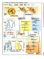
chronic complications (17) (Fig. 1).
Microvascular Dysfunction and Diabetic Neuropathy
Microvascular reactivity is further reduced at the foot level in presence of peripheral
diabetic neuropathy. Endothelial nitric oxide synthase (eNOS) is a key regulator of vas-
cular nitric oxide production. Immunostaining of foot skin biopsies in our unit, with
antiserum to human eNOS glucose transporter I, which is a functional marker of the
endothelium and von Willebrand factor, an anatomical marker, showed no difference
among patients with diabetes with or without peripheral neuropathy in the staining of
glucose transporter I and von Willebrand factor, whereas the staining for the eNOS was
reduced in neuropathic patients (Fig. 2) (18). Another study documented increased levels
of iNOS and reduced eNOS levels in skin from the foot of patients with diabetes with
severe neuropathy and foot ulceration (19).
It has also been suggested that polymorphism of the eNOS gene is implicated in car-
diovascular and renal diseases, thus indicating its potential role as a genetic marker of
susceptibility to both type 2 diabetes and its renal complications (20,21). However, a
relationship between eNOS gene polymorphism and diabetic neuropathy has not been
clearly demonstrated (22). Nonetheless, all these findings suggest that the reduced
eNOS expression/activity might be related to the development of diabetic peripheral
neuropathy.
Differences in the microcirculation between the foot and forearm levels have also been
investigated, the main hypothesis being that increased hydrostatic pressure in distal
Micro- and Macrovascular Disease in Diabetic Neuropathy 261
262 Veves and Caselli
Fig. 1. New concepts in the pathogenesis of diabetic neuropathy.
Fig. 2. Expression of eNOS in patients with diabetic neuropathy (black columns), patients
with both diabetic neuropathy and peripheral vascular disease (hatched columns) and healthy
subjects (white columns). The expression of eNOS was reduced in both the diabetic groups com-
pared with the healthy subjects (data from ref. 18).
microcirculatory beds, related to the orthostatic posture, affects the foot microcirculation
more than at the forearm level. The endothelium dependent and independent vasodilation
is in fact lower at the foot level when compared with the forearm in healthy subjects and
both nonneuropathic and neuropathic patients with diabetes (23). This forearm-foot gra-
dient exists despite a similar baseline blood flow at the foot and forearm level. Therefore,
it is reasonable to believe that erect posture might be a contributing factor for the early
development of the nerve damage at the foot, in comparison with the forearm.
Role of Autonomic Neuropathy
Autonomic neuropathy can compromise the diabetic microcirculation because of the
development of arterio–venous shunting because of sympathetic denervation. The open-
ing of these shunts might lead to a maldistribution of blood between the nutritional
capillaries and subpapillary vessels, and consequent aggravation of microvascular ischemia.
Studies using sural nerve photography and fluorescein angiography as well as other
elegant techniques seem to support this concept (24,25).
A loss of sympathetic tone is also responsible for an increased capillary permeability
in patients with diabetes with neuropathy (26). This might cause endoneurial edema, as
demonstrated by using magnetic resonance spectroscopy, which can in turn represent
another mechanism leading to a reduction of endoneurial perfusion and a worsening of
the nerve damage (27). The increased lower extremity capillary pressure upon assum-
ing the erect posture, because of early loss of postural vasoconstriction (mediated by the
sympathetic fibers), might amplify this edematous effect.
Role of Somatic Neuropathy: The Neurovascular Response
Diabetic somatic neuropathy can further affect the skin microcirculation by the
impairment of the axon reflex related-vasodilatation (Lewis’ flare) (28). Under normal
conditions, the stimulus of the c-nociceptive nerve fibers not only travels in the normal
direction, centrally toward the spinal cord, but also peripherally (antidromic conduction)
to local cutaneous blood vessels, causing a vasodilatation by the release of vasoactive
substances, such as calcitonin gene-related peptide (CGRP), Neuropeptide Y, substance P,
and bradykine by the c-fibers and initiates neurogenic inflammation (Fig. 3). This short
circuit, or nerve axon reflex, is responsible for the Lewis’ triple flare response to injury
and plays an important role in increasing local blood flow when it is mostly needed, i.e.,
in condition of stress.
This neurovascular (N–V) response is significantly reduced at the foot level in
patients with diabetes with peripheral somatic neuropathy, autonomic neuropathy, and
peripheral artery disease in comparison with patients with diabetes without complica-
tions and healthy control subjects (Fig. 4) (23,29). Moreover, local anaesthesia signifi-
cantly reduces the nerve axon reflex-related vasodilation at the foot of patients without
peripheral neuropathy, whereas it has no effect on the amount of the preanesthesia N–V
vasodilation—which is already very low—at the foot of neuropathic patients (30). This
suggests that the main determinant of the presence of the neurovascular vasodilation is
c-fiber function and that its measurement could be used as a surrogate measure of the
function of these fibers.
Micro- and Macrovascular Disease in Diabetic Neuropathy 263
As a matter of fact, it has been shown that the N–V response significantly correlates
with different measures of peripheral nerve function (30,31). Studies in our units have
shown that a N–V response lower than 50% is highly sensitive (90%) and adequately
specific (74%) in identifying patients with diabetes with peripheral neuropathy (31).
264 Veves and Caselli
Fig. 3. The nerve axon reflex-related vasodilation or neurovascular response: stimulation of the
c-nociceptive nerve fibers by acetylcholine or other noxious stimuli leads to antidromic stimulation
of the adjacent c-fibers, which secrete CGRP that causes vasodilation and increased local blood flow.
Fig. 4. The neurovascular response (expressed as percentage of blood flow increase over the
baseline blood flow) is significantly reduced at the foot level of patients with diabetes with
peripheral somatic neuropathy (DN), autonomic neuropathy (DA) and peripheral artery disease
(DV) compared with patients with diabetes without complications (DC) and healthy controls (C)
*p < 0.001 (data from ref. 29).
Besides, the finding that this response is significantly reduced even in the early stages
of peripheral neuropathy supports the hypothesis that small fiber damage is a precocious
event in the clinical history of diabetic neuropathy—even preceding large fibers’impair-
ment (Fig. 5). This leads to impaired vasodilation under conditions of stress, such as
injury or inflammation. Therefore, it is possible to speculate that small fiber neuropathy
might further contribute to nerve hypoxic damage by the impairment of this hyperemic
response, determining a vicious cycle of injury.
The previous conclusions are supported by recent studies in experimental diabetes
which have demonstrated that epineurial arterioles of the sciatic nerve are innervated by
sensory nerves that contain CGRP and mediate a hyperemic response at this level (32).
Furthermore, it has been shown that in long-term diabetic rats the amount of CGRP
present in epineurial arterioles is diminished, which could be because of a denervation
process (33). Exogenous CGRP-mediated vasodilation of these arterioles is also
impaired in experimental diabetes, indicating a reduced CGRP bioactivity (33). All
these findings furthermore support a role of small sensory nerve fibers’ impairment in
the development and progression of diabetic neuropathy.
The impairment of the nerve axon reflex-related vasodilation is not affected by
successful bypass surgery in patients with peripheral arterial disease. In addition, the
endothelium-dependent and -independent vasodilation that are not related to the nerve
axon reflex, remain impaired after successful revascularization. Therefore, despite cor-
rection in obstructive lesions and restoration of normal blood flow in the large vessels,
the changes in microcirculation continue to be present and cause tissue hypoxia under
conditions of stress (34).
Micro- and Macrovascular Disease in Diabetic Neuropathy 265
Fig. 5. The nerve axon reflex-related vasodilation at the foot level in a population with dia-
betes stratified on the basis of the degree of peripheral somatic neuropathy in patients without
neuropathy (D), with mild neuropathy (DN mild), with moderate neuropathy (DN moderate) and
with severe neuropathy (DN severe) compared with healthy controls (C). Median (25–75 per-
centile). The nerve axon reflex-related vasodilation is already significantly reduced in the early
stages of neuropathy (subclinical neuropathy), supporting the belief that small fiber dysfunction
might precede large fiber impairment in the natural history of diabetic nerve damage.
Anatomical Changes
Although the structural alterations detected in diabetic capillaries do not cause vessel
occlusion, their role in causing a reduction of nerve blood flow supply can not be com-
pletely ruled out. According to Pouiselle’s law, in fact, the blood flow is proportional to
the fourth power of the radius of a vessel. Therefore, the capillary blood flow can be
significantly reduced by even slight narrowing of the capillary lumen. Many studies
have now confirmed the presence of endoneurial microangiopathy, characterized by
basement membrane thickening, endothelial cell hyperplasia and hypertrophy, and peri-
cyte cell degeneration in patients with diabetes with peripheral neuropathy, the degree
of which correlates with the severity of the clinical disease (35,36).
In summary, both the functional and structural changes observed in diabetic micro-
circulation contribute to the shift of blood flow away from the nutritive capillaries to
low resistance arterio–venous shunts leading to functional ischemia of tissues including
peripheral nerves and, consequently, to the development of diabetic peripheral neuro-
pathy and other diabetic chronic complications.
TECHNIQUES TO ASSESS MICROVASCULAR DYSFUNCTION
AND THEIR LIMITATIONS
Endothelial dysfunction, assessed at the macrocirculation, has been proven as an
early marker of vascular complications in several diseases, including diabetes, dyslipi-
demia, and hypertension. The development of techniques capable to measure the skin
blood flow has also enabled the study of the vascular reactivity at the microcirculation
level. More specifically, the noninvasive measurement of cutaneous blood perfusion can
be performed by the laser Doppler.
Currently, laser Doppler flowmetry is the most widely accepted technique for evalu-
ating blood flow in the skin microcirculation. Basically, it measures the capillary flux,
which is a combination of the velocity and the number of moving blood cells. This is
achieved by using red laser light, which is transmitted to the skin through a fiberoptic
cable. The frequency shift of light back-scattered from the moving blood cells beneath
the probe tip is computed to give a measure of the superficial microvascular perfusion.
There are mainly two different types of instruments available: the laser Doppler per-
fusion imager (LDPI) and the laser Doppler blood flow monitor (LDM). The LDPI, or
laser scanner, enables the quantification of superficial skin blood perfusion in a multi-
ple number of adjacent sites on the skin and calculates the mean blood perfusion in a
particular region (Fig. 6). The LDM, which is characterized for having two single-point
laser probes is capable to measure the blood flow changes only in a small skin area
(about 2–3 mm diameter)—that corresponds to the area where the probes are placed—
and records the blood flow changes in response to the vasodilatory stimulus in a con-
tinuous way (Fig. 7).
The LDPI is best-suited for studying the relative changes in flow induced by a vari-
ety of physiological manoeuvres or pharmaceutical intervention procedures. The single-
point laser probe is used mainly for evaluating the hyperemic response to heat stimulus
or for evaluating the nerve-axon related hyperemic response. Both these two laser
Doppler instruments have been extensively used to evaluate the skin microcirculatory
flow of patients with diabetes in response to the delivery of two vasodilatory substances
266 Veves and Caselli
by iontophoresis: a 1% acetylcholine chloride solution (endothelium-dependent vasodi-
lation) and a 1% sodium nitroprusside solution (endothelium-independent vasodilation).
To use these methods for longitudinal analysis, a certain degree of confidence is
needed to ensure that the results are not skewed for instrumental inaccuracies or other
experimental factors. The main limitation of both techniques is, in fact, the variability,
which is higher for the single-point laser Doppler than for the LDPI. The single-point
technique has been validated against direct measurements of the capillary blood flow
velocity (37). The day-to-day reproducibility of the technique was evaluated in healthy
subjects who were repeatedly tested at their foot and arm for 10 consecutive days in
our lab. The coefficient of variation for the maximal response to heat was 27.9%,
whereas for the maximal hyperemic response after Ach and/or SNP-iontophoresis was
35.2% (18). The variability of this technique is mostly a spatial one, i.e., it is mainly
because of the high heterogeneity of the skin microcirculation and not to the technique
itself. In fact, the technique reproducibility can be significantly enhanced if one pays
attention to place the laser probe approximately at the same skin area for repeated
measurements (38).
The laser scanner has a significantly better reproducibility (which is mainly because
of the minor spatial variation of blood flow assessment) with the coefficient of variation
at the foot and forearms level being between 14 and 19%, and can therefore be used for
Micro- and Macrovascular Disease in Diabetic Neuropathy 267
Fig. 6. The LDPI or laser scanner: a helium-neon laser beam is emitted from the laser source
to sequentially scan the circular hyperemic area of the skin (surrounding the laser beam) where
the hyperemic response is produced by the iontophorized vasoactive substance.
blood flow assessment in prospective studies (39,40). Nevertheless, some factors, other
than the accuracy of the device itself, might also potentially affect the LDPI readings,
namely the scanner head height and inclination, tissue heating, prevalence of arm hair,
and arm movement.
MACROVASCULAR DISEASE AND DIABETES: AN OVERVIEW
Both type 1 and type 2 diabetes are powerful and independent risk factors for coro-
nary artery disease (CAD), stroke, and peripheral artery disease. More specifically, the
Framingham study showed that type 2 diabetes is associated with approximately a
twofold increase in CAD in men and a fourfold increase in women (41). It is also known
that patients with diabetes have the same risk of acute myocardial infarction than
patients without diabetes with a history of previous myocardial infarction, thus all
patients with diabetes have to be considered in secondary prevention for CAD (42).
Mortality from CAD in individuals with diabetes is also higher than in subjects without
diabetes (43).
268 Veves and Caselli
Fig. 7. The LDM or single-point laser doppler: it enables to quantify both the direct and indirect
vasodilatory responses to a vasoactive substance. One probe (no. 1) is placed in direct contact to the
iontophoresis solution chamber (colored ring) and sequentially measures the blood flow changes in
response to the iontophorised solution (direct response). The center probe (no. 2) measures the indi-
rect vasodilatory response which derives from the activation of the nerve axon reflex. Both responses
are expressed as percentage of mean blood flow increase over the baseline blood flow.
As opposed to the clear influence of hyperglycemia in the development of microvas-
cular complications in diabetes, hyperglycemia plays a less strong role in the develop-
ment of macrovascular disease, in particular CAD, as shown by the UKPDS (10). Thus,
the risk for macrovascular disease in diabetes seems to rely to a considerable degree on
other associated abnormalities, such as hypertension, dyslipidemia, altered fibrinolysis,
and obesity, all components of the insulin resistance syndrome (44). Endothelial dys-
function/activation, detected in most of the clinical abnormalities associated to the
insulin resistance syndrome, is now considered a precocious event in the clinical history
of both micro- and macrovascular complications, contributing to the initiation and pro-
gression of the vascular damage in diabetes.
LOWER EXTREMITY ARTERIAL DISEASE AND DIABETES
The concomitant occurrence of atherosclerotic peripheral vascular disease and
peripheral neuropathy in patients with diabetes is the main factor in the development
of diabetic foot pathology. Although neuropathy has proven the main risk factor for
foot ulceration, peripheral arterial disease of the lower extremities is considered the
major risk factor for lower-extremity amputation and it is also accompanied by a high
likelihood for cardiovascular and cerebrovascular diseases (45). The rate of lower
extremity amputation in the population with diabetes is 15 times that seen in the popu-
lation without diabetes and within 4 years of the first amputation about 50% of con-
tralateral limbs are lost (46,47). Life expectancy is also consistently reduced, as a
result (48).
Although the underlying pathogenesis of atherosclerotic disease in diabetics is simi-
lar to that noted in nondiabetics, there are significant differences. As previously men-
tioned, diabetics have a fourfold higher prevalence of atherosclerosis, which progresses
at a more rapid rate to occlusion. Patients with diabetes present with the sequelae of ath-
erosclerotic disease at a significantly younger age than their counterparts without dia-
betes. Occlusive disease in patients with diabetes has a unique distribution, having the
propensity to occur in the infrageniculate arteries in the calf. The typically affected
arteries are the anterior tibial, posterior tibial, and peroneal. Equally important is the
observation that the arteries of the foot, specifically the dorsalis pedis, are often spared
of occlusive disease. This provides an excellent option for a distal revascularization
target (49).
The clinical presentation of PVD in diabetes is also different because of the coexist-
ence of peripheral neuropathy. In fact, while in patients without diabetes intermittent
claudication—defined as pain, cramping or aching in the calves, thighs or buttocks that
appears with walking exercise and is relieved by rest—is the initial presenting symp-
tom, followed by rest pain, patients with diabetes might not complain of any ischemic
symptom because of the loss of sensitivity or their symptoms can be confused with neu-
ropathic pain. As a consequence, the development of tissue loss (foot ulceration or gan-
grene) might represent the first sign of lower limb ischemia and because of its
limb-threatening potential, it is termed as critical limb ischemia. Therefore, patients
with diabetes with a foot ulcer should always be evaluated for ischemia, irrespective of
their symptoms, particularly for the increased risk of limb-threatening infection and
faulty healing related to PVD (50).
Micro- and Macrovascular Disease in Diabetic Neuropathy 269
The observations that pedal vessels are often spared from arterial occlusive disease
had a crucial impact on the manner in which peripheral vascular disease is approached
in the population with diabetes. In the past, based upon the false presumption of small
vessel disease, diabetics were not treated as aggressively with revascularization as is
now standard. A more aggressive attempt to correct the vascular deficit in diabetic
ischemic limbs in addition to more aggressive measures to control local infection has
radically altered the prognosis of peripheral vascular disease in the diabetic extremity.
PRINCIPLES OF ARTERIAL RECONSTRUCTION
Patients with diabetes at risk of lower limb amputation because of the presence of a
peripheral vascular disease are a growing population because of the higher prevalence
of diabetes and to the longer life expectancy of the general population. There is increa-
sing evidence that distal arterial revascularization is effective in preventing major ampu-
tations in the population with diabetes (51). The indications for limb revascularization
are disabling claudication (not common in patients with diabetes, as previously men-
tioned) and critical limb ischemia (rest pain or tissue loss), refractive to conservative
therapy (52).
Bypass to the tibial or pedal vessels with autogenous veins is the longest experienced
technique. In a series of more than 1000 dorsalis pedis bypasses, 5-year secondary
potency and limb salvage rates were 62.7 and 78.2%, respectively (53). The increased
use of this revascularization option showed to correlate with a decline in the incidence
of all levels of amputations. Dorsalis pedis artery bypass can therefore be performed
with a high rate of success and low morbidity and mortality, certainly equivalent to that
achieved with other lower extremity grafts.
In addition to the traditional approach based on distal bypass surgery, it is gaining
importance in terms of feasibility and effectiveness the less invasive approach by percu-
taneous trasnsluminal angioplasty. This technique allows to dilate also very distal arte-
rial stenosis/obstructions, it can be repeated in case of failure and it allows to spare
peripheral veins which might be used in other vascular districts (i.e., the coronary vas-
cular bed) (54,55). In a recently published series of 933 patients with diabetes (mean
follow-up 26 ± 15 months) in which this revascularization procedure has been used as a
first choice, the 5 years primary patency was 88% (56). Therefore, percutaneous translu-
minal angioplasty as the first choice revascularisation procedure is feasible, safe, and
effective for limb salvage in a high percentage of patients with diabetes.
MACROVASCULAR DISEASE AND DIABETIC NEUROPATHY
Conventional risk factors for macrovascular disease, such as hypertension, raised
triglyceride levels, body mass index, and smoking have been shown to be independent
predictors of the development of diabetic neuropathy (57). The link between these clas-
sical cardiovascular risk factors and diabetic microvascular complications, including
neuropathy is not clear, but the development of atherosclerosis of the lower extremities
might be one possible explanation. Several of the risk factors associated with neuropathy
are also markers of insulin resistance, which is in turn associated with endothelial dys-
function. The latter, as previously discussed, causes tissue functional ischemia and is
believed to be a pivotal factor in the development of diabetic neuropathy.
270 Veves and Caselli
It is clear that impaired blood flow and endoneurial hypoxia are the major pathogenic
factors in the development of diabetic peripheral neuropathy. Thus, arterial obstructive
lesions, even occurring at the large vessels of the lower extremities might theoretically
be responsible for nerve tissue damage by limiting adequate endoneurial oxygenation.
This hypothesis was firstly tested by Price more than 100 years ago who detected patchy
areas of nerve degeneration in the posterior tibial nerve trunks as a consequence of
proximal large vessels atherosclerosis (58). More recent studies in patients without dia-
betes with peripheral vascular disease confirm the occurrence of significant demyelina-
tion and axonal degeneration together with an endoneurial microangiopathy (59,60).
Such studies provide support for the role of acute/chronic ischaemic injury resulting in
neuronal death.
The most direct evidence of a strict relationship between lower extremity atherosclero-
sis and diabetic neuropathy is derived from large vessel revascularization studies, which
have shown an improvement in nerve conduction velocity in one but not another study
(61,62). A longer-term follow-up of the latter study did however show that reversal of
hypoxia slows the progression of peroneal nerve conduction velocity deterioration (63).
The efficacy of a number of pharmacological treatments that can achieve a similar effect,
in improving peripheral nerve function has also been tested. In a double-blind placebo-
controlled clinical trial with a vasodilator, Trandalopril, for more than 12 months, peroneal
motor nerve conduction velocity, M-wave amplitude F-wave latency, and sural nerve
amplitude improved significantly (64). Recently, the appropriate blood pressure control in
diabetes trial, aimed to assess the effects of intensive against moderate blood pressure con-
trol with either Nisoldipine or enalapril, failed to show any benefit on the progression of
diabetic nephropathy, retinopathy, and neuropathy (65).
In summary, despite some evidence that tissue hypoxia related to obstructive athero-
sclerotic disease can contribute to the development of peripheral neuropathy, the exact
mechanisms are not known. Furthermore studies will be required to delineate these
mechanisms and the potential of new therapeutic interventions.
CONCLUSION
Changes in the micro- and macrocirculation, both anatomical and functional, con-
tribute to the development of diabetic neuropathy. On the other hand, the development
of diabetic neuropathy also affects the vasodilatory capacity of the microcirculation and
can interfere with the clinical presentation of peripheral obstructive arterial disease.
Thus, the interaction between changes in the vasculature and peripheral nerves is bidi-
rectional and results in changes in both blood flow and neuronal function.
REFERENCES
1. Goldenberg SG, Alex M, Joshi RA, et al. Nonatheromatous peripheral vascular disease of
the lower extremity in diabetes mellitus. Diabetes 1959;8:261–273.
2. LoGerfo FW, Coffman JD. Current concepts. Vascular and microvascular disease of the
foot in diabetes. Implications for foot care. N Engl J Med 1984;311:1615–1619.
3. Strandness DE, Priest RE, Gibbons GE. Combined clinical and pathologic study of dia-
betic and nondiabetic peripheral arterial disease. Diabetes 1964;13:366–372.
4. Conrad MC. Large and small artery occlusion in diabetics and nondiabetics with severe
vascular disease. Circulation 1967;36:83–91.
Micro- and Macrovascular Disease in Diabetic Neuropathy 271
5. Parving HH, Viberti GC, Keen H, Christiansen JS, Lassen NA. Hemodynamic factors in
the genesis of diabetic microangiopathy. Metabolism 1983;32:943–949.
6. Flynn MD, Tooke JE. Aetiology of diabetic foot ulceration: A role for the microcircula-
tion? Diab Med 1992;8:320–329.
7. Morgensen CE, Schmitz A, Christensen CR. Comparative renal pathophysiology relevant
to IDDM and NIDDM patients. Diab Metab Rev 1988;4:453–483.
8. Cunha-Vaz JG. Studies on the pathophysiology of diabetic retinopathy. The blood-retinal
barrier in diabetes. Diabetes 1983;32:20–27.
9. Diabetes Control and Complications Trial Research Group. The effect of intensive treat-
ment of diabetes on the development and progression of long-term complications in
insulin-dependent diabetes mellitus. N Engl J Med 1993;329:977–986.
10. Intensive blood-glucose control with sulphonylureas or insulin compared with conven-
tional treatment and risk of complications in patients with type 2 diabetes (UKPDS 33).
UK Prospective Diabetes Study (UKPDS) Group. Lancet 1998;352:837–853.
11. Stevens MJ, Feldman EL, Green DA. The aetiology of diabetic neuropathy: the combined
roles of metabolic and vascular defects. Diab Med 1995;12:566–579.
12. Feldman EL, Stevens MJ, Green DA. Pathogenesis of diabetic neuropathy. Clin Neurosci
1997;4:365–370.
13. Malik RA, Tesfaye S, Thompson SD, et al. Transperineurial capillary abnormalities in the
sural nerve of patients with diabetic neuropathy. Microvasc Res 1994;48:236–245.
14. Tooke JE. Possible pathophysiological mechanisms for diabetic angiopathy in type 2 dia-
betes. J Diab Compl 2000;14:197–200.
15. Stevens MJ. Nitric oxide as a potential bridge between the metabolic and vascular hypothe-
ses of diabetic neuropathy. Diab Med 1995;12:292–295.
16. Morris SJ, Shore AC, Tooke JE. responses of the skin microcirculation to acetylcholine and
sodium nitroprusside in patients with NIDDM. Diabetologia 1995;38:1337–1344.
17. Cameron NE, Eaton SE, Cotter MA, Tesfaye S. Vascular factors and metabolic interactions
in the pathogenesis of diabetic neuropathy. Diabetologia 2001;44:1973–1988.
18. Veves A, Akbari CM, Primavera J, et al. Endothelial dysfunction and the expression of
endothelial nitric oxide synthetase in diabetic neuropathy, vascular disease, and foot ulcer-
ation. Diabetes 1998;47:457–463.
19. Jude EB, Boulton AJ, Ferguson MW, Appleton I. The role of nitric oxide synthase isoforms
and arginase in the pathogenesis of diabetic foot ulcers: possible modulatory effects by
transforming growth beta 1. Diabetologia 1999;42:748–757.
20. Monti LD, Barlassina C, Citterio L, et al. Endothelial nitric oxide synthase polymorphisms
are associated with type 2 diabetes and the insulin resistance syndrome. Diabetes 2003;52:
1270–1275.
21. Noiri E, Satoh H, Taguchi J, et al. Association of eNOS Glu298Asp polymorphism with
end-stage renal disease. Hypertension 2002;40:535–540.
22. Szabo C, Zanchi A, Komjati K, et al. Poly(ADP-Ribose) polymerase is activated in sub-
jects at risk of developing type 2 diabetes and is associated with impaired vascular reac-
tivity. Circulation 2002;106:2680–2686.
23. Arora S, Smakowski P, Frykberg RG, et al. Differences in foot and forearm skin microcircu-
lation in diabetic patients with and without neuropathy. Diabetes Care 1998;21:1339–1344.
24. Tesfaye S, Harris N, Jakubowski J, et al. Impaired blood flow and arterio-venous shunting
in human diabetic neuropathy: a novel technique of nerve photography and fluorescein
angiography. Diabetologia 1993;36:1266–1274.
25. Flynn MD, Tooke JE. Diabetic neuropathy and the microcirculation. Diab Med 1995;12:
298–301.
26. Lefrandt JD, Bosma E, Oomen PH, et al. Sympathetic mediated vasomotion and skin capillary
permeability in diabetic patients with peripheral neuropathy. Diabetologia 2003;46:40–47.
272 Veves and Caselli
27. Eaton RP, Qualls C, Bicknell J, Sibbitt WL, King MK, Griffey RH. Structure-function rela-
tionships within peripheral nerves in diabetic neuropathy: the hydration hypothesis.
Diabetologia 1996;39:439–446.
28. Lewis T. The blood vessels of the human skin and their responses. Shaw and Sons,
London, 1927.
29. Hamdy O, Abou-Elenin K, LoGerfo FW, Horton ES, Veves A. Contribution of nerve-axon
reflex-related vasodilation to the total skin vasodilation in diabetic patients with and with-
out neuropathy. Diabetes Care 2001;24:344–349.
30. Caselli A, Rich J, Hanane T, Uccioli L, Veves A. Role of C-nociceptive fibers in the nerve
axon reflex-related vasodilation in diabetes. Neurology 2003;60:297–300.
31. Caselli A, Spallone V, Marfia G, Battista C, Veves A, Uccioli L. Validation of the nerve
axon reflex for the assessment of small nerve fibers’ dysfunction. Diabetologia 2004;
47:A34.
32. Calcutt NA, Chen P, Hua XY. Effects of diabetes on tissue content and evoked release of
calcitonin gene-related peptide-like immunoreactivity from rat sensory nerves. Neurosci
Lett 1998;254:129–132.
33. Yorek MA, Coppey LJ, Gellett JS, Davidson EP. Sensory nerve innervation of epineurial
arterioles of the sciatic nerve containing calcitonin gene-related peptide: effect of
streptozotocin-induced diabetes. Exp Diabesity Res 2004;5:187–193.
34. Arora S, Pomposelli F, LoGerfo FW, Veves A. Cutaneous microcirculation in the neuro-
pathic diabetic foot improves significantly but not completely after successful lower
extremity revascularization. J Vasc Surg 2002;35:501–505.
35. Malik RA, Newrick PG, Sharma AK, et al. Microangiopathy in human diabetic neuropathy:
relationship between capillary abnormalities and the severity of neuropathy. Diabetologia
1989;32:92–102.
36. Britland ST, Young RJ, Sharma AK, Clarke BF. Relationship of endoneurial capillary
abnormalities to type and severity of diabetic polyneuropathy. Diabetes 1990;39:909–913.
37. Tooke JE, Ostergren J, Fagrell B. Synchronous assessment of human skin microcirculation
by laser doppler flowmetry and dynamic capillaroscopy. Int J Microcirc Clin Exp 1983;
2:277–284.
38. Vinik AI, Erbas T, Park TS, Pierce KK, Stansberry KB. Methods for evaluation of periph-
eral neurovascular dysfunction. Diabete Technol Ther 2001;3:29–50.
39. Agewall S, Doughty RN, Bagg W, Whalley GA, Braatvedt G, Sharpe N. Comparison of
ultrasound assessment of flow-mediated dilatation in the radial and brachial artery with
upper and forearm cuff positions. Clin Physiol 2001;21(1):9–14.
40. Gaenzer H, Neumayr G, Marschang P, Sturm W, Kirchmair R, Patsch JR. Flow-mediated
vasodilation of the femoral and brachial artery induced by exercise in healthy nonsmoking
and smoking men. J Am Coll Cardiol 2001;38(5):1313–1319.
41. Kannel WB, McGee DL. Diabetes and cardiovascular disease. The Framingham study.
JAMA 1979;241:2035–2038.
42. Haffner SM, Lehto S, Ronnemaa T, Pyorala K, Laakso M. Mortality from coronary heart
disease in subjects with type 2 diabetes and in nondiabetic subjects with and without prior
myocardial infarction. N Engl J Med 1998;339(4):229–234.
43. Pyorala K, Laakso M, Uusitupa M. Diabetes and atherosclerosis: an epidemiologic view.
Diabete Metab Rev 1987;3:463–524.
44. Reaven GM, Laws A. Insulin resistance, compensatory hyperinsulinaemia, and coronary
heart disease. Diabetologia 1994;37:948–952.
45. Reiber GE, Pecoraro RE, Koepsell TD. Risk factors for amputation in patientswith dia-
betes mellitus. A case-control study. Ann Intern Med 1992;117:97–105.
46. Morris AD, McAlpine R, Steinke D, et al. Diabetes and lower-limb amputations in the
community. A retrospective cohort study. DARTS/MEMO Collaboration. Diabetes Audit
Micro- and Macrovascular Disease in Diabetic Neuropathy 273
and Research in Tayside Scotland/Medicines Monitoring Unit. Diabetes Care 1998;21:
738–743.
47. Ebskov B, Josephsen P. Incidence of reamputation and death after gangrene of the lower
extremity. Prosthet Orthot Int 1980;4(2):77–80.
48. Lee JS, Lu M, Lee VS, Russell D, Bahr C, Lee ET. Lower-extremity amputation.
Incidence, risk factors, and mortality in the Oklahoma Indian Diabetes Study. Diabetes
1993;42(6):876–882.
49. Van Damme H. Crural or pedal artery revascularisation for limb salvage: is it justified?
Acta Chir Belg 2004;104:148–157.
50. Weitz JI, Byrne J, Clagett GP, et al. Diagnosis and treatment of chronic arterial insuffi-
ciency of the lower extremities: a critical review. Circulation 1996;94:3026–3049.
51. Holstein P, Ellitsgaard N, Olsen BB, Ellitsgaard V. Decreasing incidence of major ampu-
tations in people with diabetes. Diabetologia 2000;43:844–847.
52. American Diabetes Association. Peripheral Arterial Disease in people with diabetes.
Diabetes Care 2003;26:3333–3341.
53. Pomposelli FB, Jr, Kansal N, Hamdan AD, et al. A decade of experience with dorsalis
pedis artery bypass: Analysis of outcome in more than 1000 cases. J Vasc Surg 2003;37:
307–315.
54. Kumpe DA, Rutherford RB. Percutaneous transluminal angioplasty for lower extremity
ischemia, in Vascular Surgery (Rutherford RB, ed.), 3rd ed., WB Saunders, Philadelphia PA,
1992, pp. 759–761.
55. London NJ, Varty K, Sayers RD, Thompson MM, Bell PR, Bolia A. Percutaneous translu-
minal angioplasty for lower-limb critical ischaemia. Br J Surg 1995;82(9):1232–1235.
56. Faglia E, Dalla Paola L, Clerici G, et al. Peripheral angioplasty as the first-choice revas-
cularization procedure in diabetic patients with critical limb ischemia: prospective study of
993 consecutive patients hospitalized and followed between 1999 and 2003. Eur J Vasc
Endovasc Surg 2005;29(6):620–627.
57. Tesfaye S, Chaturvedi N, Eaton SE, et al. Vascular risk factors and diabetic neuropathy.
N Engl J Med 2005;352(4):341–350.
58. Pryce TD. On diabetic neuritis, with a clinical and pathological description of three cases
of diabetic pseudo-tabes. Brain 1893;16:416.
59. Nukada H, van Rij AM, Packer SG, McMorran PD. Pathology of acute and chronic
ischaemic neuropathy in atherosclerotic peripheral vascular disease. Brain 1996;119:
1449–1460.
60. McKenzie D, Nukada H, van Rij AM, McMorran PD. Endoneurial microvascular abnor-
malities of sural nerve in non-diabetic chronic atherosclerotic occlusive disease. J Neurol
Sci 1999;162:84–88.
61. Young MJ, Veves A, Walker MG, Boulton AJ. Correlations between nerve function and tis-
sue oxygenation in diabetic patients: further clues to the aetiology of diabetic neuropathy?
Diabetologia 1992;35:1146–1150.
62. Veves A, Donaghue VM, Sarnow MR, Giurini JM, Campbell DR, LoGerfo FW. The impact
of reversal of hypoxia by revascularization on the peripheral nerve function of diabetic
patients. Diabetologia 1996;39:344–348.
63. Akbari CM, Gibbons GW, Habershaw GM, LoGerfo FW, Veves A. The effect of arterial
reconstruction on the natural history of diabetic neuropathy. Arch-Surg 1997;132:148–152.
64. Malik RA, Williamson S, Abbott CA, et al. Effect of angiotensin-converting enzyme
(ACE) inhibitor trandalopril on human diabetic neuropathy: randomised double-blind con-
trolled trial. Lancet 1998;352:1978–1981.
65. Estacio RO, Jeffers BW, Gifford N, Schrier RW. Effect of blood pressure control on
diabetic microvascular complications in patients with hypertension and type 2 diabetes.
Diabetes Care 2000;23:B54–B64.
274 Veves and Caselli
16
Clinical Diagnosis of Diabetic Neuropathy
Vladimir Skljarevski and Rayaz A. Malik
SUMMARY
Diabetic neuropathies are among most common long-term complications of diabetes. Clinical
assessment of diabetic neuropathies typically involves evaluation of subjective symptoms and
neurological deficits since an alteration in the former does not necessarily reflect an improvement
in nerve function. A number of clinical symptom and/or deficit scales have been developed for
either mass screening or focused research purposes. The assessment may additionally be quanti-
fied using more or less sophisticated tools. The Semmes-Weinstein monofilaments and graduated
tuning fork can detect patients with advanced neuropathy, while quantitative sensory testing and
nerve conduction studies are much more sensitive to subtle changes in nerve function. Sophisticated
techniques like axon reflex, magnetic resonance imaging and corneal confocal microscopy are
rarely used outside research environment. Recent years have brought a significant progress in
symptomatic treatment of painful diabetic neuropathies. However, an effective treatment of the
underlying pathology is still lacking.
Key Words: Clinical assessment; diabetic neuropathies; clinical trials; symptoms; deficits;
screening tools.
INTRODUCTION
The neuropathies are among the most common of the long-term complications of dia-
betes, affecting up to 50–60% of patients. Progressive loss of nerve fibres might affect
both somatic and autonomic divisions, producing a wide range of symptoms and signs,
which can be assessed using an array of measures, that differ when used for screening
as opposed to detailed quantification for research or when assessing the benefits of ther-
apeutic intervention. For the latter, two major types of end point are utilized: (1) those
which assess symptoms for defining efficacy in painful diabetic neuropathy and (2)
those which assess neurological deficits. An alteration in symptoms does not necessar-
ily reflect an improvement in nerve function. Furthermore, tests which might accurately
detect structural repair on repeat nerve or skin biopsy might not necessarily translate to
improved neuronal function and vice versa. Thus, although there is considerable enthu-
siasm to develop new therapies for both symptoms and deficits, the criteria used to
determine therapeutic efficacy are varied and lacking consensus.
From: Contemporary Diabetes: Diabetic Neuropathy: Clinical Management, Second Edition
Edited by: A. Veves and R. Malik © Humana Press Inc., Totowa, NJ
275
CLINICAL SYMPTOMS
Symptomatic diabetic neuropathy might affect 30–40% of diabetic patients with neu-
ropathy. The most commonly reported symptom is pain in the distal extremities, in the
legs more than in the arms with nocturnal exacerbation. Patients report deep aching
pain, a burning feeling, sharp “shock-like” pain, or a more constant squeezing sensation
(pressure myalgia). These symptoms are called positive sensory symptoms because of
apparent “hyperactivity” of nerves and perceived as a presence of something that is nor-
mally absent. Negative sensory symptoms include “numbness,” “wooden, rubber, or
dead feet” feeling and commonly used descriptors are “a wrapped feeling,” “retained
sock feeling,” “cotton wool under soles,” and so on. Hyperalgesia and allodynia are also
prominent elements of the neuropathic sensory symptom complex and are defined as
hypersensitivity to a normally mild painful stimulus and painful sensation evoked by a
normally nonpainful stimulus, respectively. In the vast majority of patients both positive
and negative sensory symptoms coexist but they are typically picked up only by systematic
questioning, as spontaneous reporting tends to favor the positive symptoms.
Because current treatments of painful diabetic neuropathy display limited efficacy
and a troublesome side effect profile it forms a major target for clinical trials of patients
with diabetic neuropathy (1). However, many patients have difficulty in describing their
symptoms accurately and consistently, and many of the symptom questionnaires do not
necessarily capture all of the many attributes of symptomatic diabetic neuropathy. Thus,
a range of symptom questionnaires are available to record symptom quality and sever-
ity, many of which have been imported from pain states in general, and are therefore not
specific to diabetic neuropathy. Although the most common outcome measure of pain
response is the 11-point Likert scale, many other measures are used and there is no gold
standard (Table 1) (2–14).
Moreover, there is no accepted cutoff for a level of pain response, which might be
deemed clinically significant, with most studies accepting responses ranging from 30 to
50%, knowing that there is about 20–30% placebo response. To assess and compare ther-
apeutic response between different drugs, responder rates should be considered across a
range of responses from 30 to 90%. Limited head-to-head studies make comparison of rel-
ative efficacy between different therapies impossible. This compels us to develop a uni-
form, validated, and internationally accepted tool to quantify painful diabetic neuropathy.
Many of the drugs for painful diabetic neuropathy can result in significant side
effects, particularly at higher doses. Therefore, in any clinical trial, adverse effects,
maximal tolerated doses, mood, and quality of life should be evaluated as secondary
outcome measures. This is particularly important in a “real world” scenario as opposed
to a clinical trial in which treatment is often stopped by the patient or switched by the
physician as a result of adverse effects.
To try and standardize and compare treatment efficacy with safety, the number-
needed-to-treat (NNT) (reciprocal of the absolute risk reduction) for one patient to
achieve at least 50% pain relief should be calculated in addition to the relative risk (RR)
and number-needed-to-harm for adverse effects and drug-related study withdrawal.
Eventhough the proposed approach is more systematic it is not without its problems par-
ticularly when combining different studies. Variable durations and numbers of patients
in different clinical trials limit the usefulness of a summated analysis and extrapolation
276 Skljarevski and Malik