Otosclerosis and Stapes Surgery - part 3 docx
Bạn đang xem bản rút gọn của tài liệu. Xem và tải ngay bản đầy đủ của tài liệu tại đây (705.12 KB, 39 trang )
Hamersma/Hofmeyr 66
canal becomes extremely narrow. The umbo of the malleus handle is often
fused to the promontory, and this compromises a malleostapedotomy.
The results of middle ear surgery are therefore very unsatisfactory, and
bone-anchored hearing aids are advised as soon as problems are encountered
wearing an ordinary hearing aid. Successful cochlear implantation has been
reported in a case of Camurati-Engelmann disease in Canada.
The neurosurgeon is an important member of the team caring for these
patients. The increased pressure on the brain is often lethal – the patient can
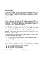
Fig. 8. Same patient as in figures 6 and 7. Encroachment of the bone onto the anterior
crus of the stapes is visible (from Dort et al. [6]). The asterisk indicates the stapes.
Fig. 9. Sclerosteosis. Part of the dome of the skull was removed from Miss. W., and
then replaced by a thin acrylic prosthesis. The posterior fossa was decompressed at a second
operation.
The Middle Ear in Sclerosing Bone Dysplasias 67
suddenly go into coma and die within hours. Emergency craniectomy is life
saving. When elective craniectomy is done, the dome of the skull is removed
and thinned by drilling on the internal surface of the skull cap [4]. This requires
extensive drilling, and we hope that laser techniques, e.g. femtosecond laser,
may be developed one day to cut this bone. The use of the presently available
lasers for middle ear surgery has not been successful because of the very thick
bone. Also, drilling on the otic capsule results in some loss of hearing in the
high tones due to the noise of the drill.
References
1 Hamersma H, Gardner J, Beighton P: The natural history of sclerosteosis. Clin Genet 2003;63:
192–197.
2 Van der Wouden A: Bone diseases of the temporal bone with hearing disorders [Leiden]. Thesis,
1971.
3 Hamersma H: Total decompression of the facial nerve in osteopetrosis (marble bone disease –
morbus Albers-Schönberg). ORL J Otorhinolaryngol Relat Spec 1974;36:21–32.
4 Du Plessis JJ: Sclerosteosis: neurosurgical experience with 14 cases. J Neurosurg 1993;78:
388–392.
5 Schuknecht HF: Pathology of the Ear, ed 2. Philadelphia, Lea and Febiger, 1993.
6 Dort JC, Pollak A, Fisch U: The fallopian canal and facial nerve in sclerosteosis of the temporal
bone: a histopathologic study. Am J Otol 1990;11:320–325.
Herman Hamersma, MD
Flora Clinic
Roodepoort (South Africa)
Arnold W, Häusler R (eds): Otosclerosis and Stapes Surgery.
Adv Otorhinolaryngol. Basel, Karger, 2007, vol 65, pp 68–74
Molecular Biology of Otosclerosis
Michael J. McKenna
a,b
, Arthur G. Kristiansen
b
a
Department of Otology and Laryngology, Harvard Medical School,
b
Department of
Otolaryngology, Massachusetts Eye and Ear Infirmary, Boston, Mass., USA
Abstract
Otosclerosis is a bone disease of the human otic capsule, which is among the most
common causes of acquired hearing loss. The pathologic process is characterized by a wave
of abnormal bone remodeling in specific sites of predilection within the endochondral layer
of the temporal bone. Although the cause of otosclerosis remains uncertain, there is a clear
genetic predisposition with half of all cases occurring in families with more than one
affected member. There is also compelling evidence that measles virus may play a role in
some cases. Ultimately, how genetic factors and viral infection result in otosclerosis must be
explained by effects on the molecular factors that control bone remodeling.
Copyright © 2007 S. Karger AG, Basel
Unlike all other bones in the body, the human otic capsule undergoes very
little remodeling following development. Otosclerosis is a process of pathologic
remodeling within a bone that is normally refractory to remodeling. Funda-
mental to elucidating the molecular biology of otosclerosis is an understanding
of the molecular factors that promote and inhibit bone remodeling. Bone is a
dynamic tissue controlled by various biochemical, hormonal and biomechanical
stimuli. Cytokine factors that include osteoprotegerin (OPG), receptor activator
of nuclear factor kappa B (RANK) and RANK ligand (RANK-L) play a major
role in the system that directly controls bone turnover. RANK-L is expressed in
a variety of cells including osteoblasts. RANK-L expressed by osteoblasts that
are involved in bone turnover promotes differentiation (in the presence of
macrophage stimulating factor) [1], activation [2] and survival [3] of osteoclasts
by activation of its specific receptor RANK on osteoclasts. OPG acts as a solu-
ble neutralizing antagonist that binds and inactivates RANK-L [4]. OPG inhibits
the differentiation, survival and fusion of osteoclastic precursor cells, suppresses
activation, and promotes apoptosis of osteoclasts [5].
Molecular Biology, Genetics, Etiopathology
Otosclerosis Molecular Biology 69
At the cellular level, bone turnover follows a pattern of bone resorption by
osteoclasts derived from monocytic/macrophagic lineage followed by new bone
formation by osteoblasts that differentiate from pluripotent mesenchymal stem
cells. The molecular coordination of the remodeling process is influenced by a
large number of factors, most of which act by influencing OPG, RANK, and
RANK-L.
Although the factors that serve to inhibit postdevelopmental remodeling
within the otic capsule have yet to be established, there is recent evidence
to suggest that OPG which is produced within the spiral ligament, secreted
into the perilymph, and diffuses into the surrounding bone may be an important
factor [6].
Genetics and Otosclerosis
Otosclerosis is most common among whites, uncommon among Asians,
and extremely rare in blacks. Otosclerosis is estimated to occur histologically in
10% of the white population and results in hearing loss in approximately 1% [7, 8].
The clinical prevalence of otosclerosis is estimated to be twice as common in
females as in males [9].
Familial aggregation of individuals affected by otosclerosis has been rec-
ognized for many years [10]. The most compelling evidence for an underlying
genetic cause for otosclerosis comes from monozygotic twins with clinical oto-
sclerosis [11, 12] in which concordance has been found in nearly all cases.
However, because information does not exist on the genetic transmission of his-
tologic otosclerosis, it is not known whether the genetic basis of inheritance is
related to the formation of an otosclerotic focus within the temporal bone or the
tendency for a lesion to progress once it has begun, or both. Most studies on
families with otosclerosis support a pattern of autosomal dominant transmis-
sion with incomplete penetrance [13–16]. A recent study on 65 pedigrees with
otosclerosis in Tunisia suggests that otosclerosis is primarily heterogenetic, and
that in 13% of the clinical cases studied, affected individuals carry a dominant
gene with nearly complete penetrance [17]. Linkage studies between otosclero-
sis and the ABO, MN, and Rh blood groups and haptoglobin genotypes have
failed to show evidence for linkage [16]. Linkage analysis of three large and
unrelated families has revealed linkage to at least three separate loci indicating
that otosclerosis is heterogenetic [18–20]. Each of these families is atypical in
that the penetrance is nearly complete with approximately half of all individuals
in each family being affected.
Although a strong familial component exists, several studies have repor-
ted that sporadic otosclerosis represents 40–50% of all clinical cases [14–16,
McKenna/Kristiansen 70
21–23]. There appears to be no significant difference in the degree of clinical
severity between sporadic and familial cases [16].
There is evidence to suggest that some cases of otosclerosis may be related
to defects in the expression of the COL1A1 gene. Association analysis has
revealed a significant association between both familial and sporadic cases of
clinical otosclerosis and the COL1A1 gene using multiple polymorphic markers
within the COL1A1 gene [24]. The association has been found to increase from
the 3-prime to the 5-prime region of the gene. Studies of the allelic expression of
the COL1A1 gene in patients with clinical otosclerosis have revealed reduced
expression of one COL1A1 allele in some cases, similar to that which has been
described in many cases of type 1 osteogenesis imperfecta [25–28]. Type 1
osteogenesis imperfecta shares both clinical and histologic similarities with oto-
sclerosis. Approximately half of all patients with type 1 osteogenesis imperfecta
develop hearing loss that is clinically indistinguishable from clinical otosclerosis
[29, 30]. It is also well known that some patients with clinical otosclerosis have
blue sclerae [31], a feature that is found in virtually all patients with type 1 osteo-
genesis imperfecta [32]. The histopathology of temporal bones from patients
with type 1 osteogenesis imperfecta is identical to that observed in patients with
otosclerosis. Most patients with mild osteogenesis imperfecta and conductive
hearing loss have mutations in the COL1A1 gene [33]. Additional studies on the
association of COL1A1 and otosclerosis have revealed an even more significant
association between clinical otosclerosis, both familial and sporadic, and an Sp1
binding site polymorphism in the first intron of the COL1A1 gene [34]. A simi-
lar and practically identical association has been described between osteoporosis
and the Sp1 binding site in the first intron of the COL1A1 gene. A preliminary
study has demonstrated that osteoporosis may be more common in patients with
otosclerosis, and these two common bone diseases may share an underlying
molecular pathologic mechanism [35].
Measles Virus and Otosclerosis
The possibility that otosclerosis may be related to a persistent viral infec-
tion of the bone was first considered because of the similarity between otoscle-
rosis and Paget’s disease of the bone, and the mounting evidence of a viral
etiology in Paget’s disease [36, 37]. The evidence which has emerged thus far is
suggestive of a possible persistent measles virus infection similar to what
occurs in the central nervous system in subacute sclerosing panencephalitis.
Support for this hypothesis comes from ultrastructural and immunohistochemi-
cal evidence of measles-like structures and antigenicity in active otosclerotic
lesions [38–40]. In addition, measles RNA has been found in archival and fresh
Otosclerosis Molecular Biology 71
footplate specimens with otosclerosis [41–44]. Elevated levels of antimeasles
antibodies have also been reported in the perilymph of patients undergoing
stapedectomy for otosclerosis as compared to controls [44]. Others have
reported lower levels of circulating antimeasles antibodies in patients with oto-
sclerosis as compared to healthy controls [45]. This hypothesis is further
strengthened by recent evidence that the incidence of otosclerosis has declined
since the introduction of measles vaccination [46].
Discussion
Otosclerosis is an abnormal remodeling process of the otic capsule, a bone
in which remodeling is extremely limited after development. It is a complex
disease with genetic heterogeneity. It could result from intrinsic abnormalities
in bone metabolism or be initiated by some other stimulus such as measles
infection, the spread and extension of which are determined by underlying
defects in bone metabolism. It is likely that a variety of gene defects result in a
similar phenotypic expression by affecting fundamental mediators of bone
remodeling.
The key factors which regulate bone remodeling are RANK which is found
on osteoclasts and their precursors, RANK-L which is produced as both a solu-
ble and membrane-bound form by osteoblasts and stromal cells in the bone
marrow, and OPG which acts as a decoy receptor for RANK-L and is produced
by osteoblasts and stromal cells. Upregulation of RANK-L results in increased
formation and activation of osteoclasts and increased bone resorption.
Upregulation of OPG results in inhibition of osteoclast formation and activity
and decreased bone resorption. Each of these factors is subject to a complexity
of upstream and downstream regulation by a variety of hormones, cytokines
and transcription factors.
Several studies have examined the effects of measles infection on bone
cells and the above-mentioned pathway. Measles infection and cells transduced
with measles gene products express increased amounts of RANK and appear to
be capable of RANK activation independent of RANK-L. Furthermore, inflam-
matory cytokines such as IL-1, TNF-␣, and IL-6 result in further upregulation
of RANK and RANK-L. It is clear from these studies that measles infection can
have direct effects that result in active resorption and remodeling.
Perhaps most fundamental to understanding the molecular biology of oto-
sclerosis is elucidation of the factors which serve to uniquely inhibit bone
remodeling in the otic capsule. The elegant studies of Frisch et al. [47, 48] have
demonstrated that otic capsule remodeling is most reduced in proximity to the
inner ear. We have recently found that OPG is produced in high quantity within
McKenna/Kristiansen 72
the spiral ligament and directly secreted into the perilymph. We have also
shown that proteins within the perilymph can diffuse into the surrounding otic
capsule bone. Since OPG is a potent inhibitor of osteoclast formation and acti-
vation, it may be one important factor that prevents otic capsule remodeling.
With a better understanding of the molecular factors which serve to inhibit
normal otic capsule remodeling and promote abnormal remodeling as occurs
with otosclerosis comes the possibility of developing better forms of treatment
for otosclerosis. We suspect that compounds that have been and are being devel-
oped for the treatment of other metabolic bone diseases such as Paget’s disease
and osteoporosis may have direct application in the treatment of otosclerosis.
Acknowledgement
This work was supported by a grant from the National Institutes of Health, the National
Institute on Deafness and Communication Disorders, RO1 DC03401.
References
1 Quinn JM, Whitty GA, Byrne RJ, Gillespie MT, Hamilton JA: The generation of highly enriched
osteoclast-lineage cell populations. Bone 2002;30:164–170.
2 Burgess TL, Qian Y, Kaufman S, Ring BD, Van G, Capparelli C, Kelley M, Hsu H, Boyle WJ,
Dunstan CR, Hu S, Lacey DL: The ligand for osteoprotegerin (OPGL) directly activates mature
osteoclasts. J Cell Biol 1999;145:527–538.
3 Lacey DL, Tan HL, Lu J, Kaufman S, Van G, Qiu W, Rattan A, Scully S, Fletcher F, Juan T, Kelley M,
Burgess TL, Boyle WJ, Polverino AJ: Osteoprotegerin ligand modulates murine osteoclast sur-
vival in vitro and in vivo. Am J Pathol 2000;157:435–448.
4 Lacey DL, Timms E, Tan HL, Kelley MJ, Dunstan CR, Burgess T, Elliott R, Colombero A, Elliott G,
Scully S, Hsu H, Sullivan J, Hawkins N, Davy E, Capparelli C, Eli A, Qian YX, Kaufman S,
Sarosi I, Shalhoub V, Senaldi G, Guo J, Delaney J, Boyle WJ: Osteoprotegerin ligand is a cytokine
that regulates osteoclast differentiation and activation. Cell 1998;93:165–176.
5 Udagawa N, Takahashi N, Yasuda H, Mizuno A, Itoh K, Ueno Y, Shinki T, Gillespie MT, Martin TJ,
Higashio K, Suda T: Osteoprotegerin produced by osteoblasts is an important regulator in osteo-
clast development and function. Endocrinology 2000;141:3478–3484.
6 Zehnder AF, Kristiansen AG, Adams JC, Merchant SN, McKenna MJ: Osteoprotegerin in the inner
ear may inhibit bone remodeling in the otic capsule. Laryngoscope 2005:115:172–177.
7 Engstrom H: Über das Vorkommen der Otosklerose nebst experimentellen Studien über chirurgis-
che Behandlung der Krankheit. Acta Otolaryngol 1940;43S:1–153.
8 Guild SR: Histologic otosclerosis. Ann Otol Rhinol Laryngol 1944;53:246–267.
9 Pearson RD, Kurland LT, Cody DT: Incidence of diagnosed clinical otosclerosis. Arch
Otolaryngol 1974;99:288–291.
10 Albrecht W: Über die Vererbung der hereditären Labyrinthschwerhörigkeit und der Otosklerose.
Arch Ohren Nasen Kehlkopfheilkd 1922;110:15–48.
11 Fowler EP: Otosclerosis in identical twins. A study of 40 pairs. Arch Otolaryngol 1966;83:
324–328.
12 Hammerschlag V: Zur Frage der Vererbbarkeit der Otosklerose. Wien Klin Radsch 1905;19:5–7.
13 Causse JR, Causse JB: Otospongiosis as a genetic disease. Early detection, medical management,
and prevention. Am J Otol 1984;5:211–223.
Otosclerosis Molecular Biology 73
14 Gapanavichyus BM, Venslauskas MI: Genetic analysis of the inheritance of otosclerosis. Sov
Genet 1974;8:251–260.
15 Larsson A: Otosclerosis. A genetic and clinical study. Acta Otolaryngol 1960;154(suppl):1–86.
16 Morrison AW: Genetic factors in otosclerosis. Ann R Coll Surg Engl 1967;41:202–237.
17 Ben Arab S, Besbes G, Hachicha S: Otosclerosis in populations living in northern Tunisia: epi-
demiology and etiology. Ann Otolaryngol Chir Cervicofac 2001;118:19–25.
18 Chen W, Campbell CA, Green GE, Van Den Bogaert K, Komodikis C, Manolidis LS, Aconomou E,
Kyamides Y, Christodoulou K, Faghel C, Giguere CM, Alford RL, Manolidis S, Van Camp G,
Smith RJ: Linkage of otosclerosis to a third locus (OTSC3) on human chromosome 6p21.3–22.3.
J Med Genet 2002;39:473–477.
19 Tomek MS, Brown MR, Mani SR, Ramesh A, Srisailapathy CR, Coucke P, Zbar RI, Bell AM,
McGuirt WT, Fukushima K, Willems PJ, Van Camp G, Smith RJ: Localization of a gene for oto-
sclerosis to chromosome 15q25–q26. Hum Mol Genet 1998;7:285–290.
20 Van Den Bogaert K, Govaerts PJ, Schatteman I, Brown MR, Caethoven G, Offeciers FE, Somers T,
Declau F, Coucke P, Van de Heyning P, Smith RJ, Van Camp G: A second gene for otosclerosis,
OTSC2, maps to chromosome 7q34–36. Am J Hum Genet 2001;68:495–500.
21 Cawthorne T: Otosclerosis. J Laryngol Otol 1955;69:437–456.
22 Nager FR: Zur klinischen und pathologischen Anatomie der Otosklerose. Acta Otolaryngol
1939;27:542–551.
23 Shambaugh GE: Fenestration operation for otosclerosis. Acta Otolaryngol Suppl 1949;79:1–101.
24 McKenna MJ, Kristiansen AG, Bartley ML, Rogus JJ, Haines JL: Association of COL1A1 and
otosclerosis: evidence for a shared genetic etiology with mild osteogenesis imperfecta. Am J Otol
1998;19:604–610.
25 McKenna MJ, Kristiansen AG, Tropitzsch AS: Similar COL1A1 expression in fibroblasts from
some patients with clinical otosclerosis and those with type I osteogenesis imperfecta. Ann Otol
Rhinol Laryngol 2002;111:184–189.
26 Willing MC, Deschenes SP, Scott DA, Byers PH, Slayton RL, Pitts SH, Arikat H, Roberts EJ:
Osteogenesis imperfecta type I: molecular heterogeneity for COL1A1 null alleles of type I colla-
gen. Am J Hum Genet 1994;55:638–647.
27 Willing MC, Deschenes SP, Slayton RL, Roberts EJ: Premature chain termination is a unifying
mechanism for COL1A1 null alleles in osteogenesis imperfecta type I cell strains. Am J Hum
Genet 1996;59:799–809.
28 Willing MC, Pruchno CJ, Atkinson M, Byers PH: Osteogenesis imperfecta type I is commonly due
to a COL1A1 null allele of type I collagen. Am J Hum Genet 1992;51:508–515.
29 Nager GT: Osteogenesis imperfecta of the temporal bone and its relation to otosclerosis. Ann Otol
Rhinol Laryngol 1988;97:585–593.
30 Ziyeh S, Berger R, Reisner K: MRI-visible pericochlear lesions in osteogenesis imperfecta type I.
Eur Radiol 2000;10:1675–1677.
31 Fowler E: The incidence (and degrees) of blue sclerae in otosclerosis and other ear disorders.
Laryngoscope 1949;59:406.
32 Garretsen TJ, Cremers CW: Clinical and genetic aspects in autosomal dominant inherited osteoge-
nesis imperfecta type I. Ann NY Acad Sci 1991;630:240–248.
33 Sykes B, Ogilvie D, Wordsworth P, Wallis G, Mathew C, Beighton P, Nicholls A, Pope FM,
Thompson E, Tsipouras P, et al: Consistent linkage of dominantly inherited osteogenesis imper-
fecta to the type I collagen loci: COL1A1 and COL1A2. Am J Hum Genet 1990;46:293–307.
34 McKenna MJ, Nguyen-Huynh AT, Kristiansen AG: Association of otosclerosis with Sp1 binding
site polymorphism in COL1A1 gene: evidence for a shared genetic etiology with osteoporosis.
Otol Neurotol 2004;25:447–450.
35 Clayton AE, Mikulec AA, Mikulec KH, Merchant SN, McKenna MJ: Association between osteo-
porosis and otosclerosis in women. J Laryngol Otol 2004;118:617–621.
36 Friedrichs WE, Reddy SV, Bruder JM, Cundy T, Cornish J, Singer FR, Roodman GD: Sequence
analysis of measles virus nucleocapsid transcripts in patients with Paget’s disease. J Bone Miner
Res 2002;17:145–151.
37 Mee AP: Paramyxoviruses and Paget’s disease: the affirmative view. Bone 1999;24(suppl 5):
19S–21S.
McKenna/Kristiansen 74
38 McKenna MJ, Mills BG: Immunohistochemical evidence of measles virus antigens in active oto-
sclerosis. Otolaryngol Head Neck Surg 1989;101:415–421.
39 McKenna MJ, Mills BG: Ultrastructural and immunohistochemical evidence of measles virus in
active otosclerosis. Acta Otolaryngol Suppl 1990;470:130–140.
40 McKenna MJ, Mills BG, Galey FR, Linthicum FH Jr: Filamentous structures morphologically
similar to viral nucleocapsids in otosclerotic lesions in two patients. Am J Otol 1986;7:25–28.
41 Karosi T, Konya J, Szabo LZ, Sziklai I: Measles virus prevalence in otosclerotic stapes footplate
samples. Otol Neurotol 2004;25:451–456.
42 McKenna MJ, Kristiansen AG, Haines J: Polymerase chain reaction amplification of a measles
virus sequence from human temporal bone sections with active otosclerosis. Am J Otol 1996;17:
827–830.
43 Niedermeyer H, Arnold W, Neubert WJ, Hofler H: Evidence of measles virus RNA in otosclerotic
tissue. ORL J Otorhinolaryngol Relat Spec 1994;56:130–132.
44 Niedermeyer HP, Arnold W: Otosclerosis: a measles virus associated inflammatory disease. Acta
Otolaryngol 1995;115:300–303.
45 Lolov SR, Encheva VI, Kyurkchiev SD, Edrev GE, Kehayov IR: Antimeasles immunoglobulin G
in sera of patients with otosclerosis is lower than that in healthy people. Otol Neurotol 2001;22:
766–770.
46 Vrabec JT, Coker NJ: Stapes surgery in the United States. Otol Neurotol 2004;25:465–469.
47 Frisch T, Sorensen MS, Overgaard S, Bretlau P: Estimation of volume referent bone turnover in
the otic capsule after sequential point labeling. Ann Otol Rhinol Laryngol 2000;109:33–39.
48 Frisch T, Sorensen MS, Overgaard S, Bretlau P: Predilection of otosclerotic foci related to the
bone turnover in the otic capsule. Acta Otolaryngol Suppl 2000;543:111–113.
Michael J. McKenna, MD
Department of Otolaryngology
Massachusetts Eye and Ear Infirmary, 243 Charles Street
Boston, MA 02114–3096 (USA)
Tel. ϩ1 617 573 3672, Fax ϩ1 617 573 3939, E-Mail
Arnold W, Häusler R (eds): Otosclerosis and Stapes Surgery.
Adv Otorhinolaryngol. Basel, Karger, 2007, vol 65, pp 75–85
The Genetics of Otosclerosis: Pedigree
Studies and Linkage Analysis
S.R. Saeed, M. Briggs, C. Lobo, F. Al-Zoubi,
R.T. Ramsden, A.P. Read
University Department of Otolaryngology-Head and Neck Surgery,
Manchester Royal Infirmary and Department of Clinical Genetics,
St. Mary’s Hospital, Manchester, UK
Abstract
Otosclerosis is one of the commonest causes of hearing loss in adults. The hereditary
nature of the disease has been acknowledged for over a century but the precise genetic basis
of the disorder has as yet not been characterised. It is currently recognised that familial oto-
sclerosis exhibits autosomal dominant inheritance with variable penetrance and expression.
More recently, family linkage studies have identified three chromosomal regions that can be
ascribed to this disorder: otosclerosis 1 on chromosome 15, otosclerosis 2 on chromosome 7
and a third locus on chromosome 6. The genes responsible for the disease within these
regions remain to be defined. The work presented in this paper firstly examined the familial
nature of the disease in a cohort of individuals that had undergone surgery for otosclerosis.
Following detailed ascertainment, pedigrees were constructed for subsequent genetic analysis.
The laboratory analysis included linkage analysis of the candidate region on the long arm of
chromosome 15, linkage analysis of the aggrecan protein gene within the 15q region and
linkage analysis to chromosome 7q. The pedigree studies confirmed the hereditary nature of
otosclerosis and the recognised mode of inheritance. Linkage to the chromosome 15 locus,
the candidate aggrecan gene and the chromosome 7 locus was excluded, confirming that oto-
sclerosis exhibits locus heterogeneity.
Copyright © 2007 S. Karger AG, Basel
The hereditary nature of otosclerosis has been recognised for nearly 150
years [1]. Despite this, the precise genetic basis of the disorder remains to be
defined. The aims of this study were firstly to confirm the familial nature of the
disease and secondly to examine two of the specific chromosomal loci that have
been described as harbouring genes implicated in the pathogenesis of otosclero-
sis, OTSC1 and OTSC2 [2, 3].
Saeed/Briggs/Lobo/Al-Zoubi/Ramsden/Read 76
Materials and Methods
Source of Data
The starting point for the acquisition of study material was individuals in whom the
diagnosis of otosclerosis had been confirmed surgically. The operating theatre records for the
period from 1990 to 1995 were examined retrospectively by the authors and prospectively
during the study at several hospitals in the North-West of England. In addition, a single large
family with otosclerosis was identified by colleagues in Leeds and made available for col-
laborative research. Details were also made available from the personal stapedectomy series
of an otologist in London. The details of all these patients were entered into a database con-
structed by the authors under the regulations of the Data Protection Act.
Identification of Potentially Informative Families
The case notes of the patients identified from the operating theatre records were indi-
vidually studied for historical evidence of a positive family history of otosclerosis. Ethical
approval for subsequent contact and entry into the study of those patients with a family his-
tory was confirmed by the Manchester Health Authority. Those patients consenting to take
part in the study were then either evaluated by the first author in the University Department
of Audiology, Manchester, or at the individual’s home.
Construction of Pedigrees
Detailed ascertainment of families through the index case was undertaken historically
and pedigrees constructed. Evaluation of index cases and relatives included otoscopic and
tuning fork examination, audiometric analysis and the collection of a venous blood sample.
Individuals were designated as affected or unaffected on this basis. The surgically confirmed
affected status of individuals in a family was also noted.
Audiometric Analysis
Pure-tone audiometry was undertaken by the authors in all individuals available for
evaluation in accordance with the guidelines of the British Society of Audiology. Where indi-
cated, masking of the non-test ear was undertaken in accordance with accepted guidelines.
Hard copies of the pure-tone audiograms were made for subsequent analysis.
Collection of Blood Samples and Extraction of Constitutional DNA
Between 10 and 20 ml of venous blood was collected from each individual studied and
anticoagulated with ethylenediaminetetraacetic acid (0.5ml 0.5 M, pH 8.0). Constitutional
DNA was extracted from leucocytes from the venous blood samples using standard techniques.
The homogenous solution of DNA was stored at Ϫ70ЊC until required.
Stock DNA was diluted with deionised water in the ratio DNA 10 l:1,000 l deionised
water. The optical density machine was calibrated with a 1-ml deionised water blank and the opti-
cal density of the DNA solution was read at 260 nm against the water blank in a quartz cuvette.
Chromosome 15q and 7q Linkage Analysis
Linkage analysis using microsatellite markers for the 14.5-cM region on chromosome
15 (15q25–q26) and the 16-cM region on chromosome 7 (7q33–q36) was undertaken in
families A–G. A summary of the oligonucleotide primers utilised for the study is presented in
Genetics of Otosclerosis 77
table 1. The forward primer in each pair was fluorescently labelled. The DNA was diluted with
deionised water to give 50–100 ng of genomic DNA per reaction. The reaction volumes were
all made up to 10 l with deionised water. The PCRs were set up and run in an automated ther-
mal cycler. Following an initial step at 93ЊC for 3 min, 40 cycles were utilised with the follow-
ing reaction conditions: denaturing at 94ЊC for 1 min, annealing at 55ЊC for 1 min and
synthesis at 72ЊC for 1 min. The fluorescent product for each primer pair was visualised on an
automated ABI PRISM 377 DNA sequencer/genotyper and computed using the Genotyper
®
1.1.1 software. Multipoint linkage analysis was undertaken using Genehunter 2.1.
Aggrecan Gene Analysis
A summary of the intragenic oligonucleotide primer utilised for this study is presented
in table 2. The DNA was diluted with deionised water to give 50–100 ng of genomic DNA
per reaction. The reaction volumes were all made up to 10 l with deionised water. The PCRs
were set up and run in an automated thermal cycler. Forty cycles were utilised with the fol-
lowing reaction conditions: denaturing at 94ЊC for 1min, annealing at 67ЊC for 1 min and
synthesis at 70ЊC for 2 min. The amplified products were separated on a 3.5% agarose gel
and visualised with ethidium bromide staining.
Results
Identification of Potentially Informative Families
The demographic details of individuals that had undergone stapes surgery
for otosclerosis were entered into a database. In total, 225 such individuals were
Table 1. Summary details for the oligonucleotides used
for the chromosome 15q and 7q studies
Locus Oligonucleotide markers
15q25–q26 D15S996
D15S127
D15S158
D15S963
D15S652
D15S531
D15S1004
D15S649
D15S130
D15S657
7q33–q36 D7S509
D7S497
D7S2560
D7S684
D7S2513
D7S2426
Saeed/Briggs/Lobo/Al-Zoubi/Ramsden/Read 78
identified and the number of subjects with a positive family history for each
source of data is summarised in table 3.
The 33 individuals in London that had undergone stapes surgery and had a
positive family history were not ascertained during the study period. The 35 indi-
viduals from the North of England that had undergone stapes surgery and had a
positive family history according to their clinical records were contacted by the
methods described previously. Of these individuals, there was no response despite
repeated attempts to contact them in 3 individuals and 11 individuals did not in
fact have a positive family history on direct questioning. The remaining 21 index
cases were ascertained in detail. Of these, 2 did not have a positive family history
and in 1 the affected status of the relative was equivocal. In 5 families, the only
living affected individual was the proband. In 3 families, there were 2 living
affected individuals including the proband and there were 6 families with 3
affected living individuals. One family contained 4 living affected individuals
including the proband and in 3 families, there were 5 or more affected individuals
still alive. These findings are summarised in table 4.
Chromosome 15q Studies
The 7 study families were typed for the candidate region using between 3
and 8 markers. One family was uninformative and one family generated a small
Table 2. Summary details for the oligonucleotides used for the aggrecan gene study
Marker Primer sequence Allele size, bp Heterozygosity, % Reference
AGC1.PCR forward: 5Ј-TAGAGGGCTCTG 775–1,915 70 [4]
CCTCTGGAGTTG-3Ј
reverse: 5Ј-AGGTCCCCTACCG
CAGAGGTAGAA-3Ј
Table 3. Patient data summary
Region Number Positive family history Sex ratio (M:F)
North of England 97 35 (36)
a
0.39
London 128 33 (26) 0.50
Total 225 68 (30) 0.46
Figures in parentheses indicate percentages.
a
Nineteen percent after more detailed ascertainment.
Genetics of Otosclerosis 79
positive lod score. The remaining 5 families generated maximum lod scores bet-
ween Ϫ3.62 and –5.00 at 0% recombination (table 5), thereby excluding linkage
to the candidate region in the study material.
Chromosome 7q and Aggrecan Studies
Parametric multipoint linkage analysis to chromosome 7q generated a
maximum lod score of –0.4, excluding linkage. Non-parametric linkage analysis
also excluded linkage to 7q.
Six of the 7 study families were also typed for linkage to the aggrecan gene.
One family was uninformative (family E) and 3 families generated a weak posi-
tive lod score (families D, F and G). The 2 remaining larger families (B and C)
generated maximum lod scores of Ϫ4.30 and Ϫ4.03 at 0% recombination,
thereby excluding linkage to the aggrecan gene.
Table 4. Family history details for the North of England
families
Family history Number of families
Not contactable 3
No true/equivocal family history 14
Only proband alive 5
2 affected alive 3
3 affected alive 6
4 affected alive 1
5 or more affected alive 3
Total 35
Table 5. Maximum lod score at 0% recombination
Family Maximum lod score at 0% recombination
A –3.62
B –5.00
C –4.21
D –4.09
E 0.40
F 0.00
G –4.33
Saeed/Briggs/Lobo/Al-Zoubi/Ramsden/Read 80
Discussion
Despite the fact that the hereditary nature of otosclerosis has been recog-
nised for 140 years [1], the precise genetic basis of the disease and its patho-
genesis remain elusive. Recent advances in molecular biology have given a
new impetus to the study of deafness genetics and this is reflected in the ongo-
ing recognition and refinement of the genetics of syndromic and non-
syndromic hereditary hearing loss [5, 6]. Whilst otosclerosis is one of the
commonest causes of familial hearing loss, there are a number of reasons why
to date, the genes implicated in the development of the disease have not been
defined. Firstly, in a given individual presenting with hearing loss, the diagno-
sis is presumptive rather than absolute, based on the history, clinical examina-
tion and audiometric findings being compatible with such a diagnosis. The
diagnosis is proven either at surgery or if high-resolution computed tomogra-
phy scanning (CT scanning) demonstrates the lesion in the oval window or otic
capsule. Since a negative CT scan does not exclude the disease and many indi-
viduals presenting with deafness due to otosclerosis elect not to undergo
surgery, the phenotype of the index case and kindred cannot be ascribed with
absolute certainty. Secondly, even if the index case is defined as one that has
undergone surgery for otosclerosis, the construction of informative pedigrees
can be difficult. The fundamental starting point in the study of a familial dis-
order is the ascertainment of cases and the construction of robust pedigrees.
For a disorder that shows an autosomal dominant mode of inheritance, the
ideal family for linkage analysis comprises around 10 informative meioses
allowing the identification of recombinant individuals with unambiguous
characterisation of the affected and unaffected individuals in the family. These
criteria are rarely met, particularly in smaller families in the Western World. In
addition, anamnestic data may be unreliable. On this basis, in order to reach
statistical significance, data from several smaller families are combined using
the lod score as a statistical tool. The identification of affected and unaffected
individuals is further hampered by the fact that the classical mendelian inheri-
tance of otosclerosis is complicated by incomplete penetrance and variable
expression. This would account for the difference between clinical and histo-
logical otosclerosis as described by Guild in 1944 [7]. Despite these difficul-
ties, otosclerosis is a recognised familial deafness disorder and as such should
be amenable to the application of modern clinical and molecular biological
techniques. Such techniques have been applied successfully to less common
familial deafness disorders such as non-syndromic hereditary hearing loss,
Usher’s disease and Wardenburg’s syndrome [8]. Many of the studies charac-
terising the genetics of these disorders have been collaborative research
projects in large kindreds emanating from areas such as the Middle East.
Genetics of Otosclerosis 81
In addition, these studies have not been hampered by incomplete penetrance
and are based on a firm audiometric diagnosis. Finally, in otosclerosis the
pathological lesion itself is not generally available for molecular analysis
unless at surgery the stapes is inadvertently removed in its entirety. This is in
contrast to the familial cancer syndrome neurofibromatosis type II (NF2) in
which the hearing loss is due to the presence of bilateral vestibular schwanno-
mas (acoustic neuromas). At the time of surgical removal, tumour samples
together with a sample of the individual’s blood can be made available for
genetic studies. This has led to novel molecular studies culminating in the
detailed genetic characterisation of this familial disorder and ongoing research
into the molecular biology of the sporadic form of the disease in which individ-
uals develop a unilateral tumour [9, 10]. Such a strategy would be immensely
helpful in the molecular study of otosclerosis.
Pedigree Studies
The fundamental basis of this study was a group of individuals that had
undergone surgery for otosclerosis in several Otolaryngology Departments in
the North of England over a 6-year period. These 225 individuals were therefore
a selected group and not representative of the population suffering with otoscle-
rosis as a whole for several reasons. The first of these is motivation on the part of
the person presenting for a specialist opinion. Such individuals would have
sought medical advice at a point when he or she felt that their hearing loss was
causing a significant disability in terms of day-to-day living or work. This is
more likely to be the case the greater the hearing loss and in particular if both
ears are affected by the disease process. The point at which a given hearing
loss constitutes a disability is variable from individual to individual and is itself
subject to differences between people depending on their age, sex, vocation,
lifestyle and expectations. In addition, some individuals may have positive or
indeed negative family experiences of the management of otosclerosis which
will also influence their own threshold for seeking medical advice. Secondly,
once a presumptive diagnosis of otosclerosis is reached by the clinician, three
management options are available to the patient: observation, provision of a
hearing aid or surgery. This process is also subject to bias depending on the sur-
geon’s understanding and experience of the management of this disorder. Finally,
any retrospective case record study will be subject to the vagaries of hospital
record keeping. This was found to be the case in several instances where the clin-
ician seeing the patient had not documented the presence or absence of a family
history despite dealing with a patient with a recognised familial disorder. The
issues raised in this discussion are therefore based on selected material subject to
analysis and cannot be extrapolated to the population suffering with otosclerosis
at an epidemiological level.
Saeed/Briggs/Lobo/Al-Zoubi/Ramsden/Read 82
The findings from this study confirm that otosclerosis can be a familial dis-
order. Interestingly, prior to detailed ascertainment, 36% of the North of England
index cases yielded a positive family history. Following detailed assessment, this
figure fell to 19%. This serves to highlight the dangers of relying on anamnestic
data, a point made by Larsson in 1960 [11]. Patients will not necessarily differ-
entiate between otosclerosis and other causes of deafness when questioned about
the presence or absence of hearing loss in their relatives. Commonly deafness in
relatives due to the aging process or suppurative middle ear disease will be
assumed to be due to otosclerosis by the index case. The issue is only clarified
once the relatives of the proband are ascertained and this study confirms the
great importance of examining the relatives of the index case to avoid erroneous
conclusions [12]. Based on this study, the prevalence of familial otosclerosis is
close to 1 in 5, which is lower than the accepted figures in the literature which
range from 30 to 50% [13, 14]. The wide range probably reflects a selection bias
and variable ascertainment of relatives of the proband. The male to female ratio
however for affected individuals in this series was close to 40%, which is com-
parable to other epidemiological studies. The number of affected individuals
within a pedigree is subject to three main factors: the size of the family, incom-
plete penetrance and variable expression. This study group confirms this obser-
vation in that the number of living affected individuals in the pedigree ranged
from 1 to 10. However, only 4 pedigrees had 4 or more living affected individu-
als and this illustrates one of the difficulties cited previously in subjecting such
families to linkage analysis. Overall family size is an important determinant and
it is well recognised that family size in part is subject to personal, religious, cul-
tural and political factors. Consanguinity however was not a complicating factor
in this study. Larger families in a population in whom otosclerosis is prevalent
such as those seen in the Indian subcontinent are in theory a good source for
linkage analysis and indeed the study by Tomek et al. [2] was based on a large
Indian kindred.
The process of detailed ascertainment was based on a clinical and audio-
metric assessment. In those individuals that had undergone surgery for otoscle-
rosis the affected status was secure. Characterisation of affected and unaffected
non-operated relatives however poses a potential problem as the characterisa-
tion is necessarily presumptive. The presence of a normal tympanic membrane
which is mobile on pneumatic otoscopy, appropriate tuning fork test results, a
type A tympanogram and a characteristic conductive hearing loss on pure-tone
audiometry is highly suggestive of a diagnosis of otosclerosis. This is particu-
larly the case if there is a known affected relative such as the proband. However,
congenital or acquired fixation of the malleus or incus will give exactly the
same picture though incudomalleolar fixation is considerably rarer than the
stapedial fixation seen in otosclerosis. In this respect, the history of the hearing
Genetics of Otosclerosis 83
loss assumes considerable importance. The sensitivity and specificity of tuning
fork tests have been debated amongst otolaryngologists in more recent times
and are probably unreliable as a single screening modality [15]. In addition,
field audiometry is subject to errors due to adverse environmental issues such
as noise. In reality, classification of relatives as affected or unaffected proved
less difficult than anticipated as the process is a constellation of clinical and
audiometric compatibility. In those cases where doubt remained, audiometry
was repeated at the university and unless unequivocal, the individual was
labelled as clinically unaffected for the purposes of this study.
Allowing for the potential sources of error outlined above, observation of the
inheritance patterns in this group is possible. An autosomal dominant mode of
inheritance was observed in 7 of the 21 families ascertained. Of these, incomplete
penetrance was noted in 2 families. X-linked dominant inheritance was observed
in 1 family with the remaining families being too small to ascribe a mode of
inheritance. However, autosomal or X-linked recessive inheritance was not
observed. These findings are consistent with the currently accepted inheritance
patterns and characteristics in this disorder despite the study group being selected
in the manner described above. Overall, the pedigree studies were consistent with
the accumulated literature and the families were deemed suitable for subsequent
molecular studies.
Chromosome 15 and Chromosome 7 Linkage
This study was unable to provide evidence of linkage to the candidate
regions on 15q and 7q. The implication of excluding linkage to these regions is
locus heterogeneity. More recently, an additional family has been studied by the
Manchester group. The family was ascertained in India and comprises 32 indi-
viduals across 4 generations. Nine affected individuals were identified and hap-
lotypes constructed utilising the markers for the 15q candidate region in the
manner described in this paper. Typing was undertaken on 21 individuals and
again linkage to the candidate region was excluded [unpubl. data]. This adds
weight to the observation of locus heterogeneity in otosclerosis, and indeed a
third locus has been identified more recently on chromosome 6 [16].
Aggrecan Gene Linkage
The most important gene that maps to the candidate region identified by the
Tomek group is the gene for the aggrecan protein. The aggrecan gene is a good
candidate gene for the pathogenesis of otosclerosis. Six of the 7 study families
were typed for linkage to this gene. Not surprisingly, linkage was excluded (max-
imum lod scores of Ϫ4.30 and Ϫ4.03 at 0% recombination). One cannot however
conclude that the aggrecan gene is not implicated in the pathogenesis of otoscle-
rosis based on this work, as this work excluded linkage to the whole 15q region.
Saeed/Briggs/Lobo/Al-Zoubi/Ramsden/Read 84
The key step will be to look for evidence of linkage to the aggrecan locus in a
family that first links to the 15q region. Such data have not been forthcoming
from the Tomek group but the search for pedigrees that link to this region should
continue in addition to a wider genome search.
Conclusions
The following conclusions may be drawn from the studies described in this
paper.
Of the 97 individuals studied that had undergone surgery for otosclerosis,
an overall positive family history of the disease was found in 19% with a male
to female ratio of 0.39. An autosomal dominant mode of inheritance was
demonstrated with evidence of incomplete penetrance.
Linkage to the candidate regions on the long arm of chromosomes 15 and
7 was excluded in the 7 study families typed for these regions. In addition, link-
age to the aggrecan protein gene was also excluded, thereby demonstrating that
familial otosclerosis exhibits locus heterogeneity.
Clearly, the current evidence both in the literature and amongst investiga-
tors interested in this particular field warrants continuing research at a molecu-
lar level utilising pedigrees that have been ascertained in addition to families
ascertained in the future.
Acknowledgements
We thank Dr. A.R.H. Grace (York), Dr. G. Woods (Leeds) and Dr. J. Hazell (London)
for clinical material, and the Royal College of Surgeons of England and the charity Defeating
Deafness for financial support for this research.
References
1 Toynbee J: Pathological and surgical observations on the diseases of the ear. Med Chir Trans
1861;24:190–205.
2 Tomek MS, Brown MR, Mani SR, Ramesh A, Srisailapathy CRS, Coucke P, Zbar RIS, Bell AM,
McGuirt WT, Fukushima K, Willems PJ, Van Camp G, Smith RJH: Localisation of a gene for
otosclerosis to chromosome 15q25–q26. Hum Mol Genet 1998;7:285–290.
3 Van Den Bogaert K, Govaerts PJ, Schatteman I, Brown MR, Caethoven G, Offeciers FE, Somers T,
Declau F, Coucke P, Van de Heyning P, Smith RJH, Van Camp G: A second gene for otosclerosis,
OTSC2, maps to chromosome 7q34–36. Am J Hum Genet 2001;68:495–500.
4 Doege KJ, Coulter SN, Meek LM, Maslen K, Wood JG: A human-specific polymorphism in the
coding region of the aggrecan gene. Variable number of tandem repeats produce a range of core
protein sizes in the general population. J Biol Chem 1997;272:13974–13979.
Genetics of Otosclerosis 85
5 Lalwani AK, Castelein CM: Cracking the auditory genetic code: nonsyndromic hereditary hearing
impairment. Am J Otol 1999;20:115–132.
6 Tseng CJ, Lalwani AK: Cracking the auditory genetic code. 2. Syndromic hereditary hearing
impairment. Am J Otol 2000;21:437–451.
7 Guild SR: Histological otosclerosis. Ann Otol Rhinol Laryngol 1944;53:246–267.
8 Van Camp G, Smith RJH: Hereditary Hearing Loss Homepage. />9 Wu CL, Thakker N, Neary W, Black G, Lye R, Ramsden RT, Read AP, Evans DG: Differential
diagnosis of type 2 neurofibromatosis: molecular discrimination of NF2 and sporadic vestibular
schwannomas. J Med Genet 1998;35:973–977.
10 Evans DG, Sainio M, Baser ME: Neurofibromatosis type 2. J Med Genet 2000;37:897–904.
11 Larsson A: Otosclerosis: a genetic and clinical study. Acta Otolaryngol 1960;154(suppl):1–86.
12 Morrison AW: Genetic factors in otosclerosis. Ann R Coll Surg Engl 1967;41:202–237.
13 Michael AG: The genetics of otosclerosis. A review. Am J Otol 1989;10:426–438.
14 Sabitha R, Ramalingam R, Ramalingam KK, Sivakumaran TA, Ramesh A: Genetics of otosclerosis.
J Laryngol Otol 1997;111:109–112.
15 Browning GG: Clinical Otology and Audiology, ed 2. London, Butterworth-Heinemann, 1998.
16 Chen W, Campbell CA, Green GE, Van Den Bogaert K, Komodikis C, Manolidis LS, Aconomou E,
Kyamides Y, Christodoulou K, Faghel C, Giguere CN, Alford RL, Manolidis S, Van Camp G,
Smith RJH: Linkage of otosclerosis to a third locus (OTSC 3) on human chromosome 6p21.3–22.3.
J Med Genet 2002;39:473–477.
S.R. Saeed, MBBS, MD, FRCS (ORL)
University Department of Otolaryngology-Head and Neck Surgery
Manchester Royal Infirmary, Oxford Road
Manchester M13 9WL (UK)
Tel. ϩ44 161 276 4426, Fax ϩ44 161 276 5003, E-Mail
Arnold W, Häusler R (eds): Otosclerosis and Stapes Surgery.
Adv Otorhinolaryngol. Basel, Karger, 2007, vol 65, pp 86–92
Measles Virus and Otosclerosis
H.P. Niedermeyer
a
, T. Gantumur
a,b
, W.J. Neubert
b
, W. Arnold
a
a
Department of Otorhinolaryngology, Klinikum rechts der Isar, Technical University
Munich, Munich,
b
Max-Planck Institute of Biochemistry, Molecular Virology,
Martinsried, Germany
Abstract
Measles virus (MeV) might play an important role as an environmental stimulus in the
etiopathogenesis of otosclerosis. Chronic inflammation was shown in morphologic investiga-
tions of otosclerotic foci and MeV N, P, and F proteins were detected within cells of the otoscle-
rotic focus by immunohistochemical investigations. MeV RNA was extracted from fresh-frozen
otosclerotic tissue by the use of in vitro RT-PCR. This result was validated through amplification
of MeV genome sequences by RT-PCR from celloidin-embedded sections with morphologically
ascertained otosclerotic foci. In searching for an immune response of the inner ear immune
system against MeV proteins, elevated anti-MeV IgG levels were detected in the perilymph of
patients with otosclerosis in comparison with the serum levels. In situ RT-PCR allowed the local-
ization of MeV sequences in osteoclasts, osteoblasts, chondrocytes, macrophages, and epithelial
cells in middle ear mucosa of otosclerotic tissue. Further evidence for MeV persistence has
recently been given. Genotyping of MeV in otosclerotic foci demonstrated the presence of MeV
genotype A, which circulated in Europe around 1960. All the above results confirm a strong
association between MeV and otosclerosis.
Copyright © 2007 S. Karger AG, Basel
Otosclerosis was described for the first time by Antonio Maria Valsalva in
1735 [1] as a disease of the human temporal bone. More than one century later,
Toynbee [2] recognized otosclerosis as a cause of hearing loss. Otosclerosis
may occur as a histological type within the human temporal bone without affe-
cting the stapes footplate. In only 10% of patients with otosclerosis is the focus
localized near the oval window niche leading to fixation of the stapes with con-
secutive (characteristic) conductive or mixed hearing loss. Women are affected
1.4 times more frequently than men and the age of onset has risen in Caucasians
in the last decades [3]. Otosclerosis is the most important cause of hearing loss
Measles Virus and Otosclerosis 87
in Europe and the USA, whereas it appears to be uncommon in developing
countries and among the Japanese population [4].
Morphologic Analysis of the Otosclerotic Focus
Otosclerosis only affects the human temporal bone, but otosclerosis-like
lesions were observed in the crura of LP/J mice leading to conductive or com-
bined hearing loss [5].
Histologically, three different phases can be distinguished: the first phase
shows bone resorption. The tissue is highly vascularized and macrophages and
activated osteoclasts are present. The second phase is characterized by new
bone formation beginning around the vessels leading to the characteristic blue
mantles of Manasse. Finally, in the last phase, the otosclerotic focus appears as
a scar with rare cells and calcification [6].
Immunohistochemistry
A variety of immunocompetent cells including macrophages (MAC 387
antigen positive), HLA-DR-positive cells, cells expressing 
2
-microglobulin, T
suppressor cells and complement C3 were found in otosclerotic tissue by
immunohistochemical investigations [7, 8]. Deposits of immunoglobulins (IgG,
IgM and IgA) and complement C3 are present along the resorption lacunae, as
well as in osteocytes and chondrocytes surrounding the destructive process [9].
What Is the Reason for This Inflammatory Process?
The etiopathogenetic hypothesis for the development of otosclerosis includes
mechanical distress, enzymatic imbalance, disease of the collagen, and viral
infection. The current hypothesis considers otosclerosis as an inflammatory
disease with a genetic background. Five otosclerosis genes have been localized in
familial cases of otosclerosis [10, 11], but the presence of these genes could not
be confirmed by case-control studies. Mutations of collagen genes are also
discussed as a cause for the otosclerotic process. However, a genetic inheritance is
accepted in up to 50% of cases. The triggering event could be an environmental
stimulus such as a common viral infection [12].
Electronmicroscopic studies in Paget’s disease, which is histologically
very similar to otosclerosis, revealed the presence of paramyxoviral structures
in pagetic bone [13]. Analogously, filamentous structures very similar to
paramyxoviral nucleocapsids were observed in otosclerotic bone specimen
[14]. Immunohistochemical studies were undertaken to characterize these
nucleocapsid-like structures. The expression of measles virus (MeV) N, F, and
Niedermeyer/Gantumur/Neubert/Arnold 88
H antigens in osteoclasts and macrophages of otosclerotic tissue strongly sup-
ports the hypothesis that MeV is involved in otosclerosis [15–17].
Detection of MeV RNA within the Otosclerotic Tissue
Studies on the RNA level were undertaken to determine the presence of
MeV-related sequences within the otosclerotic tissue since specificity and sen-
sitivity of MeV antigen detection have been discussed controversially. Total
RNA from fresh-frozen otosclerotic bone chips obtained during stapes surgery
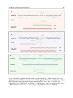
was analyzed by RT-PCR for MeV RNA (fig. 1) [18]. MeV-related sequences
were amplified in an average of 84% in several studies, whereas the negative
controls always remained negative [19–21]. A possible explanation for the neg-
ative cases is primarily the true absence of MeV in the otosclerotic tissue.
Alternatively, the absence of an otosclerotic focus might explain the negative
results, since RNA has been extracted from the stapes fragments without histo-
logic controls. Finally, technical problems and limitations of the RNA extrac-
tion technique from small eburnized bone chips have to be considered.
McKenna et al. [22] were able to detect MeV RNA in 8 out of 11 temporal
bones with morphologically confirmed otosclerotic foci. All negative controls
remained negative. The true absence of MeV within the otosclerotic tissue might
explain the 3 negative cases. However, false-negative results could be related to
technical problems which may occur dealing with celloidin-embedded tissues.
ϽϪ120bp
1234ϩϪMW
Fig. 1. Detection of MeV by RT-PCR. Lanes 1, 2, and 4 with the amplicons of the expected
length (120 bp). MW ϭ Molecular weight marker; ϩϭpositive control; Ϫϭnegative control.
Measles Virus and Otosclerosis 89
Recently, Karosi et al. [23] have found MeV RNA in 14 out of 20 fresh-frozen
footplates from patients with otosclerosis. They amplified RNA from minced
and crushed bone chips by in vitro RT-PCR. In contrast, Grayeli et al. [24] could
not confirm the presence of MeV neither in cells cultured from the otosclerotic
foci nor in bone chips after RNA extraction and amplification by RT-PCR. They
concluded that MeV is not involved in otosclerosis. However, it cannot be
excluded that the negative results are due to the absence of otosclerotic foci in
the examined tissue, as morphologic controls were not available. Furthermore,
only few copies of MeV RNA are expected in a persisting infection and highly
sensitive techniques including RNA extraction procedures are needed.
The controversial discussion about MeV RNA within the otosclerotic focus
asked for a technique such as in situ RT-PCR, which combines morphology and
amplification of the genetic material. In situ RT-PCR has been successfully used
in research on hematologic tumors, but only few studies with bony tissue are
available [25]. These studies were related to a paramyxoviral etiopathogenesis in
Paget’s disease and performed on decalcified bone. The authors demonstrated
the presence of canine distemper virus in all cases examined [26, 27]. Up to now,
we had analyzed stapes footplate specimens of 15 patients with clinical otoscle-
rosis by in situ RT-PCR. The bone chips were decalcified and paraffin embedded
and the histological examination demonstrated the presence of otosclerotic foci
within the decalcified and paraffin-embedded tissue. In all cases, osteoblasts,
osteoclasts, chondrocytes, and epithelial cells of the middle ear mucosa close to
the otosclerotic focus contained MeV RNA amplification products [unpubl. data].
Recently, we have managed to genotype the MeV within the otosclerotic
tissue. Cells cultured from otosclerotic bone chips of 5 patients had the mor-
phological and biochemical characteristics of preosteoblasts. After RNA extrac-
tion and reverse transcription, the C-terminal part of the MeV N gene was
amplified and sequenced by two independent companies. The phylogenetic
analysis revealed that all MeV were of the genotype A. This genotype was pre-
sent in Europe before the vaccination era and contains several wild-type strains
isolated before 1970. Sequencing enabled us to distinguish MeV found in our
patients from all other strains known up to now [unpubl. data]. This result
proves the persistence of the MeV genome for more than 40 years within the
temporal bone of patients with otosclerosis and excludes any speculation of
contamination or false-positive results.
MeV Antibodies within the Perilymph
The otosclerotic focus usually has intimate contact with the perilymph spa-
ces so that antigens from the otosclerotic focus might reach the immune target
Niedermeyer/Gantumur/Neubert/Arnold 90
organ localized in the endolymphatic sac [28, 29]. It is known that antigenic
stimulation of the endolymphatic sac via the perilymph can trigger a specific
immune reaction [30]. We analyzed the perilymph and serum of patients with
otosclerosis or Ménière’s disease, and of controls for the content of albumin,
total IgG and specific MeV IgG by nephelometric assay and ELISA. The MeV
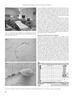
IgG fraction of total IgG was significantly higher in the perilymph compared to
the serum of patients with otosclerosis (fig. 2) [21]. In contrast, evidence for
local production of antibodies against herpes simplex virus type I was found in
patients with Ménière’s disease [31]. The reactivity of antibodies against MeV
is decreased in patients with otosclerosis [32].
Conclusion
There is convincing evidence for a chronic inflammatory reaction in oto-
sclerosis. MeV involvement was demonstrated in morphological, biochemical
and immunological studies. Epidemiological data show a decrease in occur-
rence of otosclerosis and an increase in the average age of onset during the past
10 years, which could be due to the introduction of MeV vaccination in 1970 in
Germany. Taken together, there is a strong association between MeV and oto-
sclerosis. Further investigations will elucidate the role of MeV in the etiopatho-
genesis of otosclerosis.
Measles
virus
Herpes simplex
virus
Cytomegalo
virus
0
10
20
30
40
50
Otosclerosis
Cochlea implant
Ménière’s disease
Index
Fig. 2. Analysis of IgG in the serum and perilymph. Perilymph samples from patients
with otosclerosis, or Ménière’s disease, and from patients subjected to cochlea implantation
were investigated by ELISA. The amounts of specific (MeV, herpes simplex virus,
cytomegalovirus) IgG from total IgG in the perilymph in comparison with the amounts in the
serum are expressed as index.