Otosclerosis and Stapes Surgery - part 9 pptx
Bạn đang xem bản rút gọn của tài liệu. Xem và tải ngay bản đầy đủ của tài liệu tại đây (960.62 KB, 39 trang )
Arnold W, Häusler R (eds): Otosclerosis and Stapes Surgery.
Adv Otorhinolaryngol. Basel, Karger, 2007, vol 65, pp 300–307
Protecting the Cochlea during Stapes
Surgery: Is There a Role for
Corticosteroids?
Jan Kiefer
a
, Qing Ye
b
, Jochen Tillein
c
, O. Adunka
b
, Wolfgang Arnold
a
,
Wolfgang Gstöttner
b
a
Klinik für Hals-Nasen-Ohrenheilkunde, Klinikum rechts der Isar, Technische
Universität München, Munich,
b
Klinik für Hals-Nasen-Ohrenheilkunde und
c
Zentrum der Physiologie, Johann Wolfgang Goethe-Universität, Frankfurt am Main,
Germany
Abstract
The aim of the present study was to evaluate possible protective effects of corticos-
teroids on the inner ear after surgical trauma and to exclude any ototoxicity. A corticosteroid
(triamcinolone, Volon A
®
) was topically applied to the inner ear of guinea pigs, either by
extracochlear application with permeation and diffusion through the round window mem-
brane or by intracochlear application with direct infusion into the inner ear via a
cochleostomy. Threshold and input/output functions of compound action potentials (CAPs)
were determined before and after application of the corticosteroid. We found that extra-
cochlear application of the corticosteroid induced insignificant mild shifts of mean CAP
thresholds, but significantly increased mean maximal amplitudes of input/output function
after the 14th day following application of the steroid. No detrimental effects on cochlear
function were noted in the extracochlear group, indicating absence of ototoxicity with the
concentrations used. In the intracochlear group, CAP thresholds and amplitudes of input/
output function recovered from partial hearing loss due to cochleostomy between 7 and 14
days after application of the steroid, whereas in controls without steroid application, no such
recovery of hearing was detected. These results suggest that topical application of triamci-
nolone has no ototoxic effect and that it leads to increased recovery of cochlear functions
after trauma in the guinea pig inner ear.
Copyright © 2007 S. Karger AG, Basel
Stapes surgery in experienced hands is a safe procedure and inner ear dam-
age with hearing loss or severe vertigo only seldom occurs. However, in these
rare instances, it represents a severe complication and should be avoided in any
Protecting the Cochlea during Stapes Surgery 301
case. The most important issue in prevention of hearing loss after stapes surgery
is a meticulous surgical technique; nevertheless, hearing loss may occur even in
technically correct and uneventful surgical procedures. Mechanisms other than
direct mechanical trauma, e.g. inflammation or infection, acoustic and meta-
bolic stress and disturbance of fluid balance, may be at the origin of postopera-
tive hearing loss. Additional pharmacological treatment for the protection of the
inner ear, e.g. with corticosteroids, has found interest and is applied in clinical
practice in an attempt to prevent or alleviate auditory dysfunction.
Corticosteroids have been shown to reduce noise-induced cochlear damage
and hearing loss [1–3], increase recovery after noise trauma [4, 5] and are the
mainstay in the treatment of sudden sensorineural hearing loss [6, 7]. However,
efficacy in stapedoplasty is still under debate. Riechelmann et al. [8] found no
positive effect but increased patient discomfort after intravenous administration
of corticosteroids in stapedoplasty; Hendershot [9] stated that short-acting cor-
ticosteroids were able to alleviate postoperative serous labyrinthitis, whereas
long-acting corticosteroids increased the incidence of postoperative vertigo and
reduced the success rate. Spandow et al. [10] reported possible ototoxicity after
local administration of hydrocortisone.
Possible ways of application are systemic and local application, either via
diffusion through the round window or by direct instillation after opening of
inner ear spaces.
Niedermeyer et al. [11] have measured concentrations of prednisolone in
the human perilymph after systemic application and found that high doses of
250 mg were necessary to obtain a significant increase of concentration. They
also noted a great interindividual variability of results.
Tobita et al. [12] were able to measure uptake of prednisolone, with a peak
1 h after application and a prolonged stay in cochlear tissue only at high doses
of 100mg/kg (corresponding to a human dosage of 7,000 mg for a 70-kg
patient); at 30mg/kg, corresponding to 1,000 mg human dosage, they could not
detect an increase in steroid concentration in the tissue with their measurement
system. Relatively high doses of corticosteroid seem to be necessary to exert
measurable effects in the inner ear, but they carry the risk of systemic side
effects. Therefore, direct instillation of drugs into the cochlea offers several
advantages, allowing delivery of high drug concentrations to the target organ
while minimizing side effects. Some attempts at steroid delivery directly into
the cochlea, such as intratympanic therapy [13] and osmotic micropump infu-
sion [14, 15], have been reported.
To further evaluate the efficacy and exclude any ototoxic effects, we
decided to design an experimental study on the safety of topically applied
steroid and protective effects on acoustic hearing in guinea pigs after specific
trauma.
Kiefer/Ye/Tillein/Adunka/Arnold/Gstöttner 302
Methods
Study Design
The design of the study included two arms. In the first study arm, steroids were applied
extracochlearly at the round window, avoiding any direct mechanical impingement on the
cochlea, to test whether steroids have any ototoxic effect. In the second arm of the study,
steroids were applied intracochlearly to evaluate possible protective effects of a locally applied
steroid on the inner ear after a specific surgical trauma to the cochlea, namely cochleostomy.
Animal Preparation and Application of Corticosteroids
This study complied with the guidelines of the Institutional Review Board. All efforts
were made to minimize both the number of animals used and their suffering.
Eleven pigmented guinea pigs (21 ears), weighing from 400 to 630 g, were used in the
study. Guinea pigs were anesthetized by intraperitoneal injection of Ketavet (ketamine,
Pharmacia & Upjohn GmbH, Erlangen, Germany; dose: 85 mg/kg), xylazine (Rompun,
Bayer, Leverkusen, Germany; dose: 8.5mg/kg), and atropine (Braun, Melsungen, Germany;
dose: 0.3 mg/kg). Body temperature was maintained at 37ЊC during the experiments. The otic
bulla was exposed via a postauricular incision and opened with a hole of 2 ϫ 2 mm to allow
visualization of the round window.
In the extracochlear study arm, a size of 1 ϫ 1 mm Gelfoam with 5l Volon A
®
(crys-
talline triamcinolone acetonide solution, 5 ears, verum group) or saline (Ringer’s solution,
5 ears, control group) was implanted in contact with the round window membrane. In the
intracochlear study arm, a cochleostomy of about 1 ϫ 1 mm in the basal turn of the cochlea
was drilled and 3 l Volon A (7 ears, verum group), or Ringer’s solution (4 ears, control
group) were infused into the scala tympani, using a microsyringe.
Measurements of Compound Action Potentials
A gold hook electrode was anchored to the bony edge of the round window and con-
nected to a percutaneous plug at the vertex to serve as recording electrodes for the acousti-
cally evoked compound action potentials (CAPs) of the auditory nerve.
Acoustic thresholds were determined in a soundproof chamber using frequency-
specific gauss pips. The acoustic stimuli were delivered to the ear via a tightly sealed
tubed earphone. The intensity of the gauss pip was changed in 5-dB steps between 20 and
119 dB SPL. Thresholds of CAPs were determined at 25 frequencies, distributed logarithmi-
cally between 250 Hz and 64 kHz. In addition, input/output (I/O) functions of CAPs in
response to click stimuli of increasing intensity were recorded to assess the cochlear function
at threshold and suprathreshold levels. CAP amplitudes were measured from the first nega-
tive peak to the subsequent positive peak of the waveform through a programmed algorithm.
Frequency-specific thresholds and I/O functions were determined after opening of the
bulla, prior to the placement of Gelfoam at the round window or before cochleostomy, and
repeated soon after the placement of Gelfoam or cochleostomy and regularly on days 1, 3, 7,
14, 21 and 28 after the operation.
Statistical Analyses
Paired t tests were used to analyze pre- and postoperative results within animals.
Unpaired t tests were used for comparison of group results. Differences were considered sta-
tistically significant when p Ͻ 0.05. Mean values are given SD.
Protecting the Cochlea during Stapes Surgery 303
Results
Extracochlear Study Arm
No significant shifts of mean CAP thresholds at different frequencies have
been observed after the application of the corticosteroid or in the control group.
Whereas thresholds remained unchanged, mean maximal amplitudes of I/O
function in response to click stimuli at suprathreshold levels increased signifi-
cantly (p Ͻ 0.05) at days 14, 21, and 28 after application of the steroid in com-
parison with amplitudes prior to the application. There were no significant
changes of amplitudes from pre- to postapplication of saline in the control
group (fig. 1). These results indicate that there are no ototoxic effects, but on
the contrary, steroids increased amplitudes of CAPs.
Intracochlear Study Arm
Soon after cochleostomy, thresholds increased up to about 10–20 dB in
both the corticosteroid group as well as in the control group. Cochleostomy
itself induced a significant hearing loss (p Ͻ 0.05) in comparison with the
0
50
100
150
200
250
Amplitude (mV)
pre 1st
day
28th
day
21st
day
14th
day
7th
day
Steroid groups
Control groups
pre 1st
day
7th
day
14th
day
21st
day
28th
day
*
*
*
Fig. 1. Mean maximum amplitudes of CAPs at different intervals after extracochlear
application of either corticosteroid or saline. Note the significant increase of amplitudes in
the steroid group at days 14, 21 and 28. Significant changes (p Ͻ 0.05) are marked with an
asterisk.
Kiefer/Ye/Tillein/Adunka/Arnold/Gstöttner 304
values prior to cochleostomy. The shift of thresholds was most prominent in the
high-frequency range from 8 to 64kHz.
In the control group without corticosteroids, thresholds continued to
increase up to day 7 and only little recovery has been seen afterwards.
In the steroid group, thresholds also increased up to day 3, but then, recov-
ery of CAP thresholds was found on day 7, and gradually continued until day
28, when it returned close to the presurgical level. In the control group, no
notable recovery occurred until the end of the experiment. Statistic comparison
of mean CAP thresholds in the steroid group showed significant differences
between pre- and postcochleostomy at the time of surgery and on days 1, 3, 7
and 14, but no longer on days 21, and 28, whereas in the control group, statisti-
cally significant differences of mean CAP thresholds persisted at all intervals
(fig. 2).
Mean maximal amplitudes of I/O function, both from the steroid and the
control group, decreased immediately after cochleostomy, and were down to the
lowest level at the 3rd day. In the steroid group, recovery could be found on day
14, which continued until day 28. Amplitudes from the control group stayed at
a low level until day 14 and recovered only after the 21st day following opera-
tion, about 1 week later than in the steroid group.
Ϫ35
Ϫ30
Ϫ25
Ϫ20
Ϫ15
Ϫ10
Ϫ5
0
1
01
3
7
14
21
28
234567
Time after cochleostomy (days)
Hearing loss (dB)
*
*
*
*
*
Steroid groups
Control groups
Fig. 2. Mean changes of thresholds after cochleostomy in the steroid and the control
group at different time intervals after operation. Significant changes (p Ͻ 0.05) are marked
with an asterisk.
Protecting the Cochlea during Stapes Surgery 305
Discussion
The safety and efficacy of topic application of steroids to the inner ear is
still controversial and few reports on possible ototoxicity are available. Doubts
about the effects of steroids on inner ear function still exist [16–18].
In this study, we investigated the safety of topically applied steroid and
possible protective effects on acoustic hearing in guinea pigs after surgical
trauma to the inner ear. Our findings from this study were consistent with those
studies which did not find ototoxic effects.
In the extracochlear group, hearing of animals in both the steroid as well as
the control group did not change significantly from the time of application of
the drug up to 28 days after surgery; on the contrary, even enhanced amplitudes
of CAPs were found in the steroid group. The latter phenomena could be par-
tially interpreted by the study of Shirwany et al. [19], in which they observed
that blood flow in the cochlea increased after application of the steroid. Another
possible interpretation could be that the surgical procedure of preparation and
opening of the bulla and placing the recording electrode already introduced a
minor trauma to the inner ear. Application of the steroid might, as in the intra-
cochlear group, have some rescuing effect on inner ear structures. Results from
this first set of experiments clearly demonstrated absence of ototoxicity of the
steroid for the substance, concentration and route of application that were used.
Applying drugs onto the round window membrane has previously been shown
to be a reliable route for the delivery of drugs to the inner ear. Nomura [20],
Bachmann et al. [21] and Parnes et al. [13] using triamcinolone, dexametha-
sone, prednisolone-21-hydrogen succinate and hydrocortisone, respectively,
demonstrated that corticosteroids permeate through the round window mem-
brane into the perilymph and they reported success in the treatment of patients
with sudden hearing loss [22, 23].
In the case of intracochlear application of steroid to the inner ear after a
surgical trauma, hearing of animals decreased soon after cochleostomy in both
groups. While hearing loss was initially similar in both groups, thresholds in the
steroid group started to recover from the 7th day and returned close to the
preapplication level on day 28, whereas recovery of thresholds in the control
group did not reach the preapplication level on day 28. Possible reasons of hear-
ing loss after cochleostomy may be loss of perilymph, acoustic trauma due to
drilling noise, and inflammation due to surgical disturbance. In principle, peri-
lymph loss should be stopped by sealing the opening of the cochleostomy and
should be compensated spontaneously by cerebrospinal fluid, which reaches the
cochlea via the open cochlear aqueduct. Influence of drilling noise on hearing
can either be a temporal threshold shift, disappearing within a few hours to a week,
or a permanent damage. Corticosteroids may contribute to related intracochlear
Kiefer/Ye/Tillein/Adunka/Arnold/Gstöttner 306
recovery processes associated with restoration of the auditory function by influ-
ence on carbohydrate metabolism, transcription of specific genes, as indicated
by an increase in specific mRNAs, and influence on potassium turnover in the
stria vascularis. Inflammation can severely impair the inner ear structure, if
without effective control. It is well known that corticosteroids have a strong
anti-inflammatory action, inhibiting the reactive processes of inflammation and
scar tissue formation. In this study, recovery of hearing in the intracochlear
steroid group may be partly attributable to the inflammation-inhibiting effect of
steroid.
In recent years, more studies on apoptosis in the auditory system have been
reported. It is agreed that any trauma associated with cochlear implant electrode
insertion has the potential to form reactive oxygen species and to result in loss
of auditory sensory cells through oxidative stress-induced apoptosis [24]. It is
hypothesized that steroids may have the ability to block the initiating pathways
of sensory cell apoptosis and inhibit the subsequent degeneration of the periph-
eral processes of the auditory neurons, thereby enhancing neural preservation
for patients receiving cochlear implants.
In conclusion, results from this study indicated that topical application of
steroid had no ototoxic effect and was able to rescue some cochlear functions in
the guinea pig after trauma to the inner ear. Moreover, it was shown that corti-
costeroids can have a protective effect against damage of inner ear structures
and hearing loss in stapes surgery. They were not able to prevent hearing loss
but to increase recovery. There are indications that local application by direct
instillation is more effective than diffusion via the round window and can avoid
the side effect of systemic application. However, possible local effects, e.g.
delayed healing, will have to be investigated.
References
1 Henry KR: Noise-induced auditory loss: influence of genotype, naloxone and methyl-
prednisolone. Acta Otolaryngol 1992;112:599–603.
2 Michel O, Steinmann R, Walger M, Stennert E: Die medikamentöse Beeinflussung der
Innenohrfunktion in einem neuen Lärmschädigungsmodell. Otorhinolaryngol Nova 1993;3:
292–297.
3 Wang Y, Libermann MC: Restraint stress and protection from acoustic injury in mice. Hear Res
2002;165:96–102.
4 Lamm K, Arnold W: The effect of prednisolone and non-steroidal anti-inflammatory agents on the
normal and noise-damaged guinea pig inner ear. Hear Res 1998;115:149–161.
5 Lamm K, Arnold W: Successful treatment of noise induced cochlear ischemia, hypoxia and hear-
ing loss. Ann NY Acad Sci 1999;28:233–248.
6 Wilson WR, Byl FM, Laird N: The efficacy of steroids in the treatment of idiopathic sudden hear-
ing loss: a double-blind clinical study. Arch Otolaryngol 1980;106:772–776.
7 Lamm K, Arnold W: How useful is corticosteroid treatment in cochlear disorders?
Otorhinolaryngol Nova 1999;9:203–216.
Protecting the Cochlea during Stapes Surgery 307
8 Riechelmann H, Tholen M, Keck T, Rettinger G: Perioperative glucocorticoid treatment does not
influence early post-laser stapedotomy hearing thresholds. Am J Otol 2000;21:809–812.
9 Hendershot EL: Corticosteroid therapy in stapedectomy: a clinical study. Laryngoscope
1974;84:1346–1351.
10 Spandow O, Anniko M, Hellström S: Hydrocortisone applied into the round window niche causes
electrophysiological dysfunction of the inner ear. ORL J Otorhinolaryngol Relat Spec 1989;51:
94–102.
11 Niedermeyer HP, Zahneisen G, Luppa P, Busch R, Arnold W: Cortisol levels in the human peri-
lymph after intravenous administration of prednisolone. Audiol Neurotol 2003;8:316–321.
12 Tobita T, Senarita M, Hara A, Kusakari J: Determination of prednisolone in the cochlear tissue.
Hear Res 2002;165:30–34.
13 Parnes LS, Sun AH, Freeman DJ: Corticosteroid pharmacokinetics in the inner ear fluids: an ani-
mal study followed by clinical application. Laryngoscope 1999;109:1–17.
14 Lefebvre P, Staecker H: Steroid perfusion of the inner ear for sudden sensorineural hearing loss
after failure of conventional therapy: a pilot study. Acta Otolaryngol 2002;122:698–702.
15 Kopke RD, Hoffer ME, Weter D, O’Leary MJ, Jackson RL: Targeted topical steroid therapy in sud-
den sensorineural hearing loss. Otol Neurotol 2001;22:475–479.
16 Nordang L, Linder B, Anniko M: Morphologic changes in round window membrane after topical
hydrocortisone and dexamethasone treatment. Otol Neurotol 2003;24:339–343.
17 Arriaga MA, Goldman S: Hearing results of intratympanic steroid treatment of endolymphatic
hydrops. Laryngoscope 1998;108:1682–1685.
18 Karlidag T, Yalcin S, Ozturk A, Ustundag B, et al: The role of free oxygen radicals in noise induced
hearing loss: effects of melatonin and methylprednisolone. Auris Nasus Larynx 2002;29:147–152.
19 Shirwany NA, Seidman MD, Tang W: Effect of transtympanic injection of steroids on cochlear
blood flow, auditory sensitivity, and histology in the guinea pig. Am J Otol 1998;19:230–235.
20 Nomura Y: Otological significance of the round window, in Pfaltz CR (ed): Advances in Oto-
Rhino-Laryngology. Basel, Karger, 1984, pp 63–71.
21 Bachmann G, Su J, Zumegen C, Wittekindt C, Michel O: Permeability of the round window mem-
brane for prednisolone-21-hydrogen succinate. Prednisolone content of the perilymph after local
administration vs systemic injection. HNO 2001;49:538–542.
22 Chandrasekhar SS: Intratympanic dexamethasone for sudden sensorineural hearing loss: clinical
and laboratory evaluation. Otol Neurotol 2001;22:18–23.
23 Hillman TM, Arriaga MA, Chen DA: Intratympanic steroids: do they acutely improve hearing in
cases of cochlear hydrops? Laryngoscope 2003;113:1903–1907.
24 Scarpidis U, Madnani D, Shoemaker C, et al: Arrest of apoptosis in auditory neurons: implications
for sensorineural preservation in cochlear implantation. Otol Neurotol 2003;24:409–417.
PD Dr. J. Kiefer
Klinik und Poliklinik für HNO-Heilkunde, Klinikum r.d. Isar der Technischen Universität
München
Ismaninger Strasse 22
DE–81675 Munich (Germany)
Tel. ϩ49 89 4140 2389, E-Mail
Arnold W, Häusler R (eds): Otosclerosis and Stapes Surgery.
Adv Otorhinolaryngol. Basel, Karger, 2007, vol 65, pp 308–313
Imaging of Postoperative
Sensorineural Complications of
Stapes Surgery
A Pictorial Essay
Denis Ayache
a
, Delphine Lejeune
a
, Marc T. Williams
b
a
ENT Department, and
b
Department of Imaging, Fondation Rothschild,
Paris, France
Abstract
Sensorineural hearing loss and/or vertigo are rare but severe complications of stapes
surgery for otosclerosis, ranging from 0.2 to 3%. Management of such complications
depends on the underlying cause: intravestibular protrusion of the prosthesis, perilymph fis-
tula, labyrinthitis, and reparative granuloma extending into the vestibule. Surgery is manda-
tory in cases of intravestibular prosthesis or of persistent perilymph fistula. In cases of
suppurative labyrinthitis or reparative granuloma extending into the vestibule, prognosis is
usually poor, despite aggressive medical therapy or revision surgery. CT scan or magnetic
resonance imaging can frequently help to determine the cause of the inner ear complication
of stapedectomy. Demonstrative cases are presented to illustrate the prominent place of
imaging in managing sensorineural complications of stapes surgery.
Copyright © 2007 S. Karger AG, Basel
Sensorineural hearing loss (SNHL) is a rare but severe complication of
stapes surgery, frequently associated with vertigo, ranging from 0.2 to 3% in
primary stapedectomy for otosclerosis [1, 2]. Management and prognosis of
postoperative SNHL are closely related to its etiology. According to previous
reports [1, 3, 4], surgical revision is mandatory in cases of intravestibular pros-
thesis or perilymph fistula (PLF). On the other hand, suppurative labyrinthitis
and granuloma extending into the vestibule are of poor prognosis despite
surgical revision [1, 3, 4]. To improve diagnosis of post-stapedectomy SNHL,
imaging plays a more and more important role [5–8].
Imaging of Postoperative Sensorineural Complications of Stapes Surgery 309
CT Findings
In case of postoperative SNHL or disabling vertigo, CT is performed in
emergency because it can show causes which need to be surgically managed
promptly. The imaging technique consists in helical CT with multiplanar recon-
structions (MPR), with particular attention to reconstructing images along the
main axis of the prosthesis.
Too Long Prosthesis
As there is no standard definition, we consider the diagnosis of too long
piston syndrome when postoperative vertigo or SNHL are associated with a
penetration of the prosthesis of more than 1 mm into the vestibule. This can
easily be depicted with CT (fig. 1), leading to revision surgery.
Pneumolabyrinth
A pneumolabyrinth is defined as the presence of an air bubble in the labyrinth,
and has been considered to be the only pathognomonic imaging sign of PLF [7, 8].
Nevertheless, a pneumolabyrinth can be observed on CT within the first post-
operative days following stapes surgery, without any pejorative meaning (fig. 2).
A pneumolabyrinth can readily suggest PLF if it is not observed in the
immediate postoperative period (fig. 3), leading the otologist to prompt revision
Fig. 1. Intravestibular protrusion of the tip of the prosthesis on axial CT with MPR.
Ayache/Lejeune/Williams 310
surgery. On the other hand, if a pneumolabyrinth is observed in the immediate
postoperative period, the decision on whether or not to perform revision surgery
must take into account the evolution of the clinical presentation and audiologic
evaluations with aggressive medical therapy.
Noncontributive CT Findings
In case of postoperative sensorineural complications of stapes surgery, CT
is considered as negative when it shows a well-located prosthesis without a
a b
Fig. 2. a A pneumolabyrinth on axial CT with MPR performed 3 days after stape-
dectomy. b Follow-up CT showing resolution of the air bubble 3 days later.
Fig. 3. Coronal CT showing a pneumolabyrinth in a patient referred for postoperative
SNHL 1 month after surgery (PLF was found at revision surgery).
Imaging of Postoperative Sensorineural Complications of Stapes Surgery 311
pneumolabyrinth or opacity of the middle ear, or when it reveals a nonspecific
opacity of the middle ear in the immediate postoperative period.
MRI Findings
When CT findings are not relevant, MRI can be helpful to assess the
underlying causes of the postoperative inner ear complications.
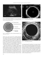
Intralabyrinthine Hemorrhage
MR examination may show an intralabyrinthine hemorrhage presenting as
a high signal intensity of labyrinthine cavities on both T
1
- and T
2
-weighted
images (fig. 4). If vestibular bleeding is the only cause to assess postoperative
complication, the prognosis is usually good with rest and medical therapy [9].
Intralabyrinthine Reparative Granuloma
Intravestibular extension of a reparative granuloma is a rare but severe
complication of stapes surgery. CT examination usually shows a nonspecific
soft tissue mass filling in the oval window fossa. Conversely, intralabyrinthine
focal hypointensity on T
2
-weighted images with associated enhancement on
postcontrast T
1
-weighted images is highly suggestive of reparative granuloma
(fig. 5). Reparative granuloma has a poor prognosis despite aggressive medical
therapy, and even revision surgery [4].
a b
Fig. 4. a In case of intravestibular postoperative hemorrhage, the axial T
1
-weighted
image shows a spontaneous hyperintense signal within the vestibule and the basal turn of the
cochlea. b The axial T
2
-weighted image demonstrates an abnormal hyperintense signal
within the vestibule, compared to normal hyperintensity of cerebrospinal fluid.
Ayache/Lejeune/Williams 312
Fig. 5. In this case of postoperative SNHL, the axial T
2
-weighted image shows the
obliteration of the labyrinthine fluids corresponding to an extension of the reparative granu-
loma into the labyrinth.
a b
Fig. 6. a In case of postoperative labyrinthitis, the axial T
2
-weighted image shows partial
obliteration of the labyrinthine cavities. b The axial T
1
-weighted image after contrast administra-
tion shows an enhancement of the cochlea, vestibule and fundus of the internal auditory canal.
Imaging of Postoperative Sensorineural Complications of Stapes Surgery 313
Labyrinthitis
Suppurative labyrinthitis is a rare complication usually associated with a
poor prognosis. CT findings are nonspecific. MRI demonstrates an obliteration
of the intralabyrinthine fluids on T
2
-weighted images associated with an exten-
sive enhancement of the labyrinth on postcontrast T
1
-weighted images (fig. 6).
In case of suppurative labyrinthitis or reparative granuloma, MRI findings
are very similar, suggestive of an inflammatory process obliterating the
labyrinthine cavities.
Conclusion
Imaging has a predominant role in managing post-stapedectomy SNHL.
CT is the first imaging technique to perform. It can depict an excessive pene-
tration of the prosthesis into the vestibule or a pneumolabyrinth caused by PLF.
In case of negative or noncontributive findings, MRI might be helpful, as it can
show reparative granuloma extending into the vestibule, labyrinthitis or import-
ant bleeding into the vestibule.
References
1 Mann WJ, Amedee RG, Fuerst G, Tabb HG: Hearing loss as a complication of stapes surgery.
Otolaryngol Head Neck Surg 1996;115:324–328.
2 Wiet RJ, Harvey SA, Bauer GP: Complications in stapes surgery. Otolaryngol Clin North Am
1993;26:471–490.
3 Lippy WH, Schuring AG: Stapedectomy revision following sensorineural hearing loss.
Otolaryngol Head Neck Surg 1984;92:580–582.
4 Betsch C, Ayache D, Decat M, Elbaz P, Gersdorff M: Les réinterventions dans la chirurgie de
l’otospongiose: à propos de 73 cas. J Otolaryngol 2003;32:38–47.
5 Williams MT, Ayache D, Elmaleh M, Heran F, Elbaz P, Piekarski JD: Helical CT findings in
patients who have undergone stapes surgery for otosclerosis. AJR Am J Roentgenol 2000;174:
387–392.
6 Swartz JD, Lansman AK, Berger AS, Wolfson RJ, Bell G, Popky GL, Swartz NG: Stapes prosthe-
sis: evaluation with CT. Radiology 1986;158:179–182.
7 Rangheard AS, Marsot-Dupuch K, Mark AS, Meyer B, Tubiana JM: Postoperative complications
in otospongiosis: usefulness of MR imaging. AJNR Am J Neuroradiol 2001;22:1171–1178.
8 Williams MT, Ayache D: Imaging of the postoperative middle ear. Eur Radiol 2004;14:482–495.
9 Ayache D, Sleiman J, Nengsu Tchuente A, Elbaz P: Variantes et incidents per-opératoires observés
au cours de la chirurgie de l’otospongiose. Ann Otolaryngol Chir Cervicofac 1999;116:8–14.
Denis Ayache, MD
ENT Department, Fondation Rothschild
25, rue Manin
FR–75019 Paris (France)
Tel. ϩ33 1 48 03 68 40, Fax ϩ33 1 48 03 65 17, E-Mail
Arnold W, Häusler R (eds): Otosclerosis and Stapes Surgery.
Adv Otorhinolaryngol. Basel, Karger, 2007, vol 65, pp 314–319
Revision Stapes Surgery
Klaus Jahnke, Daniela Solzbacher, Philipp Dost
Department of Otorhinolaryngology, University Hospital of Essen,
Essen, Germany
Abstract
We present the results of our revision stapes operations from 1989 to 2004 (n 217).
Long-term follow-up was performed in the first 135 cases. Eighteen of these patients were
revised because of inner ear symptoms, predominantly within the first year. One hundred and
sixteen cases underwent revision surgery due to conductive hearing loss, on average after
10.6 years. One patient was operated because of dysgeusia. In 1999, we first described inner
ear damage after implantation of gold prostheses. Therefore, we developed a titanium
implant that was initially investigated in cell culture and subsequently tested in a clinical
trial. We report on the most frequent causes that led to revision surgery such as adhesions,
prosthetic problems, erosions of the long process of the incus, or refixation of the footplate,
and on the different surgical techniques. In a first series of patients with a conductive hearing
loss, a significant hearing improvement of 69.4% of these cases was obtained. However, this
result very much depends on the selection of cases. There was no case of additional sen-
sorineural hearing loss. Since 1999, we had mainly used titanium implants for replacement in
stapes revision surgery. In a second series, a significant hearing improvement of 76.2% was
found. One patient with a platinum Teflon implant had to be revised because of vertigo and
conductive hearing loss which was observed during MRI.
Copyright © 2007 S. Karger AG, Basel
Stapes surgery is one of the most standardized and successful procedures
in otology. This does not apply to revision surgery, which is performed after
shorter or longer periods of time due to very different causes of failure. The
results of the revision surgery are considerably influenced by the selection of
cases and the specific surgical techniques used.
The aim of this study was to describe pre- and intraoperative findings in
217 cases, our general surgical rules and special techniques as well as the post-
operative results of the first 135 cases.
Revision Stapes Surgery 315
Patients and Methods
From April 1989 to December 2004, one author (K.J.) performed 217 revision stapes
surgeries. The mean age of the patients was 48.7 years (range 11–78 years). Long-term
follow-up was performed in the first 135 cases, done until December 2000.
Eighteen of these patients were revised because of inner ear symptoms, predominantly
within the first year (n 11), after an interval of 1–10 years (n 6) and after more than
10 years (n 1). Six cases mainly complained of progressive sensorineural hearing loss;
in 10 cases, sensorineural hearing loss together with vertigo was observed, and 2 patients
exclusively had vertigo.
One hundred and sixteen cases underwent revision surgery due to significant conduc-
tive hearing loss; the interval between the two operations was less than 1 year in 12 cases,
1–10 years in 60 cases and more than 10 years in 44 cases. One patient was operated because
of dysgeusia. In total, 76% of the patients had previously been operated on elsewhere. All
patients had otosclerosis except 5 with minor ear malformations and 1 with a stapes fracture
after skull base trauma.
Additionally, the short-term results of the last 82 cases will be reported.
Results
Surgical Procedures
In stapes revision surgery, there are some important rules according to
Dietrich Plester. First, all operations should be performed under local anesthe-
sia whenever possible so that there is control of vertigo when carefully touching
the implant inserted during the first procedure. During the last few years, two
medialized implants had to be left in place because of vertigo while trying to
lateralize them. In addition, it is an advantage to examine hearing improvement
at the end of surgery. Everything has to be explained to the patient prior to
surgery. Furthermore, there must be an informed consent that the surgeon has
the option to postpone surgery at any stage.
During the last few years, the erbium:YAG laser proved to be advantageous
when scars and bony fragments were noted near the vestibulum.
Intraoperative Findings
The causes of inner ear symptoms were inner ear otosclerosis, middle ear
inflammation, excessively long prostheses, gold incompatibility, adherent
swab/particles and a fistula. In the 2 patients sent to our department with
middle ear inflammation within 2 weeks postoperatively, the residual hearing
capacity could be saved and slightly improved by rinsing the middle ear with
antibiotics and corticoid solution.
In 1999, we first described inner ear damage after implantation of gold
stapes prostheses [1]. Usually, sensorineural hearing loss developed within the
Jahnke/Solzbacher/Dost 316
first postoperative weeks starting in the high frequencies (n 4). Early revi-
sion showed significant granulation tissue formation around the implant veri-
fied by histology (fig. 1a). Topical and systemic corticoid therapy resulted in
partial recovery of the high-tone loss in 3 cases (fig. 1b). Dermal tests with such
implants demonstrated a metal allergy only in 1 of 4 cases. Therefore, it is more
likely that the high current conductibility of this metal – in contrast to titanium –
plays a significant role in such cases [Helm, J., pers. comment].
After our report, we got the information that in some other departments
single cases of complete deafness were observed. Therefore, we developed
stapes prostheses made of titanium in co-operation with an industrial partner. In
a first step, the biocompatibility of titanium was confirmed in in vitro studies
with human stapes osteoblasts (fig. 2a) [2]. It is well known that the potential of
titanium to form a tight connection with bone depends on the surface structure.
Consequently, the piston was polished and the loop grasping around the incus
was roughened (fig. 2b). The loop was altered so that it could not hinder itself
when pinched around the long process. During that time, we noticed that Fisch
et al. [3] had also used titanium implants for stapes replacement in a few
selected cases.
Within a prospective clinical study, the initial 47 implants were analyzed. It
was seen that the titanium stapes prosthesis was at least as good in practical use
0
10
20
30
40
50
60
70
80
90
100
110
120
dB
0.125
0.25
0.5
0.75
1
1.5
2
3
4
6
8
12
kHz
Pre revision
Post revision
ab
Fig 1 Sensorineural hearing loss after implantation of gold stapes prostheses.
a Granulation tissue close to the implant. b An example of a ‘gold incompatibility’ audiogram,
bone conduction only.
Revision Stapes Surgery 317
as the conventional implants used until now. We have observed no hints of any
side effects or incompatibility since autumn 1999, when this new implant was
introduced [4].
The clinical trial demonstrated an excellent inner ear compatibility. Up to
now, we have not observed any sensorineural hearing loss in more than 300
cases. Therefore, we think that a possible advantage of titanium implants is an
excellent compatibility when the implant is in contact with the perilymph of the
vestibulum as well as with the long process of the incus. On the other hand, it
cannot be judged if there is an increased risk of an osseous fixation by recurrent
otosclerosis of the oval window niche.
In many revision cases on the basis of conductive hearing loss (n 116),
different causes were seen (table 1).
According to the findings, the operation techniques were very different: in
102 cases, the adhesions were cut, e.g. between the long process of the incus
and the tympanic membrane, or in other cases, scar tissue was carefully
removed from the oval window niche, sometimes with the erbium:YAG laser. In
28 cases, only a new adjustment of the prosthesis was necessary; however, in
the majority of cases, a new implant was inserted (n 86), mostly a piston.
When the end of the long process of the incus was completely cut, which was
particularly observed with the platinum band Teflon piston, the angled implant
a b
Fig 2 Titanium stapes prostheses. a Excellent biocompatibility was confirmed in in
vitro studies with human stapes prostheses. b Essen titanium stapes prosthesis.
Jahnke/Solzbacher/Dost 318
was used. In cases in which the Schuknecht wire prosthesis had been used in the
first intervention according to Plester, it proved to be very advantageous to cut
the wire with scissors and leave the end in place (n 9) (fig. 3). In a next step,
the vestibulum had to be opened carefully at the posterior circumference, where
a new piston was inserted.
Ninety-eight of the patients could be followed up in the long term. There
was no single case of a significant additional sensorineural hearing loss. The
hearing improvement in patients with conductive hearing loss was more than
Table 1. Most frequent causes leading to revision
surgery
Adhesions n 102
Prosthetic problems n 60
Lateralization n 26
Loose implant n 19
Malpositioned implant n 12
Too short implant n 6
Erosion of the long process of the incus n 42
Refixation of the footplate n 28
Dysfunction of the eustachian tube n 5
Malleus head fixation n 3
Fig 3 Plester’s technique: Divide the
shaft of the Schuknecht wire prosthesis,
leave the end in place, open the vestibulum
posteriorly (arrow).
Revision Stapes Surgery 319
20 dB in 32 cases and 5–20 dB in 36 cases, i.e. a significant hearing improve-
ment of 69.4% in this series.
From 2001 to 2004, stapes revision surgery was performed in a further 82
patients. Six of these revisions were performed on the basis of inner ear prob-
lems. All had vertigo, 2 had additional sensorineural hearing loss, 1 of these 2
additional conductive hearing loss.
For replacement in stapes revision, we mainly used titanium implants
(n 50). The short-term results in this second series showed a significant hear-
ing improvement of 76.2%.
In the case of an 81-year-old lady, transient vertigo and a significant com-
bined hearing loss occurred during NMR spectroscopy with 1.5 T. Revision
stapes surgery was performed 9 months after this event and 10 years after the
first procedure. Intraoperatively, the platinum Teflon implant was found to be
lateralized and the long process of the incus was completely divided, so appar-
ently the implant was first medialized and then lateralized during NMR spec-
troscopy, resulting in the above-mentioned symptoms. With the implant
removed it was shown that it could not have been dislocated by the magnetic
field. Therefore, it is more likely that this event was due to changes in the gra-
dient fields. After that, a titanium implant was inserted and hearing signifi-
cantly improved.
References
1 Jahnke K, Dost P, Missfeldt N: Revisionen nach Stapes-Chirurgie. HNO 1999;47:427.
2 Dost P: Biomaterialien in der rekonstruktiven Mittelohrchirurgie. Laryngorhinootologie 2000;
79(suppl 2):53–72.
3 Fisch U, Acar GO, Huber AM: Malleostapedotomy in revision surgery for otosclerosis. Otol
Neurotol 2001;22:776–785.
4 Dost P, Arweiler-Harbeck D, Jahnke K: A prospective evaluation of the Essen titanium stapes pros-
thesis. Clin Otolaryngol 2005;30:21–24.
Klaus Jahnke, MD
Department of Otorhinolaryngology, University Hospital
Hufelandstrasse 55
DE–45122 Essen (Germany)
Tel. ϩ49 201 723 2481, Fax ϩ49 201 723 5903, E-Mail
Arnold W, Häusler R (eds): Otosclerosis and Stapes Surgery.
Adv Otorhinolaryngol. Basel, Karger, 2007, vol 65, pp 320–322
Vibrant Soundbridge Middle Ear
Implant in Otosclerosis
Technique – Indication
Thibaud Dumon
J. Causse Clinic, Béziers, France
Abstract
With our growing experience with the Vibrant Soundbridge (VSB) middle ear implant,
the question emerged of its indication in mixed hearing loss due to advanced otosclerosis.
We describe the VSB implantation technique in primary otosclerosis performed together
with a stapedotomy piston procedure. Hearing results under headphone and free-field condi-
tions show that the stapedotomy piston procedure closes the air-bone gap as expected and
that the VSB provides comparable gain to that usually recorded for pure sensorineural hear-
ing loss. The gains of the two procedures add up. These results open the field of mixed hear-
ing loss to the VSB middle ear implant.
Copyright © 2007 S. Karger AG, Basel
With our growing experience with the Vibrant Soundbridge (VSB) middle
ear implant, the question emerged of its indication in advanced otosclerosis.
The feasibility and indication of the VSB implantation together with a success-
ful stapedotomy piston procedure have to be considered in primary nonoperated
and in previously operated otosclerosis.
Acute trials during classical VSB implantation demonstrate that there is
enough space to fit the loop of a Teflon piston and the clip of the floating mass
transducer (FMT) of the VSB on the long process of the incus. In primary oto-
sclerosis, the loop of the Teflon piston has to be fitted over the clip of the FMT,
whereas in previously operated otosclerosis, the clip of the FMT has to be fitted
over the loop of the piston.
Vibrant Soundbridge Middle Ear Implant in Ostosclerosis
Vibrant Soundbridge Middle Ear Implant in Otosclerosis 321
Primary Otosclerosis: Stapedotomy Piston and
VSB Single-Stage Procedure
For the first case, we decided to associate a retroauricular facial recess
approach for the VSB with an ear canal approach for the stapedotomy piston
procedure, as the reference technique for the two [1, 2].
The surgical technique requires a combined ear canal and retroauricular
approach. The retroauricular approach was performed with a facial recess
approach. The internal part of the VSB was fitted in the retroauricular field, and
the FMT was placed in the posterior tympanotomy opening, ready to be clipped
on the incus. Then the ear canal approach was performed. The stapes super-
structures were removed, the calibrated stapedotomy was performed and cov-
ered with the vein interposition. Coming back to the facial recess approach, the
FMT was fitted on the long process of the incus and its clip tightened. Coming
back to the ear canal approach, the Teflon piston was placed in the stapedotomy
opening, on the vein, and its loop fitted on the long process of the incus, over
the clip of the FMT, and tightened (fig. 1). Classical closure and ear canal pack-
ing were observed.
The results of the first case show that the stapedotomy piston procedure
closes the air-bone gap as usual. The VSB gives a gain comparable to that in
classical indications. The gains of the two procedures add up (fig. 2).
We demonstrated the feasibility and described the surgical technique to
implant a VSB middle ear implant in mixed hearing loss due to otosclerosis.
Fig. 1. VSB in primary otosclerosis: loop of the piston over the clip of the FMT.
Dumon 322
The indication of the VSB has to be decided according to the sensorineural part
of the mixed hearing loss, with the same criteria as for pure sensorineural hear-
ing loss. The implantation of the FMT by the ear canal approach, without the
facial recess approach, may be discussed.
References
1 Fisch U, Cremers CW, Lenarz T, Babighian G, Uziel A, Proops DW, O’Connor AF, Helms J,
Fraysse B: Clinical experience with the Vibrant Soundbridge implant device. Otol Neurotol
2001;22:962–972.
2 Vincent R, Gratacap B, Causse JB: Argon laser and Gherini-Causse Endo-Otoprobe in otologic
surgery. Ear Nose Throat J 1996;75:773–780.
Dr. Thibaud Dumon
Clinique J. Causse, Traverse de Béziers
FR–3444 Colombiers (France)
Tel. ϩ33 4 67 35 66 22, Fax ϩ33 4 67 35 62 00, E-Mail
Warble tone thresholds preop./Piston/PistonϩVSB
Vibrat Soundbridge in Otosclerosis Stapedotomy pistonϩVSB gain
0
20
40
60
80
100
120
0.125 0.25 0.5 124810
kHz
LE preop. AC
LE preop. BC
LE Piston
LE PistonϩVSB
Preop. ABG : 32 dB HL
AC gain (0.5Ϫ4 kHz):
Piston: 32 dB HL
PistonϩVSB: 62 dB HL
Postop ABG : 11 dB HL
Speech gain (50% recognition):
Piston: 35 dB HL
PistonϩVSB: 50 dB HL
Fig. 2. Primary otosclerosis in a 42-year-old man (first case). ABG ϭ Air-bone gap;
AC ϭ air conduction.
Arnold W, Häusler R (eds): Otosclerosis and Stapes Surgery.
Adv Otorhinolaryngol. Basel, Karger, 2007, vol 65, pp 323–327
Cochlear Implantation and
Far-Advanced Otosclerosis
I. Mosnier
a,b
, D. Bouccara
a
, E. Ambert-Dahan
a
, E. Ferrary
a
, O. Sterkers
a,b
a
Department of Otolaryngology, Hôpital Beaujon, AP-HP, Faculté Xavier-Bichat,
Université Paris-7, Clichy,
b
Department of Otolaryngology, Hôpital Louis-Mourier,
AP-HP, Faculté Xavier Bichat, Université Paris-7, Colombes, France
Abstract
Objective: To evaluate results of cochlear implantation in patients with far-advanced
otosclerosis. Methods: Sixteen patients with far-advanced otosclerosis had undergone uni-
lateral (n 13) or bilateral (n 3) cochlear implantation. Surgical difficulties, incidence of
complications and postoperative benefit were analyzed. Results: A full electrode insertion
was achieved in all patients without surgical difficulties. All patients demonstrated excellent
benefit of cochlear implantation. Binaural implantation still improves speech performances,
compared to unilateral implantation. In case of residual cochlear function of one nonoperated
side, a stapes surgery, performed during the same surgical time as cochlear implantation, can
improve speech scores and restore bilateral hearing. Facial nerve stimulation occurred only
in 1 patient. Conclusion: Cochlear implantation is the method of choice for rehabilitation of
patients with otosclerosis, presenting profound or total hearing loss. Patients obtain excellent
benefit with a low rate of complications.
Copyright © 2007 S. Karger AG, Basel
Cochlear implantation in far-advanced otoscerosis may represent a sur-
gical challenge because of a possible partial ossification of the cochlea.
Moreover, a high incidence of facial stimulation is reported in this population
[1, 2]. In addition, some authors report a satisfactory benefit obtained by a
stapes surgery and hearing aid amplification in this population, even in patients
with no cochlear reserve [3].
This study presents our management of patients with otosclerosis and
severe or profound hearing loss, showing the excellent performances of cochlear
implantation in this population and the low incidence of complications.
Mosnier/Bouccara/Ambert-Dahan/Ferrary/Sterkers 324
Materials and Methods
Between 1991 and 2003, 134 adult patients had undergone cochlear implantation at our
institution. The cause of the hearing loss was otosclerosis in 16 cases (12%), selected for this
study. Thirteen patients had been implanted with a Nucleus device (5 Nucleus 22 and
8 Nucleus 24; Cochlear AG, Basel, Switzerland) and 3 patients had been bilaterally
implanted with a Med-El
®
Combi-40 device (Innsbruck, Austria). The data were reviewed
regarding preoperative patient history, radiological findings (CT scan and MRI available for
all patients), surgical difficulties, complications, and preoperative and postoperative audio-
logical assessments. For comparison of speech perception performances between otosclero-
sis patients and patients with other causes of hearing loss, a one-way ANOVA was applied.
Results
The characteristics of the population are detailed in table 1. In 8 cases, an
important rarefaction of the otic capsule bone was observed on CT scan. MRI
did not show objective cochlear duct obliteration in any patient. Six patients
required a partial drill-out of the basal turn of the cochlea because of a partial
ossification. A full electrode insertion into the scala tympani was achieved in all
patients.
The incidence of side effects was very low. A reimplantation was required
in 1 patient because of a traumatic device failure. Facial nerve stimulation
occurred only in this patient, after reimplantation, controlled after deactivation
of the 6 offending electrodes. Speech recognition scores in quiet were not
modified, but the patient reported alteration of the sound quality and speech
intelligibility in noisy environments.
Table 1. Characteristics of the study population
Male: female ratio 7:9
Mean age, years 61 (range 44–74)
Mean duration of profound 2.5 (range 0–5)
deafness, years
Stapes surgery before implantation 11
Unilateral 4
Bilateral 7
Hearing aid at the time of implantation 9
Device implanted
Nucleus (unilateral) 13
Med-El Combi-40 (bilateral) 3
Stapes surgery during implantation 2
Blindness 1