The Foot in Diabetes - part 10 pot
Bạn đang xem bản rút gọn của tài liệu. Xem và tải ngay bản đầy đủ của tài liệu tại đây (961.46 KB, 32 trang )
International Consensus and Practical Guidelines 335
336 The Foot in Diabetes
International Consensus and Practical Guidelines 337
338 The Foot in Diabetes
International Consensus and Practical Guidelines 339
340 The Foot in Diabetes
International Consensus and Practical Guidelines 341
342 The Foot in Diabetes
International Consensus and Practical Guidelines 343
344 The Foot in Diabetes
22
The Foot in LeprosyÐLessons
for Diabetes
GRACE WARREN
Westmead Hospital, Sydney, NSW, Australia
``The diabetic foot'' is a term that implies impaired physiological function
that may result in damaged tissues, ulceration, deformity, destruction and
amputations. Many of these problems are the result of neuropathy, which is
slowly progressive and may be ®bre-selective, with pain ®bres frequently
affected early, well before there is clinical loss of touch or pressure
sensation. It is often unaccompanied by de®nite symptoms, so that many
patients do not realise that a neural de®cit is developing until they are
confronted with ulceration or other resultant problems.
A similar problem may occur in leprosy, in which the nerves are
parasitized early without any symptoms. Over a period of many years there
may be increasing ®brosis and slow loss of neural function until the limb is
totally neuropathic, motor and autonomic as well as sensory. The sensory
neuropathy is the main problem, allowing the possibility of unnoticed and
hence untreated trauma because the patient does not have enough pain
perception to demand care. The resultant problems are virtually the same as
those seen in diabetes. In leprosy, as in diabetes, the autonomic
involvement causing altered skin physiology makes the skin more prone
to trauma from stress, bumps or dehydration.
Together with many other neuropathies, these diseases have several
problems in common. The most important is the so-called ``non-healing
ulcer''. In 1877 John Hilton
1
wrote: ``pain was made the prime therapeutic
agent . . . After injury, pain suggested the necessity of, and indeed compelled
The Foot in Diabetes, 3rd edn. Edited by A. J. M. Boulton, H. Connor and P. R. Cavanagh.
& 2000 John Wiley & Sons, Ltd.
The Foot in Diabetes. Third Edition.
Edited by A.J.M. Boulton, H. Connor, P.R. Cavanagh
Copyright
2000 John Wiley & Sons, Inc.
ISBNs: 0-471-48974-3 (Hardback); 0-470-84639-9 (Electronic)
man to seek for, rest''. Because of the damage to sensory nerve pain ®bres,
these patients lack the natural sparing re¯ex that makes man seek for that rest.
In the middle of the twentieth century, large institutions provided
prolonged outpatient care for thousands of leprosy patients and residential
care for hundreds who lived in these institutions for many years. This
provided an excellent opportunity for studying the effects of all degrees of
neuropathy and of observing the results of neglect and the effects of
methods of management.
Previously, it was assumed that the ulcers of leprosy patients were part of
the disease and would never heal. However, with the introduction of
effective bactericidal drug therapy, it was realised that these ulcers were not
part of the disease itself but were mainly due to the loss of pain perception.
Dr Paul Brand, working at the Christian Medical College Hospital in
Vellore, South India, in the late 1940s, was challenged with the question,
``Why do ulcers continue to occur when the disease is cured?'' He did not
know; no-one knew. He gathered around him a group of researchers who
played a major role in identifying the reason for continuing ulcers and
determining methods of management that literally save hands and feet, not
only of leprosy patients, but of persons with neuropathy from any cause.
The story of the battle to understand the problem of ``no pain'' is a
fascinating one of how many people, working together, eventually solved
the problem of why neuropathic ulcers appear not to heal
2
. The under-
standing we now have, and the methods that we have been using for
leprosy for over 40 years, stem from this research. In the last 40 years I have
been asked to treat neuropathy from many causes and I have found that the
same methods are effective, irrespective of the cause of the neuropathy
3
.
The principles laid down by Dr Brand and his colleagues are still applicable
world-wide in saving limbs and improving the quality of life of those who
have damaged nerve function from any cause.
In diabetes, as in leprosy, there are often no characteristic symptoms that
indicate that a nerve de®cit is developing. Hence, the patient may not
realise that the ability to feel pain has been lost until some accident occurs
that results in a lesion that is surprisingly painless. This may be a burn, a
blister or a fracture. The common factor is that a lesion caused by trauma is
neglected because it is painless and the patient does not automatically
respond by protecting the traumatized area, and hence the lesion may
become a non-healing ulcer. However, in leprosy it had been shown that if
an affected limb was completely rested, ulcers healed
4
as quickly as similar
lesions in a sensate limb. This understanding resulted in the use of total
contact casts
5
, which encouraged healing by preventing excess pressure on
the traumatized areas but enabled the patient to continue walking.
Special testing of nerve function is often requested following examination
of the patient. Nerve conduction studies may show the speed with which an
346 The Foot in Diabetes
impulse travels along a nerve, but do not tell what information those
impulses pass to the brain. Many patients with neuropathy have
paraesthesiae, but what do those paraesthetic feelings indicate? Is the
body trying to indicate what a person with normal sensation would interpret
as pain? It is the ability of perceiving pain as pain that is the important factor.
Hence, electrical testing may give false ideas of the patient's ability to protect
him/herself. Loss of pain perception is the biggest problem. The use of
Semmes±Weinstein mono®laments
6
to test skin sensation is helpful to chart
variations in neural function. But it is not a measure of protective sensation,
as it does not test for pain perception. A patient may have normal perception
of a 10 g ®bre but have no discomfort when a sliver of glass cuts the foot. The
wisest rule is to treat any patient with any suspicion of neuropathy as though
there was complete loss of sensory perception and to start teaching the
patient self-care as soon as neuropathy is suspected.
Leprosy patients may show marked motor and autonomic nerve
dysfunction, even when there is little obvious sensory de®cit
5
. It is advised
that multiple neuropathies be assumed to be present whenever any neural
de®cit is detected. Over the past 40 years the writer has managed patients
with neuropathy from many causes, using the same principles as those
indicated by Coleman and Brand
7
. Teach the patient how to protect the limbs as
though there was no sensation at all. The patient may say that feeling is present,
but who can know exactly what that patient means by ``feeling''. Is it
paraesthesia, numbness or one of a multitude of feelings, such as ``burning'',
``cutting'', or ``compression'' that do not include protective sensation and do
not provide the stimulus needed for the patients to protect themselves? Many
patients say they have pain but describe what may be ``tingling'', or ``pins and
needles'' that may be due to abnormal nerve activity and may even indicate
regrowth of damaged nerves. Young patients may call this ``pain'' because
they have no previous experience of real pain. It is advisable to inquire into
the quality of ``pain'' and perhaps record the feelings as discomfort rather
than pain. If deformity and disability are to be prevented, it is essential that
the patient realises that there is a de®ciency in sensory perception.
Our work with leprosy patients brought to our attention other problems
arising from nerve de®cits that may be relevant to diabetic patients. The
involvement of motor nerves may result in clawing of the toes which, in
turn, causes excessive pressures over proximal interphalangeal joints and
on the plantar surface of the metatarsal heads. In diabetic patients this is
often dealt with by orthoses and moulded shoes. These have also been used
in leprosy patients, but it was found that surgery
8
, such as that for
correcting clawed toes or a dropped foot, could correct the problem
permanently. This could eliminate the constant need for new footwear by
straightening the toes yet leaving them mobile, ¯exing the metatarsal joints
and so spreading the stresses of weightbearing. It reduces the risk of
The Foot in LeprosyÐLessons for Diabetes 347
ulceration. The excessive stresses caused by muscle imbalance in the lower
limbs may stimulate excessive callus formation and may result in
ulceration. In leprosy, many of the consequences of muscle imbalance are
minimized by tendon surgery and, in my experience, these procedures are
just as effective in neuropathy from other causes
9
and have often resulted in
the salvage of a limb that might otherwise have been amputated.
Unfortunately, it is uncommon for diabetic patients to bene®t from this
type of surgery, although for the diabetic patients on whom I have
performed reconstructive surgery, the results have been well worthwhile
3
.
In leprosy patients a large proportion of ulcers originate under callus or
scarred skin. This callus, if not regularly removed, builds up and forms a thick
mass, some of which dehydrates and becomes very hard. If on the sole, this
may cause excessive pressures in the deeper tissues during walking and result
in ulceration. A similar situation exists in diabetes
10
and other neuropathies
9
.
After removalof the callosity, leprosy patientsare taughtto rub oil into the area
on a daily basis, which keeps moisture in the skin, preventing dehydration
3
.
Skin treated in this way improves in texture and resilience and, by becoming
less fragile, is better able to withstand trauma. The same principles have been
applied to patients with diabetes presenting with dry, fragile skin. Regular
rehydration and oiling results in improved smoothness and suppleness and
ability to withstand the stresses of daily use. Rehydration and oiling help to
compensate for the effect of autonomic neuropathy on the sweat and
sebaceous glands when the secretion of both sets of glands may be greatly
reduced or completely lost. Brand
4
observed that feet that sweat normally
rarely become ulcerated and that rehydration is possible. In 1966, Harris and
Browne
11
published observations showing that the application of cosmetic
moisturisers alone did not improve skin quality, but soaking in water,
followed by oil, was effective as long as it was continued regularly. Tovey
10
suggested that, for diabetic patients, oilatum emulsion be added to the water
used for soaking dry skin and aqueous cream be rubbed into the skin
afterwards.
There are an estimated 15 million people affected by leprosy in the world,
mostly in areas where there are minimal medical facilities, and it was
necessary to devise treatment plans that patients could do themselves at
minimal cost. The following daily routine has been taught in many areas so
that the patient provides his/her own home care
3
.
DAILY CARE FOR PERSONS
WITH NEUROPATHIC LIMBS
It is important to teach all patients who are suspected of having
neuropathy to start daily care as soon as possible in order to maintain
348 The Foot in Diabetes
the affected areas, especially the feet, in good condition. This teaching
should be given by demonstration. Do not just tell the patients; show
them how and then get them to do it themselves so that they really
know how to continue at home
3
. As the feet are the most likely areas to
be affected, the care of the feet will be described but the principles can
be adapted to other areas of the body.
1. LOOK at their own feet every day, preferably at night, so that any
wounds can be treated that night. If they cannot see the sole of the foot
they can use a mirror or arrange for a partner or carer to do it for them.
Their feet and shoes should be inspected by a staff member every time
they attend clinic. This helps to impress importance of foot care upon
the patients and their relatives and may reveal trauma that has been
considered unimportant. Many patients have stated, ``my feet are ®ne'',
yet painless lesions are found on removal of shoes and socks.
2. SOAK the feet and legs in plain water every day. This remains a
controversial point in diabetes but is used by some clinicians treating
diabetes
10
. In leprosy, as stated above, it has been shown time and time
again that soaking is bene®cial
5
. Dryness is very obvious when people
walk barefoot or wear open sandals, and a dry atmosphere constantly
increases the dehydration of the skin. In leprosy clinics it has been
observed that healing occurs more rapidly on ulcerated feet that are
soaked daily and in which dehydration is prevented by oiling, than in
those feet that are left dry. The application of moisturising creams and
lotions does not actually rehydrate the deeper layers of dry skin. They
may improve hydration of super®cial layers and reduce further
dehydration, hence they keep in what water there is and make the
skin feel moist for a period.
3. SCRAPE off hard, dry or rough callus that may irritate or increase
local pressure: smooth dry hard callus splits and cracks, traumatizing
tissues. Most patients can learn to keep callus under control
themselves at home (in the case of those visually impaired, a
relative can do it for them). However, this is controversial and not all
authorities recommend self-care of callus in neuropathic patients. If
the clinician or podiatrist commences by removing the excessive
amounts of callus, it should be possible for the patient to prevent a
new build-up of callus. However, it is still advisable for the patient
to visit the podiatrist regularly to ensure that new masses of callus
do not build up. Many patients have used a pumice stone or nylon
pot scraper which, to be effective, needs to be rough. Other
commitments often mean there may be long intervals between visits,
allowing excessive amounts of callus to develop unless the patient
can help by doing a little every day.
The Foot in LeprosyÐLessons for Diabetes 349
The ®rst time a patient is seen with an ulcer surrounded or
covered by callus, it is essential that the callus be removed in order
to determine the size and severity of the ulcer
3
. The pressures caused
by localized masses of callus are one of the most likely causes of
ulceration in the insensate limb and the patient needs to understand
that thick, dry, hard, irregular or cracked callus causes problems.
4. OIL the skin to keep the water in. Any oil or moisturising cream will be
adequate to help prevent evaporation and dehydration. There is
evidence that ®sh oil or animal oil, such as lanoline, may be absorbed
and improve the quality of the skin as well as keeping it hydrated, and
these oils are often rubbed in regularly to improve quality of scars after
burns.
5. DRESS WOUNDS with simple dressings to keep them clean. Expensive
dressings have no advantage over saline, simple ointment, Ungvita or
Magnoplasm
3
. Most neuropathic wounds will heal with anything on
them except the patient's weight! It is not the dressing that heals the
ulcer. The ulcer will heal if it is kept clean and protected from further
trauma. Pressure on an ulcer causes local anoxia and this damages the
healing tissues.
6. PROTECT from trauma. If there is no wound or ulcer on the limb,
proper protection would be the wearing of suitable footwear and the
use of other protective appliances. If there is trauma or an ulcer on a
vulnerable site, some form of splinting or other protection should be
instituted once the dressing is in place.
7. EXERCISE to maintain mobility of ankles, toes and hands, and to gain
optimal ef®ciency of any functioning muscle.
THE ORIGIN AND TREATMENT OF ULCERS
How do ulcers start
7
? The initial ulcer is usually the result of primary
trauma. It may be due to:
1. Sudden very high pressure, as when jumping from a height, or stepping
on a sharp object.
2. Lower pressure occurring intermittently for a long period, as in
walking.
3. Low pressure occurring continuously, as with bed sores, or when
wearing tight shoes.
4. Burns, cuts, bruises and friction.
Once an ulcer has healed, an area of scar will remain that will be more
prone to trauma than normal tissue.
350 The Foot in Diabetes
Most ulcers need only to be kept clean and rested and they will heal. The
ideal is that an ulcer on a weightbearing surface should not be walked on
7
.
The best method of treating an ulcer is ``REST and PROTECTÐNOT ONE
STEP PER DAY'' on the ulcer unless it is protected in a suitable walking
cast. Ulcers on neuropathic feet will heal as rapidly as would a similar ulcer
on a sensate foot, if adequately rested and protected. The provision of
healing shoes does encourage ulcers to heal more quickly than they would
in a normal shoe, but while walking in a shoe they will heal more slowly
than if fully rested. In leprosy it was found that the use of total contact
walking casts resulted in the healing of most ulcers within 6 weeks,
although very gross ulcers may take much longer.
However, many clinicians are not prepared to ``hide'' an ulcer under a
cast for 6 weeks. Hence, total contact casting has now been modi®ed by
bivalving the total contact cast, as shown in Figure 22.1
3
, so that it can be
removed, as needed, for dressings. When replaced correctly and held ®rmly
by Velcro or bandages, it still functions as a total contact cast and eliminates
stress and friction on the wounds, by spreading the patient's weight over
the whole inside of the cast while allowing the patient to walk. These casts
are worn 24 hours a day and, in many patients, it is best that they are
removed only for inspection or dressings, i.e. not removed regularly for
bathing or at night. These casts have proved popular with patients and staff
and are very effective in obtaining healing of ``non-healing ulcers''
3
. A total
contact cast enables the patient to have full rest of the ulcerated area and yet
be at home and mobile. It is not the dressing that heals the ulcer. The ``rest
and protect'' allows the body to proceed with healing without the constant
interruption of repeated trauma.
Diabetic patients with ``non-healing'' ulcers are frequently sent to a
vascular surgeon: however, there may be no de®ciency in the blood supply.
The autonomic nerve damage results in a warm limb, which is also very
common in leprosy patients
7,10,11
. The capacity of increased ¯ow in the
presence of infection is as great as normal, producing the normal signs of
in¯ammation when appropriate.
Special shoes are often prescribed to provide relief of pressures on the
ulcer
4
. If these are total contact shoes and correctly made they will assist in
healing the ulcer, but healing will be slower than in a total contact walking
cast (TCC). In a special shoe the area of weightbearing is much less, so
pressures are greater. Also, patients remove shoes at night, when trauma
can occur. One step is enough to undo any healing that has occurred during
that day. If a moulded insole is provided and the shoe is not correctly ®tted
to the foot, it may do more harm than good as peak pressure points may fall
on incorrect areas of the traumatized foot. A dressing on the ulcer may
destroy the ®t of the shoe, causing increased pressure to occur at the site of
the dressing, and this may increase the anoxia and tissue breakdown at that
The Foot in LeprosyÐLessons for Diabetes 351
352 The Foot in Diabetes
Figure 22.1 A total contact cast that has been bivalved. (a) The use of a football
sock as a liner to replace the minimal padding used to make the cast. This
padding is discarded when the cast is bivalved. The two halves of the cast are
side-by-side, ready to be closed round the leg. (b) The two pieces of the cast held
®rmly by Velcro. The patient was able to walk well even though both feet
required casting
site or elsewhere. A foot which is still basically anatomically normal does
not require a moulded sole
4
.
The so called ``non-healing ulcer'' is usually an ulcer that has not been
given the opportunity to heal. It has not been adequately rested and trauma
still continues to damage the healing tissues. The non-healing ulcer is a
compromised ulcer; either the clinician has not offered the best treatment or
the patient has not complied
3
.
HOT SPOTS
A common presenting symptom of patients with neuropathy is altered
temperature perception. Yet that limb is still able to become warmer than
normal as a response to in¯ammation. The natural response to in¯amma-
tion from any cause is heat, redness, swelling and pain. In the neuropathic
limb the ability to appreciate pain is frequently defective, so we must teach
patients to depend on heat, redness and swelling to inform themselves of
any form of in¯ammation and to make the patients think how to deal with
the problem and prevent it becoming worse. For practical purposes we term
this a ``hot spot'', and the patient with neuropathy needs to look for ``hot
spots'' every day during foot examination and know how to deal with them
without delay
3
.
A hot spot indicates the presence of some tissue pathology
3
. It may be a
sprain or a torn ligament or tendon, an infection either super®cial or deep,
soft tissue in¯ammation or osteomyelitis, or a fracture that, if neglected,
may result in bone disintegration. Unfortunately, when many clinicians see
a warm swollen painless foot, either they do nothing or they diagnose
osteomyelitis, even when there is no other evidence of infection (Figure
22.2). A full examination and history are mandatory. If a hot spot persists
for days or weeks it should be regarded as serious. If it settles rapidly while
the patient is resting, it is obviously not osteomyelitis. However, if it returns
as soon as use is resumedÐbeware. The oedema of travel or heart failure is
not usually hot. A patient with a neuropathic foot may feel no pain on
slipping or twisting a foot, so usually there is no known history of trauma
9
.
A hot spot on the sole of the foot usually indicates incipient blistering or
ulceration, and if that foot is rested at once it may be possible to prevent a
blister or an ulcer occurring. Many hot spots that indicate potential
ulceration will completely resolve in a week of total rest or in a total contact
cast. If the hot spot occurs on the dorsum of the foot, without obvious signs
of infection, it is more likely to indicate a lesion of neuropathic boneÐsuch
as a fracture or neuropathic disintegration. These lesions will settle very
quickly on complete rest but return within 24 hours on resumption of
normal activity. A radiograph may not show any bone lesion initially, as
also happens with a stress fracture. The latter often requires 6±8 weeks
The Foot in LeprosyÐLessons for Diabetes 353
before there is enough osteoporosis to make the fracture obvious. If it is
considered that it may be a neuropathic bone lesion, it is best to immobilize
in a total contact cast for 6±8 weeks and then review with new radiographs.
To allow the patient unprotected walking at this time is to risk bone
disintegration and increasing deformity.
BONE LESIONS
Neuropathic bone lesions are relatively common problems in the neuropathic
foot
12
. They are usually secondary to trauma and not to infection, although
infection may be present when the disintegration is secondary to the
osteopenia that occurs in association with an ulcer or infected lesion. In
adults it is rare to ®nd blood-borne osteomyelitis affecting an ulcer-free area.
Hence, a ``painless hot spot'' on a neuropathic non-ulcerated foot should be
considered to be neuropathic bone disintegration (NBD), unless proved
otherwise. The treatment for NBD is a ®xed total contact cast for a prolonged
354 The Foot in Diabetes
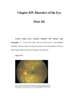
Figure 22.2 This diabetic patient had a history of at least twelve months duration of
a warm swollen painless foot, a ``hot spot''. She was admitted and treated as
osteomyelitis although no proof of that diagnosis was forthcoming. (a) Lateral
radiograph showing marked variations in the density of calci®cation of the talus and
some lucencies. There is also bone haziness of the posterior tubercle of the talus. The
AP view showed a crack in the talus that was not reported but was present 12
months earlier. The patient was treated with prolonged antibiotics but the foot was
never casted or protected. (b) The same view, taken 12 months later, shows some
dense bone but is dominated by the collapsing talus and loss of ankle joint space.
This case is typical of the progressive deformities that result from neuropathic bone
disintegration
period
3
. To give antibiotics ``just in case of osteomyelitis'' will not do any
harm to the NBD if it is in a total contact cast, but to treat the hot spot with
antibiotics alone, assuming osteomyelitis, and not provide a protective cast is
asking for increasing deformity and disability.
Patients should be taught to examine their feet every day looking for heat
and swelling. If any is found at night then check again in the morning. Heat
and swelling that persist overnight are warnings of trouble. Radiographs
should be taken as soon as de®nite symptoms or signs occur, and inform
the radiologist that you suspect a bone problem at such and such a point. If
the hot spot persists and there is no obvious fracture, it is advisable to apply
a total contact walking cast for 6±8 weeks and then re-X-ray. That time will
allow the more simple things like sprains and twists to heal uninterrupted.
It will also allow the osteoporosis around the fracture to develop until it can
be seen. If the patient does not wear a total contact cast, there is danger of
wearing away the osteoporotic bone and the fracture will turn into bone
disintegration which, if allowed to progress without restriction, may
eventually develop into a neuro-arthropathic foot. The observation of many
leprosy patients has enabled us to say that many neuro-arthropathic feet are
really neuropathic disintegration that started as a neglected fracture
because of little or no pain (see Figure 22.3)
3
. Some show no real
disintegration but are deformed because an undiagnosed fracture
displaced, and then healed producing a deformity that caused an ulcer-
prone stress point.
In the neuropathic foot, any fracture must be treated adequately. Because
of the lack of pain perception the patient will not limp to spare the
traumatized limb and may cause displacement of the fragments, impaction
or disintegration that results in a deformed limb, which then becomes ulcer-
prone. Our experience suggests that the limb requires total immobilization
(which can be in a total contact walking cast once the swelling has subsided)
for two or three times as long as would be required for the normal sensate
foot
3
. In neuropathy there appears to be a normal ability to heal but,
because of the lack of pain perception, the patient overstresses the healing
tissue too early and fractures it. Hence, for a midtarsal fracture it is
recommended that the foot be immobilized for 8±12 months before a trial of
free walking is allowed. The foot shown in Figure 22.4 indicates the typical
ability to heal.
There is no place for surgical interference in early true disintegration
without displacement, but in diabetic patients the displacement of fractures
in the Lisfranc area is common and, if seen early enough, it may be
advisable to internally ®x them, if this can be done adequately, rather than
allow displacement. Prolonged supported immobilization is still essential to
obtain bone healing. Where NBD has resulted in marked deformity, it is
advisable to treat in a total contact cast for 6 months or until the bones have
The Foot in LeprosyÐLessons for Diabetes 355
reconstituted, and then to perform wedge osteotomies or other surgery to
reconstruct a functional shape
3
as shown in Figure 22.5 (for further
discussion of this somewhat controversial area, see Chapters 17 and 18).
Neuropathic bone disintegration and the effects of chronic infection, such
as osteomyelitis, frequently leave local pressure points that are predisposed
to ulceration. It is practical to consider ostectomies to remove such bumps,
rough bones and irregular periosteum that may predispose to further
356 The Foot in Diabetes
Figure 22.3 This diabetic patient presented with a painless, warm swollen foot. (a)
Radiograph showing a fracture of the navicular, which was reported as a ``Charcot
foot''. The surgeon stated that a Charcot foot would not respond to treatment, but
the patient should wear an orthopaedic shoe. The patient attempted to do this, but
the condition of the foot deteriorated. Six months later he presented with an ulcer
over the cuboid, and radiograph (b) shows impaction of the head of the talus into the
navicular, producing a boat-shaped deformity. The bone did heal in a total contact
cast, after which the deformity was corrected
The Foot in LeprosyÐLessons for Diabetes 357
Figure 22.4 (a) Radiograph of a painless foot that had been hot and swollen for
many months and shows generalized marked osteoporosis and a midtarsal fracture.
Complete immobilization in a ®xed total contact walking cast for 12 months resulted
in union of all the bony fragments. Most of the joints fused into one solid bone mass.
(b) Radiograph taken 4 years after full healing had occurred
358 The Foot in Diabetes
Figure 22.5 (a) This woman presented with a chronic ulcer. (b) This ulcer was seen
on radiograph to be over the point of a ``boat-shaped foot'' that had resulted from
impaction of a fracture involving the dorsal surface of the foot. A mid-tarsal
osteotomy to realign the foot resulted in a functional non-ulcerating foot that could
be shod with a normal shoe. An extra resilient insole ensured no recurrence of the
ulceration. (c) Photograph taken about 4 years after the surgery
ulceration in the future. Basic surgical principles should be strictly observed
and all incisions on the weightbearing surface should be kept to a
minimum
3
. Moreover, as much good-quality weightbearing surface as
possible must be preserved in order to spread the load of body weight as
widely as possible.
TRIAL WALKING
Whenever a patient with neuropathy resumes free walking, such as after
removal of a cast or a prolonged period of rest in bed without walking, it is
advisable that a trial of walking is undertaken
3
. This is a slowly graded
daily increase in the duration of walking allowed, starting with only 3±5
minutes at a time, with the foot checked for hot spots 2 hours after the walk.
If there is no heat and swelling, the duration of walking can be regularly
increased until the patient is walking for 40 minutes at a time without
problems. Persistent heat and swelling will indicate that the bones have not
fully healed or that the newly healed scars are not strong enough to
withstand use.
FOOTWEAR
Patients with neuropathy frequently have muscle wasting and, as a result,
may develop clawed toes and prominent metatarsal heads. There are many
``extra-depth'' shoes to accommodate the clawed toes, and it is essential that
they have a resilient insole to compensate for the lack of normal padding
3,4
.
Poron, which is a material commonly used by podiatrists, does not offer
enough resilience for an insensate foot. Brand
4
showed in leprosy that a
material of 15 degrees Shore provided a excellent insole to minimize
pressure points. Alternatively, moulded shoes can be made to accom-
modate the deformities
9
. When these are used it is essential that the patient
always wears the shoes for every step and that they are always worn
fastened up. Moulded shoes, when not ®rmly ®xed to the correct place on
the foot, can do more harm than good. For some patients the best solution is
to ``make the foot ®t the shoe'' by surgeryÐin other words, remove rough
bones and bumps and correct gross deformities so that the patient does not
need moulded shoes. Even then, the patient will be better off if wearing
resilient insoles in adequately large shoes and wearing them all the time.
It is not what you use but how you use it that counts. Brand showed that
a suitably resilient insole will reduce the rate of ulceration by 50%,
without any other measure, in a foot that still has basically normal
anatomy
4
. There are suitable insoles that can be added to convert less ideal
soles into reasonable ones, providing there is adequate depth. It is
important to ensure that you know how to check that your patients have
The Foot in LeprosyÐLessons for Diabetes 359