ENCYCLOPEDIA OF ENVIRONMENTAL SCIENCE AND ENGINEERING - DISINFECTION pptx
Bạn đang xem bản rút gọn của tài liệu. Xem và tải ngay bản đầy đủ của tài liệu tại đây (307.06 KB, 20 trang )
224
DISINFECTION
INTRODUCTION
Disinfection is a term which has for many years been used
with different shades of meaning. It has frequently been con-
fused with antisepsis which, although analogous to disinfec-
tion (see later), does not strictly have the same interpretation.
This, particularly when considered in conjunction with other
terminology, means that any article dealing with disinfec-
tion must clearly define the sense in which the term is being
used. Davis (1968) rather vaguely defines a disinfectant as
“a material having powerful germicidal activity and suitable
for use as such.”
Fortunately, at least some of the confusion relating to
disinfection and to similar, but not identical, terms has now
been resolved.
The terms defined in this report have been classified as
shown in Table 1, but only those applicable to this chapter
will be defined here, together with the term chemosterilizer
(Borick, 1968).
Sterilization is the process of destroying or removing all
microbial life.
A sterilant (sterilizer) is an agent used in sterilization
which destroys microbial life, including bacterial spores,
and is thus distinct from a disinfectant. The term “sterilant”
may itself be somewhat confusing, however, for a “chemo-
sterilant” is sometimes used in the Untied States to denote
a “chemical substance used to sterilize insects and render
them incapable of reproduction on mating with non-sterile
partners” (Borick, 1968). Davis (1968) defines a sterilant as
a disinfectant suitable for use in the food industry.
A sporicide is a chemical agent that kills bacterial
spores.
A chemosterilizer (Borick, 1968) refers to a chemical
compound which is used to destroy all forms of microbial
life, and is thus the same as a sterilant defined above. The
term has not been widely used.
Disinfection is the destruction of microorganisms but
not usually bacterial spores. Commercially, the term applies
solely to the treatment of inanimate objects, and does not
necessarily imply that all microorganisms are killed, but
rather that they are reduced to a level not normally harmful
to health.
Antisepsis is the destruction of microorganisms, but not
bacterial spores, on living tissues—not necessarily killing all
microorganisms, but reducing them to a level not normally
harmful to health. The term is thus analogous to disinfection.
A sanitizer is a disinfectant with the connotation also
of cleansing; it is used mainly in the food and catering
industries.
The suffices “-cide” and “-stat” may be added to vari-
ous words to give a precise meaning, e.g., bactericide means
a substance which kills bacteria but not spores, bacteriostat
(bacteristat) a substance which inhibits the growth of bacte-
ria, thereby producing the state of bacteriostasis. Other terms
which are frequently used in this context include the follow-
ing: sporicide (see earlier), fungicide, fungistat, virucide,
microbiocide and biocide.
Not all authorities would agree with all of the defini-
tions listed, and one term in particular which might be hotly
disputed is “antiseptic.” This is often used to denote a chemi-
cal agent usually applied to human skin and acting either
by destroying microorganisms or inhibiting their growth
(Olivant and Shapton, 1970).
Another term which is frequently employed is “detergent -
sterilizers” or “detergent-sterilants”; these consist of two
components, one of which has a cleansing action, and the
other an antimicrobial activity. Unfortunately, “sterilizer”
or “sterilant” has an absolute meaning (see above) and this
would imply that a detergent sterilizer (sterilant) is spo-
ricidal as well as being lethal to other microorganisms,
whereas those compounds which comprise the “sterilizer”
or “sterilant” component are usually not sporicidal. There is
probably, however, need of a term which includes the word
“detergent.” Foster et al. (1953) use “sanitization” to denote
the application of a bacterial process sufficient to render
dairy equipment approximately sterile, this also implying
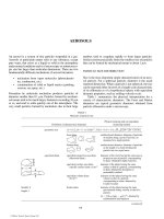
TABLE 1
Terminology
a
used in sterilization and disinfection
I. Definitive terms II. Ter ms in common use
Sterile Disinfectant
Sterilization Disinfectant
Sterilizing agent Antiseptic
-cide
b
Antisepsis
-stat Sanitizer
-statis Sanitization
a
British Standard Glossary of terms.
b
Not germicide.
© 2006 by Taylor & Francis Group, LLC
DISINFECTION 225
that pathogens likely to be associated with such equipment
and with eating and drinking vessels will be killed. Davis
(1968) has employed the name “detergent-sterilant” through-
out his review, although, as pointed out earlier, his definition
of a sterilant differs from that above. Throughout the present
chapter, “sanitizer,” without quotes, will be employed rather
than these other terms.
KINETICS
It is tempting to imagine that the application of an appropriate
disinfection procedure will result in immediate elimination of
all microorganisms from the site of interest. This temptation
is often fostered by various advertising interests in pursuance
of their sales campaigns. However, a cursory inspection of
the literature soon dispels this cosy, over-simplified view.
Disinfection has been shown repeatedly to be not only a
gradual or even prolonged, process, but also a complex one.
Almost invariably, investigation into the course of disin-
fection processes have involved the study of purified cultures
of microorganisms (usually bacteria) under specified condi-
tions. This has led to certain criticisms that such systems are
too far removed from reality to be of practical significance.
While it is true that considerable caution must be exercised
in applying the results of these studies to practical situations,
the experimental systems are still far from simple, and have
yielded much useful information.
Survivor Curves
The most common method of monitoring the progress of
a disinfection process is by means of viable counting tech-
niques. These suffer from certain inherent limitations. In
particular, the absolute values obtained are dependent on the
specific technique and the experimental conditions associated
with it; and in addition, cells which have been exposed to the
process, may respond quite differently from those examined
prior to exposure. In order to obviate this difficulty, alter-
native methods of assessing “vital activity” have been sug-
gested, usually biochemical in nature. Unfortunately, while
the greater simplicity of these methods allows more precise
measurements to be made, the killing of microorganisms
usually involves a whole series of complex reactions, which
makes correlation of the results rather difficult. Despite their
faults, viable counting methods do reflect the complexity of
the killing process.
The usual scheme of events is to expose the chosen cul-
ture of microorganisms to the disinfection process of inter-
est, under controlled experimental conditions. Estimates of
the viable population density of the system are made by per-
forming viable counts on representative samples removed
convenience, these estimates are usually plotted graphically
against time of exposure or occasionally dosage of the dis-
infection agent employed. While the estimated numbers of
organisms may be plotted directly, they are usually converted
to a proportional basis such as “surviving fraction” or “per-
centage survivors,” since this facilitates visual comparison
of the results.
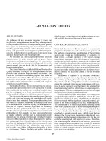
The simplest graph so obtained is the arithmetic plot
which invariably exhibits a curve of similar general form to
Figure 1. The main point of interest about this curve is that
it indicates that the rate of disinfection varies inversely with
the number of surviving organisms. This is interpreted as
an indication that the individual cells of the culture exhibit
differing sensitives to the process, i.e., there is a distribution
of resistances. Unfortunately, curves of this type are difficult
to analyze or to compare visually, and so the survivors are
often plotted in a logarithmic fashion. This results in a whole
Figure 2(a) shows the simplest result, the familiar
straight line which is often prized for its ease of charac-
terization. It also possesses the sometimes dubious advan-
tage of ease of extrapolation; a property which should be
utilized only with extreme caution. This graph indicates
that the rate of disinfection is inversely proportional to
the logarithm of the number of surviving organisms. The
similarity between this situation and the kinetics of a first-
order chemical reaction has caused this type of response
to be described as unimolecular or monomolecular. It is
important to stress, however, that the description applies
to the graphical response of the system; for it would be
extremely naive to assume from this that the mode of death
of the cells is attributable to a first-order chemical reac-
tion. The straight line may be described mathematically
by the equations:—
k
t
N
N
ϭ
1
0
log
⎛
⎝
⎜
⎞
⎠
⎟
where
k ϭ rate constant or slope of line
t ϭ time elapsed
N
0
ϭ number of viable cells initially
N ϭ number of viable cells at time t
time
Surviving Fraction
10
FIGURE 1
© 2006 by Taylor & Francis Group, LLC
“family” of possible results, as shown in Figure 2.
from the system: see, for example, Prince et al. (1975). For
226 DISINFECTION
or, if natural logarithms are used:—
N
N
kt
0
ϭ exp( ).
These equations have led to the alternative and preferred
descriptions of the response as being exponential or logarith-
mic. Since the number of viable cells decreases throughout
the process, then the rate constant, k, is always negative in
character.
The two curves shown in Figures 2(b) and 2(c) illus-
trate similar, though opposite, deviations in response from
the straight line case. These are described, according to their
shape, as concave or convex, and qualified by the additional
designation upward or downward, to indicate orientation. The
graphs indicate that the rate of disinfection changes gradu-
ally in the early stages, but then assumes a more steady state
of change similar to that in the straight case. In Figure 2(b),
the rate of disinfection is relatively slow at first, but gradu-
ally increases to a steady, limiting value. Various interpre-
tations have been suggested to account for this change;
among them, that the distribution of resistances between the
individual cells exhibits a relative deficiency of cells of low
resistance; or alternatively, that the cells must pass through
one or more intermediate stages before becoming sensitive
to the disinfection process in question. The weakness of such
interpretations is underlined by the fact that changes in the
experimental conditions often result in a change in the shape
of the graphical response. Figure 2(c) illustrates a relatively
fast rate of disinfection initially, which gradually decreases
to a steady, limiting value.
In Figure 2(d) is illustrated the most common deviation
from the straight line response, the sigmoid curve. Responses
of this type are more easily demonstrated in systems employ-
ing a moderate rate of disinfection. As the rate of disinfection
is increased, the limitations of viable counting techniques
make it more and more difficult to monitor the progress of
the process with any precision. This results in the apparent
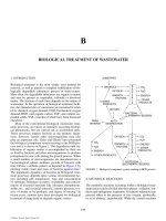
response becoming indistinguishable from the straight line
case. It is sometimes suggested that the sigmoid response is
the most common situation encountered, but that it is often
unrecognized due to the practical difficulties experienced.
The sigmoid response is usually interpreted as an indication
that the distribution of resistances between the individual
cells is of the log normal type. Complete agreement with this
model distribution is indicated when the sigmoid curve is
symmetrical.
Figure 2(e) illustrates a response of particular interest.
The graph consists of two parts, both of which are linear
but of different slope, with a fairly sharp transition between
the two. This would appear to indicate a fairly rapid rate of
disinfection initially, followed by a fairly sharp transition to
a lower, but steady, rate. Such a sudden transition naturally
engenders interest, if not suspicion. It has been suggested that
this type of response indicates the presence of two distinct
groups of cells, each of which exhibits its own characteristic
distribution of resistances. Experiments with a mixture of
two bacterial cultures of different identity, whether obtained
different species, or consisting of spores and vegetative
cells of the same species, can be shown to yield this type of
response. The first part of the graph corresponds to the usual
response of the more sensitive component, and the second
part to that of the more resistant component. However, at
relatively low temperatures and humidities, exposure of
nominally homogeneous cultures to ethylene oxide gas often
yields this type of response. While it is sometimes suggested
that this indicates the presence of two distinct groups of cells,
as discussed above, it must also be considered that not only
can this phenomenon be demonstrated with cultures appar-
ently homogeneous to other sterilization methods, but also
that this two part response reverts to the exponential type on
increasing the temperature or humidity of the system.
As indicated in the foregoing discussion, consideration
of the shapes of survivor curves may provide useful circum-
stantial evidence on which to base hypotheses relating to
the response of cell populations to disinfection processes.
However, as also indicated in the discussion, the operative
word is “circumstantial.”
Empirical Parameters
While a survivor curve illustrates the response of a cell popu-
lation in terms of variation in number of survivors with time
(or dose) of disinfection treatment, this is essentially a static
time
(Log)
Surviving Fraction
1.0
(a) (b)
(c) (d)
(e)
FIGURE 2
© 2006 by Taylor & Francis Group, LLC
DISINFECTION 227
situation since all other factors are held constant. Often, it is
the variation in response of the system to changes in experi-
mental conditions which is of particular interest. This varia-
tion in response may be monitored by constructing families
of survivor curves, one curve for each level of the factor
being varied. Comparison of curves within these families
indicates the nature of the change in response.
The two factors most prominent in the regulation of
disinfection processes are temperature and concentration of
disinfectant. For both of these factors, it is found that their
influence on the disinfection process varies in a regular
manner over a fairly wide range of conditions. As a result,
empirical parameters have been derived which enable the
influence of these two factors to be characterized in a con-
venient form.
Temperature Coefficient
In general, it is found that the activity of a disinfectant varies
directly with the temperature, i.e., the higher the tempera-
ture the greater the activity. This change in activity may be
quantified by expressing the disinfection rates observed at
two different temperatures as a ratio. The value of this ratio
is found to remain reasonably constant over a wide range of
temperature. This may be expressed mathematically:
u
TT
kk
21
21
Ϫ
ϭ /
where k
1
and k
2
are the rate constants at temperatures T
1
°C
and T
2
°C, respectively, with T
2
greater than T
1
. The con-
stant, u, is termed the temperature coefficient, and assumes
a numerical value which is characteristic of the agent and,
to a certain extent, of the organism, employed. The super-
script ( T
2
− T
1
)°C is necessary to indicate the temperature
difference. Since the disinfection rate is inversely related to
killing or extinction time, the latter may be used instead. The
expression then becomes
u
TT
tt
21
12
Ϫ
ϭ /
where t
1
and t
2
are the extinction times at temperatures T
1
°C
and T
2
°C, respectively. At high value for u indicates that the
process is relatively sensitive to temperature changes.
It is usual to find two versions of the temperature coeffi-
cient in most common use: the coefficient for a 1°C tempera-
ture change, u, when the superscript is usually omitted; and
the coefficient for a 10°C temperature change, u
10
, which
may be sometimes expressed as Q
10
. The popularity of the
10°C coefficient follows from its use to characterize changes
in reaction rates in chemical systems. This enables interest-
ing comparisons to be made between disinfection processes
and non-living chemical reactions. Attempts have been made
to deduce modes of death of the organisms by such compari-
sons. However, subsequent biochemical investigations have
tended to disagree with these deductions.
Dilution Coefficient
The activity of disinfectants under otherwise constant condi-
tions is found to vary directly with the concentration of dis-
infectant employed over a considerable concentration range.
As before, killing or extinction times are usually employed
as a measure of disinfection rate. The effect of concentration
may be expressed mathematically as:
c
h
t ϭ a constant
or:
h log c ϩ log t ϭ a constant
where
c ϭ concentration of disinfectant
t ϭ extinction time
h ϭ dilution coefficient.
A high value for the dilution coefficient indicates that the
process is relatively sensitive to changes in concentration of
disinfectant. The dilution coefficient is sometimes referred
to as the concentration exponent.
Other Factors Influencing Activity
The antimicrobial activity of several disinfectants is influenced
to a considerable extent by changes in pH. For example, a rise
in pH results in a decrease in the activity of phenols (Bennett,
1959), organic, acids, compounds liberating chlorine, benzoic
acid and iodine, although iodine is less affected by acidity
than is chlorine. An increase in pH increases the dissociation
of phenols and benzoic acid (Wedderbern, 1964); chlorine in
water forms HClO, which is dissociated with a rise in pH with
a concomitant loss of activity. In contrast to the above, how-
ever, there is an increased microbicidal activity of the quater-
nary ammonium compounds (QACs) and of acridines (Foster
and Russell, 1971); in the case of QACs, the effect of pH is
considered by Salton (1957) to be on the cell rather than the
disinfectant molecule, since the number of negatively-charged
groups on the bacterial surface will be increased as the pH
rises, thus influencing the number of positively charged mol-
ecules which can be attached.
Another factor which influences the activity of certain
antimicrobial agents is organic matter, e.g., the presence of
blood, serum, pus, etc. In general terms, the more chemically
reactive a compound, the greater the effect of organic matter
on its activity. This is particularly true with the hypochlorites.
other examples are provided under individual compounds
later.
Mathematical Models
A considerable number of mathematical models have been
derived at various times, in attempts to reconstruct the disinfec-
tion process. These have utilized deterministic, probabilistic,
and thermodynamic approaches to the problem, and have been
reviewed in detail by Prokop and Humphrey (1970).
In general, these models are based on attempts to recon-
struct the survivor curves obtained, even though, as already
discussed, these are liable to be changed by changes in
© 2006 by Taylor & Francis Group, LLC
228 DISINFECTION
experimental conditions. In addition, they usually involve
preliminary assumptions concerning the nature of the
disinfection process. Consequently, attempts to deduce
mechanisms of killing from these models tend to be rather
disappointing. They are, however, useful in terms of con-
cise descriptions of the results of the process; and as fur-
ther biochemical information on modes of killing becomes
available, their usefulness and reliability should increase
further.
DISINFECTANT TESTING
The testing of disinfectants is a topic with a long history of
controversy. Differing opinions on the merits and relevance
of various methods are legion, and have been the cause of
many heated exchanges between those holding them. The
wide variety of methods may most conveniently be consid-
ered under the headings of screening, standardization and
“in-use” tests, although there is a degree of overlap between
the three categories. The interested reader should consult the
papers by Forsyth (1975), Miner et al. (1975) and Reybrouck
(1982) for further information.
Screening Tests
These are usually of the simplest type, since they are specula-
tive by nature, and often involve the testing of large numbers
of disinfection agents or formulations; both time and cost
usually dictate this simplicity. The actual methods employed
vary according to the physical characteristics of the disinfec-
tant and the type of use envisaged. Ideally, any test should be
as realistic as possible, although for screening purposes this
will be subject to the requirements of simplicity discussed
above.
Disinfectants which are soluble or miscible in water are
often incorporated in microbiological culture media which
are then inoculated with suitable microorganisms. After
incubation under appropriate conditions, the inoculated
media are examined for growth of the organisms, absence
of growth indicating that the inoculum has been inhibited.
Where the continuous presence of the disinfectant is inap-
propriate, then the method is modified to include a suitable
means of removing or inactivating the disinfectant after a
predetermined exposure time.
Other disinfectants may be tested in a similar manner,
by arranging for inocula of suitable organisms to be sub-
jected to standardized exposure to them. After exposure, the
organisms are inoculated into suitable culture media, and
incubated as before. Since the end-point in these tests is the
death of all the organisms involved, this type of test is often
referred to as an Extinction Test.
The phrase “suitable organisms” used above, represents
one of the key factors in the testing of disinfectants. A disin-
fectant is expected to kill all undesirable organisms, which
usually refers to organisms injurious to health (see previous
section). Test organisms may be chosen, therefore, either
for their resistance to disinfection or, like Salmonella typhi,
for their medical significance. Although the designation
“undesirable organisms” covers a wider range of life-forms,
most disinfectant testing has employed various species of bac-
teria for reasons of convenience. In retrospect, this does not
appear to have been a significant disadvantage, since activity
against bacteria usually coincides with that against the other
life-forms. However, although it is recognized that bacterial
spores are much more resistant to disinfection than the veg-
etative forms, the latter have, with few exceptions, invariably
been used in testing. This is partly due to convenience, but it
should be noted that there are many disinfectants marketed
which under normal conditions of use are incapable of killing
bacterial spores. This has been countered, in certain quarters,
by re-defining a disinfectant as a chemical agent capable of
killing bacteria but not necessarily bacterial spores (see pre-
vious section).
Standardization Tests
This is the area which has given rise to the greatest amount
of contention. In most cases, this has been due to misin-
terpretation and misapplication of the results of testing
in situations where they have little, if any, relevance. The
standardization of disinfectants usually requires the use
of microbiological techniques rather than chemical assay.
Even when chemical characterization of the active agent(s)
involved is possible, the activity of a disinfectant will usu-
ally be significantly influenced by factors concerned with
the formulation and method of use of the product. However,
although the disinfectant may be standardized in terms of its
intended biological effect, this is carried out under specific,
controlled conditions. Depending on the similarity of their
circumstances, the results from standardization tests may, or
may not, be capable of extrapolation to practical situations.
This is where the controversy tends to arise.
The most popular methods of testing for standardiza-
tion purposes have been extinction tests. These are basically
similar to the general method discussed in the previous
section, except that the procedure and materials used are
rigidly standardized in order to achieve best reproducibility
of results. In addition, the most widely used tests employ
the pure chemical, phenol, as a control of the resistance
of the test organisms used. Since results are expressed by
comparison of the activities of phenol and the disinfectant
being tested, these tests are referred to as Phenol Coefficient
methods. The most popular and official versions in use are
the Rideal–Walker Method (as modified by B.S. 541), the
Chick–Martin Method (as modified by B.S. 808), and the
Association of Official Agricultural Chemists (AOAC)
Phenol Coefficient Method (1970). Details of these tests
may be found in the appropriate publications.
While the use of phenol as a control on the resistance
of the test organisms is extremely valuable, it has led to
widespread assumptions that the ratio of activities of disin-
fectant and phenol indicated by the phenol coefficient will
hold true in all circumstances. This, of course, is far from
true. The AOAC (1970) attempted to improve this situation
by introducing a Use-Dilution Method. This test is based on
© 2006 by Taylor & Francis Group, LLC
DISINFECTION 229
the hypothesis that disinfectants in use shall be at least as
efficient as 5% phenol, and that a dilution of twenty times
the phenol coefficient should achieve this. Accordingly, this
dilution is tested against standardized cultures in order to
confirm this, or alternatively, in order to determine a correc-
tion factor.
Whereas the extinction methods discussed above evaluate
the extinction concentration corresponding to predetermined
exposure times, Berry and Bean (1954) devised a test for
evaluating extinction times for chosen disinfectant concentra-
tions. While the test is only applicable to phenolic and other
easily inactivated disinfectants, it has been claimed to be at
least as reproducible as other methods in common use (Cook
and Wills, 1954). The method of assessing the end-point of
the reaction has been further improved by Mathei (1949).
Methods other than extinction tests for standardizing dis-
infectants include various methods based on assessment of
cessation of vital enzyme activities. The enzymes involved
have generally been certain oxidases or dehydrogenases
(Sykes, 1939; Knox et al. , 1949). In a less specific manner,
inhibition of respiration has been used as a method of assess-
ment (Roberts and Rahn, 1946). The controversy surround-
ing these methods centers around the problem of correlating
enzyme activity with viability.
Other methods which have been proposed include mea-
surement of post-incubation opacity corresponding to stan-
dard survivor levels (Needham, 1947), and also measurement
of cell volume increase following post-exposure incubation
(Mandels and Darby, 1953).
The range of tests discussed above is by no means
exhaustive, and only covers general purpose tests appli-
cable to water-misible disinfectants. Any number of alter-
native tests could be devised by appropriate selection and
standardization of the various parameters. In addition, there
are numerous tests which can be, and have been, devised in
order to standardize disinfectants intended for specific uses
such as sporicidal or tuberculocidal duties (AOAC 1970).
Methods of standardizing disinfectants other than those
to which the above discussion applies are usually designed
more closely around the particular use envisaged. Thus, a
considerable element of actual, or simulated, in-use testing
is usually involved. For convenience, they are best discussed
in the following section.
A recent test is the Kelsey–Sykes test (Kelsey and Maurer,
1974), which is a form of capacity test. In this, incremental
additions of test organisms are made to appropriate dilutions
of test disinfectant and aliquots are removed for detecting
survival immediately prior to the next addition of organisms.
On the basis of this method, use-dilutions of the test dis-
infectant (which need not necessarily be a phenolic) under
clean and dirty conditions can be recommended to hospitals,
which should then check them during in-use tests (see also
In-Use Tests
The previous two sections have dealt with testing methods
applied in the laboratory, which yield information primarily
of value to the disinfectant manufacturer. The “consumer,”
however, is almost solely concerned with the performance
of disinfectant materials under the conditions of use which
are associated with the application envisaged. To this end,
he is more interested in testing methods which resemble, as
closely as possible, these practical conditions. However, the
foregoing remarks should not be taken to imply that labo-
ratory standardization methods are completely arbitrary. In
view of the greater difficulty experienced in killing organ-
isms which are present as dried surface films, as opposed to
those in fluid suspension, many standardization tests have
employed films on various surfaces as their inocula. Surfaces
used have included silk thread (Koch, 1881), garnets (Kronig
and Paul, 1887), glass cover-slips (Jensen and Jensen, 1933),
glass cylinders (Mallmann and Hanes, 1945), glass slides
(Johns, 1946), stainless steel cylinders (AOAC Use-Dilution
Confirmatory Test, 1960), and glass tablet tubes (Hare, Raik
and Gash, 1963). It should be noted that while these sur-
faces represent a step toward the practical situation, they
nevertheless comprise a collection of laboratory “artifacts”
when compared with real situations. A nearer approach was
achieved by use of surfaces such as rubber strips (Goetchins
and Botwright, 1950) and glazed, waxed, and rubber tiles
(Rogers, Mather and Kaplan, 1961).
Where the physical size of articles required to be disin-
fected is fairly small, it is quite feasible to carry out in-use
testing in the laboratory. Hence metal trays were used by
Neave and Hoy (1947), 10 gallon milk churns by Hoy and
Clegg (1953), and small drinking glasses by Gilcreas and
O’Brien (1941). Similarly, scalpels, syringes and similar
small items may conveniently be subjected to in-use testing
in the laboratory. Where the physical size of the system is
somewhat greater, then the laboratory must be forsaken in
order to carry out on-site testing. Typical examples of such
situations include hospital walls and floors, and industrial or
dairy processing machinery. The outstanding value of in-use
testing arises from the fact that results obtained are directly
applicable to the system without the need for interpreta-
tion and extrapolation, and that practical difficulties such as
short contact times or inaccessibility of certain areas can be
accounted for.
The choice of test organisms usually reflects either of
two main approaches. The simplest approach is to inoculate
the system artificially with organisms considered to be of
practical significance in the particular application. This sig-
nificance may be due to the resistance to disinfection of the
organism, or alternatively, to its practical effect on the system
should it survive the disinfection process. In order to simu-
late the practical system, the organisms may be suspended
in appropriate materials before inoculation. An example
would be the use of milk as a suspending fluid in tests on
dairy disinfection. A slight variation on this approach is to
inoculate with indeterminate mixtures of likely organisms
obtained from natural sources, such as low quality, raw milk.
The second approach involves the use of the normal, pre-
existing flora of the system which has arisen during normal
use. Investigations of this flora before and after the disin-
fection process would provide direct evidence as to whether
© 2006 by Taylor & Francis Group, LLC
Coates, 1977; Cowen, 1978).
230 DISINFECTION
the process has achieved its object. One difficulty with this
approach is that of providing suitable growth conditions for
all of the possible species of organisms which may be pres-
ent. Also, the normal flora is likely to vary with time, and so,
ideally, this method is best applied in the form of a routine
monitoring procedure. Both of these difficulties produce an
increase in the financial cost of this type of approach, but the
reliability of the results obtained is correspondingly high.
The ideal method would involve a combination of these two
approaches: an initial test with known resistant organisms
in order to indicate the upper levels of activity which may
be required; this to be followed by routine monitoring tests
to guard against both unsuspected resistance amongst the
normal flora, and unforeseen breakdowns in the method of
application.
Any testing method which involves the use of surface
films is subject to the problem of physical recovery of the
organisms. In the case of small surfaces, the articles can often
be placed in or on a suitable nutrient medium, and provided
this allows contact between the medium and the organisms
in the film, then growth takes place. Where larger or less
accessible surfaces are concerned, then the simple direct
method will have to be replaced by some kind of sampling
technique. Sampling techniques vary widely and not all are
applicable in all situations. In the case of accessible surfaces,
simple methods such as wiping with sterile swabs may be
used; or alternatively, flooding the surface with a sterile
liquid, some or most of which is then removed by swab or
pipette. An interesting alternative consisting of a sterile, agar
medium, “sausage” was devised by Ten Cate (1965). The
exposed transverse surface of this sausage is pressed against
the surface to be sampled. After sampling a slice is cut from
the sausage and incubated with the exposed side uppermost.
A simple method which is used for sampling skin surfaces
may also be used in other applications. This method employs
adhesive cellophane tape. The adhesive surface is pressed
onto the surface to be sampled, and then removed for trans-
fer to a suitable medium. Where surfaces are inaccessible,
as in pipes and processing machinery, then a rinsing tech-
nique is usually most convenient. The resulting liquid may
be added directly to suitable nutrient media, or, if present
in large bulk, may be filtered through membrane filters to
remove the organisms. The resulting filter membrane is then
transferred to a suitable medium.
One final problem which is of importance in all meth-
ods of testing which involve assessment of viability, is that of
recovery and growth of the test organisms following exposure
to the disinfectant. Organisms which have survived a disin-
fection process often show altered requirements for optimal
growth. Response to physical conditions such as incubation
temperature, as well as to biochemical conditions such as
dependence on certain nutrients, may be completely altered
from that of unexposed cells. While considerable effort has
been made to derive efficient recovery methods (Flett et al. ,
1945; Jacobs and Harris, 1960; Harris, 1963; Russell, 1964)
the problem is so variable and so many combinations of fac-
tors must be considered, that it is far from being solved. There
is general agreement that recovery methods should be selected
which will allow maximum recovery of exposed organisms;
but putting this into effect can be extremely difficult.
LIQUID DISINFECTANTS
Several chemical agents have long been employed for destroy-
ing microorganisms, although it is frequently asserted that
such substances are without effect on bacterial spores. With
many agents, however, this is untrue (Sykes, 1970; Russell,
1971, 1982). The most important substances are phenols and
cresols, biguanides, chlorine-releasing compounds and other
halogens, aldehydes, alcohols, quaternary ammonium com-
pounds, mercury compounds, strong acids and alkalis and
hydrogen peroxide. The majority of these are considered in
detail below. Further information is provided by Hugo and
Russell (1982).
Phenols and Cresols
Although Kronig and Paul (1897) and Chick (1908) showed
that phenol was active against spores, the concentrations,
5%, employed, were considerably higher than those needed
to kill vegetative bacteria. More recent studies have indicated
that bacterial spores are not killed even after long exposure to
phenols (Sykes, 1958, 1965; Loosemore and Russell, 1963,
1964; Russell and Loosemore, 1964; Russell, 1965, 1971;
Rubbo and Gardner, 1965; Briggs, 1966). Of the bacterial
spores. Bacillus stearothermophilus is the most resistant to
phenol and B. megaterium the most sensitive (Briggs, 1966).
However, in contrast to its lack of sporicidal activity, phenol
is sporostatic at low concentrations.
Several factors influence the antimicrobial activity of
phenols and cresols (Bennett, 1959; Cook, 1960; Bean,
1967):
1) Concentration. These compounds have high con-
centration exponents, h, which, as described above,
indicates that they rapidly lose their antibacterial
activity on dilution. This also means that dilution
procedures can be used to prevent the carryover of
inhibitory concentrations into recovery media when
viable counts or sterility tests are being carried out
(Russell, 1982; Russell et al. , 1979). Studies from
Bean’s laboratory (Bean and Walters, 1955; Bean
and Das, 1966; Bean, 1967) are of interest, for they
show that with dilute solutions of disinfectants
with high intrinsic activity, e.g. benzylchlorphenol,
the high proportion taken up by the cells means that
the concentration remaining is only weakly bacte-
ricidal, so that the surviving cells do not meet lethal
conditions.
2) Temperature. The bactericidal activity of the phe-
nols and cresols increases rapidly with an increase
in temperature. Examples of temperature coeffi-
cient ( u
10
) against E. coli at 30–40° (Tilley, 1942)
are: phenol 8.4, o -cresol 6.9, p -cresol 5.6.
© 2006 by Taylor & Francis Group, LLC
DISINFECTION 231
Of importance, also, is the finding that these
substances are sporicidal at elevated temperatures
(Berry et al. , 1937; Russell and Loosemore, 1964).
As a result of the findings of Berry et al. (1937),
one method of sterilizing certain injections by
heating them with 0.2% w/v chlorocresol is still an
official method in Britain (British Pharmacopoeia,
1980).
3) pH. The phenols are more active at an acid pH
than in alkaline solution, as phenates (phenox-
ides) are formed at high pH. Acid pH also results
in a more effective, although still slow, sporicidal
action (Sykes and Hooper, 1954).
4) Organic matter. The presence of organic matter
may decrease the antimicrobial activity of these
compounds; this was early recognized in the
design of the Chick–Martin test for evaluating
phenolic disinfectants (Chick and Martin, 1910;
Garrod, 1934, 1935). The results do, however,
depend on the actual phenol used and on the kind
of organic matter, and the interference is less than
with other disinfectants such as the quaternary
ammonium compounds (Cook, 1960).
5) Oxygen tension. Anaerobic bacteria are generally
more resistant than aerobes to phenols. Moreover,
facultative organisms, e.g., E. coli, are more resis-
tant when grown under anaerobic conditions.
6) Type of organism. As described above, these
compounds are bactericidal and sporostatic at
low concentrations, and sporostatic and not spo-
ricidal even at high concentrations. As a group,
however, they are also fungicidal to several
moulds, and use is made of this in the inclusion
of cresol and chlorocresol as preservatives in
creams which are liable to fungal contamination
(Wedderbern, 1964).
Morris and Darlow (1971) have pointed out
that phenolic compounds with high R-W coeffi-
cients are effective against some viruses, but that
they are generally too variable in their activity to
be suitable as general virucidal agents.
7) Chemical nature. Dihydric and trihydric phenols
(Figure 3) are generally less active than phe-
nols, and alkylation of monohydric phenols to
give cresols potentiates the antimicrobial activ-
ity. Also halogenation of the phenols increases
their activity (although to a lesser extent if the
halogenation is in the ortho- than in the parapo-
sition) and this is even more pronounced when
accompanied by the introduction of aliphatic or
aromatic groups into the nucleus, e.g., p -chloro-
m -cresol (chlorocresol). This increase in anti-
microbial activity is, however, paralleled by a
decrease in water solubility.
To overcome this decrease in aqueous solubility, vari-
ous soaps have been used to render water-soluble (solubi-
lize) these substances. However, the effect of soap on the
biological efficacy of the phenols depends on two factors,
firstly the nature of the soap, and second the proportion of
soap to phenol. Solution of cresol with soap (Lysol, BP), for
example, contains 50% of cresol in a saponaceous solvent
and has a bactericidal activity which depends on the nature
and amount of soap used. The ratio of cresols to soap may
be critical, the optimal cresol–soap ratio being of the order
of 2:1. In lysol, the soap content is c. 22%, and the ratio is
thus 2.2:1. The soap solutions are able to solubilize insoluble
phenols in the micelles; the critical micelle concentrations
(cmcs) of different soaps vary, and this explains differences
in bactericidal action noted above.
Lysol and the so-called “black-fluids,” which consist of
the lower coal tar phenols, are formulated in sufficient soap
so that they are retained in solution when diluted with water.
In contrast, the white fluids consist of concentrated emul-
sions of high boiling phenols stabilized with protective col-
loids; they can be diluted with hard or soft waters, whereas
black fluids should be diluted with soft waters only.
Micelles have been considered as being reservoirs of phe-
nols, so that when the concentrated solution is diluted before
use, the phenols are released by dilution below the cmc to
give a highly active solution. Two types of system have been
investigated experimentally in attempts to assess the exact
role of the micelles; these are (a) constant phenol concentra-
tion, and (b) a constant phenol/soap ratio (Berry, 1951; Berry
and Briggs, 1956; Berry, Cook and Wills, 1956; Cook, 1960).
With a constant phenol system, there is a rapid increase in
bactericidal activity below the cmc of the soap which could
be the result of an increased uptake of phenol together with
an increased permeability of the bacterial surface (Mulley,
1964); however, above the cmc in this system, there is a
decrease in bactericidal activity, as the increasing numbers
of micelles being formed compete for the phenol with the
cell surface. In systems where there is a constant phenol/soap
ratio, there is likewise an increase in activity below the cmc,
OH
OH
OH
OH
OH
OH
Pyrogallol
Resorcinol
m-Cresol Chlorocresol
Phenol
OHOH
CH
3
CH
3
Cl
FIGURE 3
© 2006 by Taylor & Francis Group, LLC
232 DISINFECTION
and a decrease in activity at the cmc. However, in this system
the activity again increases at higher soap concentrations;
thus, this increased bactericidal activity parallels a saturation
of the soap micelles with the phenol. The reasons for this
remain unclear, for although the soap itself could contrib-
ute to or be responsible for the increased activity, systems
in which the soap has a low activity give similar results. The
actual role of the micelles is thus difficult to assess. Of the
two systems described, it is apparent that the system with
the constant phenol/soap ratio is more important from the
viewpoint of practical disinfection. It is necessary to use the
lowest possible proportion of soap to phenol, thus giving a
comparable situation to bactericides in two-phase systems, in
which the bactericidal efficiency is related to the concentra-
tion in the aqueous phase (Bean and Heman-Ackah, 1965).
Phenol itself is an effective bactericide and anti-fungal
agent, which is used as a preservative in some injections and
creams; it is also the standard reference substance in some
methods of testing phenolic bactericides (see earlier). Cresol,
a mixture of o -, m - and p -cresol, in which the meta -isomer
predominates, and of other phenols obtained from coal tar,
is highly bactericidal and fungicidal. Apart from being the
active constituent of Lysol, it is also used as a preservative
in certain injections and creams. Chlorocresol is a power-
ful antimicrobial agent which, in conjunction with heat, is
employed for the sterilization of certain injections. It is also
used as a preservative in certain cosmetic creams and lotions,
and is included in Sodium Benzoate and Chlorocresol
Solution, which may be used for the storage of sterilized
surgical instruments, the sodium benzoate delaying rusting.
Chloroxylenol has low water solubility and is solubilized
by means of soap, the solution being known as Roxenol. Its
antimicrobial activity is markedly reduced in the presence of
organic matter. Thymol is a potent bactericide and fungicide
which, in the form of Glycerin of Thymol, is employed as an
oral antiseptic. Hexachlorophane [2,2Ј-methylenebis(3,4,6-
trichloro-phenol)] is most frequently used as a soap contain-
ing 2% of the substance; the effectiveness of this soap as
a skin disinfectant may depend upon the accumulation of
hexachlorophane on the skin. Pentachlorophenyl dodecano-
ate is extensively used as a fungicide and insecticide in the
textile and packing industries. Unlike the parent molecule,
pentachlorophenol, it is nontoxic to humans.
None of the above compounds can be relied upon to kill
bacterial spores at ordinary temperatures.
Another group, related to the phenols, consists of the
esters (parabens) of p -hydroxybenzoic acid (3-hydrobenzoic
acid). Unlike benzoic acid, the dissociation of which increases
with increasing pH with a corresponding decrease in activity,
the parabens are active against bacteria and fungi over a fairly
wide pH range. Their bactericidal and fungicidal properties
increase within the homologous series, but this is paralleled
by a decrease in aqueous solubility. The parabens are not spo-
ricidal, and although on their discovery they were heralded
as being the ideal preservatives, they are now used (often
in combination of two or more) mainly as preservatives in
various pharmaceutical and cosmetic products (Russell,
Jenkins and Harrison, 1967; Parker, 1982).
The phenols and parabens have an effect on the cytoplas-
mic membranes of bacteria and fungi (Hugo, 1976a,b).
Biguanides and Bisbiguanides
Biguanides have the general formula
R
1
R
2
H
.
NHNH
NH
NC
CN
(5)(1)
R
2
Three distinct antimicrobial actions of N
1
, N
5
-substituted
biguanides have to date been recognized (Weinberg, 1968):
germicidal, antiviral and antimalarial. Unfortunately, no one
compound is generally active in more than one of these three
categories. The requirements for each type of activity are
fairly unique, e.g., for maximum broad-spectrum germicidal
activity, both N
1
and N
5
should have a halogen-substituted
aralkyl substituent.
Bisbiguanides have the general formula
R
1
.
NH
NH
NH NH
NH NH
NH
NH NH
NHCCCC
(CH
2
)
6
R
For maximum broad spectrum germicidal activity R and R
1
should consist of a halogen-substituted aryl group. A number
of bisbiguanides are known to be germicidal, and the most
important member of this group is chlorhexidine (R and R
1
are both C
6
H
5
Cl).
Chlorhexidine was first described in 1954 (Davies et al. ,
1954), and it is bacteriostatic in low concentrations; higher
concentrations are bactericidal to several bacterial species,
including strains of the genus Proteus, of which Pr. mirabilis
is the most resistant, and Pr. rettgeri and Pr. morganii are
the most sensitive to chlorhexidine, with Pr. vulgaris occu-
pying an intermediate position. Some strains of Ps. aerugi-
nosa may be highly resistant. Chlorhexidine also possesses
antifungal activity. It is a membrane-active agent (Hugo,
1976a,b, 1982).
Chlorhexidine, the active constituent of “Hibitane,” is
used in surface disinfection, as an antimicrobial agent in
eye-drops, and, in the presence of sodium nitrite to prevent
corrosion, for the storage of surgical instruments.
Chlorine-Releasing Compounds
It was observed by Dakin (1915, 1916) that the commercial
hypochlorites then in use were not of constant composition
and contained free alkali and sometimes free chlorine. He
thus developed a solution (Dakin’s Solution or Chlorinated
Soda Solution, Srugical) which is still in use today. The sta-
bility of free available chlorine in solution is dependent on a
number of factors, in particular on the chlorine concentration,
pH of the solution, the presence of catalysts, temperature, the
presence of organic matter, and light (Dychdala, 1977).
© 2006 by Taylor & Francis Group, LLC
DISINFECTION 233
The types of chlorine compounds which are frequently
used are (1) Hypochlorites. These are cheap and convenient
to use, and have a wide antibacterial spectrum (Davis, 1963).
They possess potent sporicidal activity (Truman, 1971; Kelsey
et al. , 1974; Waites, 1982) which may be potentiated by alco-
hols (Coates and Death, 1978; Death and Coates, 1979).
The hypochlorites are moderately effective against animal
viruses.
The antibacterial activity of the hypochlorites decreases
with increasing pH (Charlton and Levine, 1937; Weber,
1950; Ito et al. , 1967; Hays et al. , 1967), e.g., whereas 99%
of spores of B. cereus are killed after 2.5 min at pH 6 by a
solution containing 25 ppm available chlorine, nearly 8 hours
are required for a comparable kill at a pH of c. 13 (Rudolph
and Levine, 1941). At constant pH, the time to kill bacteria
depends on the concentrations of available chlorine. The spo-
ricidal activity of sodium hypochlorite may be potentiated by
various compounds, e.g., by the addition of ammonia (Weber
and Levine, 1944) or 1.5–4% sodium hydroxide (Cousins
and Allan, 1967), notwithstanding the earlier comment
about pH. In the presence of bromide, hypochlorite has an
enhanced effect in bleaching cellulosic fibres as compared
with hypochlorite alone, possibly because of a continuous
generation of hypobromite when hypochlorite is in excess.
A potentiation of the bactericidal effect of hypochlorite has
been achieved by the addition of small amounts of bromide
(Farkas–Himsley, 1964).
The antimicrobial activity of hypochlorites is considerably
reduced by organic matter. However, the hypochlorites are
used in the disinfection of water, dairy equipment and eating
utensils. (2) Chloramine-T (sodium p -toluene sulphonchlo-
ramide). Dakin et al. (1916) considered that chloramine-T had
a powerful germicidal action. It is bactericidal and sporicidal,
although the rate of kill is slower than with the hypochlorites.
Its activity is considerably higher at acid than at alkaline pH
(Weber, 1950), and a drop of 10°C in the reaction tempera-
ture results in a 3–4 fold increase in the time necessary to kill
microorganisms (Weber and Levine, 1944). Chloramine-T is
employed as a wound “disinfectant,” and as a general surgi-
cal disinfectant. It is nonirritant and nontoxic, in contrast to
Dichloramine-T (toluene- p -sulphon-dichloramide) which
although a powerful disinfectant is not used because of its
toxicity and instability.
The mode of action of chlorine compounds is unknown,
although several proposals have been made, e.g., the informa-
tion of chloramines as a result of combination of chlorine with
bacterial protoplasm, halogenation or oxidation reactions of
chlorine with bacterial cells, changes in cellular permeability
and an effect on enzyme systems. It has also been found,
however (Bernarde et al. , 1967) that chlorine dioxide causes
a marked and immediate cessation of protein synthesis in
growing cells.
Iodine and Iodophors
Iodine in aqueous or alcoholic solution is considered by
most authors (Gershenfeld and Witlin, 1950; Gershenfeld,
1956; Report, 1965; Sykes, 1970) to be a reliable and
effective germicide which is lethal to vegetative bacteria,
bacterial spores and acid-fast bacilli. Spaulding et al. (1977),
however, consider that alcoholic iodine (0.5% iodine in 70%
alcohol) possesses good activity against non- sporing bacte-
rial and M. tuberculoses but none against bacterial spores,
whereas Rubbo and Gardner (1965) state that bacterial
spores are moderately resistant to iodine. Viruses are consid-
ered by Rubbo and Gardner (1965) to be moderately sensi-
tive to iodine.
Iodine has a high fungicidal or fungistatic activity against
yeasts and various moulds, but its antimicrobial properties
are to a great extent inhibited in the presence of organic
matter, since iodine is a highly reactive element.
Iodine is sparingly soluble in cold water, but more solu-
ble in hot water. Stronger solutions can be made in potassium
iodide solution or in aqueous alcohol. Iodine is more effective
as a germicide at acid than at alkaline pH, but is less affected
by pH than are chlorine compounds. The concentration of
iodine to disinfect does not vary greatly with different types
of microorganisms. Various types of iodine solution are used
for the first-aid treatment of small wounds and abrasions,
and in pre-operative skin “disinfection.” Iodine has also been
employed for the sterilization of surgical catgut (although
this method is now little used) and is nowadays used for the
disinfection of drinking and swimming pool water, the disin-
fection of instruments and of clinical thermometers, and the
sanitization of eating and drinking utensils.
Unfortunately, iodine solutions stain fabrics and tissues
and tend to be toxic. However, certain non-ionic surface-
active agents can solubilize iodine to form compounds, the
iodophors (Blatt and Maloney, 1961; Davis, 1962, 1963,
1968) which retain the germicidal activity of iodine, but
not its undesirable properties; these iodophors are literally
“iodine-carriers.” They are active against bacterial spores,
including pathogenic anaerobic spores (Lawrence et al. ,
1957; Gershenfeld, 1962). It is the concentration of free
iodine in an iodophor which is responsible for its microbial
action; this has been well demonstrated by Allawala and
Riegelman (1953) who made a log-log plot of killing time
against amount of free iodine, and found that the 99% killing
time of B. cereus spores was a function of the concentration
of free-iodine in the presence or absence of added surface-
active agent. The bactericidal properties of the iodophors are
increased at low pH values, but their stability is unaffected
(cf. hypochlorites). They may thus be employed with acids,
e.g., phosphoric acid, to enhance their microbial action and
also to assist in preventing the formation of film or milkstone
(see later) in the dairy industry. The iodophors are consid-
ered (Davis, 1968) to be powerful detergents, although they
do not dissociate protein as readily as do alkalis. The formu-
lation of acidic solutions of iodophors is particularly useful
when calcium or magnesium scale is encountered, but they
can be corrosive, especially with galvanized iron.
Surface-Active Agents
Surface-active agents have 2 regions in their molecular
structure, one a hydrocarbon, water-repellent (hydrophobic)
© 2006 by Taylor & Francis Group, LLC
234 DISINFECTION
group, and the other a water-attracting (hydrophilic or polar)
group. Depending on the basis of the charge or absence of
ionization of the hydrophilic group, surface-active agents are
classified into (James, 1965):
1) Anionic agents, which usually have strong deter-
gent, but weak antimicrobial properties, e.g.,
sodium lauryl sulphate.
2) Cationic agents, which have strong bactericidal,
but weak detergent, properties. The term “cationic
detergent” usually signifies a quarternary ammo-
nium compound (QAC, onium compound), but
this is not strictly accurate, as the concentration
at which a QAC is germicidal is so low that its
detergent activity is negligible.
3) Non-ionic agents, which consist of a hydrocar-
bon chain attached to a non-polar water-attracting
group, which is usually a chain of ethylene oxide
units, e.g., cetomacrogols. Non-ionic agents have
no antimicrobial properties.
4) Ampholytic (amphoteric) agents, which are com-
pounds of mixed anionic–cationic character, in
which the charge can be similarly positive and
negative. They are effective antimicrobial com-
pounds, which are extensively used in the dairy
industry.
The QACs are organically substituted ammonium com-
pounds, in which the nitrogen atom has a valency of 5,
four of the substituent radicals (R
1
–R
4
) are alkyl or het-
erocyclic radicals, and the fifth is a small anion. The sum
of the carbon atoms in the 4R groups is more than 10.
For a QAC to have a high activity, at least one of the four
organic radicals must have a chain length in the range C
8
to C
18
. The germicidal activities of the QACs were origi-
nally recognized in 1916, but they did not attain promi-
nence until Domagk’s work in 1935. They are primarily
active against Gram-positive bacteria, with concentrations
as low as about 1 in 200,000 being lethal; higher concentra-
tions ( c. 1 in 30,000) are lethal to Gram-negative bacteria,
although Pseudomonas aeruginosa is highly resistant. The
QACs possess antifungal properties, are sporostatic and
not sporicidal (Russell, 1971) and are inactive against the
tubercle bacillus. Their antimicrobial activity is markedly
affected by organic matter, and they are incompatible with
anionic surface-active agents, some of the non-ionic agents
(such as Lubrols and Tweens) and the phospholipids, e.g.,
lecithin and other fat-containing substances (Russell, 1982;
Russell et al. , 1979). The QACs exert their maximal bac-
tericidal activity under alkaline conditions. Although the
QACs are effective bactericidal and fungicidal agents, it
has been found (Grossgebauer, 1970) that viruses are rather
more resistant than bacteria or fungi.
R
1
R
4
R
3
R
2
N
+
X
–
Although several hundred QACs have been prepared
and tested for antimicrobial activity, only a few are regularly
used. These are:
1) Cetrimide (cetyltrimethylammonium bromide,
CTAB), a mixture of dodecyl, tetradecyl- and
hexadecyl-trimethylammonium bromide. In addi-
tion to it uses for pre-operative skin disinfec-
tion and treatment of seborrhoea of the scalp,
cetri mide is also employed, in conjunction with
sodium nitrite, which delays or prevents rusting,
for the storage of sterilized surgical instruments,
although this practice should be discontinued
(Rubbo and Gardner, 1965).
2) Benzalkonium chloride (Zephirol, Zephiran) is
the active constituent of a general purpose and
skin sterilizing agent, “Roccal.” It is also used to
alleviate or prevent napkin rash caused by urea-
splitting organisms, as a preservative in eye-drop
preparations (Pharmaceutical Codex, 1979) and
for the disinfection of blankets.
Other important QACs are domiphen bromide and cetyl-
pyridinium chloride.
The value of QACs in the disinfection of woollen blan-
kets has been observed by various authors (Frisby, 1957;
International Wood Secretariat, 1961), although they cannot be
incorporated in the wash with an anionic compound, because
of incompatibility. In addition, it must be emphasized that res-
idues of anionic agents on blankets from previous washings
could interfere with the action of QACs. To overcome this,
blankets may be washed in an appropriate non-ionic deter-
gent, with a final prolonged rinse in a QAC, or they may be
treated with a non-ionic detergent incorporating a QAC.
The QACs are also of considerable value as disinfectants
in food and dairy plant. If an alkali detergent containing
anionic surface-active wetting agents is used prior to sanitiz-
ing, then utensils and equipment must be thoroughly rinsed
(Barrett, 1969). The QACs at their normally used concentra-
tions are odourless and noncorrosive, but many are not free-
rinsing, and undesirable traces may remain on equipment or
even be present in dairy food (Clegg, 1967, 1970). However,
a combination of a free-rinsing type of QAC and a suitable
non-ionic agent may be usefully employed for washing
instruments and cutlery, etc. (Barrett, 1969).
The cytoplasmic membrane of bacteria and fungi is the
site of action of the QACs. The membrane is composed of
lipoprotein, and it is considered (Russell, 1971) that the lipid
moiety is involved in the lethal action of these compounds
There appears to be a clear relationship between the ther-
modynamic and antibacterial activities of QACs, with solu-
tions having equal antimicrobial activity against an organism
having surface concentration values of the same order of
magnitude. Thermodynamic activities of QACs at two bac-
terial survivor levels (1% and 0.01%) have been shown to
be sufficiently constant (Laycock and Mulley, 1970) to sup-
port the Ferguson (1939) principle that compounds with the
© 2006 by Taylor & Francis Group, LLC
(for more recent information, see Hugo, 1976a,b).
DISINFECTION235
same thermodynamic activity will have an equal bactericidal
activity.
Amphoteric compounds, as already stated, are of mixed
anionic-cationic character, and they combine the detergent
properties of anionic compounds with the bacterial proper-
ties of the cationic substances; their bactericidal properties
remain virtually constant over a wide pH range (Barrett, 1969)
and they are less readily inactivated by proteins than are the
QACs (Clegg, 1970). Examples of amphoteric surface-active
agentsare dodecyl- b -alanine, dodecyl- b -aminobutyric acid
and dodecyldi(aminoethyl)-glycine (Davis, 1960a,b, 1968), the
last named being a “Tego” compound. The Tego series of com-
pounds have a high molecular weight, and in addition to being
recommended for use in pre-operative hand cleansing and pre-
operative skin preparation it has also been found that they are
suitable for the cleansing of surgical operating theatre floors,
walls and equipment and for ward cleansing (Frisby, 1959,
1961). It has, however, recently been shown that Tego 103S in
1% solution is less active than a 0.5% solution of chlorhexidine
in 70% alcohol (Kuipers and Dankert, 1970). Amphoteric sur-
face-active agents are inactivated by soaps and other anionic
compounds (Frisby, 1959), but they are non-irritating and non-
corrosive. Unfortunately, they tend to be expensive.
Aldehydes
The two most important aldehydes are glutaraldehyde
(Pentanedial) and formaldehyde (methanal).
CH
2
· CHO
CH
2
· CHO
HOOOH
CH
2
Hydrated Ring Structure
Glutaraldehyde is a dialdehyde which has been used for
several years as a fixative in electron microscopy investigations
and its antimicrobial activity has been comparatively recent
1968), but they do indicate that this substance has a valuable
role to play. A 2% solution of glutaraldehyde buffered with
sodium bicarbonate (0.3% w/v is considered to be the optimum
bicarbonate concentration) is effective in killing nonsporing
bacteria within 2 min, M. tuberculosis, fungi and viruses in 10
min, and spores of Bacillus and Clostridium spp. in 3 hours.
Aqueous solutions of glutaraldehyde are acid, and are consider-
ably less active against microorganisms than are alkaline ones
(Pepper and Chandler, 1963; Stonehill et al. , 1963; Snyder and
Cheatle, 1965; Lane et al. , 1966; Rubbo et al. , 1967; Munton
and Russell, 1970a,b), but solutions become progressively less
stable at pHs above 7. Concentrated solutions of glutaraldehyde
(25%) can be purchased, diluted to the required concentration
(2%) and “activated” by the addition of sodium bicarbonate.
(Alternatively, 2% solutions ready for use when “activated”
can also be purchased.) When made alkaline, glutaraldehyde
solutions gradually undergo polymerization with a consequent
loss of activity, this polymerization proceeding rapidly at pH
values above 9. At pH 7.5–8.5, however, activity is maintained
for at least 2 weeks.
Serum does not affect the antimicrobial activity of glu-
taraldehyde, but the dialdehyde is considerably less active in
nutrient broth at pH 7.5 than it is at the same pH in buffer
(Rubbo et al. , 1967; Munton and Russell, 1970a), the reason
being that glutaraldehyde combines with the peptone present
in broth (which is thereby discolored).
Glutaraldehyde is used as a fixative in the preparation
of microbial cells for electron microscopy. It is a useful
hospital disinfectant, particularly for articles which cannot
be sterilized by physical means (Report, 1965). It has been
employed in the sterilization of cytoscopes in urology (Lane,
McKeever and Fallon, 1966) and of endoscopic instruments,
such as bronchoscopes (Snyder and Cheatle, 1965), as
it has no deleterious effect on the cement or lens coating.
Glutaraldehyde is also employed as a tanning agent in pref-
erence in glyoxal and formaldehyde (Fein et al. , 1959), and
has been shown to inactivate rapidly influenza virus and a
coliphage in mouse tissue blocks (Sabel et al. , 1969).
Glutaraldehyde is non-corrosive, and does not affect
rubber and plastic articles or the sharpness of cutting instru-
ments; because it does not coagulate protein matter, such as
blood and mucus, it does not render the cleaning of blood-
covered instruments more difficult. It is obvious, therefore,
that glutaraldehyde is most useful.
Rubbo et al. (1967) have proposed that the microbicidal
activity of glutaraldehyde is due to the presence of two free
aldehyde groups in the molecule. In solution, glutaraldehyde
exists in an equilibrium between the open chain structure and
the hydrated ring structure (see above), and there is a com-
plete loss of activity if one or both of the aldehyde groups
is altered, whereas a substitution elsewhere in the molecule
reduces, but does not abolish, its activity. It is thus essential
to have free aldehyde groups, which may react with cell sul-
phydryl or amino groups. Glutaraldehyde is about 10 times as
active as glyoxal, with succinaldehyde occupying an interme-
diate position (Pepper and Chandler, 1963). Certain bacteria
treated with glutaraldehyde become pink in color (Munton
and Russell, 1970b) as a result of cell-aldehyde interaction.
Formaldehyde has long been employed as a disinfectant.
Formaldehyde solution is rapidly sporicidal to B. subtilis
various Clostridia (Ortenzio et al. , 1953; Klarmann, 1956,
1959). Ethanol (Rubbo et al. , 1967) and methanol (Willard
and Alexander, 1964) cannot be recommended as vehicles for
formaldehyde, as there is a reduction in antibacterial activity.
Formaldehyde is an important virucidal agent which
finds its greatest use in the preparation of certain sterile vac-
cines, e.g., Poliomyelitis Vaccine (Inactivated). As a result of
the experimental evidence accumulated over several years,
a considerable amount of information is now available on
the kinetics of the virus inactivation by formaldehyde. This
particular vaccine consists of poliovirus Types I, II and III,
and each is inactivated separately and then blended to give
the trivalent vaccine. It is thus essential that formaldehyde
treatment be sufficient to destroy the viruses without affect-
ing their antigenicity; prolonged exposure to the aldehyde
© 2006 by Taylor & Francis Group, LLC
(Ortenzio et al. , 1953; cf. Klarmann, 1956, 1959) but not to
(see Rubbo and Gardner, 1965; Rubbo etal., 1967; Borick,
236 DISINFECTION
will, in fact, destroy the antigenic potency also (Morris and
Darlow, 1971). The inactivation of poliovirus by formalde-
hyde has been considered to be a first-order reaction so that
extrapolation of the death curve would give a point at which
the probability of any infective particles remaining would
approach zero. However, first-order kinetics cannot be used
with any degree of safety to extrapolate the inactivation
curve. Similarly, although the inactivation of SV
40
virus
by formaldehyde has been shown by Sweet and Hilleman
(1960) to be linear, it is now known that this linear inactiva-
tion is followed by a flattening of the curve indicating the
persistence of a residual fraction which resists inactivation.
Formaldehyde is rapidly lethal to vegetative bacteria,
and is sometimes used for this purpose in the preparation of
inactivated bacterial vaccines.
Metals
Because of their antibacterial and antifungal activity, com-
pounds of mercury, silver, copper and tin are of importance
from both medical and industrial points of view (Hugo and
Russell, 1982).
Mercury Compounds These are of two types, the
inorganic mercuric and mercurous salts and the organic
substances. Mercuric salts are primarily bacteriostatic and
fungistatic and contrary to earlier findings are not sporicidal
salts do not find widespread use in modern medicine, but are
preservation of wood, leather and paper, and in the control
of fungal infections in seeds and bulbs. Mercurous salts have
no application as preservatives.
The most important organic mercury compounds are the
phenylmercuric salts (nitrate, acetate and borate) and thi-
omersal. Phenylmercuric nitrate (PMN) and acetate (PMA)
are now mainly employed as preservatives in various phar-
maceutical and cosmetic products. PMN is also used as a
spermicide in certain contraceptive formulations, as a plant
fungicide and for the disinfection of leather and timber.
However, because of their lack of sporicidal activity at ordi-
nary temperature, the organic mercury compounds cannot be
recommended as sterilizing agents. Some plasmid-containing
gram-negative bacteria are resistant to mercury compounds,
which are vaporized (Chopra, 1982).
Various sulphydryl compounds, such as cysteine and
thioglycollic acid, can reverse mercury-induced bacteriosta-
sis, which led Fildes (1940) to propose that these compounds
combined with, and displaced, mercury from its combina-
tion with the —SH group of an enzyme (E).
E
EE
S
S
SH
SH
SH
SH
+ Hg
Hg
H
2
S
+ HgS
+
Silver Compounds Silver compounds have long been
used in medicine for their antimicrobial activity, which
extends to Gram-positive and Gram-negative bacteria and
fungi. Of the silver compounds available, silver protein
and silver nitrate are the most important. The latter, in the
form of compresses, is highly effective in preventing the
colonization of burns with Ps. aeruginosa and Proteus
species.
Copper Compounds These are bactericidal and fungicidal.
They have been used for the latter purpose for more than 200
years, and their sole use nowadays is as industrial preserva-
tives against fungal spoilage. The most frequently used sub-
stances are copper naphthenate, oxinate, 1-phenylsalicylate
and sulphate; the last-named, in combination with a lime mix-
ture, is known as Bordeaux mixture.
Dialkyldithiocarbamates are considered (Albert, 1963)
to be converted into active bactericides and fungicides in the
presence of copper. Such salts are highly successful, widely
used, agricultural fungicides (Owens, 1969).
Tin Compounds Stannous and stannic salts have little
antimicrobial ctivity. However, when tin is coupled with
organic radicals, forming what are known as the organo-
tins, potent antimicrobial activity results. If R represents the
organic radical linked directly to a tin atom, by C—Sn bond,
and X an inorganic or organic radical not so linked, various
types of compounds can be obtained, of which R—SnN
3
is
most active. Gram-negative bacteria are less sensitive than
Gram-positive bacteria to organotin compounds. Triphenyltin
acetate and hydroxide are important agricultural fungicides.
Dyes
The acridines have held a valuable place in medicine for sev-
eral years, although with the advent of the antibiotics and
other chemotherapeutic agents, they are now less widely used
than hitherto. The acridines are active against several Gram-
positive and Gram-negative bacteria, and have been used
mainly in treating infected wounds. Their uses and mode of
action have been reviewed (Foster and Russell, 1971).
The most important members of the triphenylmethane
group of dyes are crystal violet, brilliant green and malachite
green. These have mainly been employed for local applica-
tion to burns and wounds.
Some members of a third group of dyes, the quinones,
are important agricultural fungicides. The quinones are
natural dyes which impart color to many forms of plant
and animal life. Chemically, the quinones are diketocy-
clohexadienes, the simplest of which is 1,4-benzoquinone
moulds and yeasts, followed (in this order) by phenanthren-
equinones, benzoquinones and anthraquinones (Figure 4).
Antimicrobial activity is increased by halogenation, and
two powerful antimicrobial agents employed as fungicides
are chloranil (tetrachloro-1, 4-benzoquinone) and dichlone
(2,3-dichloro-1,4-naphthaquinone).
Alcohols
Ethyl alcohol, although active against Gram-positive and
Gram-negative bacteria, is devoid of lethal activity against
bacterial spores, and thus cannot be relied upon as a steril-
izing agent (Russell, 1971). Methyl alcohol is likewise not
© 2006 by Taylor & Francis Group, LLC
(Figure 4). Naphtaquinones are the most toxic to bacteria,
extensively employed as industrial preservatives, e.g., in the
(see Russell, 1971). Because of their toxicity, the mercuric
DISINFECTION 237
sporicidal. Moreover, alcohols may reduce the sporicidal
activity of aldehydes.
GASEOUS ANTIMICROBIAL AGENTS
Gaseous agents may play an important role in sterilization of
certain types of medical equipment and of components used
in outer space research. However, only two gases (ethylene
oxide and formaldehyde) are used extensively for the ster-
ilization of medical products. Other gases (methyl bromide,
b -propiolactone and propylene oxide) are not used as routine
methods. Appropriate measures must be taken to counteract
toxicity to humans (Christensen and Kristensen, 1982).
Ethylene Oxide
Ethylene oxide (EO) exists as a gas which is soluble in rubber,
water, oils and several organic solvents. Chemically, it is
CH
2
CH
2
.
O
Its inflammability when in contact with air is overcome by
using mixtures of EO with carbon dioxide or with fluoro-
carbon compounds (Phillips and Kaye, 1949; Barwell and
Freeman, 1959; Freeman and Barwell, 1960). EO diffuses
freely through paper, cellophane, cardboard and some plas-
tics, but less rapidly through polythene; it cannot penetrate
crystalline materials (Opfell and Miller, 1965). The antimi-
crobial activity of EO has been dealt with in many papers and
reviews (Phillips, 1949, 1958, 1961, 1977; Phillips and Kaye,
1949; Kaye, 1949, 1950; Kaye and Phillips, 1949; Kaye
et al., 1952; Phillips and Warshowsky, 1958; Thomas, 1960;
Bruch, 1961; Sutaria and Williams, 1961; Russell 1971; 1976;
Kelsey, 1961, 1967; Sykes, 1970; Hoffman, 1971; Kereluk,
1971; Ernst, 1974, 1975; Hugo and Russell, 1982).
Several factors are known to influence the antimicrobial
activity of EO (Christensen and Kristensen, 1982):
1) Concentration. As would be expected, the higher
the concentration of EO (expressed as mg/l,
which refers to the actual amount present in the
sterilizer) the more rapid is the rate at which
microorganisms are killed. However, even at
high concentrations, EO is only slowly lethal,
and long periods may, therefore, be needed for
sterilization to be achieved.
2) Temperature. The lethal activity of EO increases
with a rise in temperature. It has a temperature coef-
ficient of 2.74 for each 10°C rise in temperature.
3) Type of organism. EO gas will kill bacteria and
their spores, yeasts, moulds and their spores,
and viruses (Griffith and Hall, 1938), and resis-
tant strains have not been developed. In contrast
to many other chemical substances which are
considerably less effective against spores than
against vegetative cells, bacterial spores are only
about 2–10 times as resistant as the latter to EO
(Toth, 1959; Phillips, 1958) and in some cases,
e.g., B. stearothermophilus (Thomas et al., 1969)
even less resistant. These results imply that EO
can freely penetrate the outer layer(s) of the
bacterial spore, although experimental results in
support of this contention are sadly lacking.
In addition to its antibacterial and antifungal
activity, ethylene oxide is also effective against
rickettsiae and viruses (Hoffman, 1971).
4) Relative humidity (RH). Of all the factors which
influence the activity of EO, RH is probably the
most important. The optimum RH is considered
to be c. 28–33% (Schley et al., 1960), and EO
is considerably less microbicidal at high RH
and in relatively dry air. The correct RH may
be achieved to prehumification of the steriliza-
tion chamber (Halowell et al. 1958; Ernst and
Schull, 1962). However, the moisture content of
microorganisms themselves, as well as the RH
of the environment, is also important. Bacterial
cells which have been desiccated and then equil-
ibrated to successively high RH values contain
less water and are more resistant to EO than cells
which have not been desiccated but have instead
been allowed to dry naturally until equilibrated to
the same RH values. To overcome this resistance
O
O
O
OO
O
O
O
Naphthaquinone
9,10-Anthraquinone
9,10-Phenanthrenequinone
Benzoquinone
FIGURE 4
© 2006 by Taylor & Francis Group, LLC
238 DISINFECTION
to EO produced by dehydration, the cells have
to be actually wetted (Perkins and Lloyd, 1961;
Winge-Heden, 1963; Gilbert et al., 1964).
Alkylating agents act through alkylation of
sulphydryl, amino, carboxyl, hydroxyl and phe-
nolic groupings, with the loss of a hydrogen atom
and the production of an alkyl hydroxyethyl
group (Phillips, 1952, 1958; Kelsey, 1967), and
it seems likely that EO kills microorganisms by
an alkylation of protein molecules (Gilbert et al. ,
1964). The influence of RH on the microbicidal
activity of EO is related to this, since the pres-
ence of insufficient water prevents alkylation,
whereas excess water causes hydrolysis of EO
to ethylene glycol, CH
2
OHCH
2
OH (Wilson and
Bruno, 1950).
5) Effect of drying medium on spore resistance.
Organisms dried from salt solution always show
some survivors after exposure to EO (Royce and
Bowler, 1961; Beeby and Whitehouse, 1965).
Moreover, organisms enclosed within crystals
are protected from the action of EO (Abbott
et al. , 1956; Royce and Bowler, 1961) as a result
of the inability of the gas to penetrate crystalline
materials.
EO has several uses in the pharmaceutical and
medical fields. These include the sterilization
of ophthalmic instruments, anaesthetic equip-
ment, heart-lung machines, disposable syringes
(Rubbo and Gardner, 1968) and hospital blankets
(Kaye, 1950), and as a decontamination proce-
dure for articles handled by tuberculous patients.
There may, however, be a “crazing” of disposable
syringes, and EO treatment is a slow, costly pro-
cess, with a “quarantine” period necessary at the
end of the process to ensure that complete dissipa-
tion of the gas has been achieved.
b -Propiolactone
b -Propiolactone (BPL) is a colorless liquid at room tem-
perature. It has been employed as a chemosterilizer in both
the liquid and gaseous states, and is microbicidal in both
forms (Allen and Murphy, 1960). Its antimicrobial activity
depends on its concentration and on the temperature and
RH at which the vapor is used (Spiner and Hoffman, 1960),
a temperature above 24°C being required for optimal activ-
ity. As with EO, RH is of considerable importance in deter-
mining the activity of BPL vapor, although with the latter
a RH in excess of 70% is required for rapid microbicidal
acivity (Hoffman and Warshowsky, 1958). Again, however,
it is not necessarily the atmospheric RH which is of greatest
importance but the location and content of water within the
microbial cell. B. globigii spores equilibrated to 98% are
rapidly killed by BPL at 45% RH, whereas they are more
resistant if preconditioned at 75% RH before treatment
with BPL at 45% RH, and a small proportion of spores
equilibrated at 1% RH are subsequently highly resistant to
BPL at 75% RH (Hoffman, 1968).
Bacterial spores are more resistant to BPL than vegeta-
tive cells, viruses or fungi (Lo Grippo et al. , 1955; Trafas
et al. , 1954; Bruch and Koesterer, 1962; Toplin, 1962)
although some strains of Staph. aureas may be almost as
resistant as spores (Hoffman and Warshowsky, 1958).
BPL is also highly active against viruses and rickettsiae
(Hoffman, 1971).
BPL has been used for the chemical sterilization of regen-
erated collagen sutures (Ball et al. , 1961), for the decontamina-
tion of enclosed spaces (Bruch, 1961b) and for the sterilization
of a variety of instruments contaminated with various sporing
and non-sporing bacteria (Allen and Murphy, 1960). However,
its reported carcinogenic effects in rats and mice (Walpole
et al. , 1954) mean that a considerable degree of caution is
needed before BPL is employed as a chemosterilizer.
Formaldehyde
Formaldehyde vapor may be obtained by evaporating appro-
priate dilutions of standardized batches of commercial for-
malin (a 40% solution of formaldehyde in water) with 10%
methanol added to prevent polymerization (Report, 1958).
Temperature affects the activity of the gas, as does the RH,
there being an increase in activity with increasing RH up to
50%, but little further increase in killing rate as the RH rises
from 50 to 90% (Nordgren, 1939; Report, 1958). In contrast,
gross wetting retards killing.
Bacteria protected by organic matter, such as blood
and sputum are less rapidly killed by formaldehyde vapor
(Nordgren, 1939; Bullock and Rawlins, 1954). Micro-
organisms may also be protected from it when they are
included in a crystal mass, in contrast to surface-contaminated
crystals (Abbot et al. , 1956).
Although bacterial spores are more resistant than vegeta-
tive cells to formaldehyde vapor, the degree of difference
is not high (Phillips, 1952; Report, 1958; Sykes, 1965).
The vapor only has weak penetration, and its application is
thus normally limited to surface sterilization (Borick, 1968;
Davis, 1968). However, the addition of formaldehyde vapor
to steam under sub-atmospheric pressure at temperatures
below 90°C results in deep penetration into fabrics with
destruction of heat-resistant microorganisms (Alder et al. ,
1966; Alder and Simpson, 1982).
Formaldehyde vapor has long been used for the disin-
fection of blankets, and is considered to be one of the best
methods available for disinfecting woolen blankets that have
not received a shrink-resist treatment (International Wool
Secretariat, 1961). It is also used to decontaminate rooms,
buildings and instruments (Hoffman, 1971).
CH
2
CH
2
C
OO
© 2006 by Taylor & Francis Group, LLC
DISINFECTION 239
PRACTICAL USES
This final section brings together some of the data presented
in the preceding sections, and also provides information in
certain specific instances. Disinfectants will be considered
from two points of view, first their medical, and second their
nonmedical uses. Some brief information on antiseptic and
preservative use will also be supplied.
Medical Uses
The use and choice of disinfectants in hospitals have been
extensively considered in the last decade in Britain by a spe-
cial committee (Report, 1965) set up for this purpose. Our
comments here are thus based on the recommendations of this
report and on the findings presented by Kelsey and Maurer
(1967). The Report (1965) recommended that two classes
of disinfecting agents were needed, (a) for general disinfec-
tion and (b) for surface disinfection of clean objects. Agents
for general disinfection should have a wide spectrum, and at
appropriate dilutions should remain active in the presence of
organic matter; the main purpose of such disinfectants, e.g.,
phenolic disinfectants based on coal-tar acids, is not neces-
sarily to kill all bacteria but to ensure that an object is free
of significant numbers of organisms. Chemicals for surface
disinfection must be quick-acting, have a wide spectrum, be
non-harmful to materials and leave no objectionable odours.
Such disinfectants, e.g., hypochlorites, should be used for
the rapid disinfection of clean surfaces such as trolley tops,
kitchen tables and clinical thermometers.
Kelsey and Maurer (1967, 1972) have presented a list
of the steps to be taken in drawing up a policy for the use
of general purpose disinfectants in hospitals, and among the
points they make is the non-usage of disinfectants in cer-
tain cases, especially where sterilization is the objective or
where other more reliable means are available. For further
and Lowbury (1982). Preoperative disinfection of the skin
(including surgeon’s hands), disinfection of operation sites
and topical prophylaxis, i.e., antisepsis in burns, are dis-
cussed by Lowbury (1982). Ayliffe and Collins (1982) pro-
vide a rational approach to hospital disinfection.
Nonmedical Uses
The main nonmedical uses of disinfectants occur in the
food, dairy, brewing and fermentation industries (Foster
et al. , 1958; Frazier, 1967). The maintenance of equipment
for use in these industries in a proper sanitary condition
cannot be overemphasized. This therefore means that the
cleaning of such equipment is of considerable importance,
since the presence of organic matter can reduce or virtually
eliminate the effect of many disinfectants (page 164). In the
dairy industry, milk stone—resulting from milk drying on
equipment, and thus consisting of fat, protein and minerals—
and milk film are a well-known problem in disinfection
(Clegg, 1967). Chemicals which are of use against micro-
organisms in liquid suspension in laboratory tests may be of
little use against such organisms on a soiled surface if they
are poor detergents (Cousins, Hoy and Clegg, 1960).
To counteract the unwanted effects of organic matter,
one of the following two methods may be employed (see
1) detergent first, followed by a disinfectant;
2) combination of detergent and disinfectant (this
corresponds to a sanitizer, or to the detergent-
sterilant of Davis (1968), as described in the
Introduction).
Harris (1969) stresses the need for using two operations,
i.e., the use of cleaning before disinfection. Cleaning is the
first essential in the sanitary care of food equipment, and
approximate sterility the last (Foster et al. , 1958). Steam
under pressure is an obvious method of sanitization, but
this is limited only to closed systems which can withstand
pressure (Frazier, 1967). Theoretically, separate procedures
would be expected to give a better result (Clegg, 1970),
because of the inactivating effect of organic matter on disin-
fectants; however, a finding made several years ago (Neave
and Hoy, 1947) suggests that a detergent-disinfectant com-
bination would be of greater use, because “the effect of the
detergent on milk solids more than outweighs the effect of
milk residues inactivating the disinfectant” (Clegg, 1970).
An effective detergent should dissociate organic and inor-
ganic solids, emulsify, saponify or suspend grease, fats and
oils, have good wetting ability, be easily rinsed and be non-
corrosive (Olivant and Shapton, 1970). Detergents are thus
of considerable importance in this field, because they can
also be responsible for the mechanical removal of bacteria
(Gilbert, 1969). The most commonly used detergents are
strong and mild alkalis, alkali salts, strong acids, anionic
alkyl sulphates and aryl sulphonates and non-ionic conden-
sates (Davis, 1968). An excellent descaling agent is nitric
acid which can be used hot at concentrations of 0.25–0.5%
or cold at 0.5–1%, and which is, in addition, a powerful
disinfectant. It is, however, less effective than alkali in the
removal of hardened protein films, and is normally employed
with a corrosion inhibitor. In actual fact, many detergents are
good disinfectants and vice versa, e.g., a detergent such as
sodium hydroxide possesses considerable germicidal power
(Whitehouse and Clegg, 1963), whereas hypochlorites have
a useful detergent effect by disintegrating protein matter
(Davis, 1968). Cleaning is an essential first part in high-
speed food and beverage processing plant, and considerable
economic benefit is achieved as a result of the production of
stable liquid detergents and disinfectants which can be deliv-
ered by tanker and then distributed to the cleaning areas by
pipeline (Hill, 1969). However, even with good precleaning,
traces of inactivating protein material may remain on equip-
ment, and it is thus important to choose a disinfectant which
has a high protein tolerance (Harris, 1969). The design of
equipment to facilitate cleaning must also be stressed, and
stainless steel is an obvious example, with glass pipelines to
give a high degree of visual cleanliness (Harris, 1969).
© 2006 by Taylor & Francis Group, LLC
also Davis, 1972a,b and BSI 1977):
information, see Lynn (1980), Ayliffe and Collins (1982)
240 DISINFECTION
The disinfection of fermentation laboratories has been
described by Darlow (1969). Gaseous disinfectants may be
employed, and aerosols appear to have an important role to
play. Local disinfection of bench tops, floors, etc. is also a
standard practice. The importance of disinfectants in water
conservation is emphasized by Fielden (1969), and in the
pharmaceutical industry by Underwood (1980).
The control of airborne microorganisms is of particu-
lar importance in the fermentation industry, in laboratories
where strict asepsis is essential (e.g., in the production of
various sterile products in the pharmaceutical industry and
in hospital pharmacy departments, as well as in the rearing
of germ-free animals), in hospital wards to reduce the inci-
dence of cross-infection, and in special wards set aside for
patients with a rare disease (hypogammaglobulinaemia) who
are particularly sensitive to infection.
This control is normally achieved by the use of special
air filters, often in conjunction with ultraviolet lamps to
irradiate the upper atmosphere. Disinfectants in the form
of aerosols are also of importance in aerial disinfection. To
be effective for air disinfection, a chemical should ideally
possess the following properties:
1) be odorless, cheap and stable
2) be without toxic or irritant properties
3) be capable of being dispersed in the air, with con-
sequent complete and rapid mixing of infected air
and chemical
4) be capable of maintaining an effective concentra-
tion in the air
5) be highly and rapidly effective against airborne
organisms
6) be unaffected by relative humidity (RH).
Aerosols consist of a very fine dispersed liquid phase
in a gaseous (air) continuous phase. The germicide must
be nebulized in sufficiently fine spray (aerosols droplets
of <1 µ m are the accepted standard) so that it will remain
airborne and thus have ample opportunity to collide with
any microorganisms present in the air. At low RH, particles
are too dry for adequate condensation of the disinfectant
that such organisms enclosed in particles, and thus bacteria
occurring on dust or on surfaces are much less susceptible
to the aerial disinfectant than such organisms enclosed
in droplets (Sykes, 1965). The optimum RH is usually c.
40–60%.
Chemical aerosols are often generated in the following
manner: if the chemical is liquid, it may be sprayed directly
into the air from an atomizer; if the chemical exists as a
solid, it may be dissolved in an appropriate solvent, e.g.,
propylene glycol, and atomized, or alternatively the solid
may be vaporized by heat from a thermostatically-con-
trolled hot-plate.
Chemicals which have been used as aerial disinfectants
include hexylresorcinol, lactic acid, propylene glycol (this
possesses antimicrobial activity in its own right), hypo-
chlorous acid, formaldehyde gas and sulphur dioxide.
Other Uses of Antimicrobial Agents
Antimicrobial agents are widely employed as preservatives
in pharmaceutical and cosmetic products. Factors influenc-
ing their activity, as well as those governing their choice in
different classes of sterile or non-sterile products have been
well considered by Bean (1972), Croshaw (1977), Parker
(1978, 1982), Kazmi and Mitchell (1978a,b), Allwood
(1980) and Beveridge (1980).
Preservation is also required in other specialized areas,
e.g., in cutting oils (Hill, 1982a), fuels (Hill, 1982b), paper and
pulp (Weir, 1982), wood (Richardson, 1982), paint (Springle
and Briggs, 1982) and textiles (Hugo, 1982b) and in the con-
struction industry (Bravery, 1982). A most important aspect
concerns the use of chemical food preservatives and this has
been comprehensively reviewed by Sofos and Busta (1982).
REFERENCES
1. Abbot, C.F., J. Cockton, and W. Jones, 1956, J. Pharm. Pharmac.,
8, 709.
2. Albert, A., 1963, Adv. Appl. Microbiol., 5, 1.
3. Alder, V.G., A.M. Brown, and W.A. Gillespie, 1966, J. Clin. Path.,
19, 83.
4. Alder, V.G. and R. Simpson, 1982, in Principles and Practice of
Disinfection, Preservation and Sterilisation, Ed., A.D. Russell, W.B.
Hugo, and G.A.J. Ayliffe, Blackwell Scientific Publications, Oxford.
5. Allawala, N.A. and S. Riegelman, 1953, J. Am. Pharm. Ass., Sci. Ed.,
42, 396.
6. Allen, H.F. and J.T. Murphy, 1960, J. Am. Med. Ass., 172, 1759.
7. Allwood, M.C., 1980, in Pharmaceutical Microbiology, 2nd Edition,
Ed., W.B. Hugo and A.D. Russell, Blackwell Scientific Publications,
Oxford.
8. Association of Official Agricultural Chemists, 1970, Official Methods
of Analysis of the AOAC, Washington, DC.
9. Ayliffe, G.A.J. and B. Collins, 1982, in Principles and Practice of
Disinfection, Preservation and Sterilisation, Ed., A.D. Russell, W.B.
Hugo and G.A.J. Ayliffe, Blackwell Scientific Publications, Oxford.
10. Ball, E.L., A.C. Dornbrush, G.M. Sieger, F.E. Stirn, J.C. Vitucci, and
J.F. Weidenheimer, 1961, Appl. Microbiol., 9, 269.
11. Barrett, M., 1969, Process Biochem., 4, 23.
12. Barwell, C.F. and M.A.R. Freeman, 1959, Lancet, i, 917.
13. Bean, H.S., 1967, J. Appl. Bact., 30, 6.
14. Bean, H.S., 1972, J. Soc. Cosmet. Chem., 23, 703.
15. Bean, H.S. and A. Das, 1966, J. Pharm. Pharmac., 18, 107S.
16. Bean, H.S. and S.M. Heman-Ackah, 1964, J. Pharm. Pharmac.,
16, 58T.
17. Bean, H.S. and V. Walters, 1955, J. Pharm. Pharmac., 7, 661.
18. Beeby, M.M. and C.E. Whitehouse, 1965. J. Appl. Bact., 28, 349.
19. Benarde, M.A., W.B. Snow, V.P. Olivieri, and B. Davidson, 1967,
Appl. Microbiol., 15, 257.
20. Bennett, E.O., 1959, Adv. Appl. Microbiol., 1, 49.
21. Berry, H., 1951, J. Pharm. Pharmac., 3, 689.
22. Berry, H., and H.S. Bean, 1954, J. Pharm. Pharmac., 7, 224.
23. Berry, H., and A. Briggs, 1956, J. Pharm. Pharmac., 8, 1143.
24. Berry, H., A.M. Cook, and B.A. Wills, 1956, J. Pharm. Pharmac.,
8, 425.
25. Berry, H., E. Jensen, and F.K. Silliker, 1937, Quart. J. Pharm. Phar-
mac., 11, 729.
26. Beveridge, E.G., 1980, in Pharmaceutical Microbiology, 2nd Edition.
Edition. Ed., W.B. Hugo and A.D. Russell, Blackwell Scientific Pub-
lications, Oxford.
27. Blatt, R. and J.V. Moloney, 1961, Surg. Gynaec. Obstet., 113, 699.
28. Borick, P.M., 1968, Adv. Appl. Microbiol., 10, 291.
29. Borick, P.M., 1970, in Disinfection, Ed., M.A. Benarde, Marcel
Dekker, New York.
© 2006 by Taylor & Francis Group, LLC
DISINFECTION 241
30. Boucher, R.M.G., 1975, Can. J. Pharm. Sci., 10, 1.
31. Bravery, A.F., 1982, in Principles and Practice of Disinfection, Pres-
ervation and Sterilisation, Ed., A.D. Russell, W.B. Hugo, and G.A.J.
Ayliffe, Blackwell Scientific Publications, Oxford.
32. Briggs, A., 1966, J. Appl. Bact., 29, 490.
33. British Pharmacopoeia, 1980, Pharmaceutical Press, London.
34. British Standards Institution, 1934, British Standard techniques for
determining the Rideal-Walker Coefficient of Disinfectants: BS 541.
35. British Standards Institution, 1938. British Standard specification for
the modified technique of the Chick-Martin Test for Disinfectants:
BS 808.
36. British Standards Institution, 1977, Recommendations for steriliza-
tion of plant and equipment used in the dairying industry: BS 5305.
37. Bruch, C.W., 1961a, Ann. Rev. Microbiol., 15, 245.
38. Bruch, C.W., 1961b, Am. J. Hyg., 73, 1.
39. Bruch, C.W., and M.G. Koesterer, 1962, Appl. Microbiol., 10, 123.
40. Bullock, K. and E.A. Rawlins, 1954, J. Pharm. Pharmac., 6, 859.
41. Cadet, J. et al., Photosensitized Reactions of Nucleic Acids, Biochi-
mie, 68, 1986.
42. Charlton, D.B. and M. Levine, 1937, Iowa Eng. Exp. Sta. Bull., No.
132, Iowa, USA.
43. Chick, H., 1908, J. Hyg. Camb., 8, 92.
44. Chick, H. and C.J. Martin, 1908, J. Hyg. Camb., 8, 698.
45. Chopra, I.C., 1982, in Principles and Practice of Disinfection, Pres-
ervation and Sterilisation, Ed., A.D. Russell, W.B. Hugo, and G.A.J.
Ayliffe, Blackwell Scientific Publications, Oxford.
46. Christensen, E.A. and K. Christensen, 1982, in Principles and Practice
of Disinfection, Preservation and Sterilisation, Ed., A.D. Russell, W.B.
Hugo, and G.A.J. Ayliffe, Blackwell Scientific Publications, Oxford.
47. Clegg, L.F.L., 1967, J. Appl. Bact., 30, 117.
48. Clegg, L.F.L., 1970, in Disinfection, Ed., M.A. Benarde, Marcel
Dekker, New York.
49. Coates, D. and J.E. Death, 1978, J. Clin. Path., 31, 148.
50. Coates, D., 1977, Pharm. J., 219, 402.
51. Cook, A.M., 1960, J. Pharm. Pharmac., 12, 19T.
52. Cook, A.M. and B.A. Wills, 1954, J. Pharm. Pharmac., 6, 638.
53. Cousins, C.M. and C.D. Allan, 1967, J. Appl. Bact., 30, 168.
54. Cousins, C.M., W.A. Hoy, and L.F.L. Clegg, 1960, J. Appl. Bact.,
23, 359.
55. Cowen, R.A., 1978, Pharm. J., 220, 202.
56. Croshaw, B., 1977, J. Soc. Cosmet. Chem., 28, 3.
57. Dakin, H.D., 1915, Br. Med. J., 2, 809.
58. Dakin, H.D., 1916, Br. Med. J., 2, 318.
59. Dakin, H.D., J.B. Cohen, and J. Kenyon, 1916, Br. Med. J., 1, 160.
60. Darlow, H.M., 1969, Process Biochem., 4, 15.
61. Davies, G.E., J. Francis, A.R. Martin, F.L. Rose, and G. Swain, 1954,
Br. J. Pharmac., 9, 192.
62. Davis, J.G., 1960a, J. Pharm. Pharmac., 12, 29T.
63. Davis, J.G., 1960b, J. Appl. Bact., 23, 318.
64. Davis, J.G., 1962, J. Appl. Bact., 25, 195.
65. Davis, J.G., 1963, The Sanitarian, October, p. 3.
66. Davis, J.G., 1968, Progr. Industr. Microbiol., 8, 141.
67. Davis, J.G., 1972a, J. Soc. Cosmet. Chem., 23, 45.
68. Davis, J.G., 1972b, Dairy Ind., 37 (4), 212.
69. Death, J.E. and D. Coates, 1979, J. Clin. Path., 32, 148.
70. Dychdala, G.R., 1977, in Disinfection, Sterilisation and Preservation,
2nd Edition, Ed., S.S. Block, Lea and Febiger, Philadelphia.
71. Ernst, R.R., 1974, Biotech. Bioeng. Symp. No. 4, p. 858.
72. Ernst, R.R., 1975, Acta Pharmaceutica Suecica, 12 (Supplement), 44.
73. Ernst, R.R. and J.J. Shull, 1962. Appl. Microbiol., 10, 337.
74. Farkas-Himsley, H., 1964, Appl. Microbiol., 12, 1.
75. Fein, M.L., E.H. Harris, J. Naghski, and E.M. Filachione, 1959, J. Am.
Leather Chem. Ass., 54, 488.
76. Ferguson, J., 1939, Proc. Roy. Soc. B, 127, 387.
77. Fielden, T.B., 1969, Process Biochem., 4, 35.
78. Forsyth, M.P., 1975, Dev. Ind. Microbiol., 16, 37.
79. Flett, L.H., C. Haring, A.F. Guiteras, and R.L. Shapiro, 1945, J. Bact.,
50, 591.
80. Foster, E.M., F.E. Nelson, M.L. Speck, R.N. Doetsch, and J.C. Olson,
1958, Dairy Microbiology, Macmillan and Co. Ltd.
81. Foster, J.H.S. and A.D. Russell, 1971, in Inhibition and Destruction
of the Microbial Cell, Ed., W.B. Hugo, Academic Press, London and
New York.
82. Frazier, W.C., 1967, Food Microbiology, 2nd Edition, McGraw-Hill,
New York.
83. Freeman, M.A.R. and C.F. Barwell, 1960, J. Hyg., Camb., 58, 337.
84. Frisby, B.R., 1959, Lancet, 2, 57.
85. Frisby, B.R., 1961, Lancet, 2, 829.
86. Garrod, L.P., 1934, J. Hyg., Camb., 34, 322.
87. Garrod, L.P., 1935, J. Hyg., Camb., 35, 219.
88. Gershenfeld, L., 1956, Am. J. Pharm., 128, 335.
89. Gershenfeld, L., 1962, Am. J. Pharm., 134, 78.
90. Gershenfeld, L. and B. Witlin, 1950, Ann. NY Acad. Sci., 53, 172.
91. Gilbert, G.L., V.M. Gambill, D.R. Spiner, R.K. Hoffman, and C.R.
Phillips, 1964, Appl. Microbiol., 12, 496.
92. Gilbert, J., 1969, Process Biochem., 4, 37.
93. Gilireas, F.W. and J.E. O’Brien, 1941, Am. J. Publ. Health, 31, 143.
94. Goetchins, G.R. and W.E. Botwright, 1950, J. Milk Technol., 13, 63.
95. Griffith, C.L. and L.A. Hall, 1938, US Patent 2, 189, 947.
96. Grossgebauer, K., 1970, in Disinfection, Ed., M.A. Benarde, Marcel
Dekker, New York.
97. Halowell, P., J.T. Murphy, and A.B. Mangiaracine, 1958, Anaesthesi-
ology, 19, 665.
98. Hare, R., E. Raik, and S. Gash, 1963, Br. Med. J., 1, 496.
99. Harris, C., 1969, Process Biochem., 4, 19.
100. Harris, N.D., 1963, J. Appl. Bact., 26, 387.
101. Hays, H., P.R. Elliker, and W.E. Sandine, 1967, Appl. Microbiol., 15,
575.
102. Hill, E.C., 1982a, in Principles and Practice of Disinfection, Preser-
vation and Sterilisation, Ed., A.D. Russell, W.B. Hugo, and G.A.J.
Ayliffe, Blackwell Scientific Publications, Oxford.
103. Hill, E.C., 1982b, in Principles and Practice of Disinfection, Preser-
vation and Sterilisation, Ed., A.D. Russell, W.B. Hugo, and G.A.J.
Ayliffe, Blackwell Scientific Publications, Oxford.
104. Hill, R.P., 1969, Process Biochem., 4, 31.
105. Hoffman, R.K., 1968, Appl. Microbiol., 16, 641.
106. Hoffman, R.K., 1971, in Inhibition and Destruction of the Microbial
Cell, Ed., W.B. Hugo, Academic Press, London and New York.
107. Hoffman, R.K. and B. Warshowsky, 1958, Appl. Microbiol., 6, 358.
108. Hoy, W.A. and L.F.L. Clegg, 1953, Proc. Soc. Appl. Bact., 16, 1.
109. Hugo, W.B., 1976a, in The Survival of Vegetative Microbes,
Ed., T.R.G. Gray and J.R. Postgate, 26th Symposium of the Society
for General Microbiology, Cambridge University Press, Cambridge.
110. Hugo, W.B., 1976b, in The Inactivation of Vegetative Bacteria, Ed.,
F.A. Skinner and W.B. Hugo, Society for Applied Bacteriology Sympo-
sium Series No. 5, Academic Press, London and New York.
111. Hugo, W.B., 1982a, in Principles and Practice of Disinfection, Pres-
ervation and Sterilisation, Ed., A.D. Russell, W.B. Hugo, and G.A.J.
Ayliffe, Blackwell Scientific Publications, Oxford.
112. Hugo, W.B., 1982b, in Principles and Practice of Disinfection, Pres-
ervation and Sterilisation, Ed., A.D. Russell, W.B. Hugo, and G.A.J.
Ayliffe, Blackwell Scientific Publications, Oxford.
113. Hutchinson, F., A Review of Some Topics Concerning Mutagenesis
by Ultraviolet Light, Photochem. Photobiol., 45, 1987.
114. International Wool Secretariat, 1961, The Sterilization of Hospital
Woollen Blankets, Wool Science Review, No. 19 and No. 20.
115. Ito, K.A., D.J. Seslar, and W.A. Mercer, 1967, in Botulism, 66.
Ed., M. Ingram and T.A. Roberts, Chapman and Hall, London.
116. Jacobs, S.E. and N.D. Harris, 1960, J. Appl. Bact., 23, 294.
117. James, A.M., 1965, Surface Activity and the Microbial Cell, Soc.
Chem. Ind., London.
118. Jensen, V. and E. Jensen, 1933, Dansk. Tidss. Farm., 7, 77.
119. Johns, C.K., 1946, Am. J. Publ. Health, 37, 1322.
120. Johns, C.K., 1954, Can. J. Technol., 32, 71.
121. Kaye, S., 1949, Am. J. Hyg., 50, 289.
122. Kaye, S., 1950, J. Lab. Clin. Med., 35, 823.
123. Kaye, S. and C.R. Phillips, 1949, Am. J. Hyg., 50, 296.
124. Kaye, S., H.F. Irminger, and C.R. Phillips, 1952, J. Lab. Clin. Med.,
40, 67.
125. Kazmi, S.J.A. and A.G. Mitchell, 1978a, J. Pharm. Sci., 67, 1260.
© 2006 by Taylor & Francis Group, LLC
242 DISINFECTION
126. Kazmi, S.J.A. and A.G. Mitchell, 1978b, J. Pharm. Sci., 67, 1266.
127. Kelsey, J.C., 1961, J. Clin. Path., 14, 313.
128. Kelsey, J.C., 1967, J. Appl. Bact., 30, 92.
129. Kelsey, J.C., and I.M. Maurer, 1967, Mon. Bull. Min. Health, 26, 110.
130. Kelsey, J.C. and I.M. Maurer, 1972, The Use of Chemical Disinfec-
tants in Hospitals, Public Health Laboratory Service Monographs No.
2, HMSO, London.
131. Kelsey, J.C. and I.M. Maurer, 1974, Pharm. J., 207, 528.
132. Kelsey, J.C. and Sykes, 1969, Pharm. J., 202, 607.
133. Kelsey, J.C., M.M. Beeby, and C.M. Whitehouse, 1965, Mon. Bull.
Min. Health, 24, 162.
134. Kelsey, J.C., I.H. Mackinnon, and I.M. Maurer, 1974, J. Clin. Path.,
27, 632.
135. Kereluk, K., 1971, in Progress in Industrial Microbiology, 10. Ed.,
D.J.D. Hockenhull, Churchill Livingstone, Edinburgh.
136. Klarmann, E.G., 1956, Am. J. Pharm., 128, 4.
137. Klarmann, E.G., 1959, Am. J. Pharm., 131, 86.
138. Knox, W.E., V.H. Auerbach, K. Zarudnaya, and M. Spirtes, 1949,
J. Bact., 58, 443.
139. Koch, R., 1881, Arb. Kaiserl. Gesundh., 1, 1.
140. Kronig, B., and T. Paul, 1897, Z. Hug. Infekt. Kr., 25, 1.
141. Kuipers, J.S. and J. Dankert, 1970, J. Hug., Camb., 68, 343.
142. Lamy, P.P. and H.L. Flack, 1962, Am. J. Hosp. Pharm., 19, 473.
143. Lane, V., J.D. McKeever, and M. Fallon, 1966, J. Irish Med. Ass.,
58, 131.
144. Lawrence, C.A., C.M. Carpenter, and A.W.C. Naylor-Foote, 1957,
J. Am. Pharm. Ass., Sci. Ed., 46, 500.
145. Laycock, H.H. and B.A. Mulley, 1970, J. Pharm. Pharmac., 22,
1515.
146. Lo Grippo, G.A., P.R. Overhulse, D.E. Szilagyi, and F.W. Hartman,
1955, Lab. Invest., 4, 217.
147. Loosemore, M. and A.D. Russell, 1963, J. Pharm. Pharmac., 15, 558.
148. Loosemore, M. and A.D. Russell, 1964, J. Pharm. Pharmac., 16, 817.
149. Lowbury, E.J.L., 1982, in Principles and Practice of Disinfection,
Preservation and Sterilisation, Ed., A.D. Russell, W.B. Hugo, and
G.A.J. Ayliffe, Blackwell Scientific Publications, Oxford.
150. Lynn, B., 1980, in Pharmaceutical Microbiology, 2nd Edition, Ed., W.B.
Hugo and A.D. Russell, Blackwell Scientific Publications, Oxford.
151. Mallman, W.L. and M. Hanes, 1947, J. Bact., 49, 526.
152. Mandels, G.R. and R.T. Darby, 1953, J. Bact., 65, 16.
153. Mather, R.L., 1949, Biometrics, 5, 127.
154. Miner, N.A., E. Whitmore, and M.L. McBee, 1975, Dev. Ind. Micro-
biol., 16, 23.
155. Morris, E.J. and H.M. Darlow, 1971, in Inhibition and Destruction
of the Microbiol Cell, Ed., W.B. Hugo, Academic Press, London and
New York.
156. Mulley, B.A., 1964, Adv. Pharm. Sci., 1, 87.
157. Munton, T.J. and A.D. Rusell, 1970a, J. Appl. Bact., 33, 410.
158. Munton, T.J. and A.D. Russell, 1970b, Experientia, 27, 109.
159. Neave, F.K. and W.A. Hoy, 1947, J. Dairy Res., 15, 24.
160. Needham, N.V., 1947, J. Hyg., Camb., 45, 1.
161. Nordgren, G., 1939, Acta Path. Microbiol. Scand. suppl., No. 40, 1.
162. Olivant, D.J. and D.A. Shapton, 1970, in Disinfection, Ed. M.A.
Benarde, Marcel Dekker, New York.
163. Opfell, J.B. and C.E. Miller, 1965, Adv. Appl. Microbiol., 7, 81.
164. Ortenzio, L.F., L.S. Stuart, and J.L. Friedl, 1953, J. Ass. Off. Agric.
Chem., 36, 480.
165. Owens, R.G., 1969, in Fungicides, 2, Ed., D.C. Torgeson, Academic
Press, New York.
166. Parker, M.S., 1978, J. Appl. Bact., 44, Sxxix.
167. Parker, M.S., 1982, in Principles and Practice of Disinfection, Pres-
ervation and Sterilisation, Ed., A.D. Russel, W.B. Hugo, and G.A.J.
Ayliffe, Blackwell Scientific Publications, Oxford.
168. Pepper, R.E. and V.L. Chandler, 1963, Appl. Microbiol., 11, 384.
169. Perkins, J.J. and R.S. Lloyd, 1961, in Recent Developments in the
Sterilizatino of Surgical Materials, Report of a Symposium, p. 76,
Pharmaceutical Press, London.
170. Pharmaceutical Codex, 1979, Pharmaceutical Press, London.
171. Phillips, C.R., 1949, Amer. J. Hyg., 50, 280.
172. Phillips, C.R., 1952, Bacteriol. Rev., 16, 135.
173. Phillips, C.R., 1958, Becton, Dickinson Lectures on Sterilization:
Seton Hall, New Jersey, USA.
174. Phillips, C.R., 1961, in Recent Developments in the Sterilization of
Surgical Materials, Report of a Symposium, p. 59, Pharmaceutical
Press, London.
175. Phillips, C.R., 1977, in Disinfection, Sterilization and Preservation,
2nd Edition, Ed., S.S. Block, Lea and Febiger, Pennsylvania, USA.
176. Phillips, C.R. and S. Kaye, 1949, Amer. J. Hyg., 50, 270.
177. Phillips, C.R. and B. Warshowsky, 1958, Ann. Rev. Microbiol.,
12, 525.
178. Prince, J., C.E.A. Deverill, and G.A.J. Ayliffe, 1975, J. Clin. Path.,
28, 71.
179. Prokop, A. and A.E. Humphrey, 1970, in Disinfection, Ed.,
M.A. Benarde, Marcel Dekker, Inc., New York.
180. Report, 1958, Report by the Committee on Formaldehyde Disinfec-
tion , J. Hyg. Camb., 56, 488.
181. Report, 1965, Report by Public Health Laboratory Service Committee
on the Testing and Evaluation of Disinfectants , Br. Med. J., 1, 408.
182. Reybrouck, G., 1982, in Principles and Practice of Disinfection, Pres-
ervation and Sterilisation, Ed., A.D. Russell, W.B. Hugo, and G.A.J.
Ayliffe, Blackwell Scientific Publications, Oxford.
183. Richardson, B.A., 1982, in Principles and Practice of Disinfection,
Preservation and Sterilisation, Ed., A.D. Russell, W.B. Hugo, and
G.A.J. Ayliffe, Blackwell Scientific Publications, Oxford.
184. Rittenburgy, M.S. and M.E. Hench, 1965, Ann. Surg., 161, 127.
185. Roberts, M.H. and O. Rahn, 1946, J. Bact., 52, 639.
186. Rogers, M.R., K. Mather, and A.M. Kaplan, 1961, Appl. Microbiol.,
9, 497.
187. Royce, A. and C. Bowler, 1961, J. Pharm. Pharmac., 13, 87T.
188. Rubbo, S.D. and J.F. Gardner, 1965, A Review of Sterilization and
Disinfection, Lloyd-Luke, London.
189. Rubbo, S.D. and J.F. Gardner, 1968, J. Appl. Bact., 31, 164.
190. Rubbo, S.D., J.F. Gardner, and R.L. Webb, 1967, J. Appl. Bact., 30, 78.
191. Rudolph, A.S. and M. Levine, 1941, Iowa Eng. Exp. Sta. Bull., No.
150, Iowa, USA.
192. Russell, A.D., 1964, Lab. Pract., 13, 114.
193. Russell, A.D., 1971, in Inhibition and Destruction of the Microbial
Cell, Ed., W.B. Hugo, Academic Press, London and New York.
194. Russell, A.D., 1976, in The Inactivation of Vegetative Bacteria,
Ed., F.A. Skinner and W.B. Hugo, Society for Applied Bacteriology
Symposium Series No. 5, Academic Press, London and New York.
195. Russell, A.D., 1982, in Principles and Practice of Disinfection, Pres-
ervation and Sterilisation, Ed., A.D. Russell, W.B. Hugo, and G.A.J.
Ayliffe, Blackwell Scientific Publications, Oxford.
196. Russell, A.D., I. Ahonkhai, and D.T. Rogers, 1979, J. Appl. Bact.,
46, 207.
197. Russell, A.D., J. Jenkins and I.H. Harrison, 1967, Adv. Appl. Micro-
biol., 9, 1.
198. Russell, A.D. and M. Loosemore, 1964, Appl. Microbiol., 12, 403.
199. Sabel, F.L., A. Hellman, and J.J. McDade, 1969, Appl. Microbiol.,
17, 645.
200. Salk, J.E., U. Krech, J.S. Younger, B.L. Bennett, and L.J. Lewis, 1954,
Amer. J. Publ. Health, 44, 563.
201. Salton, M.R.J., 1957, 2nd Int. Congr. Surface Activity, Butterworths,
London.
202. Schley, D.G., R.K. Hoffman, and C.R. Phillips, 1960, Appl. Micro-
biol., 8, 15.
203. Snyder, R.W. and E.L. Cheatle, 1965, Amer. J. Hosp. Pharm., 22, 321.
204. Sofos, J.N. and F. Busta, 1982, in Principles and Practice of Disinfec-
tion, Preservation and Sterilisation, Ed., A.D. Russell, W.B. Hugo,
and G.A.J. Ayliffe, Blackwell Scientific Publications, Oxford.
205. Spaulding, E.H., 1958, Becton, Dickinson Lectures on Sterilization,
p. 51.
206. Spaulding, E.H., K.R. Cundy, and F.J. Turner, 1977, in Disinfection,
Sterilization and Preservation, 2nd Edition, Ed., S.S. Block, Lea and
Febiger, Philadelphia.
207. Spiner, D.R. and R.K. Hoffman, 1960, Appl. Microbiol., 8, 152.
208. Springle, W.R. and M.A. Briggs, 1981, in Principles and Practice of
Disinfection, Preservation and Sterilisation, Ed., A.D. Russell, W.B.
Hugo, and G.A.J. Ayliffe, Blackwell Scientific Publications, Oxford.
209. Stonehill, A.A., S. Krop, and P.M. Borick, 1963, Am. J. Hosp. Pharm.,
20, 458.
210. Sutaria, R.H. and F.H. Williams, 1961, Pharm. J., 186, 311.
211. Sykes, G., 1939, J. Hyg. Camb., 39, 463.
© 2006 by Taylor & Francis Group, LLC
DISINFECTION 243
212. Sykes, G., 1958, J. Pharm. Pharmac., 10, 40T.
213. Sykes, G., 1965, Disinfection and Sterilization, 2nd Edition, Spon,
London.
214. Sykes, G., 1970, J. Appl. Bact., 33, 147.
215. Sykes, G. and M.C. Hooper, 1954, J. Pharm. Pharmac., 11, 235T.
216. Ten Cate, L., 1965, J. Appl. Bact., 28, 221.
217. Thomas, C.G.A., 1960, Guy ’ s Hospital Rep., 109, 57.
218. Thomas, C.G.A., B. West, and H. Besser, 1959, Guy ’ s Hospital Rep.,
108, 446.
219. Tilley, F.W., 1942, J. Bact., 43, 521.
220. Toplin, I., 1962, Biotechn. Bioeng., 4, 331.
221. Toth, L.Z.J., 1959, Arch. Mikrobiol., 32, 409.
222. Trafas, P.C., R.E. Carlson, G.A. LoGrippo, and C.R. Lam, 1954, Arch.
Surg., 69, 415.
223. Truman, J.R., 1971, in Inhibition and Destruction of the Microbial
Cell, Ed., W.B. Hugo, Academic Press, London and New York.
224. Underwood, E., 1980, in Pharmaceutical Microbiology, 2nd Edition, Ed.,
W.B. Hugo, and A.D. Russell, Blackwell Scientific Publications, Oxford.
225. Waites, W.M., 1982, in Principles and Practice of Disinfection, Pres-
ervation and Sterilisation, Ed., A.D. Russell, W.B. Hugo, and G.A.J.
Ayliffe, Blackwell Scientific Publications, Oxford.
226. Walpole, A.L., D.C. Roberts, F.L. Rose, J.A. Hendry, and R.F. Homer,
1954, Br. J. Pharmac., 9, 306.
227. Weber, G.R., 1950, Publ. Health Rep., 65, 503.
228. Weber, G.R. and M. Levine, 1944, Am. J. Publ. Health, 34, 719.
229. Wedderbern, D.L., 1964, Adv. Pharm. Sci., 1, 195.
230. Weinberg, E.D., 1968, Ann. NY Acad. Sci., 148, 587.
231. Weir, B., 1982, in Principles and Practice of Disinfection, Preser-
vation and Sterilisation, Ed., A.D. Russell, W.B. Hugo, and G.A.J.
Ayliffe, Blackwell Scientific Publications, Oxford.
232. Whitehouse, R.L. and L.F.L. Clegg, 1963, J. Dairy Res., 30, 315.
233. Willard, M. and A. Alexander, 1964, Appl. Microbiol., 12, 229.
234. Wilson, A.R. and P. Bruno, 1950, J. Exp. Med., 91, 449.
235. Winge-Heden, K., 1963, Acta Path. Microbiol. Scand., 58, 225.
A.D. RUSSELL
P. J. DITCHETT
Welsh School of Pharmacy
University of Wales
Institute of Science & Technology
Cardiff, UK
DRAINAGE-SURFACE: see URBAN RUNOFF
© 2006 by Taylor & Francis Group, LLC