ENCYCLOPEDIA OF ENVIRONMENTAL SCIENCE AND ENGINEERING - INSTRUMENTATION: WATER AND WASTEWATER ANALYSIS ppt
Bạn đang xem bản rút gọn của tài liệu. Xem và tải ngay bản đầy đủ của tài liệu tại đây (1.57 MB, 52 trang )
538
INSTRUMENTATION: WATER AND WASTEWATER ANALYSIS
INTRODUCTION
In the observation of our pollution problems there seems
to be an attitude of separation on the part of the human
observer from the polluted lake or stream. In reality water
is so pervasive in our life; it is such a large part of our
bodily mass and surrounds us in clouds, fog, rain, snow,
lakes, rivers, and oceans. We seem to accept its presence
without much thought. However, we all are part of the eco-
system and, therefore, pollution is an intimate condition
of our lives—not something unconnected to us. Much of
the human population appears to have been separated from
their ecological heritage and membership. Perhaps this
is the reason pollution is so endemic to our world; many
people had seen pollution as something displaced from
their intimate reality.
In the last thirty years the threat and cause of damage
to ecological and human health from polluting surface and
ground water and acid rain and snow, as well as air pollution,
global warming, and the destruction of the ozone layer has
increasingly occupied our consciousness and our everyday
life. The society from young school children to adults read-
ing newspapers and watching television are aware that we
are heirs to serious environmental problems. Polls indicate
the great extent of this concern. Recently the concerns of
various national governments have led to international con-
ferences dealing with the ozone problem and discussion of
global warming. Perhaps the convergence of several envi-
ronmental conditions that threaten to change planet earth’s
ecological system have awakened the irresponsible amongst
the citizenry, government administrators, scientists and engi-
neers, and the industrial establishment to finally realize that
we are all part of the ecological system and have a vital inter-
est in the control of pollution.
The Clean Water Acts of the U.S. Congress and envi-
ronmental action of various States and similar actions in
Canada have resulted in some improvement in natural
water quality in North America. The role of the Green par-
ties and the citizenry has had a similar effect in Western
European nations. In Eastern Europe there is increasing
concern about pollution problems. Much remains to be
done in the areas of irrigation, non-point source pollution,
acid rain and snow, the effect of air pollution on water
pollution, protection of ground water from hazardous
wastes, and the further reduction of pollution from indus-
trial sources. Extensive human effort and resources have
been dedicated to detect and measure water pollutants and
understand their effect on human populations and on the
ecological system, as well as on the collection and rec-
tification of wastewater in treatment facilities. However,
much more remains to be done.
A realistic primer may help us to visualize the overall
effects of water pollution. Sitting by an ecologically healthy
lake or stream, we observe a proliferation of life—plants and
animals familiar and cherished by us. Comparing that to our
experience of being next to a polluted water body, we would
notice different plants, not attractive to us and the presence
of foul offensive odors. (However for a lake acidified by acid
rain, very clear waters, devoid of life, are observed.) The
system has changed from being aerobic (presence of dis-
solved oxygen) to anaerobic (lack of dissolved oxygen). The
water body has changed so that it is no longer attractive to
us nor can it serve as a water resource. A lack of dissolved
oxygen in the water has changed the living conditions so that
anaerobic fauna and flora can reside there. Two conditions
can cause this situation: i.e., an excess of nutrients (such as
nitrates or phosphates) serving to facilitate the growth of
plants and an excess of biodegradable organic matter serv-
ing as food for the microbial population. These pollutants
originate from human biological waste and human activities
such as agriculture and industry.
An excess of biodegradable organic matter leads to an
accelerated growth of the microbial population. Since they
are aerobic and require dissolved oxygen in the water for
respiration, a large population could deplete the dissolved
oxygen supply leading to the asphixiation of fish, other ani-
mals and insects and the death of plants. Then anaerobic
fauna and flora will flourish producing reduced gaseous
substances, such as ammonia and hydrogen sulfide. These
gases are toxic and unpleasantly odiferous. Although water
can be reaerated by the air above its surface to provide a
supply of dissolved oxygen, the process is very slow allow-
ing for the conditions of oxygen depletion to exist for long
periods of time.
Another mechanism leading to the same result is caused
by an excess of nutrients. The presence of excessive amounts
of nitrates and phosphates spur algae growth in the water
body. The upper layers of algae shield the lower layers from
sun light. This situation causes death of the lower layers
of algae adding large amounts of biodegradable organic
matter to the water body and an explosion in microbiologi-
cal growth. Thus, through the action described above the
© 2006 by Taylor & Francis Group, LLC
INSTRUMENTATION: WATER AND WASTEWATER ANALYSIS 539
dissolved oxygen content is greatly reduced and anaero-
bic conditions develop. Another category of pollution are
the toxic substances entering water bodies, such as some
synthetic organic materials and toxic metals and non-metals:
they cause the death of aquatic plants and animals disrupting
the water ecosystem. Non-biodegradable substances may be
toxic, cause problems due to their physical nature, or detract
from the beauty of nature.
From a consideration of the foregoing descriptions of the
mechanisms of pollution effects, a number of parameters for
the determination and control of water pollution can be listed.
For example degradable organic matter, non-biodegradable
substances, dissolved oxygen, nitrates and phosphates, and
toxic metals, non-metals and organic matter are classes of
substances requiring methods of analysis.
However the previous and rather bare outline of the pol-
lution scenario does not expose the complex problems in
describing the ecological mechanisms affected by pollution
and their attendant solutions. The definition of a problem is
necessary if one is to prescribe a solution. The more com-
plete the definition, the more precise and comprehensive the
proposed interpretation can be. Unfortunately, we do not
have the luxury of unlimited time to adequately define the
various environmental problems; we must institute actions
using the knowledge at hand and update and improve our
interim solutions as we approach a more complete definition
of each of the problems. Indeed, the answers to the problems
of water pollution and abatement have been undertaken in
this vein.
The large question, what do we measure, brings us to the
complexity of the issue, since what we measure is connected
to why we measure a particular property or component. The
attempt to answer these questions cannot be undertaken in
this relatively short article, however, a very limited response
will be given to these questions.
This article will describe the operation and use of
chemical instrumentation both in the laboratory and in
monitoring instrumental systems, for data collection nec-
essary for refining the definition of the environmental
water problems, monitoring of processes to treat waste-
waters and drinking water, and the ecological monitoring
of natural waters.
WATER AND WASTEWATER ANALYSIS
In the last fifty years the advances in electronics have made
possible the development of the sophisticated instrumenta-
tion and computer systems which serve very well the pur-
poses described herein. Development of chemical sensors
and their combination with instrumentation has resulted in
the laboratory and monitoring chemical measurement instru-
ments so commonly found in laboratories, environmental
monitoring systems and manufacturing plants. In addition
the interfacing of these instruments and computer systems
results in effective and creative data handling, computation,
and prediction.
A general consideration of the analysis of water and
wastewater samples brings forth several factors to consider.
What characteristics need to be monitored and for what
reasons? How do we obtain a representative sample of
the source to be analyzed and how do we preserve its
integrity until an analysis is completed? What constitutes
our present methodology and with what biological, chemi-
cal, physical and instrumental means do we carry out
these measurements? However, a primary consideration in
answering these questions relates to the nature of water
and wastewater samples.
The Nature of Water-Related Samples and Sampling
Considerations
Nature of Water-Related Samples The category of water
and wastewater samples can include water samples, sludges,
benthic muds, plant matter and so forth. Samples may be
taken from a number of systems: for example, natural water
bodies, process streams from wastewater treatment and
manufacturing plants, benthic environments, marshes, etc.
Different procedures for sampling can be required for each
variety of sample based on their unique chemical, physical,
and/or biological nature.
Water is alluded to as the universal solvent for good
reasons; it is the best solvent humans experience. In addi-
tion to dissolved substances water can also transport insol-
uble, suspended, and/or colloidal matter. Thus, a sample
can contain components in a number of physical states:
i.e.; dissolved, in ionic and molecular form; insoluble, as
are bubbles of gas, and suspended and colloidal chemical
substances; and biological organisms in a variety of sizes.
The determination of the identity and concentration of
unique chemical and biological components is important.
The presence of these components give to the water sample
biological, chemical, and physical characteristics—such as
physiological qualities, acidity, alkalinity, color, opacity and
so forth.
Sludges and mud samples are heterogeneous mixtures
containing water with dissolved matter and the sampling
procedure must not change the composition of the mix-
ture. Plant matter, having its own unique characteris-
tics, requires the proper procedures for sampling.
1
Many
samples display time-based changes once taken from the
source for a host of reasons. Changes are evident over
various time scales. For example suspended matter settles
during a time period as determined by particle size giving
a change in opacity and/or color, chemical reactions may
occur amongst components, gases and volatile substances
may diffuse to the surface of the sample and evaporate,
gases or volatile substances in the air space above the
sample may condense and dissolve at the sample interface,
etc. Substances in benthic samples can experience air oxi-
dation and plant matter can lose moisture and so forth. All
of these changes present a deviation of the sample’s com-
position and characteristics from the source. The serious-
ness of the changes depend on the purpose of the analysis
© 2006 by Taylor & Francis Group, LLC
540 INSTRUMENTATION: WATER AND WASTEWATER ANALYSIS
and its use. There must be no perturbation of the sample
composition or physical characteristics which will nullify
or seriously distort the analysis results and be detrimental
to the purposes of the analysis.
Preservation is a means of preventing decomposition
and change of various biological or chemical components in
the sample. Standard Methods provides a treatment for the
preservation of specific components in a variety of samples
where preservation is possible (see Table 1).
2
Sampling Considerations Two types of changes must be
considered in order to define and carry out a proper sam-
pling program, i.e., time-based changes in the source to be
sampled and in the sample taken from the source.
Time-based changes in the source may be short or long
term. The short-term changes taking place are indicative of
biological, chemical, and/or physical interactions in the time
span assigned to repetitive sampling. Long-term changes
in the sources are related to ecological trends. It is impor-
tant to be aware of each of these types of changes so that
sample storage changes are not mistaken for sample source
changes.
In order to obtain a legitimate sample of water or waste-
water for analysis one must understand the nature the sample
and its time-based changes. Once a sample taken from the
source it may begin to change for many reasons as discussed
in the previous section, IIA, 1. The sample must be represen-
tative of the sample source, that is it must have the same bio-
logical, chemical, and physical characteristics of the source
at the time the sample is taken. Therefore the sampling pro-
cedure must not cause a change in the sample relative to the
source if there is to be an accurate correspondence between
the sample analysis and the nature of the sample source at
the time the sample was taken.
To prevent confusing the two variables, ecological changes
in the source and interactions in the sample that can occur in
the same time span in a series of samples, preservation of
the samples is undertaken to deter changes after sampling.
However, preliminary sampling and testing may be necessary
to indicate the type of time-based changes occurring. Then a
reliable program can be established with some certainty.
Three kinds of sampling schemes are undertaken in
consideration of the sample source characteristics—grab,
composite and integrated samples.
A grab sample is one taken at a given time and place.
If the source doesn’t change greatly during a long passage
of time or within a large distance in all directions from
the sampling point, the grab sample is useful. The results
from grab samples are said to represent the source for the
given values of distance from sampling location and time.
However a time and place series of grab samples is needed
to establish the constancy of analytical values for different
times and distances from the original grab sampling point.
With that information the sampling frequencies and times
for a sampling program can be established. The sampling of
solids, such as, benthic muds and sludges, requires great care
in order to obtain truly representative samples.
A series of samples collected and blended to give a
time-averaged sample is known as a composite or time-
composite sample. In another procedure the volume of
each sample of the series collected is proportional to flow
of the sample source, namely the water body or waste
stream. The samples of various volumes are composited
to provide the final time-composite sample. This type of
sample gives an average value over a time period and saves
analysis time and cost. Sampling frequencies and the total
time span of the series depends on the source. Composite
sampling may be used for process streams to determine
the effect of unit processes or to monitor a plant outfall
for daily or shift changes. However, biological, chemical,
and physical parameters that changes on storage during
the sampling time period can’t be reliably determined in
time-composited samples and another sampling protocol is
needed (see Table 1).
At times, simultaneous samples are needed from various
locations within a given source, such as a river or lake. Grab
samples are then composited, usually based on volumes pro-
portional to flow, and are called integrated samples. These
samples are used to determine average composition or total
loading of the source which varies in composition in its
breadth and depth. The sampling program for such sources is
complex and requires careful consideration for each unique
source.
Water and Wastewater Parameters
A large number of water quality parameters are utilized in
the characterization, management and processing of water
and wastewater. Table 2 lists a number of these param-
eters separated into three categories—physical, chemical
and biological. It is obvious that only some parameters are
considered to be pollution factors because they indicate con-
ditions of water during processing or in the natural state.
The STORET system of the USEPA lists more than four
hundred parameters separated into six major groups and is
used for the analysis, collection, processing and reporting of
data.
3
Table 3 gives a sampling of groups of parameters in
this system. Not all of these parameters are used frequently,
since many are rather unique to particular waste effluents. In
actuality, a very small number are used in the analysis of a
particular sample.
Water quality parameters may be divided into two groups,
specific and non-specific water quality parameters. Specific
parameters refer to chemical entities of all types, e.g., ions,
elements, compounds, complexes, etc. For example, in
Table 2 some specific parameters are ammonia, all metals
listed, dissolved oxygen, nitrates, sulfates, and so forth. Non-
specific parameters are included in three categories and some
examples are as follows: chemical (hardness, alkalinity, acid-
ity, BOD [biochemical oxygen demand], TOC [total organic
carbon], COD [chemical oxygen demand], chlorine demand),
physical (salinity, density, electrical conductance, filterable
residue), and physiological (taste, odor, color, turbidity, sus-
pended matter). Many of these non-specific parameters are
© 2006 by Taylor & Francis Group, LLC
INSTRUMENTATION: WATER AND WASTEWATER ANALYSIS 541
TABLE 1
Summary of special sampling or handling requirements
a,*
Determination Container
†
Minimum
Sample
Size mL
Sample
Type
‡
Preservation
§
Maximum Storage
Recommended/
Regulatory
Acidity P, G(B) 100 g Refrigerate 24 h/14d
Alkalinity P, G 200 g Refrigerate 24 h/14 d
BOD P, G 1000 g Refrigerate 6 h/48 h
Boron P 100 g, c None required 28 d/6 months
Bromide P, G 100 g, c None required 28 d/28 d
Carbon, organic,
Total
G 100 g, c Analyze immediately: or refrigerate and
add H
3
PO
4
or H
2
SO
4
to pH Ͻ 2
7 d/28 d
Carbon dioxide P, G 100 g Analyze immediately stat/N.S.
COD P, G 100 g, c Analyze as soon as possible, or add
H
2
SO
4
to pH Ͻ 2; refrigerate
7 d/28 d
Chloride P, G 50 g, c None required 28 d
Chlorine, residual P, G 500 g Analyze immediately 0.5 h/stat
Chlorine dioxide P, G 500 g Analyze immediately 0.5 h/N.S.
Chlorophyll P, G 500 g, c 30 d in dark 30 d/N.S.
Color P, G 500 g, c Refrigerate 48 h/48 h
Conductivity P, G 500 g, c Refrigerate 28 d/28 d
Cyanide:
Total P, G 500 g, c Add NaOH to pH Ͼ 12, refrigerate
in dark
#
24 h/14 d; 24 h if sulfide present
Amenable to
chlorination
P, G 500 g, c Add 100 mg Na
2
S
2
O
3
/L stat/14d; 24 h if sulfide present
Fluoride P 300 g, c None required 28 d/28 d
Hardness P, G 100 g, c Add HNO
3
to pH Ͻ 2 6 months/6 months
Iodine P, G 500 g, c Analyze immediately 0.5 h/N.S.
Metals, general P(A), G(A) 500 g For dissolved metals filter immediately,
add HNO
3
to pH Ͻ 2
6 months/6 months
Chromium VI P(A), G(A) 300 g Refrigerate 24 h/24 h
Copper by
colorimetry*
Mercury P(A), G(A) 500 g, c Add HNO
3
to pH Ͻ 2, 4ЊC, refrigerate 28 d/28 d
Nitrogen:
Ammonia P, G 500 g, c Analyze as soon as possible or add
H
2
SO
4
to pH Ͻ 2, refrigerate
7 d/28 d
Nitrate P, G 100 g, c Analyze as soon as possible or refrigerate 48 h/48 h (28 d for chlorinated
samples)
Nitrate ϩ nitrite P, G 200 g, c Add H
2
SO
4
to pH Ͻ 2, refrigerate none/28 d
Nitrite P, G 100 g, c Analyze as soon as possible or refrigerate none/48 h
Organic,
Kjeldahl* P, G 500 g, c Refrigerate; add H
2
SO
4
to pH Ͻ 2 7 d/28 d
Odor G 500 g Analyze as soon as possible; refrigerate 6 h/N.S.
Oil and grease G, wide-mouth
calibrated
1000 g, c Add HCl to pH Ͻ 2, refrigerate 28 d/28 d
Organic compounds:
MBAS P, G 250 g, c Refrigerate 48 h
Pesticides* G(S), TFE-lined
cap
1000 g, c Refrigerate; add 1000 mg ascorbic
acid/L if residual chlorine present
7 d/7 d until extraction; 40 d
after extraction
Phenols P, G 500 g, c Refrigerate, add H
2
SO
4
to pH Ͻ 2 */28 d
(continued)
© 2006 by Taylor & Francis Group, LLC
542 INSTRUMENTATION: WATER AND WASTEWATER ANALYSIS
very important in water and wastewater characterization and
instruments are available to measure specific and non-specific
parameters.
Methodology
The large variety of tests carried out on water and waste-
water samples and sources have been codified and are
included in the laboratory reference in the United States
entitled “Standard Methods for the Examination of Water
and Wastewater” and is commonly referred to as Standard
Methods. This compendia of methods is regularly updated.
At the present time the 19th edition published in 1995 is in
use
2
and a supplement was issued in 1996. Supplements are
used to update methods on an ongoing basis in order not to
unduly prolong the publication of the new edition. However
not more than one supplement appears to have been published
for each edition.
Three professional organizations jointly write and edit
this manual—the American Public Health Association,
the American Water Works Association and the Water
Environment Federation (formerly the water pollution
Control Federation). It is published by the American
Public Health Association. Over five hundred profession-
als belonging to these organizations and others participate
in Standard Methods. It was first published in 1905 and an
interesting history of its genesis is given in the preface to
the 19th edition.
2
At one time methods were segregated between water and
wastewater test methods, however, since the 14th edition in
1976, that division ceased. In the 19th edition, methods are
classified in ten groups: Introduction, Physical Aggregate
Properties, Metals, Inorganic Nonmetallic Constituents,
Aggregate Organic Constituents, Individual Organic
Constituents, Radioactivity, Toxicity, Microbiological
Examination, and Biological Examination.
TABLE 1
Summary of special sampling or handling requirements
a,*
(continued)
Determination Container
†
Minimum
Sample
Size mL
Sample
Type
‡
Preservation
§
Maximum Storage
Recommended/
Regulatory
#
Purgeables* by
purge and trap
G, TFE-lined cap 2 ϫ 40 g Refrigerate; add HCl to pH Ͻ 2; add
1000 mg ascorbic acid/L if residual
chlorine present
7 d/14 d
Oxygen, dissolved: G, BOD bottle 300 g
Electrode Analyze immediately 0.5 h/stat
Winkler Titration may be delayed after acdification 8 h/8 h
Ozone G 1000 g Analyze immediately 0.5 h/N.S.
PH P, G 50 g Analyze immediately 2 h/stat
Phosphate G(A) 100 g For dissolved phosphate filter
immediately; refrigerate
48 h/N.S.
Salinity G, wax seal 240 g Analyze immediately or use wax seal 6 months/N.S.
Silica P 200 g, c Refrigerate, do not freeze 28 d/28 d
Sludge digester gas G, gas bottle — g — N.S.
Solids P, G 200 g, c Refrigerate 7 d/2–7 d; see cited reference
Sulfate P, G 100 g, c Refrigerate 28 d/28 d
Sulfide P, G 100 g, c
Refrigerate; add 4 drops 2N zinc acetate/
100 mL; add NaOH to pH Ͼ 9
28 d/7 d
Taste G 500 g Analyze as soon as possible; refrigerate 24 h/N.S.
Temperature P, G — g Analyze immediately stat/stat
Turbidity P, G 100 g, c Analyze same day; store in dark up to
24 h, refrigerate
24 h/48 h
* See text for additional details. For determinations not listed, use glass or plastic containers; preferably refrigerate during storage and analyze as soon as
possible.
†
P ϭ plastic (polyethylene or equivalent); G ϭ glass; G(A) or P(A) ϭ rinsed with 1 ϩ 1 HNO
3
; G(B) ϭ glass, borosilicate; G(S) ϭ glass, rinsed with organic
solvents or baked.
‡
g ϭ grab; c ϭ composite.
§
Refrigerate ϭ storage at 4ЊC, in the dark.
#
Environmental Protection Agency, Rules and Regulations. 40 CFR Parts 100–149, July 1, 1992. See this citation for possible differences regarding container
and preservation requirements. N.S. ϭ not stated in cited reference; stat ϭ no storage allowed; analyze immediately.
a
If sample is chlorinated, see text for pretreatment.
© 2006 by Taylor & Francis Group, LLC
INSTRUMENTATION: WATER AND WASTEWATER ANALYSIS 543
Twenty-five years ago the dearth of instrumentation was
used in Standard Methods.
4
However, in the present edition
the following instrumentation is employed in the method-
ologies: molecular spectroscopy (visible, uv, ir), atomic
spectroscopy (absorption, flame, ICP), chromatography
(gas, ion, liquid), mass spectrometry (GC/MS, gas chroma-
tography/mass spectrometry), electro-analytical techniques
(polarography, potentiometric and amperometric titrations,
selective ion electrodes), radio-activity counters (gas filled
and semiconductor detectors and scintillation counters), and
automated continuous-flow methods.
Also included in Standard Methods are aspects such as
safety, sampling, mathematical treatment of results, reagents,
apparatus etc. It is fortunate, indeed, that such a compre-
hensive work is available and that it is regularly revised.
Enlightened editorial leadership and the many members of
the Standard Methods committees in the last twenty years
can be credited for the steady increase in the inclusion of
instrumentation in Standard Methods. In the increasing com-
plexity of environmental and ecological problems and guid-
ance of Standard Methods is a valuable and practical support
in obtaining necessary analytical data.
The American Society for the Testing of Materials,
ASTM, is an important compendium for the analysis of raw
and finished material products. A large section is devoted
to the analysis of water and wastewater in the context of
processing and usage.
5
The EPA has published instrumental methods for the
analysis of priority pollutants and other substances controlled
by Federal legislation.
6
Types and Purposes of Instrumentation and Computer
Systems
The development of a large variety of analytical instru-
mentation has been a boon to water and wastewater char-
acterization, research, management, and process control.
In addition to the requirements of the process industries,
the needs of the water and wastewater area have spawned
the development of some specialized laboratory, monitor-
ing and process control instrumentation. Some examples
are the total carbon and organic carbon analyzers, biologi-
cal oxygen analyzers and the residual chlorine analyzer.
Monitoring and data acquisition systems, in conjunction
with this instrumentation, are increasingly used in waste-
water management and plant process control. Certainly a
number of physical parameters such as temperature, flow
rate, pressure and liquid level have been measured instru-
mentally in the process industries, including wastewater
treatment plants, predating the development of this wide
variety of analytical instrumentation.
7
Instruments utilized in the measurement of parameters
important to wastewater analysis, treatment and manage-
ment can be divided into two categories based on application.
Monitoring of water bodies and waste treatment processes
require monitoring instruments which are characterized by
ruggedness and capability of unattended operation and data
storage and/or transmission. A second category, laboratory
instruments, in many instances, may be more sophisticated,
sensitive to the surrounding environment and also have data
storage and transmission capabilities. Each type has its spe-
cific utility in the scheme of analysis and data acquisition
for wastewater characterization and processing. In many
instances monitoring instruments are laboratory devices
which were ruggedized and prepaared for field use. Thus
the variables measured and the principles of operation are
the same in many cases. Some examples of variables mea-
sured by laboratory and monitoring instrumentation are pH,
conductivity, DO (dissolved oxygen), specific cations and
TABLE 2
Some water quality parameters
a
Physical Chemical Biological
Color Acids or alkali Algae
Conductivity Ammonia Bacteria
Odor Biochemical oxygen demand Pathogens
Radioactivity (BOD) Protozoa
Solar radiation intensity Calcium Viruses
Suspended solids or Chloride
sludges Chlorophyll
Temperature Chemical oxygen demand
Turbidity (COD)
Dissolved oxygen
Hardness
Heavy metals:
Chromium
Copper
Iron
Lead
Manganese
Mercury
Magnesium
Nitrate, nitrite
Organic compounds:
Detergents
Herbicides
Pesticides
Phenol
Oils and greases
Oxidation–reduction potential
pH
Phosphates
Potassium
Sodium
Sulfate
Total organic carbon (TOC)
a
Reprinted from Ref. (4), p. 1438 by courtesy of Marcel Dekker, Inc.
© 2006 by Taylor & Francis Group, LLC
544 INSTRUMENTATION: WATER AND WASTEWATER ANALYSIS
anions, and a variety of specific and nonspecific parameters
by automated analyzers. However, the instrumental appear-
ance and some unique functions related to unattended oper-
ations may differ.
Monitoring and data acquisition systems are also con-
sidered in this article. In the control of wastewater treatment
systems and plants, data may be obtained exclusively from
monitoring instrumental systems or a combination includ-
ing data from laboratory instruments via a laboratory data
system and/or from data entered through terminals.
Analytical instrumentation can be classified according
to principles based on various physical phenomena. These
general categories are spectroscopy, electrochemistry, radio-
chemistry, chromatography, and automated chemical analy-
sis. The instrumentation described in this article is organized
according to these categories.
INSTRUMENTATION
Structure of Instruments
An instrument is a device that detects a physical property
or chemical entity through the conversion of a physical or
chemical analytical signal to an energy signal, usually elec-
trical, with subsequent readout of the energy signal.
Three main parts comprise an instrument: that is a chemi-
cal or physical sensor, signal conditioning circuits, and read-
out devices. The sensor develops a signal, usually electrical,
in response to a sample property and the signal conditioning
circuit modifies the signal in order to allow convenient read-
out display of the signal. Finally, a readout device displays
the signal, representative of the sample, in terms of a reading
on an analogue or digital meter, a recorder chart, an oscil-
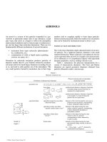
loscopic trace, etc. Figure 1 delineates the three major parts
and functions of an instrument, sample properties (measur-
and) to be measured, and instrumental criteria.
Sensors A sensor, the primary contact of the instrument
with the sample, is a device that converts the input energy
derived from a sample property to an output signal, usually
electrical in nature. The relationship between the input energy
(measurand), Q
1
, and the output energy, Q
0
, is expressed in
the form:
Q
0
ϭ f ( Q
1
) (1)
and is known as the transfer function. The sensitivity is given
in the equation
S ϭ dQ
0
/ dQ
1
. (2)
When the transfer function is linear, the sensitivity is constant
throughout the sensor’s range. However, the sensitivity (gain
or attenuation factor) is dependent on the value of the dif-
ferential fraction in equation 2. The sensor threshold is the
smallest magnitude of input energy necessary to obtain a
measurable change in the output.
Readout signals may be digital, D (discrete), or analog,
A (continuous), in form and are a function of the nature of the
input signal and the sensor and the design of the signal condi-
tioning circuits. These signals are interconvertible using A / D
or D / A devices. Fast reacting sensors and circuits, however,
are utilized for producing digital signals, where, formerly,
analog signals were obtained.
Two varieties of sensors, chemical and physical, are in
use on various instruments. The physical sensor allows the
conversion of physical energy from one to another. One
example is a photocell that converts an impinging light beam
TABLE 3
Number of STORET listings for water analysis
Parameters by groups Example Number of
parameters in group
General physical and chemical Alkalinity, COD, iron turbidity, zirconium 149
Physical observations Algae, foam, oil 12
Radionuclides Gross alpha and beta, strontium-90 141
Microbiological Coliform by MPH and MF, total plate count 18
Organic materials
Carbon adsorption data Chloroform and alcohol extractables 12
Natural organics Chlorophyll, tannins 4
Synthetic organics ABS, phenols 2
Halogenated hydrocarbons Aldrin, heptachlor, toxaphene 62
Phosphorated hydrocarbons Malthion, parathion 10
Miscellaneous pesticides Silvex 8
Treatment-related observations Available chlorine 6
© 2006 by Taylor & Francis Group, LLC
INSTRUMENTATION: WATER AND WASTEWATER ANALYSIS 545
to an electrical signal and is used in spectrometers. A second
example is a piezoelectric crystal-based sensor that converts
a mechanical force to an electrical charge translatable to a
potential. The piezoelectric effect is reversible; an electric
charge will cause a mechanical dislocation in the crystal.
Another example of a physical sensor is a platinum resis-
tance thermometer where the resistance of a platinum wire is
altered by a change in temperature.
Chemical sensors are devices that allow the analyte or
target material through one of its specific chemical param-
eters to ultimately generate an energy signal, usually electri-
cal, in a transducer through the agency of a selective chemical
or physical chemical reaction. A transducer is a material
structure inside of which or on whose surface the specific
chemical or physical chemical reaction takes place leading
to the generation of the energy signal. Thus, there are two
parts to the chemical sensor, the interface zone or area where
the selective reaction takes place and the usually non-specific
transducer.
8
Figure 2 illustrates, functionally, the parts of a
chemical sensor.
An example of a chemical sensor is a potentiometric
electrode. Here the selective chemical reaction, the redox
reaction of the analyte, is in equilibrium at the electrode sur-
face imposing a potential that is proportional to the loga-
rithm of the concentration of the analyte as described by the
Nernst equation. For example, a copper electrode in a solu-
tion of copper ions will take on a potential in response to the
concentration of copper ions. The logarithm of the copper
ion concentration is proportional to the electrode potential.
Another illustration of a chemical sensor is an amperometric
electrode, where a current arises due to the redox reaction of
the analyte when the electrode is at the appropriate poten-
tial. The concentration of the analyte is proportional to the
magnitude of the current. A platinum electrode maintained
at the redox potential for the silver/silver ion redox system
will detect the concentration of silver ions. A membrane
electrode is another type of chemical sensor. The fluoride
electrode consists of a lanthanum fluoride (LaF
2
), thin, crys-
tal membrane. On the outside surface, the sample side of
the membrane, the fluoride ions, F
Ϫ
, from the sample are
attracted electrostatically to the lanthanum ion, La
3ϩ
, at the
surface of the membrane to form a complex. The complexed
entities do not penetrate very deeply into the surface. The
amount of F
Ϫ
complexed is a direct function of its activity
(see Section III,B,2, a ) and represents a selective physical
chemical reaction. A membrane potential arises because
the opposite side of the membrane is exposed to a standard
activity of F
Ϫ
giving a net difference in potential between
the two sides. The membrane potential is the non-specific
electrical signal of the sensor.
Signal-Conditioning Circuits These circuits modify the
signal produced by the sensor so as to provide an accurate
representation of the sensor signal with optimal electrical
characteristics to drive the readout device. In Figure 1 a
number of signal conditioning modes are given and can be
Measurand
Sample property
Input
Signal
Sensor
Energy transducer
Transducer
Signal
Signal conditioning
Signal modification
Output
Signal
Readout
Types
Light
Absorption
Emission
Thermal
Conductivity
Temperature
Heat capacity
Electrical
Redox
Electrolytic conductivity
Ionic activity
Mechanical
Mass
Density
Viscosity
Surface tension
Nuclear
(X-ray, b.g)
Emission
Absorption
Light
Photocell
Photographic plate
Thermal
Thermocouple
Katharometer
Thermister
Bolometer
Electrical
Electrode pair
Electrodes, AC system
Membrane electrode
Mechanical
Balance force transducer
Force transducer
Hydrometer
Viscosity pipet
Nuclear
Ionization tubes
Scintillation counters
Photographic plates
Cloud chamber
Semiconductor detectors
Amplification
Arithmetic operation
Chopping
Comparison to reference
Digitization
Rectification
Stabilization
Analog
Meter
Oscilloscope
Recorder
Digital
Nixie display
Point plotter
Printer
Tape, paper, or
magnetic
Criteria
Band width
Noise figure
Sensitivity
Signal-to-noise
Time constant
Error
Hysteresis
Nonlinearity
Scale
Zero displacemen
t
Instrument
FIGURE 1 Diagram of instrumental functions. Reprinted from Ref. (4), p. 1442 by courtesy of Marcel Dekker, Inc.
© 2006 by Taylor & Francis Group, LLC
546 INSTRUMENTATION: WATER AND WASTEWATER ANALYSIS
placed in four categories—modification of sensor output,
amplification, mathematical operation, and signal modifica-
tion for readout.
The electrical components used in these circuits are of
two types, active and passive elements. Active elements,
such as solid state devices add energy to a circuit; whereas
passive elements, such as resistors, capacitors, inductors,
diodes add no energy. Both elements are combined to form
active and passive circuits. Active circuits change signals in
a complex way. Passive elements are used in active circuits
to provide necessary conditions for the proper functioning
of active circuits. Some active devices are ionization cham-
bers, vacuum phototubes, operational amplifiers and gas dis-
charge tubes.
Readout Devices The sensor signal modified by the
conditioning circuits is ultimately converted into a visual
form by the readout device or output transducer. The read-
out signal may be analog or digital requiring a compatible
readout device. Analog readout devices comprise record-
ers, meters, oscilloscopes, photographic plates and integra-
tors; printers, computers and digital meters with optical
displays provide digital readouts. A digital computer may
be interfaced to an instrument, in order to compute values
from a digital output signal and produce a hard (printed)
copy of the data using a printer. Analog output signals may
be digitized in order to utilize a computer. The advantages
of digital outputs are the statistical benefit derived from
counting and analog outputs are advantageous in feedback
control systems.
Analog Devices The automatic recording potentiometer
or potentiometric recorder has been, over the years, the
most frequently used readout device providing a continu-
ous trace on a chart of an analog signal. Its operation is
based on a low power servomechanism utilizing a feedback
system. The instrumental signal to be measured is com-
pared to a standard reference signal. The amplified, differ-
ence or error signal activates the pen-drive motor moving
the pen on the chart to a position representing the magni-
tude of the analog signal. The control of the pen, based on
the error signal, denotes the feedback system and the total
system is referred to as a servomechanism.
9,10
Two types of
recorders, the Y -time or X – Y, allow the recording of a signal,
Y, as a function of time or of two signals representing the
ordered (data) pair, x, y, respectively. In the Y -time device,
a constant-speed motor moves the chart in the x direction
while the servomechanism deals with the y signal. The X – Y
recorder has two servo- systems, one for each signal, x and y.
However, recorders may be limited by the rate that the data
flows from the instrument. Some recorders can adequately
respond to signals during fast scans. For example fast scans
in cyclic voltammetry of about 1 volt/sec. can be transcribed
using a recorder, however, at faster rates an oscilloscope is
necessary.
Almost any instrument can utilize a potentiometric
recorder. A Y -time analog recorder is commonly used to
trace gas and liquid chromatograms; the abscissa, X axis,
is for retention volume or time and the ordinate is for the
detector response.
The oscilloscope is a measuring device with complicated
circuitry that allows accurate display and measurement of
non-sinusoidal or complex waveforms. The oscilloscope’s
basic part is the cathode ray tube, CRT. A CRT is a vacuum
tube containing an electron gun pointing to a fluorescent
screen at the tube’s end. The electron gun provides a beam
whose movement is controlled by two sets of deflector plates
perpendicular to each other. The plates receive the signals
representing the waveforms. These analog signals are dis-
played on a fluorescent screen as Y -time or X-Y curves. The
display is photographed to provide a hard copy of the analog
data. The oscilloscope can display data that is generated at
high rates, since there are no mechanical movements used
in manipulating the electron beam. Where very fast events
must be recorded, an oscilloscope is an effective readout
device.
11
(See the previous paragraph on the potentiometric
recorder.) Oscilloscopes have facilities to store, compare,
and manipulate signals.
Analog meters are based on the D’Arsonval meter move-
ment. The electrical current signal passing through a moving
coil, to which is fixed a pointer, induces a magnetic field in
the coil. A static magnetic field from a permanent horseshoe
magnet surrounds the coil. The interaction between the two
fields causes the movement of the coil: the degree of move-
ment is determined by the magnitude of the signal current.
Analog meters require the analyst to interpret or read the
output signal value by the position of the indicator needle
or pointer using a calibrated scale mounted on the meter.
A resistance placed in series with the meter movement allows
FIGURE 2 Chemical sensor.
CHEMICAL SENSOR
INTERFACE ZONE
TARGET
or
ANALYTE
selective reaction
(chemical or physical
TRANSDUCER
electrical
signal
© 2006 by Taylor & Francis Group, LLC
INSTRUMENTATION: WATER AND WASTEWATER ANALYSIS 547
the measurement of voltage. Resistance may also be mea-
sured with the meter. One weakness of this device is its low
internal resistance causing loading errors by high impedance
signals.
11
A meter is used in the analysis of single samples or
samples analyzed, serially, at a slow rate on a spectroscopic
instrument at one frequency or wavelength. Meters also
are employed to indicate proper adjustment of potentials,
currents, temperatures, etc. for various instruments.
An electronic voltmeter, EVM, is more sensitive and
accurate than the D’Arsonval-based meter previously
described, particularly for signals with high impedance.
The internal resistance is 10 Mohms (megaohms, 10
6
ohms)
or more for d.c. (direct current) signals and 1 Mohm for
a.c. (alternating current) signals. The circuits use solid state
devices compared to the earlier device, a VTVM (vacuum
tube voltmeter). Current and resistance is measurable with
the EVM. Its application parallels those for the D’Arsonval-
based meter.
11
A photographic plate or film may be used to collect data
in the time domain where all the data are displayed simul-
taneously, that is a spectrum in emission spectroscopy. The
radiation in the dispersion pattern of the sample reflected or
transmitted from the prism or grating impinges on the pho-
tographic plate.
Electronic integrators determine the area under a curve
and are superior in precision to the ball and disk integra-
tor and the several hand methods widely utilized. They
may be based on operational amplifier or transistor cir-
cuitry. Some potentiometric recorders have a second pen
controlled by an integrator and the density of the pen’s
excursions determine the area under the curve. This last
type is not as convenient as the electronic integrators that
can correct for baseline changes. Chromatographic peak
areas for GC and HPLC (high performance liquid chroma-
tography), anodic stripping analysis peaks, spectroscopic
curves, etc. are integrated as a means of quantitation and
analysis of an analyte.
Analog computers are available but are not used now to
any great extent.
Digital Devices The digital computer or microprocessor
interfaced to the instrument brings a broad capability to the
display and processing of instrumental data. Data reception
and storage is convenient when real time computation and
display are not required. Mathematical calculations, includ-
ing the areas under curves, graphic and tabular displays,
correlation with previously collected data, and many other
operations can be carried out at one’s convenience. Real
time processing can be accomplished on a time-sharing
basis or with a dedicated computer. The visual display is at
a video monitor and a printer provides a hard (printed) copy
of the raw and calculated data, graphs, and other informa-
tion. Computer devices include microprocessors and micro-,
mini-, and mainframe computers. The instrument must be
carefully interfaced to the computer and this task requires
much electronic skill. Instruments providing spectral read-
outs, the need for number crunching and repetitive analyses
can benefit greatly from a computer interface. Some instru-
ments that utilize Fourier transform analysis require a com-
puter capability and many instrumental techniques have been
revolutionalized by computer use. The use of the computer
12
in the reduction of noise in instrumental signals by ensemble
and boxcar averaging has greatly improved the quality of
instrumental data.
12
Digital meters measure analog signals and provide
a digital readout. A/D conversion of the analog input is
accomplished electronically. The digital data is displayed
as numeric images using solid state devices such as LEDs,
light emitting diodes, and LCDs, liquid crystal displays, and
lamps such as, NIXIE, neon, and incandescent bulbs. The
LED is the more convenient device because its seven seg-
ment readout display uses lower currents and voltages than
the lamp displays. The LED’s red image, due to the semi-
conductor gallium arsenide doped with phosphorus, may
be increased in intensity by using more semiconductor in
the LED. The image color of LEDs may be fabricated to be
green or yellow, also.
11,13
LCDs operate by means of polar-
izing light. They use reflected light for viewing, a seven-
segment and dot matrix readout display, an a.c. voltage,
consume very little power and are more fragile than LEDs.
14
The LCDs and LEDs are the newest and most convenient
display devices.
Digital meters can be used in place of the analog variety.
The former are more accurate and easier to read.
Instrumental Parameters and Definitions Instrumental
characteristics of operation and data treatment and statistics
are defined by a number of parameters. A definition of each
term is as follows:
• The range of frequencies (information) in the
signal is called the bandwidth. During amplifica-
tion, some amplifiers cannot respond to the range
of frequencies in the signal producing an amplified
signal with a narrower bandwidth.
• The baseline is the signal obtained when no
sample is being examined and reflects the noise
inherent in the instrument.
• Calibration is the process relating instrument
response to quantity of analyte. In general a
series of standard solutions or quantities of ana-
lyte are analyzed on the instrument taking reagent
blanks into account and using a similar matrix
as the sample under consideration. The quantity-
response data are plotted to provide a calibration
curve where error bars indicate the precision of
the method.
15
Other calibration procedures such as
the methods of standard additions
16
and of internal
standards
17
have advantages in specific situations.
The former is helpful in ameliorating interferences
from the sample matrix and the latter in correcting
for changes in instrument response particularly in
GC, and ir (infrared) and emission spectroscopy.
18
• The gain refers to the ability to amplify a signal
and is the ratio of the output to input signal. The
© 2006 by Taylor & Francis Group, LLC
548 INSTRUMENTATION: WATER AND WASTEWATER ANALYSIS
gain may refer to voltage, current or power ampli-
fication and its input and output impedances.
• Noise refers to random signals, usually continu-
ous, that restricts the lower detection limit and
accuracy of the signal. Noise arises from elec-
tronic components and environmental sources and
cannot, at times, be completely eliminated.
• The ratio of the amplitude of the signal to that of
the noise is called the signal-to-noise, S/N, ratio.
This ratio gives the ability to distinguish between
signals and noise, that is a measurement of the
quality of an instrument. One cannot usually dis-
tinguish the signal from the noise when the ratio
is less than about 2 or 3.
• Resolution or resolving power is the capabil-
ity of displaying two signals differing slightly
in value. The resolving power, R, of a mono-
chrometer concerns absorption of emission spec-
tral signals,
R ϭ λ / d λ (3)
is the wavelength under consideration and d λ is the differ-
ence of wavelength between the two signals. In mass spec-
trometry resolution refers to the separation of two mass
peaks Ms and Ms ϩ dMs, where dMs is the difference in
masses so that
R ϭ Ms / dMs. (4)
For resolution for chromatographic methods see Part Two
Section III,B,4, a.
• Response time refers to the time needed for a pen
of a potentiometric recorder to travel the total ver-
tical distance on the Y axis.
• Sensitivity, S, describes the ratio of the change in
the response or output signal, dI
0
of the instrument
to a small change in the concentration or amount
of the analyte, dC. The ratio is given as follows:
S ϭ dI
0
/ dC. (5)
• Linear dynamic range, LDR, describes the
mathematical relationship between amount or
concentration of the analyte and the response of
the instrument. An increase in the analyte quan-
tity results in a linear increase in response. The
size of the range of quantities accommodated
by the instrument response is the key factor for
this parameter. For example in voltammetry
the LDR is 10
Ϫ8
to 10
Ϫ3
M (molar), five orders
of magnitude, in (ultraviolet) uv–visible spec-
trophotometry, about 10 to 100, and for a GC
with a FID (flame ionization detector) the LDR
extends from 10
Ϫ1
to 10
7
ng (nanograms, 10
Ϫ9
grams) or eight orders of magnitude. Obviously
the sensitivity remains constant in contrast to a
non-linear dynamic relationship.
• The reagent blank or blank in a spectroscopic
determination is the signal obtained by the solution
of the reagents without any analyte. The sample
matrix is important to include, if known, in the
blank. In many instances the effect of the matrix is
determined indirectly.
• Accuracy defines, mathematically, the absolute
error, e
a
, inherent in the method when comparing
the analytical result, x
i
, with the true value, x
t
, of
the analyte content of the sample.
e
a
ϭ ( x
i
Ϫ x
t
). (6)
Preparing a standard sample containing an accurately known
concentration of the analyte is required. This is not a simple
task, because homogeneity of any mixture is difficult to
obtain and ascertain.
• The precision of a method is concerned with the
repeatability of the analytical results for a number
of analyses on the same sample. There are several
ways of expressing precision; standard deviation
is a very effective and meaningful measure. The
standard deviation, sd, for small sets of data is
given as follows:
sdxxNo
ia
i
N
ϭϪϪ
ϭ
()
.
,
2
1
12
1
∑
⎡
⎣
⎢
⎤
⎦
⎥
(7)
Here x
i
is the experimental value, x
a
, the average of the exper-
imental values, and No, the number of values. The standard
deviation is a measure of the average uncertainty of all the
measurements in the data set, x
i
, that is x
1
, , x
N
.
18,19
Types of Instruments
Analytical instruments can be classified according to cat-
egories based on various physical phenomena. The general
categories used in this article are spectroscopy, electrochem-
ical analysis, radiochemical analysis, chromatography, and
automated analysis. Table 4 illustrates these categories.
Spectroscopy
Introduction Spectroscopic instruments include optical and
other types of instruments. The optical instruments analyze
electromagnetic radiation, emr, while other spectroscopic
instruments deal with sound, mixtures of ions, electrons, and
other forms of energy. Other optical methods utilize instru-
ments that make refractometric and polarimetric measure-
ments. Refractometric measurements will be discussed in
the section on liquid chromatography.
Spectroscopy, classically, is that area of science where
the electromagnetic radiation, emr, emitted from or absorbed
© 2006 by Taylor & Francis Group, LLC
INSTRUMENTATION: WATER AND WASTEWATER ANALYSIS 549
by a substance is resolved into its component wavelengths
indicating its intensity and presented as a spectrum. In
this category absorption, emission, and photoluminescence
(fluorescence and phosphorescence) spectroscopy using
x-ray, ultraviolet–visible (uv–vis), and ir radiation, and the
measurement of turbidity, or suspended matter by neph-
elometry and turbidimetry, are included. However, today in
a broader sense, spectroscopy includes the following: reso-
lution of electrons of many energies by uv and x-ray photo-
electron, Auger etc. spectroscopy; sound waves by acoustic
spectroscopy; ions by mass number by mass spectroscopy;
and absorption of radiowaves by atoms and electrons exposed
to a magnetic field in nuclear magnetic resonance and
electron spin resonance spectroscopy. The phenomena of
absorption, emission, photoluminescence (fluorescence and
phosphorescence), and scattering are the bases of spectro-
scopic instruments.
b. Spectroscopic instruments
Spectroscopic instrumentation is differentiated with
respect to the wavelength range of the instrument, that is
x-ray, uv, visible, and ir and type of instrument, i.e. absorp-
tion, emission, photoluminescence (fluorescence and phos-
phorescence), and turbidity. The energy sources, sample
cells, wavelength selection devices (gratings, prisms, filters,
crystals) and sensors may differ for these various instru-
ments. These parts are listed in Figure 3 for the wavelength
regions of from 100 to 40,000 nm (nanometer, 10
Ϫ9
meters).
X-ray and non-optical spectroscopic instruments are not
included.
TABLE 4
Bases for instrumental methods
Energy interaction Process Instrumental method
EMR, range Absorption of emr x-ray, uv/vis atomic & ir spectrophotometry
Emr/magnetic field Absorption of emr in a magnetic field NMR spectroscopy
e
Ϫ
, ions, or electric field Ion formation/seperation in electric or
magnetic field
Mass spectroscopy
Electricity (arc, spark), heat (flame,
plasma)
Emission of emr x-ray, uv/vis, flame emission spectroscopy
Emr, x-ray Emission of electrons x-ray photoelectron spectroscopy (XPS or
ESCA)
Emr, uv Emission of electrons UV photoelectron spectroscopy (UPS)
x-ray or e
Ϫ
Emission of electrons Auger spectroscopy
Emr, uv/vis Emission of acoustic energy Photoacoustic spectroscopy
None Emission via radioactive decay Radiochemical methods
Emr, range Fluorescence & phosphorescence of emr x-ray uv/vis
1
, & atomic fluorescence
spectroscopy
Emr, vis Scattering of emr by particles Nephelometry, turbidimetry
Emr, vis Scattering of emr by molecules Raman spectroscopy
Emr, x-ray Diffraction x-ray diffraction
Emr, vis Refraction (bending of light beam) Refractometry
Emr, uv/vis Rotation of plane-polarised light Polarimetry
Emr, uv/vis Rotation as a function of wavelength Optical rotatory dispersion
Emr, uv/vis Rotation using circularly polarized light Circular dichroism
Electricity current measurement Amperometry, coulometry, polarography,
voltammetry
Electricity pass current/weigh-plated material Electrogravimetry
Electricity potential measurement Chronopotentiometry, potentiometry
Electricity resistance/conductance measurement Conductometry
Heat weight loss vs increasing temperature
differential temperature vs increasing
temperature heat flow to sample vs
increasing temperature. Temperature vs
volume of reagent
Thermogravimetric analysis. Differential thermal
analysis. Differential scanning calorimetry.
Enthalpimetric methods
© 2006 by Taylor & Francis Group, LLC
550 INSTRUMENTATION: WATER AND WASTEWATER ANALYSIS
The types of instruments can be characterized by some
simple diagrams regardless of wavelength range as given in
Figure 4. The basic difference between absorption and emis-
sion spectroscopy is the use of a transmitted emr energy
source in the former, while in the latter, the sample is stimu-
lated in a thermal or electrical energy source to emit radiation.
Photoluminescence (fluorescence and phosphorescence),
stimulated by emr, is observed usually perpendicular to the
stimulating beam. In nephelometry instrumentation similar
to photoluminescence is utilized; turbidimetry can employ
absorption instrumentation.
A brief description of the basic parts of the instruments
using these phenomena follows.
(1) Energy sources
As noted from the diagrams above, all but emission
instrumentation use energy sources that irradiate the sample.
In Figure 3 the sources are indicated as a function of the
wavelength range of their radiant emissions. Various lamps,
i.e., argon, xenon, H
2
(hydrogen) and D
2
(deuterium), and
solid state radiators give continuous emissions, i.e., a range
of contiguous wavelengths and are used in molecular spec-
troscopic instrumentation. Photoluminescence and nephelo-
metric instruments use these sources.
For atomic absorption instruments hollow cathode
lamps are utilized. They are line (discontinuous) sources
providing unique radiation with a narrow bandwidth char-
acteristic of particular element. An individual lamp is usu-
ally employed for each element. Some multielement lamps
are available.
X-ray sources include x-ray tubes or radioactive sources.
The x-ray tube consists of a tungsten cathode that emits elec-
trons when heated. The electrons accelerated by a large poten-
tial strike the metal anode generating x-rays characteristic of
(a) Sources
Continuous
Wavelength, nm
Discontinuous
Spectral region
100 200 400 700 1000 20004000
7000 10,000
70,000
40,000
VAC UVUVVISIBLEIR
Argon
lamp
(b) Wavelength
selectors
Continuous
Discontinuous
fluorite
prism
(c) Materials for
cells, windows,
& lenses
(d) Transducers
Photon
detectors
Heat
detectors
Pyroelectric cell (capacitance)
Golay pneumatic cell
Thermocouple (volts) or Bolometer (ohms)
Photoconductor
Silicon diode
Tungsten lamp
Nernst glower (ZrO
2
+ Y
2
O
3
Nichrome wire (Ni + Cr)
Globar (SiC)
Hollow cathode
lamps
Photomultiplier
Phototube
Photocell
TIBr - TII
KBr
NaCl
Silicate glass
Corex glass
Fused silica or quartz
LiF
filters
Glass absorption
Interference filters
Interference wedgers
Gratings with various number of lines/mm
50 lines/mm
KBr prism
NaCl prism
Fused silica or quartz prism
xanon lamp
3000 lines/mm
Glass prism
NEAR IR
FAR IR
H
2
or O
2
lamp
FIGURE 3 Components and materials for optical spectroscopic instruments. (Courtesy
of Prof. A. R. Armstrong, College of William and Mary.)
© 2006 by Taylor & Francis Group, LLC
INSTRUMENTATION: WATER AND WASTEWATER ANALYSIS 551
the particular metal target. Metals such as chromium, copper,
iron, molybdenum, rhodium, silver, tungsten and others com-
prise the anode target. A number of radioisotope sources emit
useful x-rays, e.g., iron-55 yields manganese K radiation,
cadmium-109 gives silver K radiation, and cobalt-57 provides
iron K radiation.
(2) Sample interface
The sample is usually presented as a solution contained
in a cell made of material transparent to source radiation
for absorption and photoluminescence (see Figure 3). Solid
samples are also used. Potassium bromide disks containing
homogeneously distributed powdered analyte are used in ir
absorption methods.
However in atomic absorption spectroscopy the sample
is atomized in a flame, plasma or thermal heat source. In
effect the sample container is that volume of flame, plasma
or heat source.
Solutions, as well as solid samples in the form of pressed
disks, pieces of solids, or solid solutions in borax, are con-
veniently analyzed in an x-ray fluorescence instrument.
Solutions of sufficient thickness are the best sample prepara-
tions because of their homogeneity; they may be contained
in mylar cells. Obviously the solvent must not contain heavy
atoms that fluoresce. Sample surfaces are directly exposed to
the x-ray beam (see Figure 5).
In emission instruments the solid sample is placed in an
energy source environment, e.g., an electrical arc or spark, a
flame, or plasma.
(3) Wavelength selectors
The wavelength selector allows isolation of a particu-
lar wavelength segment of the source or transmitted beam.
A monochrometer is a selector comprising a grating or a
prism which disperses or separates the radiation continuously
over a considerable wavelength region. The effective band-
width of the wavelength, isolated by slits placed before the
sample, is quite narrow, 1 nm or less. The grating operates on
the principle of interference and the prism by dispersion.
Other wavelength selectors are interference and absorp-
tion filters. Their effective bandwidths are about 20 to 50 nm,
respectively; they are not continuous. An interference wedge
is continuous over a region with an effective bandwidth
of 20 nm.
The dispersing device, a single crystal mounted on a
rotating table or goniometer (see Figure 5a), is the wave-
length selector used in x-ray spectrometers. A specific
wavelength and its second and third orders of reflection are
diffracted at a given angle of the beam to a particular plane
of the crystal. The angle of diffraction depends on the “d” or
interplanar spacing of the crystal and the wavelength and is
defined by Bragg’s law. Some examples of diffracting crys-
tals with their unique wavelength ranges are topaz—0.24 to
2.67 Å, sodium chloride—0.49 to 5.55 Å, and ammonium
diphosphate—0.93 to 10.50 Å. (An angstrom, Å, is 10
Ϫ8
cm.) Unlike a prism or a grating that disperses a total spec-
trum in the spectral regions of the source of radiation, the
x-ray monochrometer diffracts a unique wavelength and its
orders of reflection depending on the angle of the beam to
the crystal plane.
(4) Detectors
In Figure 3 the variety of transducers are listed with their
wavelength range of detection. Following is a description
of the most commonly used detectors grouped according to
their wavelength range.
(a) Uv/visible
(i) Photovoltaic (barrier layer) cells
This detector, which generates its own signal, is sensitive
to radiant energy in the visible (350 to 750 nm) region. Light
shining on a semiconductor coating, such as selenium or
copper(I) oxide plated on an iron or copper electrode, gener-
ates a current at the metal–semiconductor interface. A second
electrode, a transparent coating of gold or silver on the outer
surface of the semiconductor, collects the electrons formed
by the action of radiant energy on the semiconductor. The
magnitude of the photocurrent is proportional to the number
of photons/sec impinging on the semiconductor. This detec-
tor is insensitive to low light levels, slow in response, shows
a tendency to suffer fatigue, and has a high temperature coef-
ficient. However, photovoltaic cells are rugged, require no
separate source of energy and are low in cost. They are used
in inexpensive filter photometers.
(ii) Vacuum photoemissive tubes
20
In a photoemissive detector two electrodes, a cathode
with an electron emissive coating and an anode, are enclosed
in an evacuated tube. When the saturation potential is applied
A. Absorption
a
& Turbidity
Energy
Source
Wavelength
Selector
Sample
Photoelectric
Detector
Signal
Processing
& Readout
Energy
Source
Energy
Source
Signal
Processing
& Readout
Signal
Processing
& Readout
Wavelength
Selector
Wavelength
Selector
Wavelength
Selector
Signal
Processing
& Readout
Photoelectric
Detector
Photoelectric
Detector
Photoelectric
Detector
Sample
Sample
Sample
Energy
Source
Filter
or
Wavelength
Selector
B. Infrared absorption
C. Emission
b
a
uv-vis and AA (flame and electrothermal)
b
arc, dc spark, inductively coupled & dc plasma, & flame
D. Fluorescence
c
& Nephelometry
c
uv, x-ra
y
FIGURE 4 Outline of spectroscopic instrumentation.
© 2006 by Taylor & Francis Group, LLC
552 INSTRUMENTATION: WATER AND WASTEWATER ANALYSIS
across the electrodes, the radiant energy or photons cause
emission of photoelectrons. The photoelectrons are collected
at the anode giving rise to a photocurrent. The photocurrent is
proportional to the power or radiant energy of the light beam
and is independent of the applied potential (see Figure 6).
The eleven chemical compositions of various photoemissive
cathode coatings determine the wavelength range and sen-
sitivity varying from the uv to the near ir spectral regions.
The window in the tube must be transparent to wavelength
of interest. The dark current is a small current flowing when
no light falls on the cathode and is due to thermal energy and
electron emission from potassium-40,
40
K, in the glass tube.
It limits the sensitivity of the detector. Although this detector
has about one tenth the sensitivity of the photovoltaic cell,
its signal may be amplified because of its large internal elec-
trical resistance compared to the photovoltaic detector. The
photoemissive detector is used for higher intensity radiation
and lower wavelength scanning rates than used with other
detectors.
(iii) Photomultiplier tubes
20
A photomultiplier tube contains a photoemissive cathode
followed by a sequential, electron multiplying assemblage
of about nine dynodes (electrodes) as illustrated in Figure 7.
The voltage of each succeeded dynode increases by 75 to
100 volts. Photoelectrons from the photoemissive cathode are
accelerated by the voltage increase of the first dynode caus-
ing the release of several electrons for each impinging pho-
toelectron. This multiplier effect continues as the electrons
Sample
changer
Sample
spinner
Rotation
of detector
Collimators
Detector
Diffracting
crystal
Goniometer
Phototube
Rotation
of crystal
100 kV
power supply
X-Ray tube
Phase
detector
Balance
indicator
Calibrated
attenuator
60-Hz
power
input
Cooling water
30-Hz
generator
60-Hz
X-ray
unit
Synchronous
motor
Chopper
Cell
Fluorescent screen
Light collector
Dial
1800
rpm
75°
150°
0°
φ
φ
2
x
s
θ
2
θ
(a)
(b)
θ
θ
Amplifier
FIGURE 5 (a) Geometry of a plane-crystal x-ray fluorescence spectrometer.
Note that the angle of the detector with respect to the beam, 2θ, is twice that of
the detector to the crystal face, θ. (Courtesy of Philips Electronic Instruments.)
(b) Nondispersive x-ray absorptiometer. (Courtesy of General Electric Co.)
© 2006 by Taylor & Francis Group, LLC
INSTRUMENTATION: WATER AND WASTEWATER ANALYSIS 553
contact the succeeding dynodes accelerated by ever higher
voltages. A cascade of a large number of electrons is collected
by the anode of the ninth dynode. The final photocurrent can
be amplified, electronically, before readout. The gain, G, can
be calculated as follows:
G ϭ ( fs )
n
(8)
where fs, the secondary emission factor for each stage,
depends on the dynode emissive coating and n is the
number of dynode stages. Using values for fs of 3 to 10 for
older dynode emissive coatings and 50 for newer coatings
and n equal to 9 results in gains of about 10
4
, 10
9
and 10
15
,
respectively. The response times can vary from 0.5 to 2
nsec (nanosec, 10
Ϫ9
sec). The dark current can be decreased
considerably by cooling the photomultiplier detector. Since
the dark current is a fairly constant value it may be sub-
tracted or automatically nulled using a potentiometer. The
–
Cathode
Phototube
Anode
hn
R=10
6
Ω
E=1V
1=10
–6
A
FIGURE 6 Simple phototube circuit.
(Reprinted from Ref. (176), p. 441
by permission of Prentice Hall, Inc.,
Englewood Cliffs, New Jersey.)
Incident radiation
Grill
Shield
Tube envelope
0
= Opaque photocathode
1–9 =Dynode = electron multiplier
10 = Anode
1
1
0
2
2
4
4
3
3
5
5
6
6
7
7
8
8
9
9
10
10
11
Focus ring
Semitransparent
photocathode
Internal conductive
coating
Incident
radiation
Faceplate
Focusing electrode
1–10 = Dynodes = Electron multiplier
11 = Anode
(b)
(a)
FIGURE 7 Photomultiplier Design. (a) The Circular-Cage Multiplier Structure in a
Side-on Tube and (b) The Linear-Multiplier Structure in a Head-on Tube. (Courtesy of the
General Electric Company.)
© 2006 by Taylor & Francis Group, LLC
554 INSTRUMENTATION: WATER AND WASTEWATER ANALYSIS
photomultiplier tube is the most widely utilized detector in
optical spectroscopic instruments.
(iv) Photodiodes
The photodiode, PD, is a small wafer of silicon dioxide
with a shallow layer of p and n material of the top and bottom
surfaces, respectively, to which are attached electrodes. The
device is reverse biased. When photons impinge on the
optically-active p diffusion region, electrons promoted to the
conduction band generate a photocurrent that is proportional
to the intensity of the optical light beam. The PD detectors
are about ten times more sensitive than the vacuum photo-
emissive tube and are mostly responsive in the visible and
near ir regions. Some tubes are sensitive to the uv region at
about 200 nm. A lens is optically coupled to each small PD
wafer.
Linear arrays or vidicon tubes of these multichannel
detectors allow nearly simultaneous detection of a spectrum
of wavelengths in instruments operated in the spatial mode
(see Section III,B,1, b,5 Instrumental ensembles), where
the detectors are swept electronically. Optical multichannel
analyzers consisting of a monochromater, a multichannel
detector and a computer are used in flame emission and uv/
visible spectrophotometers.
21
(b) Infrared detectors
22,23
There are two categories of detectors used for the spec-
tral region above 1.2 m (micrometer, 10
Ϫ6
m) namely, heat
and semiconductor detectors.
(i) Thermocouples and thermopiles
A thermocouple is formed when two wires of a metal
are separately joined to the opposite ends of a wire of a
dissimilar metal. If the two dissimilar metal junctions are
maintained at different temperatures, a thermoelectric cur-
rent will flow in the circuit. Therefore, if one junction is
maintained at a constant temperature, a thermoelectric cur-
rent will be generated proportional to the temperature of the
second junction. The changes in the intensity of incident
ir radiation can be detected in ir spectrophotometers using
this type of detector. The sensitivity is 6 to 8 microvolts per
microwatt and a temperature difference of 10
Ϫ6
ЊC is detect-
able (see Figure 8). A thermopile, consisting of a number
of series-connected thermocouples, may be miniaturized
through thin film techniques to provide an effective ir detec-
tor. It has an 80 msec (millisec, 10
Ϫ3
sec) response time with
a flat response below a frequency of 0.35 Hz (Hertz).
(ii) Golay cell
The Golay cell is a pneumatic device similar to a gas
thermometer. The ir radiation shining on the blackened sur-
face of a sealed cell containing xenon gas causes the gas to
expand and distort a diaphragm, a part of the cell wall. The
moving diaphragm may be coupled to one plate of a capaci-
tor transducing an ir intensity to a capacitance. In another
mechanism the beam of ir radiation is reflected from the mir-
rored diaphragm surface to impinge on a photocell. The area
of coverage of the beam on the photocell changes as a func-
tion of the movement of the diaphragm. The intensity of the
ir radiation affects the area of the beam that impinges on the
photocell and ultimately the magnitude of the photocurrent.
The ir beam must be optically focused on the detector. Its
response time is 20 msec. Sensitivity is about equal to that
of the thermocouple detector. In the far ir it is an excellent
detector (see Figure 3).
+15V
Irradiated junction
Reference junction
–15V
Negative
feedback
To
amplifier
+
–
FIGURE 8 Thermocouple and preamplifier (Reprinted from Ref. (180). With permission from
the Journal of Chemical Education.)
© 2006 by Taylor & Francis Group, LLC
INSTRUMENTATION: WATER AND WASTEWATER ANALYSIS 555
(iii) Pyroelectric detector
The pyroelectric detector is a thin wafer of material
such as, LiTaO
3
and LiNbO
3
, placed between two elec-
trodes to form a capacitor. The detector material is a non-
centrosymmetrical crystal whose internal electric field is
changed as a function of temperature when it is below its
Curie temperature. This detector is based on the change of
the capacitance of a substance with temperature and is sen-
sitive to the rate of change of the detector temperature. The
changing radiation is modulated by chopping or pulsing
because the detector ignores steady unchanging radiation.
Therefore this detector has a much faster response time than
those dependent on temperature directly. Depending on
the circuit parameters, the response times may be 1 msec
or 10 sec and the responsivity (detector output/incident
radiation) is 100 or 1, respectively. This detector has a large
ir range (see Figure 3).
(iv) Photoconductive and photovoltaic detectors
Photoconductive detectors are crystalline semicon-
ductor devices that experience an increase in conductivity
upon interaction with a photon. The increase of conduc-
tivity is due to the freeing of bound electrons by energy
absorbed from the radiation. A Wheatstone bridge is used
to measure the change in conductance (see following
Section III,B, 2,d. ). The semiconductor materials include
the metallic selenides, stibnides or sulfides of cadmium,
gallium, indium or lead.
Lead sulfide is commonly used as a detector in the near
infrared, 800 to 2000 nm, where it exhibits a flat response.
The cell consists of a thin layer of the compound on a thin
sheet of quartz or glass kept under vacuum.
Photovoltaic detectors have been discussed for uv/vis
spectroscopic instruments (see Section III,B,1, b.(4), (a), ( i )).
For ir applications the p -type indium antimonide detector,
cooled by liquid nitrogen, is available with a sensitivity limit
at 5.5 m. However the two types of lead in telluride detec-
tors extend the ir range. One detector, cooled with liquid
nitrogen, has a range of 5 to 13 m and a second one, cooled
with liquid helium, has a range of 6.6 to 18 m. A minimum
response time of 20 nsec (nanosec. 10
Ϫ9
sec) is achieved
with these detectors.
(c) X-ray detectors
Gas-filled and semiconductor detectors and signal
processors and readout used in the measurement of
radioactivity (see Sections III,B, 3, b,( 1 ),(a) and (b) and
(2)) are the applicable in x-ray spectroscopy.
(5) Instrument ensembles
The design of an instrument depends on its use and mon-
etary considerations. The several main modes of design are
designated temporal, spatial and multiplex. In turn each of
these are of the dispersive or nondispersive type.
24
In the temporal category the instrument scans sequentially,
in time, the wavelength in order to determine the intensity.
Dispersive systems employ monochrometers that are rotated
so as to position the selected wavelength on an aperature or
slit preceding the sample or detector. Nondispersive systems
utilize a series of absorption or interference filters that are
interchangeable.
Spatial systems display the total spectrum with simul-
taneous determination of the radiation intensities. For a dis-
persive instrument a monochrometer provides the dispersed
radiation and a multichannel detector to detect their inten-
sities. Multichannel detectors utilized are a detector array
(silicon diode array or vidicon tube), a number of individual
detectors properly positioned, or a photographic plate. In
nondispersive systems the radiation beam is divided into a
number of beams and each passes through a unique filter
followed by a detector.
Multiplex systems employ a single data channel where
all the components of the signal are observed simultane-
ously. A Fourier transform is usually employed to resolve
the complex signal into its components requiring the use of a
computer. There are distinct advantages to Fourier transform
spectroscopy: namely, increased S/N ratio, increased energy
throughput, large precision in wavelength measurement, and
facility in its use. However, thus far the instrument is costly
to acquire and maintain. No further comment will be made
in this article about these instruments.
25,26
Dispersion instruments give more spectral detail because
the wavelength selected has a narrower bandwidth wave-
length spread. However non-dispersive instruments are usu-
ally cheaper, more rugged and have a higher signal to noise
ratio. Filter instruments are used frequently in monitoring
equipment.
(6) Absorption instrumentation
In the absorption process, radiation passes through the
sample and a specific pattern of absorption of different
wavelengths occurs leading to a spectrum for that sample.
For each wavelength the amount of light absorbed will differ
and therefore the amount of transmitted light vary for each
wavelength. The intensity of transmitted light is inversely
proportional to the concentration of sample and is measured
in absorbance, Ab, units. The spectrum is the qualitative
factor of identification while the intensity of the transmit-
ted radiation is the quantitative measure. Light is absorbed
in the uv and visible region by electrons in the atoms or
molecules of a sample. Some elements are identified by
atomic absorption, AA, and some functional groups and
species by uv/vis spectroscopy. Absorption in the ri is due
to vibrational and rotational activity of atoms in molecu-
lar groupings, such as functional groups, double, triple, and
conjugated bonds, etc.
The spectroscopic curve or an instrument reading provides
an absorbance value for a chosen wavelength from which the
concentration of the absorbing substance can be computed.
The absorbance value represents the degree of attenuation of
the radiation of specific wavelength by absorbing substances
in the sample solution in the cell. A constant, the absorptivity,
a, or molar absorptivity, e, can be calculated for a pure sub-
stance for a given wavelength and solvent. The mathematical
relationship between the concentration of a substance, C, the
© 2006 by Taylor & Francis Group, LLC
556 INSTRUMENTATION: WATER AND WASTEWATER ANALYSIS
path length of the sample cell, b, and its absorbance units, Ab,
is expressed as follows:
molar absorptivity, e ϭ Ab /( b, cm)( C, moles/L) (9)
absorptivity, a ϭ Ab /( b, cm)( C, g/L).(10)
The absorptivity, a, can be expressed in a number of con-
centration terms (g/L, grams/Liter and mg/ml, milligrams/
milliliter) and path length terms (cm, centimeters and mm,
millimeters).
Equations 9 and 10 are a statement of Beer’s Law indicating
a linear relationship between the absorbance and the con-
centration for fixed conditions. In analytical determinations
the concentration can be calculated using equations 9 or 10.
Also used in analysis is a calibration curve, whose slope is
absorptivity. It can be drawn using concentrations and cor-
responding absorbance values. Beer’s Law prevails for many
substances, but there are deviations for some substances due
to chemical, instrumental, and physical phenomena.
(a) Uv/visible instrumentation
27
A uv/visible instrument consists mainly of uv and visi-
ble energy sources (lamps), a wavelength selector (grating,
prism or filter), reference and/or sample cell, a detector
(photodetector or photomultiplier) and a readout device
(recorder, analog or digital meter, etc.) (see Figure 4A).
Figures 9 and 10 illustrate the arrangement of these parts
for photometers and spectrophotometers, respectively. The
distinction between the two types of instruments is that a
photometer uses a filter and a spectro photometer a grat-
ing or prism as a wavelength selector. In Figure 9 the dif-
ference between a single and double beam instrument is
shown and the arrangement also refers to a spectrophotom-
eter. The various parts that transmit the light beam such as
lens, cells, mirrors, transmission gratings and prisms must
be transparent to uv light and be fabricated of fused silica
or quartz. Flint glass can be used in the visible region.
(b) Infrared instrumentation
28
Figure 11 is a schematic of a double beam ir spectropho-
tometer. The parts and functions are similar to a un/visible
instrument, however the arrangement differs—the light beam
passes through the sample and then the wavelength selector
in contradistinction to the uv/visible instrument (see Figures
4A, 9, and 10). Materials transparent to ir are the alkali metal
chloride, bromide, and iodide salts.
Filters
Grating
Visible
source
UV Source
Slits
Sample
Compartment
Focal
Point Detector
Tungsten
lamp
Deuterium
lamp
Concave
grating
Sector
mirror
Sample
Reference
Photomultiplier
Grid
mirror
(a)
(b)
FIGURE 9 Single- and double-beam uv-visible spectrophotometers.
(a) Beckman DU
R
Series 60, single-beam. (Courtesy of Beckman Instruments,
Inc., Fullerton, CA.) (b) Hitachi Model 100–60, double-beam. (Courtesy of
Hitachi Instruments, Inc., Danbury, CT.)
© 2006 by Taylor & Francis Group, LLC
INSTRUMENTATION: WATER AND WASTEWATER ANALYSIS 557
(c) Atomic absorption instrumentation
29
A schematic for a single beam atomic absorption
spectrophotometer is given in Figure 12. Both flame and
electrothermal atomizers may be utilized in this instrumenta-
tion (see Figure 4A). The hollow cathode lamp is the energy
source generating uv or vis radiation that passes through
the sample. The flame or thermal area of the electrothermal
device acts as the sample cell where the sample solution that
has been nebulized (formed into a fine aerosol) is atomized.
(Atomization is the formation of free atoms through thermal
energy in the flame or thermal area.) The flame is generated
from various fuel mixtures: acetylene and the oxidants air,
oxygen or nitrous oxide; hydrogen and the aforementioned
oxidants; and natural gas and air or oxygen. Each of these
mixtures as well as the fuel/oxidant ratio determines the
flame temperature, a critical condition for atomization.
(7) Emission instrumentation
30,31
Emission of emr by elements and some chemical enti-
ties, energized by flames, plasmas, and arc, is the basis of
emission spectroscopy. The electrons are energized and
move to higher energy levels on absorption of the energy.
On relaxation, the electron returns to a lower energy level
and the absorbed energy is emitted as radiation.
Emission methods give rise to atomic spectra by a series
of atomization techniques: namely, flame, inductively cou-
pled argon plasma (ICP), electric arc and spark, and direct
current argon plasma, DCP (see Figure 4B). An emission
spectrophotometer capable of using plasma and arc and
spark sources is illustrated in
Figure 13.
(8) Photoluminescence instrumentation
The occurrence of fluorescence and phosphorescence
(photoluminescence) refer to substances which on excitation
by radiation emit light on relaxation of the excited species.
In resonance fluorescence the wavelengths of excitation and
emission are the same. However, in many cases the wave-
length of emitted radiation is longer than that of the exciting
radiation. The difference in fluorescence and phosphores-
cence is the time delay between excitation and emission. The
former is quite small (Ͻ10
Ϫ6
sec), while the latter has a time
delay of several seconds or longer. Fluorescence can occur in
a number of organic and metal-organic complex molecules,
and gases on excitation with uv light.
(a) Uv/visible
32
A general schematic for this instrumentation is given in
Figure 4C. Fluorescence of molecular substances is measured
Field lens
Entrance slit
Objective lens
Grating
Wavelength
cam
Light control
Exit
slit
Occluder
Sample
Filter
Lamp
Measuring
phototube
FIGURE 10 An example of a simple spectrophotometer. The SPECTRONIC
R
20. (Courtesy of the Milton Roy Co.,
Rochester, NY)
Reference
Beam
Sample
Beam
Source
Comb
M5
M6
M8
M7
M9
G2
G1
M12
M13
S2
S1
Filters
Thermocouple
M14
C
+
Sampling
Area
Attenuator
M2
M1
M
M
M11
M10
FIGURE 11 Schematic diagram of a typical double-beam infrared spectrophotometer. The sym-
bol M1 through M14 indicate mirrors; S1 and S2 indicate slits; and G1 and G2 indicate gratings.
(Courtesy of the Perkin-Elmer Corporation.)
© 2006 by Taylor & Francis Group, LLC
558 INSTRUMENTATION: WATER AND WASTEWATER ANALYSIS
in a fluorometer (see Figure 14) or a spectrofluorometer (see
Figure 15).
(b) Atomic
33,34
Atomic fluorescence is a fairly new instrumental
method that has been used for environmental samples. The
instrumental arrangement is given in Figure 4C. Because
of the dearth of commercial sources of the instrument and
no large benefits compared to other atomic spectroscopic
instruments there is a small number of literature citations
for its use.
(c) X-ray
35
X-ray fluorescence spectroscopic instrumentation uti-
lizes an x-ray source, i.e., x-ray tube or radioactive source,
to energize electrons of the inner orbitals of atoms which on
+
–
Power
supply
Motor
Fuel
Sample
Oxygen
Flame
Rotating
chopper
Hollow
cathode
tube
Monochromator
Detector
amplifier
Read-out
a–c
FIGURE 12 Components of an atomic absorption spectrophotometer. The flame may be
replaced by a furnace. (Reprinted from Ref. (176), p. 464 by permission of Prentice Hall,
Inc., Englewoods Cliffs, NJ.)
Rowland circle
Photomultiplier tubes
Mirror
–1000 V dc
power supply
Integrating
capacitor
Measuring
electronics
Microprocessor
A/D
Quartz
window
Lens
Aperture
Pivoted mirror
Mirror
Moveable slit
Stepper
motor
Concave
diffraction
grating
Mercury lamp
Source
Lens
Prealigned
exit slit
Computer
FIGURE 13 A plasma multichannel spectrometer based upon Rowland circle optics. (Courtesy of Baird Corp./
IMC Bedford, MA.)
© 2006 by Taylor & Francis Group, LLC
INSTRUMENTATION: WATER AND WASTEWATER ANALYSIS 559
Emission monochromator
Grating
Sample
photomultiplier tube
White
reflector
Reference
photomultiplier tube
Grating
Absorbance
compensating
cell
Bean
splitter
Sample
compartment
Xenon
lamp
Excitation monochromator
FIGURE 15 A spectrofluorometer. (Courtesy of SLM Aminco Instruments, Urbana, IL.)
REFERENCE
LIGHT PATH
PHOTOMULTIPLIER
LIGHT INTERRUPTER
MOUNTING BLOCK
LUCITE LIGHT
PIPES
FORWARD LIGHT PATH
BLANK
SHUTTER
BLANK KNOB
SAMPLE
MOTOR
COOLING FAN
OPTICAL FILTER
OPTICAL FILTER
RANGE SELECTOR
LIGHT
CAM
DIFFUSE
SCREEN
LIGHT
SOURCE
Secondary; emission
Primary; excitation
Four apertures
FIGURE 14 The Model 112 Turner Digital Filter Fluorometer. (Courtesy of the Turner Division of Unipath, Mountain View, CA.)
© 2006 by Taylor & Francis Group, LLC
560 INSTRUMENTATION: WATER AND WASTEWATER ANALYSIS
relaxation emit fluorescence emission. The wavelengths in the
fluorescence emission are unique for different elements. This
information may be delineated by wavelength or energy dis-
persion instruments (see Figure 4C). Wavelength dispersion
is carried out with a crystal as a diffraction grating with sub-
sequent detection by a gasfilled detector (see Figure 5), while
energy dispersion may be accomplished by a lithium-drifted
silicon detector and energy-discriminative electronic circuits
(see Figure 16). Non-dispersive instruments use filters.
Elements with atomic numbers greater than 7
(oxygenϭ 8) fluoresce on irradiation with x-rays. Useful
irradiating wavelengths extend from 0.5 to 2.5 Å. Due to
the large absorption of wavelengths greater than 2.5 Å by
air and spectrometer windows, elements of atomic numbers
below 22 (titanium) cannot be detected. Elements with lower
atomic numbers can be detected with a change of atmo-
sphere; down to aluminium (atomic no. ϭ 13) in helium and
to boron (atomic no. ϭ 5) in a vacuum.
(9) Nephelometric & turbidimetric devices
36
The presence of turbidity, suspended matter and col-
loids, in liquid results in the scattering of a beam of incident
light passing through the liquid. The scattering process is
elastic; the wavelength of incident and scattered light is the
same. Particle shape and size distribution, size relative to the
wavelength of the incident light, concentration of particles,
and molecular absorption effect the angular distribution of
scattered light intensity. Since the scattering phenomenon is
so complicated analytical results are empirical depending on
the use of standards. However, differences in the design of
instruments leads to different values for the same standard.
Turbidimetry refers to the measure of the decrease in the
intensity of a beam of light undergoing scatter by suspended
or colloidal particles in a liquid. If the transmittance is less
than 90%, this method is effective. A filter photometer illus-
trated in Figure 9 is a suitable instrument.
If the intensity of the scattered beam is measured at
an angle to the transmitted beam, then the phenomenon is
known as nephelometry. Right angle scatter is commonly
used for a number of readings, although forward scatter
is more sensitive to large particles. Stray light caused by
scratches, dirt or condensation on cell walls leads to a positive
error. Figure 14, a simple fluorometer, measuring scattered
light at 90Њ can be employed for nephelometric measure-
ments. A surface scatter instrument shown in Figure 17 is
used to eliminate stray light. No cell is employed since the
flowing water sample surface is directly illuminated with the
light beam. Figure 18 illustrates a low range turbidimeter.
Electron
column
Si(Li)
detector
Electrons
Sample
X-rays
Preamp
Pulse
processor
Energy-to-
digital
converter
Video
Keyboard
Multi-
channel
analyzer
Disk
storage
system
Mini-
computer
FIGURE 16 Components of a typical energy-dispersive microanalysis system. The Si(Li) detector is cooled in
a liquid nitrogen cryostat. The charge pulse from the Si(Li) detector is converted in the preamp to a step on a volt-
age ramp. The pulse processor converts the signal to a well-shaped voltage pulse with an amplitude proportional
to the energy of the x-ray. (Courtesy of the Kevex Instruments, Inc.)
© 2006 by Taylor & Francis Group, LLC
INSTRUMENTATION: WATER AND WASTEWATER ANALYSIS 561
(The names turbidimeter and nephelometer appear to be used
interchangeably to describe a device measuring turbidity from
the intensity of scattered light.) The unit of measurement is
the NTU, nephelometric turbidity unit.
Colored constituents in the sample can cause error by
absorbing light. A correction can be made in a number of ways:
namely, by using a wavelength of light not absorbed by the
solution, by making an absorption reading of the clarified solu-
tion, or using an instrument that combines both readings.
37
(10) Other spectroscopic instruments
Mass spectrometry is treated in the gas chromatography/
mass spectrometry Part Two Section, 4,c, ( 2 ),( a ). NMR
(nuclear magnetic resonance) spectroscopy is not discussed
in this article. The technique is, indeed, a most fruitful
means of identifying chemical entities and their structures.
However, the use in water analysis is not a primary activity.
In the identification for known natural and anthropogenic
materials its use would be invaluable. No doubt the next edi-
tion of this article or an expansion of this article will contain
a section on NMR spectroscopy.
(c) Applications of spectroscopic instruments
Standard Methods
2
includes a number of spectroscopic
methods using various instruments for the analysis of metals
(see Table 5). Colorimetric methods using uv/vis absorption
spectroscopy are available in Standard Methods for the fol-
lowing non-metals: bromide, fluoride, iodide, residual chlo-
rine, cyanide, ammonia, nitrate, nitrite, phosphate, sulfide
and sulfite. The determination of turbidity in water by neph-
elometry and the analysis of sulfate ion by a turbidimetric
method appears in Standard Methods
.
2
See Part Two Section C
for more applications.
2. Electroanalytical instrumentation
Electroanalytical chemistry encompasses a wide variety of
analytical measurements and includes three different types of
correlations. The first type concerns the relationship between
potential, current, conductance (or resistance), charge (or capac-
itance) and the analyte. For the second is the determination,
during the titration, of the analyte and, ultimately, the endpoint
by electrochemical means. The conversion of the analyte by an
electric current to a convenient gravimetric or volumetric form
is the third type. In this section a number of methodologies and
their corresponding instruments will be discussed. They include
potentiometry, voltammetry, amperometry, coulometry, con-
ductance measurements, and titrations using potentiometry and
amperometry for endpoint detection.
WEIR
SAMPLE
OUT
LAMP
LENS
VENT
WATER
SURFACE
PHOTOCELL
SAMPLE IN
BUBBLE TRAP
FIGURE 18 Low range turbidimeter. (Used with permis-
sion of Hach Co., Loveland, CO.)
PHOTOCELL
REFLECTED LIGHT
TURBIDIMETER
BODY
REFRACTED
LIGHT
SCATTERED
LIGHT
OVER-FLOWING
SAMPLE
LENS
LAMP
LIGHT
BEAM
INST.
DRAIN
SAMPLE IN
DRAIN
FLOW DIAGRAM
FIGURE 17 Surface scatter turbidimeter. (Used with
permission of Hach Co., Loveland, CO.)
© 2006 by Taylor & Francis Group, LLC
562 INSTRUMENTATION: WATER AND WASTEWATER ANALYSIS
Potentiometric Instruments An instrument consisting of
an electrochemical cell containing indicating and reference
electrodes and electronic means for measuring cell poten-
tial to within 0.001 volt (depending on the accuracy desired)
may be optimized to measure concentrations of various ions
and molecules. The indicating electrode is the sensor which
gives the specific electrochemical detection of the analyte
in question, while, the function of the reference electrode
is to provide a stable reference potential for the indicating
electrode. The potential of the indicating electrode changes
as a function of the concentration of the analyte according to
the Nernst equation,
E ϭ E
0
Ϫ (0.0591/n)Log α
red
/ α
ox
. (11)
The activities of the reduced and oxidized species are α
red
and α
ox
, respectively, n is the number of electrons in the
redox reaction and E
0
is the standard reduction potential of
the redox couple when the activity is one.
For species, Sp, its activity, α
Sp
, is a variable related to
its concentration in Moles/L. The symbol, [Sp], represents
the concentration of Sp in Moles/L. The α
Sp
is defined by
the equation,
α
Sp
ϭ f[Sp]. (12)
The activity coefficient, f, varies inversely with the ionic
strength of the solution. (Ionic strength is a function that
can be calculated from the concentration of ions in solution
and their ionic charges.) Therefore, at high concentrations
of electrolytes, the ionic strengths are high, and the activity
coefficients, small, less than one. Activity and concentration
are approximately equal when the ionic strength is low and f
is equal to values between 0.90 to 1.
TABLE 5
Metals Analysis by Spectroscopy
a
Metals Atomic Absorption Method Flame
Sb, Bi, Cd, Ca, Cs, Cr direct aspiration air-acetylene
Co, Cu, Au, Ir, Fe, Pb
Li, Mg, Mn, Ni, Pd, Pt
K, Rh, Ru, As, Na, Sr
Tl, Sn, Zn
Al, Ba, Be, Mo, Os direct aspiration N
2
O-acetylene
Rh, Si, Th, Ti, V
b
Al, Sb, As, Ba, Be electrothermal none
Cd, Cr, Co, Cu, Fe, Pb
Mn, Mo, Ni, Se, Ag, Sn
c
Cd, Cr, Co, Cu, Fe chelation
d
/ air-acetylene
Pb, Mn, Ni, Ag, Zn extraction
e
c
Al, Be chelation
f
/N
2
O-acetylene
extraction
e
Hg cold vapor none
As, Se hydride formation argon-hydrogen
or N
2
-hydrogen
Inductively Coupled Plasma (atomic emission)
Al, Sb, As, Ba, Be, B, Cd, Ca, Cr, Co, Cu, Fe,
Pb, Li, Mg, Mn, Ni, K, Se, Si, Ag, Na, Sr,
Tl, V, Zn
Flame Emission
Li, K, Na, Sr
Colorimetric/Spectrophotometric
Al, As, Be, B, Cd, Cr, Cu, Se, Si, Ag, V, Zn
a
From Ref. (2);
b
microquantities;
c
low conc.;
d
ammonium pyrrolidinedithiocarbamate;
e
methyl isobutyl ketone;
f
8-hydroxyquinoline.
© 2006 by Taylor & Francis Group, LLC