Fundamentals of Clinical Ophthalmology Cataract Surgery - part 1 doc
Bạn đang xem bản rút gọn của tài liệu. Xem và tải ngay bản đầy đủ của tài liệu tại đây (407.61 KB, 23 trang )
Series Editor:
Susan Lightman
Fundamentals of
Clinical Ophthalmology
Cataract Surgery
Andrew Coombes and David Gartry
Fundamentals of Clinical Ophthalmology:
Cataract Surgery
Fundamentals of Clinical Ophthalmology series
Cornea
Edited by Douglas Coster
Glaucoma
Edited by Roger Hitchins
Neuro-ophthalmology
Edited by James Acheson and Paul Riordan-Eva
Paediatric Ophthalmology
Edited by Anthony Moore
Plastic and Orbital Surgery
Edited by Richard Collin and Geoffrey Rose
Scleritis
Edited by Paul McCluskey
Strabismus
Edited by Frank Billson
Uveitis
Edited by Susan Lightman and Hamish Towler
Fundamentals of Clinical Ophthalmology:
Cataract Surgery
Edited by
ANDREW COOMBES
St Bartholomew’s Hospital and The Royal London Hospital, London, UK
DAVID GARTRY
Moorfields Eye Hospital, London, UK
Series editor
SUSAN LIGHTMAN
Department of Clinical Ophthalmology,
Institute of Ophthalmology/Moorfields Eye Hospital,
London, UK
© BMJ Publishing Group 2003
BMJ Books is an imprint of the BMJ Publishing Group
All rights reserved. No part of this publication may be reproduced, stored in a retrieval system, or transmitted,
in any form or by any means, electronic, mechanical, photocopying, recording and/or otherwise,
without the prior written permission of the publishers.
First published in 2003
by BMJ Books, BMA House, Tavistock Square,
London WC1H 9JR
www.bmjbooks.com
British Library Cataloguing in Publication Data
A catalogue record for this book is available from the British Library
ISBN 0 7279 1201 1
Typeset by SIVA Math Setters, Chennai, India
Printed and bound in Malaysia by Times Offset
v
Contents
Contributors vii
Preface to the Fundamentals of Clinical Ophthalmology Series ix
Preface xi
Acknowledgements xiii
1 Teaching and learning phacoemulsification 1
2 Incision planning and construction for phacoemulsification 11
3 Capsulorhexis 25
4 Phacoemulsification equipment and applied phacodynamics 36
5 Phacoemulsification technique 46
6 Biometry and lens implant power calculation 66
7 Foldable intraocular lenses and viscoelastics 84
8 Non-phacoemulsification cataract surgery 102
9 Anaesthesia for cataract surgery 115
10 Cataract surgery in complex eyes 125
11 Vitreous loss 158
12 Postoperative complications 168
CONTENTS
vi
13 Cataract surgery in the Third World 193
14 Cataract surgery: the next frontier 200
Index 208
vii
Charles Claoe
Consultant Ophthalmologist
Harold Wood Hospital
Essex, UK
Andrew Coombes
Consultant Ophthalmologist
St Bartholomew’s Hospital and The Royal London Hospital
London, UK
Jack Dodick
Chairman of the Department of Ophthalmology
Manhattan Eye, Ear and Throat Hospital
New York, USA
Jonathan Dowler
Consultant Ophthalmologist
Moorfields Eye Hospital
London, UK
David Gartry
Consultant Ophthalmologist
Moorfields Eye Hospital
London, UK
Peter Hamilton
Consultant Ophthalmologist
Moorfields Eye Hospital
London, UK
Colm Lanigan
Consultant Anaesthetist
Lewisham Hospital
London, UK
Thomas Neuhann
Consultant Ophthalmologist
Munich, Germany
Contributors
Marie Restori
Consultant Medical Physicist
Moorfields Eye Hospital
London, UK
Paul Rosen
Consultant Ophthalmologist
The Radcliffe Infirmary
Oxford, UK
Helen Seward
Consultant Ophthalmologist
Croydon Eye Unit
Surrey, UK
Hamish Towler
Consultant Ophthalmologist
Whipps Cross Hospital
London, UK
Sarah-Lucie Watson
Specialist Registrar
Moorfields Eye Hospital
London, UK
David Yorston
Specialist Registrar
Moorfields Eye Hospital
London, UK
CONTRIBUTORS
viii
ix
Preface to the
Fundamentals of Clinical Ophthalmology series
This book is part of a series of ophthalmic monographs, written for ophthalmologists in training and
general ophthalmologists wishing to update their knowledge in specialised areas. The emphasis of
each is to combine clinical experience with the current knowledge of the underlying disease processes.
Each monograph provides an up to date, very clinical and practical approach to the subject so that
the reader can readily use the information in everyday clinical practice. There are excellent
illustrations throughout each text in order to make it easier to relate the subject matter to the patient.
The inspiration for the series came from the growth in communication and training opportunities
for ophthalmologists all over the world and a desire to provide clinical books that we can all use. This
aim is well reflected in the international panels of contributors who have so generously contributed
their time and expertise.
Susan Lightman
Preface
Cataract surgery is a dynamic and complex field and is, without doubt, a fundamental part of
ophthalmology. This book aims to cover the subject comprehensively, particularly the technical
aspects of learning, performing, and teaching phacoemulsification. The inclusion of chapters on the
Third World and the future of cataract surgery provide the reader with a broader perspective.
The structure of the text, cross-referencing between chapters, and a detailed index minimise
repetition. For example, intraoperative complications are discussed within the relevant individual
chapters on technique (although vitreous loss and the dropped nucleus have a chapter devoted to
them), whereas postoperative complications are grouped together. For those who would like more
detail, the text has been thoroughly referenced.
Inevitably, some knowledge has been assumed and some detail omitted, but we hope that this book
will be useful to both trainees and established cataract surgeons.
Andrew Coombes and David Gartry
Acknowledgements
We must first acknowledge the contributing authors, without whom this book would not exist.
Professor Susan Lightman and all at BMJ Books, particularly Mary Banks, must also be thanked for
their part (and patience).
Many individuals have contributed photographs and their help has been very much appreciated. These
include David Anderson (Figures 2.14, 3.3, 3.5, 5.3, 5.6, 5.14, and 7.20), Bill Aylward (Figure 10.21),
Caroline Carr (Figure 9.2a–f), Emma Hollick (Figures 7.4, and 12.21), Alex Ionides (Figure 10.29), James
Kirwan (Figure 8.13b, 10.23, 10.24, 10.26, 12.13, and 12.22b), Frank Larkin (Figure 12.14),
Graham Lee (Figure 7.3a,b), Ordan Lehmann (Figures 8.14, 12.12, 12.15, 12.18, 12.22a, 12.24,
and 12.26), Martin Leyland (Figure 10.16), and Chris Liu and Babis Eleftheriadis (Figure 7.13). The
staff in the day surgery unit at Chelsea and Westminster Hospital should also be thanked for their help
with many of the photographs.
A large number of companies have allowed their equipment, instruments, and lenses to be
photographed, and we are grateful for their involvement. This book was originally developed from the
Moorfields Eye Hospital phacoemulsification courses, and Alcon (and their wet laboratory facilities)
deserve particular mention for their support of these courses over many years.
We should like to take this opportunity to thank those cataract surgeons who have taught us in the
past and those who continue to inspire us. Finally, we thank our families (especially Sarah) for the
support and tolerance that has been essential in completing this book.
phacoemulsification over the past 10 years has
been well documented by Leaming,
1
who has
conducted an annual survey of the practice styles
and preferences of US cataract surgeons. In the
UK a similar shift toward phacoemulsification
has occurred
2
and is likely to continue. For the
surgeon in training, phacoemulsification is no
longer an option but an essential surgical skill
to acquire. For the trained surgeon the ability
to teach phacoemulsification in a structured
manner has also become necessary.
Structured training and
phacoemulsification courses
Phacoemulsification acquired an undeservedly
poor reputation in the past. Surgeons did not
spend sufficient time on structured training
programmes and there was a lack of suitably
qualified surgeons to supervise. Complications
during the learning curve have been reported,
3
but with better training and a wider availability of
simulated surgery these can be reduced.
Structured training for phacoemulsification
requires time that may not be readily available in
a busy eye department, but provision must be
made for both trainer and trainee if safe surgery
is to be provided for our patients. Teaching and
learning phacoemulsification should be an
enjoyable, if challenging, experience and should
not increase morbidity.
The success of the structured training plan
described below depends on the trainee having
already mastered microscope skills, including
the ability to use the microscope foot control
with the non-dominant foot. It also assumes
knowledge of instrument handling and the
ability to carry out delicate procedures using a
microscope. For the teacher it is easy to forget
what learning phacoemulsification was like.
Teaching is a skill like any other; it requires
patience and insight into the learning process.
Courses designed to teach the trainer to teach
are becoming more widespread, and these can
help to improve the effectiveness of teaching and
minimise the stress it can involve.
Teaching and learning phacoemulsification
can be divided into three sections:
● Phacoemulsification theory (see Chapter 4)
● Simulated surgery practice (wet lab)
● Surgical learning programme (in vivo).
Where possible the trainer should be involved
at each stage. For the trainee, each section should
be mastered before progressing to the next. A well
organised course that combines theory with an
introduction to phacoemulsification surgery using
a wet lab is an interesting and effective entry
point. An introductory course should consist of
several key lectures, including the following:
● The physics of phacoemulsification
● Phacoemulsification incisions (corneal and
scleral)
● Capsulorhexis
1
1 Teaching and learning
phacoemulsification
● Principles of nuclear sculpting
● Nuclear management
● Aspiration of soft lens matter following
phacoemulsification
● Rigid, folding, and injectable lens insertion
● Management of complications.
All trainees should leave a phacoemulsification
course with a training plan based on their existing
surgical skills.
Simulated surgery practice
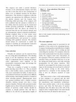
Equipment
A well equipped surgical wet laboratory (wet
lab) (Figure 1.1) is an ideal environment in which
to practice phacoemulsification, and this should
be supervised by an experienced surgeon. A wet
lab station should consist of the follwing items:
● Microscope
● Phacoemulsification machine with phaco, and
irrigation and aspiration hand pieces
● A mannequin’s head, for example the
Maloney head or a polystyrene head
● Plastic eyes with disposable cataracts and
corneas (Figure 1.2), or fresh animal eyes
● Irrigating solutions
● Disposable knives
● Cystotome and forceps for capsulorhexis
● Spatula to use in the non-dominant hand
● Rigid, folding, or injectable intraocular lenses
and instruments.
Neither postmortem animal eyes nor plastic
model eyes are able to simulate all the attributes
of the human cataractous eye. Each represents
a compromise, and their advantages and
disadvantages are summarised in Table 1.1.
Although postmortem human eyes can be used,
ethical and legal restrictions exist.
Animal eyes (most commonly from the pig)
are ideal for practicing incisions and suturing, but
because the anterior capsule is thick and elastic
they do not always simulate capsulorhexis well.
Also, the lens is soft and not ideal for practicing
nuclear fracture techniques. Attempts have been
made to harden the pig lens by injecting the eye
with a mixture of formalin and alcohol,
4
using a
microwave oven,
5
or replacing the lens with
vegetable matter.
6
Animal eyes have the
disadvantage that they are not always available
and need to be refrigerated for storage. They are
CATARACT SURGERY
2
Figure 1.1 A typical wet lab. Note the use of the
Maloney head (latrotech) to hold the artificial eyes.
Figure 1.2 Artificial eyes (bottom) with disposable
cataract (top left) and cornea (top right; Karlheinz
Hannig Microsurgical Training Systems Company).
non-sterile and may potentially be infected with,
for example, prions. In the absence of a dedicated
wet lab, the operating theatre, with its microscope
and phaco machine, is often used to provide a wet
lab facility out of hours. Unlike plastic model
eyes, animal tissue should not be used in this
environment. Plastic model eyes consistently
simulate the human cataract during sculpting,
7
and some systems have the facility to vary the
density of the “nucleus”. In contrast, rotating and
cracking the lens are less like they are in surgery
in vivo. The artifical cataract is contained within
a capsule (that may be supplied as coloured) that
allows capsulorhexis and intraocular lens
implantation to be practised. Unfortunately, the
thin plastic cornea of the model eye does not
behave like the human eye when attempting
incisions and is prone to trapping air bubbles.
Wet lab training
The set up sequence for the machine and
equipment should be understood before
commencing simulated surgery practice in the
wet lab. The following is a suggested programme
for wet lab learning and teaching.
Foot pedal control
Trainees should spend time familiarising
themselves with foot pedal function and control
(also see Chapter 4).
● Foot position 1 engages irrigation only.
● Foot position 2 engages irrigation together
with aspiration (the sound of aspiration can
be heard from the machine).
● Foot position 3 engages phacoemulsification
as well as irrigation and aspiration (the hand
piece emits a high pitched sound).
Additional audible cues may be generated by
some machines, which act as a guide to the
surgeon’s foot position. The trainee should be
able to move comfortably from one foot position
to the next without watching the screen and
should know which foot pedal position has been
engaged. It is important to explain and
understand the need to remain consistently in
foot position 1 while the phaco tip is in the eye.
This maintains the anterior chamber depth
throughout the procedure. When the three foot
positions have been mastered, the use of reflux
should be taught (usually a kick to the left once
the foot is taken off the pedal). The use of the
vitrectomy foot position should also be
explained, as should the use of the bipolar pedal.
Before moving to the next step, it is essential
that the trainer observe the trainee using the foot
pedal. The trainee needs to be able to simulate
sculpting by engaging foot position 3 for a few
seconds and then move comfortably back to foot
position 1 or 2. The use of complex pedal
TEACHING AND LEARNING PHACOEMULSIFICATION
3
Table 1.1 Comparison of plastic model and animal eyes
Eye type Advantages Disadvantages
Plastic model eyes Relative sterility (can be used in the Plastic cornea poorly simulates incision
operating theatre)
Consistent nucleus density (stimulates Air bubbles are trapped within the
sculpting well) anterior chamber during phacoemulsification
Capsular bag for practising capsulorhexis/ Lens cannot easily be rotated
intraocular lens implantation
Readily available Nucleus difficult to crack
Animal eyes Excellent for incision and suturing practice Non-sterile (cannot be used in operating
theatre)
“Normal” lens capsule for capsulorhexis, Soft nucleus, which is mainly aspirated
hydrodissection and nucleus rotation Variable availabilty and require refrigrated
storage
CATARACT SURGERY
4
movements, such as those required for dual
linear control, is best reserved for the more
accomplished surgeon.
Holding the phacoemulsification
hand piece
The hand piece should be held like a pencil,
and it is important to bring the index finger quite
close to the tip (Figure 1.3). This gives good
control of the phacoemulsification hand piece in
the eye (in the USA many surgeons hold a
phaco hand piece like a screwdriver). It is
important that the tubing and lead rest over the
arm to prevent kinking of the irrigation and
aspiration lines. The trainer should emphasise
the importance of relaxing the hand and
maintaining the horizontal position of the wrist
at this stage.
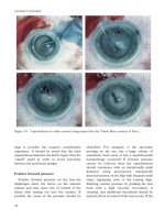
Balancing infusion and aspiration
Before inserting the phacoemulsification tip
into the eye, the trainee should check that the
hand piece is working and that the level of the
infusion is matched to the rate of aspiration. This
is achieved by putting the plastic test chamber
(the “condom”) over the phacoemulsification
needle and filling it with irrigation fluid using
foot position 1. The hand piece is then held
horizontally and foot position 2 is engaged while
the hand piece is raised. The chamber should
collapse at approximately the level of the
microscope eyepiece (Figure 1.4). If it collapses
at the level of the patient’s eye, then the
aspiration rate is too high for that level of
infusion or the infusion bottle is too low.
Conversely, if the chamber does not collapse
until well above the level of the microscope
eyepiece, then either the infusion bottle is too
high or the aspiration level is too low and this
should be rectified. The sound of the
phacoemulsification hand piece should be heard
and should be vigorous when the foot pedal is
fully depressed. If, for example, the needle is
loose, then the sound will not be normal. This
process is a quality control procedure that
Figure 1.3 Holding the phacoemulsification hand
piece.
Figure 1.4 Balancing irrigation and aspiration
before inserting the phacoemulsification tip into the
eye (the plastic test chamber should collapse at
approximately the level of the microscope eyepieces).
TEACHING AND LEARNING PHACOEMULSIFICATION
5
ensures that the phaco hand piece is in working
order before the tip is inserted into the eye.
Inserting the phaco tip into the eye
Before inserting the phaco tip into the eye the
correct position of the plastic sleeve should be
checked. This may vary depending on the nuclear
disassembly technique employed, but usually
approximately 1 mm of the phacoemulsification
needle will be exposed beyond the irrigation
sleeve. The infusion apertures in the sleeve
should be directed laterally to ensure that fluid is
not directed against the endothelium or the
capsule. Foot pedal position 1 is engaged as the
phacoemulsification tip is inserted into the eye,
with the bevel down to prevent the sharp edge of
the needle catching the iris. Once the tip is in the
eye, the non-dominant hand turns the hand piece
through 90° while the dominant hand supports
the hand piece.
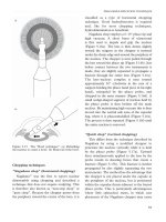
Simple “Divide and conquer” (Figure 1.5)
Sculpting the nucleus The plastic cataract
is ideally suited for learning sculpting. The
phaco tip is used to sculpt or “shave” the surface
of the nucleus. Sculpting commences within the
capsulorhexis, starting close to the incision. As
the phaco tip touches the lens surface the foot
pedal is depressed to position 3, and at the end
of the stroke the foot pedal is moved back to
position 1. The sequence is then repeated. The
tip of the phaco needle should never be
completely occluded, although the amount of
the lens engaged by the phaco needle depends
on the density of the nucleus. In a soft nucleus
up to half of the needle can be engaged and the
phaco tip can be moved reasonably quickly.
Conversely, in a hard nucleus only about one
fifth of the needle should be engaged and it must
be moved slowly. This avoids pushing the
nucleus, which may apply stress to the zonules.
If the needle moves the nucleus, then increasing
the phaco power should prevent this. For the
learning surgeon using a relatively soft plastic
nucleus, the phaco power should be set to
20–30%. Hence, when foot position 3 is
engaged 20–30% phaco power will result. With
more experience, linear power should be used so
that the surgeon may vary the power between
1 and 100%, depending on the density of the
nucleus.
A groove, approximately one and a half phaco
tips in width, should be sculpted from the
nucleus. The surgeon should be encouraged to
groove the nucleus to at least 75–80% of its
depth. A clue to groove depth is the red reflex
appearing in the groove base (even in a plastic
eye). Also the depth can be gauged by
comparison with the phaco tip diameter. When
the surgeon feels a depth of at least 75% has
been reached, the cataract should then be
removed from the eye for inspection. If the
surgeon successfully grooves two or three
cataracts to 75% depth, then it is no longer
necessary to remove the cataract from the eye
while learning.
Rotating and cracking the nucleus A
second instrument should be inserted through a
side port incision and time should be spent
working with two instruments within the eye.
Although the plastic cataract may not rotate
within the bag, the second instrument should be
inserted and an attempt should be made to
rotate it in order to familiarise oneself with this
movement. Usually the cornea has to be
removed to rotate the nucleus. Also, if air
bubbles become a problem during the sculpting,
then the remainder of the procedure can be
performed without the cornea in place. A further
groove should be made 90° from the original
groove and sculpting should be continued until
a cross has been made in the cataract. This
usually requires the nucleus to be rotated several
times through 90°. The trainee should ensure
that the phaco tip is never buried within the
nucleus and that it is always visible while using
only short bursts of phacoemulsification. Once
the two grooves have been made (at right angles
to each other) an attempt is made to crack the
CATARACT SURGERY
6
a) b)
j)
c)
d) e) f)
i)
g)
h)
Figure 1.5 Basic elements of “divide and conquer” phacoemulsification. (a) Basic sculpting: microscope view.
(b) Basic sculpting: cross-section of anterior segment. (c) Positioning the second instrument prior to nuclear
rotation. (d) Rotating the nucleus through 90°. (e) Creating the second groove. (f) Further rotation and sculpting
to create a cross. (g) Positioning the second instrument and phaco probe prior to cracking. (h) Bimanual
cracking, generating two halves (repeated after 90° rotation to create four quadrants). (i) The phaco probe is
driven into a quadrant of the nucleus. (j) Once the phaco tip is buried, suction is maintained to grip and extract
the quadrant, allowing removal with phacoemulsification in the “central safe zone”.
nucleus, ensuring that both instruments are
deep within the groove. Even if the procedure is
difficult to perform, this is an excellent learning
experience in the simultaneous use of two
instruments within the eye. Cracking is much
more difficult in the plastic cataract and will take
time. The plastic cataract will tend to break into
smaller pieces rather than into four complete
quadrants.
Nucleus quadrant removal To remove
the quadrants higher aspiration rate and vacuum
level are used. The consistency of the plastic
cataract is often chalky, and this may cause the
hand piece and the aspiration tubing to block.
Despite the higher vacuum level, the hand piece
may require regular washing through with water.
To remove a quadrant it is first engaged with a
short burst of phaco using foot position 3. Foot
position 2 is then used to maintain aspiration,
gripping the quadrant, to allow it to be drawn
into the mid-pupil or safe area where it can be
emulsified. During removal of the quadrants the
trainee should be taught the use of pulsed phaco
and the use of a higher vacuum level.
Irrigation and aspiration, and
lens insertion
If the cornea has been removed it should then
be replaced, and time should be spent becoming
familiar with the various irrigation and aspiration
hand pieces. Plastic cataracts do not usually
leave any soft lens matter behind but it is still
worthwhile inserting these hand pieces, in
particular the angled hand piece that provides
easier access to the subincisional cortex.
A complete capsular bag should remain and
lens insertion can then be attempted. For the
trainee surgeon, a variety of rigid, folding, and
injectable lenses should be kept in the wet lab
so that experience may be gained in their
insertion techniques. The intricacies of the
different folding instruments and systems can
then be mastered in the wet lab before their use
in vivo.
Surgical learning programme
The structure of a surgical learning
programme will depend on whether the surgeon
is a trainee or an experienced extracapsular
cataract surgeon making the transition to
phacoemulsification. Whatever the level of
previous experience, an individually structured
training plan should be drawn up, with specific
goals for the training period agreed by trainer
and trainee. This plan may need to be flexible
and regular appraisal should take place,
allowing problem areas to be identified and
remedied.
During the transition from wet lab to
operating theatre, it is important to involve all
members of the theatre team, particularly
because time will be required for training and the
organisation of the operating list will have to
reflect this. The choice of patients for the trainee
to operate on must also be addressed. This all
requires advance planning if it is to be successful.
For all levels of experience, video recording is
an extremely effective and useful tool for learning
and improving phacoemulsification surgery.
Trainees can benefit from watching their own
technique, and the trainer has the opportunity to
emphasise good technique and discuss errors
(constructive criticism). Recording every
procedure should become a routine event.
Ever-increasing numbers of methods
and surgical instruments are used for
phacoemulsification. It is important that a
trainee become competent and comfortable with
one surgical technique using familiar instruments
before moving on to trying different incisions,
nucleus fragmentation techniques, and varying
methods of intraocular lens insertion. Returning
to the wet lab to practice specific surgical
techniques in conjunction with time spent in
theatre should be encouraged.
TEACHING AND LEARNING PHACOEMULSIFICATION
7
Surgeons in training
For the surgeon in training who has mastered
a step in the wet lab, that step can then be put
into action in the operating theatre under the
supervision of an experienced surgeon. A period
of 40–45 minutes dedicated to training, at the
start of each operating list, enables the trainee to
have regular teaching time. This also ensures
that a patient is not subjected to a particularly
long operation, both in terms of the need for
them to lie still and of macular light exposure
(turning the operating light off or only using
axial illumination when it is required also
minimises this). Trainees often find operating
stressful, and the knowledge that the training
will take a finite time can help to allay their fears.
An alternative allocation of time is for the trainee
to repeat the same step of an operation in a series
of cases. For example, a trainee can perform the
incision for each case on the list, with the trainer
completing the remainder of the operation. This
can be applied to the initial stages of a “reverse”
training pattern (Table 1.2), in which the trainee
performs the latter stages of the operation, the
earlier stages having been performed by the
trainer. Thus, the trainee first starts by aspirating
the viscoelastic after lens insertion and then
progresses to soft lens matter aspiration, perhaps
combining this with lens insertion and removal
of the viscoelastic. Phacoemulsification can then
be practiced by performing hydrodissection,
sculpting and, if appropriate, nucleus rotation.
Capsulorhexis is left until later when the trainee
has become competent with the other steps (see
Chapter 3).
Experienced microsurgeons
For the extracapsular surgeon making a
transition to phacoemulsification, a different
training plan is suggested (Table 1.3).
Capsulorhexis and hydrodissection are essential
aspects of phacoemulsification techniques, and
for the experienced surgeon they should become
part of their extracapsular surgery. Following
capsulorhexis, relieving incisions in the capsule
opening allow expression of the crystalline lens,
or it may be possible to simply viscoexpress or
hydroexpress the nucleus.
Using automated irrigation and aspiration to
remove soft lens matter familiarises the surgeon
with the phaco machine and helps with
developing foot pedal control. It also enables the
nursing staff to practice machine set up. When
capsulorhexis and automated irrigation and
aspiration have been mastered, phaco incision
and basic nucleus sculpting can be practised.
CATARACT SURGERY
8
Table 1.2 Reverse chain: sequence of steps for
trainees
Step Details
1 Viscoelastic irrigation and aspiration
2 Soft lens matter irrigation and aspiration
3 Intraocular lens insertion
4 Hydrodissection and nucleus sculpting
5 Nucleus rotation and cracking, and
quadrant removal
6 Continuous curvilinear capsulorhexis
7 Incision
8 Complete case
Note that steps can be combined within a single case. For
example, once step 1 has been learnt steps 2, 3, and 1 may
be combined. Similarly once steps 4 and 5 are learnt, they
can be combined with steps 1, 2, and 3 to build up to a
complete case.
Table 1.3 Sequence of steps for experienced cataract
surgeons
Step Details
1 Continuous curvilinear capsulorhexis through
extracapsular incision or paracentesis
2 Automated irrigation and aspiration for
soft lens matter
3 Phaco incision (modified extracapsular
cataract extraction) and nucleus sculpting
4 Nucleus rotation and cracking, and quadrant
removal
5 Complete case
Note that steps can be combined within a single case. For
example, step 1 combined with step 2, followed by steps 1,
2, and 3 together.