Báo cáo y học: "Current findings for recurring mutations in acute myeloid leukemia" pdf
Bạn đang xem bản rút gọn của tài liệu. Xem và tải ngay bản đầy đủ của tài liệu tại đây (1.4 MB, 11 trang )
REVIEW Open Access
Current findings for recurring mutations in acute
myeloid leukemia
Shinichiro Takahashi
Abstract
The development of acute myeloid leukemia (AML) is a multistep process that requires at least two genetic
abnormalities for the development of the disease. The identification of genetic mutations in AML has greatly
advanced our understanding of leukemogenesis. Recently, the use of novel technologies, such as massively parallel
DNA sequencing or high-resolution single-nucleotide polymorphism arrays, has allowed the identification of several
novel recurrent gene mutations in AML. The aim of this review is to summarize the current findings for the
identification of these gene mutations (Dnmt, TET2, IDH1/2, NPM1, ASXL1, etc.), most of which are frequently found
in cytogenetically normal AML. The cooperative interactions of these molecular aberrations and their interactions
with class I/II mutations are presented. The prognostic and predictive significances of these aberrations are also
reviewed.
Keywords: gene mutations, acute myeloid leukemia, cooperative interactions
Introduction
The identification of mutations in certain genes, such as
the fms-related tyrosine kinase 3 (FLT3), CCAAT/
enhancer binding protein alfa (C/EBPa), runt-related
transcription factor 1 (RUNX1), myeloid-lymphoid or
mixed lineage leukemia (MLL), Wilms tumor (WT1)
and nucleophosmin (NPM) 1 genes, in acute myeloid
leukemia (AML) has significantly improved our under-
standing of leukemogenesis [1-4]. This is particularly the
case for patients with normal cytogenetics, who com-
prise the largest subgro up (approximately 45%) of AML
patients [5-7]. In fact, assessments of the presence of
internal tandem duplications in the FLT3 receptor gene
(FLT3-ITD) [8] and mutations in the NPM1 gene [4]
are currently routine practices in guiding therapeutic
decisions in AML patients with a normal karyotype [9].
Recent studies h ave revealed prevalent mutations, such
as DNA methyltransferase (Dnmt) 3a mutations [10],
ten-eleven-transloca tion oncogene family member 2
(TET2) [11] mutations and isocitrate dehydrogenase
(IDH) 1 gene mutations [3], using novel t echnologies
like high-throughput massively parallel DNA sequencing
[12]. Indeed, AML is increasingly subclassified as a
unique entity in the 2008 revision of the World Health
Organization classification of myeloid neoplasms and
acute leukemia [13], based on specific recurring genetic
abnormalities that can predict the prognosis and
response to therapy [7].
AML development is considered to be a multistep
process that requires the collaboration of at least two
classes of mutations to obtai n full-blown leukemia.
Almost a decade ago, Gilliland and Griffin [14] pre-
sented a paradigm model for this process, designated
the “two-hit model”. This model comprises class I muta-
tions that activate signal transduction pathways and
confer a proliferation advantage on hematopoietic cells,
and class II mutations that affect transcription factors
and primarily serve to impair hematopoietic differentia-
tion [15,16]. Mutations leading to activation of the
receptor tyrosine kina se (RTK) FLT3, c-kit (KIT) and
Ras signaling pathway are considered to be class I muta-
tions. Recurring chromosomal aberrations such as t(8;
21), inv(16) and t(15; 17), w hich generate fusion tran-
scripts of RUNX1/ETO, CBFb/MYH11 and PML/RARa,
respectively, fall into class I I mutations. Not only chro-
mosomal abnormalities b ut also mutations of the tran-
scription factors RUNX1, C/EBPa and MLL are
classified into this group. These “classical” class I an d
Correspondence:
Division of Molecular Hematology, Kitasato University Graduate School of
Medical Sciences and Division of Hematology, Kitasato University School of
Allied Health Sciences, 1-15-1 Kitasato, Minami-ku, Sagamihara 252-0373,
Japan
Takahashi Journal of Hematology & Oncology 2011, 4:36
/>JOURNAL OF HEMATOLOGY
& ONCOLOGY
© 2011 Takahashi; licensee BioMed Central Ltd. This is an Open Access article distributed under the terms of the Creative Commons
Attribution License ( licenses/by/2.0), which permi ts unrestricted use, distribution, and reproduction in
any medium, provided the original work is properly cited.
class II mutations are presented in Figure 1. However,
most of the newly identified genetic alterations, such as
those in Dnmt, TET2, IDH1, IDH2, NPM1, ASXL1,
which are addressed mainly in this review, have not
been classified because the consequences of these muta-
tions have not been identified. In this review, each of
these “unclassified mutations” is detailed individually.
The author summarizes the current findings of these
novel gene mutat ions, most of which are frequentl y
found in cytogenetica lly normal AML (CN-AML). The
cooperative interactions of the molecular aberrations are
also presented.
Dnmt mutations
Dnmts are enzymes that catalyze the ad dition of a
methyl group to the cytosine residue of CpG dinucleo-
tides. Aberrant CpG island methylation has long been
hypothesized to contribute to the pathogenesis of cancer
[17]. Recently, using massively parallel DNA sequencing
[12], Ley et al. [10] identified a somatic mutation in
Dnmt3a by sequencing 116.4 billion base pairs of the
sequence with 99.6% diploid coverage of the genome of
cells from an AML patient with a normal karyotype.
Further analyses revealed the presence of Dnmt3a muta-
tions in 62 of 281 AML patients (22.1%). These muta-
tions were highly enriched in a group of patients with
an intermediate-risk cytogenetic profile, as well as muta-
tions in FLT3 (either ITD or tyrosine kinase domain
[TKD] mutations), NPM1 and IDH1. The median over-
all survival (OS) among patients with Dnmt3a mutations
was signifi cantly shorter than that among patients with-
out such mutations (12.3 vs. 41.1 months, p < 0.001)
[10] (Table 1). Walter et al. [18] also described relatively
frequent mutations in myelodysplastic syndrome (MDS).
They carried out sequencing for 150 patients with MDS,
and identified 13 heterozygous mutations with predicted
transl ational consequences in 12 patients (8.0%). Yan et
al. [19] discovered Dnmt3a mutations in 23 of 112 cases
(20.5%) with the M5 subtype of AML. They revealed
that, although Dnmt3a mutations do not dramatically
alter global D NA methylation levels in AML genomes
[10], there were alterations of specific gene DNA methy-
lation patterns and/or gene expression profiles, such as
HOXB genes, in samples with Dnmt3a mutations com-
pared with those without such changes [19]. Consistent
with these observations, the Dnmt3a mutations, which
frequently occurred in arginine (R) 882, caused reduced
enzymatic activity in vitro [19].
TET2 mutations
In 2009, Delhommeau et al. [11] conducted high-res olu-
tion single-nucleotide p olymorphism and comparative
genomic hybridization arrays to identify a candidate
tumor-suppressor gene common to patients with MDS,
myeloproliferative disorders and AML. They identified
inactivating mutations of the TET2 gene in about 15%
of patients with various myeloid malignancies, such as
MDS (19%), myeloproliferative disorders (12%), second-
ary AML (24%) and chronic myelomonocytic leukemia
(CMML) (22%) [11]. TET2 can convert 5-methylcyto-
sine (5-mC) to 5-hydroxymethylcytosine (5-hmC) [20],
which to be an intermediate in DNA demethylation.
Bone marrow samples from patients with TET2 muta-
tions displayed uniformly low levels of 5-hmC in
Class I Class II
FLT3-ITD
Runx1 mut.
FLT3-TKD
MLL rearr.
PML/RAR
C/EBP mut.
CBF/MYH11
CBL mut.
KIT mut.
Runx1
mut
.
Runx1
m
MLL
rearr
.
RUNX1/ETO
NRAS mut.
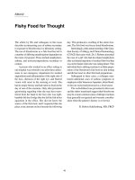
Figure 1 The model of the “ classical” class I and class II
mutations in AML. This model comprises class I mutations that
activate signal transduction pathways and confer a proliferation
advantage on hematopoietic cells, and class II mutations that affect
transcription factors and primarily serve to impair hematopoietic
differentiation [15,16]. The development of AML is a multistep
process that requires at least these two genetic abnormalities for
the development of the disease. Class I mutations are shown in
yellow boxes and class II mutations are in red boxes. The
combination of each mutation is demonstrated as blue rings. Runx
1 mutations and MLL rearrangements may be exception in this
model, as shown in orange boxes, since co-occurrence is observed
between these two mutations.
Takahashi Journal of Hematology & Oncology 2011, 4:36
/>Page 2 of 11
genomic DNA compared with bone marrow samples
from healthy controls [20]. Metzeler et al. [21] recently
analyzed 427 patients with CN-AML, and revealed that
TET2 mutations were detected in 95 of 418 (23%) of
the patients, and associated with older age (p < 0.001)
and higher pretreatment white blood cell counts (p =
0.04) compared with wild-type TET2. IDH1 and IDH2
mutations were less frequent in TET2-mutated patients
than in TET2-wild-type patients (p < 0.001), suggesting
that these mutations are mutually exclusive. They also
observed a tr end toward a higher prevalence of C/EBPa
mutations among TET2-mutated patients (p = 0.07)
[21]. In the European Leukemia Net (ELN) favorable-
risk group (patients with CN-AML who have mutated
CEBPa and/or mutated NPM1 without FLT3-ITD),
TET2-mutated patients had shorter event-free survival
(EFS) (p < 0.001), because of a lower complete remission
(CR) rate (p = 0.007), shorter disease-free survival (DFS)
(p = 0.003) and s horter OS (p = 0.001) compared with
TET2-wild-type patients (Table 1). TET2 mutations
were not associated with outcomes in the ELN inter-
mediate-I-risk group (CN-AML with wild-type CEBPa
and wild-type NPM1 and/or FLT3-ITD). In multivariate
models, TET2 mutations were associated with shorter
EFS (p = 0.004), lower CR rate (p = 0.03) and shorter
DFS (p = 0.05) only among favorable-risk CN-AML
patients [21]. Abdel-Wahab et al. [22] evaluated the
mutational statuses of TET1, TET2 and TET3 in myelo-
proliferative neoplasms (MPNs), CMML and AML.
They identified TET2 mutations in 27 of 354 MPN
patients (7.6%), 29 of 69 CMML patients (42%), 11 of 91
AMLpatients(12%)and1of28M7AMLpatients
(3.6%). Although they identified several single nucleotide
polymorphisms in TET1 and TET3, they did not iden-
tify somatic TET1 or TET3 mutations in 96 MPN
patients examined.
IDH1 and IDH2 mutations
Mardis et al. [3] used massively parallel DNA sequen-
cing to obtain a very high level of coverage of a primary,
cytogenetically normal, de novo genome for AML with
minimal maturation (AML M1) and a matched normal
skin genome, and identified 12 acquired mutations
within the coding se quences of genes and 52 somatic
point mutations in conserved or regulatory portions of
the genome. Many of these were mutations that had
already been identified, such as those in NRAS and
NPM1, but they also found novel mutations of the
IDH1 gene [3]. They further found that IDH1 gene
mutations were pre sent in 15 of 187 AML genomes and
strongly associated with a normal cytogenetic status.
IDH1 and IDH2 function at a crossroads involving cel-
lular metabolism in lipid synthesis, cellular defense
against oxidative stress, oxidative respiration and oxy-
gen-sensing signal transdu ction [23]. Recently, AML
patients harboring IDH1 and IDH2 mutations were
found to show aberrant hypermethylation [24]. In fact,
these mutations led to the production of an abnormal
cellular metabolite, 2-hydroxyglutarate, which can inhi-
bit the hydroxylation of 5-mC by TET2 [24]. Consistent
with Metzeler et al. [21], IDH1 and IDH2 mutations
were mutually excl usive with TET2 mutat ions (p =
0.009) [24]. Boissel et al. [25] analyzed the prognosis of
patients with IDH1 mutations and IDH2 mutations in a
Table 1 Clinical features of gene mutations in AML (unclassified mutations)
Gene Clinical Features Selected
Ref.
Frequency
DNMT3a The median OS among patients with Dnmt3a mutations was significantly shorter than wild type
patients.
[10] 22.1% in AML
TET2 In the European Leukemia Net (ELN) favorable-risk group, TET2-mutated patients had shorter EFS (EFS; p
< 0.001) because of a lower CR rate (p = 0.007), and shorter DFS (p = 0.003), and also had shorter OS
(p = 0.001) compared with TET2-wild type patients. (TET2 mutations were not associated with
outcomes in the ELN intermediate-I-risk group.)
[21] 23% in CN-AML
IDH1 IDH 1 mutation was associated with normal cytogenetics, a higher RR and a shorter OS. Prognosis was
adversely affected by IDH1 mutations with trend for shorter OS (p = 0.110), a shorter EFS (p < 0.003)
and a higher cumulative risk for relapse (p = 0.001). Clear prevalence in intermediate risk karyotype
group (10.4%, p < 0.001).
[25-27] 6.6-9.6% in AML
IDH2 In IDH2 mutation CN-AML patients, there is a higher risk of induction failure, a higher RR and shorter
OS.
[25,26] 3.0-8.7% in AML
NPM1 The analysis of the clinical impact in 4 groups (NPM1 and FLT3-ITD single mutants, double mutants,
and wild-type for both) revealed that patients having only an NPM1 mutation had a significantly better
OS and DFS and a lower cumulative incidence of relapse.
[28,29] 27.5-35.2% in AML
45.7-53% in CN-AML
ASXL1 Patients with ASXL1 mutations had a shorter OS than patients without, but the mutation was not an
independent adverse prognostic factor in multivariate analysis.
[39] 10.8% in AML
WT1 Multivariate analysis demonstrated that the WT1 mutation was an independent poor prognostic factor
for OS and RFS among total patients and the CN-AML group.
[41,42] 8.3-10.7% in CN-AML
6.8% in de novo
non-M3 AML
Takahashi Journal of Hematology & Oncology 2011, 4:36
/>Page 3 of 11
cohort of 520 adults with AML homogeneously treated
in the French Acute Leukemia French A ssociation
(ALFA)-9801 and -9802 trials. The prevalences of IDH1
mutations and IDH2 mutations were 9.6% and 3.0%,
respectively, and the mutations were mostly associated
with CN-AML. In patient s with C N-AML, IDH1 muta-
tions were associated with higher risk of relapse (RR)
and shorter OS (Table 1). In CN-AML patients with
IDH2 mutations, they ob served a higher risk of induc-
tion failure, higher RR and shorter OS. Similar results
were reported by Paschka et al. [26], who evaluated 805
adults with AML enrolled in the German-Austrian AML
study group, and found IDH mutations in 129 patients
(16.0%), IDH1 mutations in 61 patients (7.6%) and
IDH2 mutations in 70 patients (8.7%). These two
reports both suggest the presence of interaction s
between IDH mutations and the genotype of mutated
NPM1 without FLT3-ITD. They also both demonstrated
that IDH mutations in AML are associated with a poor
prognosis. Schnittger et al. [27] conducted a larger
study. They analyzed IDH1R132 mutations in 1414
AML patients, and detected IDH1 mutations in 6.6% of
the patients, with a clear prevalence in the intermediate-
risk karyotype group (10.4%; p < 0.001). They also
showed that IDH1 mutations had strong associations
not only with NPM1 mutations (p < 0.001), but with
MLL-partial tandem duplicaton (PTD) as well (p =
0.020) (Table 1). In addition, they revealed that the
prognosis was adversely affected by IDH1 mutations
with trends for shorter OS (p = 0.110), shorter EFS (p <
0.003) and higher cumulative RR (p = 0.001) (Table 1).
NPM1 mutations
Falini et al. [4] described abnormal localization of
NPM1 in AML patients. Cytoplasmic NPM1 w as
detected in 208 of 591 specim ens (35.2%) from patients
with primary AML, but not in 135 secondary AML spe-
cimens or in 980 hematopoietic or extrahematopoietic
neoplasms other than AML [4]. Thiede et al. [28] per-
formed a larger study. They investigated 1485 AML
patients for NPM1 exon 12 mutations, and found that
the C-terminus of this protein was mutated in approxi-
mately 27.5% of the patients. NPM1 mutations were
more prevalent in patients with a normal karyotype (324
of 709; 45.7%) than in patients with karyotype abnorm-
alities (58 of 686; 8.5%; p < 0.001). They suggested that
NPM1 mutations are strongly associated with FLT3-ITD
mutat ions in patients with a normal karyotype (mutated
NPM1/FLT3-ITD, 43.8% vs. wild-type NPM1/FLT3-
ITD, 19.9%; p < 0.001) [28]. Analyses of the clinical
impactsinfourgroups(NPM1singlemutants,FLT3-
ITD single mutants, NPM1/FLT3-ITD double mutants,
and wild-type for both) revealed that patients with only
NPM1 mutations had si gnificantly better OS and DFS
and a lower cumulative incidence of relapse [28] (Table
1). Schlenk et al. [29] foc used their analyses on CN-
AML patients. A mong 872 pat ients examined, they
found that 53% of the patients had NPM1 mutations. In
addition, 31% had FLT3-ITD, 11% had FLT3-TKD, 13%
had C/EBPa mutations, 7% had MLL-PTD and 13% had
NRAS mutatio ns. They further demonstrated that
FLT3-ITD (p < 0.001) and FLT3-TKD mutations (p =
0.03) were significantly associated with NPM1 muta-
tions, while NRAS mutations were not (p = 0.46).
NPM1 is thought to have relevant roles in diverse cel-
lular functions, including ribosome bioge nesis, centro-
some duplication, DNA repair and response to stress
[30]. NPM1 is also involved in the funct ions of p53 and
p19ARF [31,32]. Li et al. [33] demonstrated that wild-
type NPM1 protects hematopoietic cells against p53-
induced apoptosis under conditions of cellular stress.
Therefore, it is possible that failure of the mutated
NPM1 to protect cells may make them more sensitive
to high-level genotoxic stress induced by chemotherapy.
Consequently, it is possible to speculate that patients
with mutated NPM1 have a better prognosis.
ASXL1 mutations
The additional sex comb-like 1 (ASXL1) gene belongs to
a family with three identified members that encode
poorly characterized proteins involved in the regul ation
of chromatin remodeling. The ASXL proteins contain a
C-terminal plant homeodomain (PHD) finger and
belong to the polycomb and trithorax complexes that
regulate the genetic program of stem cells. ASXL1 is
involved in the regulation of histone methylation by
cooperation with heterochromatin protein-1 to modu-
late the activity of lysine-specific demethylase (LSD) 1
[34], a histone demethylase for H3K4 and H3K9 that is
also important for global DNA methylation [35]. How-
ever, the hematopoietic function of ASXL1 is still
unclear, since an ASXL1 knockout mouse model shows
only a mild hematopoietic phenotype [36]. In 2009,
Gelsi-Boyer et al. [37] first identified mutations of
ASXL1in40MDS/AMLsamplesusinghigh-density
comparative genomic hybridization array s. They found
mutations in the ASXL1 gene in 4 of 35 MDS patients
(11%) and 17 of 39 CMML pa tients (43%). Another
study identified mutations of the ASXL1 gene in 12 of
63 (19%) secondary AML patients transformed from
MPN [38]. Recently, Chou et al. [39] examined ASXL1
gene mutations in exon 12 in 501 adults with de novo
AML. ASXL1 mutations were detected in 54 patients
(10.8%), with 8.9% among patients with a normal karyo-
type and 12.9% among patients with abnormal cytoge-
netics. The mutations were closely associated with older
age, male sex, isolated trisomy 8, RUNX1 mutations,
and expression of human leukocyte antigen-DR and
Takahashi Journal of Hematology & Oncology 2011, 4:36
/>Page 4 of 11
CD34, but inversely associated with t(15; 17), complex
cytogenetics, FLT3-ITD, NPM1 mutations, WT1 muta-
tions and expression of CD33 and CD15. Patients with
ASXL1 mutations had shorter OS than patients without
such mutations, but the mutations were not an indepen-
den t adverse prognostic factor in a multivariate analysis
[39] (Table 1).
WT1 mutations
Although mutations of WT1 were first discovered in
hematological malignancies more than a deca de ago, the
precise roles of WT1 in normal and malignant hemato-
poiesis remain elusive [40]. It has been implicated in the
regulation of cell survival, proliferati on and differentia-
tion, and may function as b oth a tumor suppressor and
an oncogene [40]. Paschka et al. [41] analyzed 196
adults aged < 60 years with newly diagnosed primary
CN-AML, who were treated similarly with Cancer and
Leukem ia Group B (CALGB) protoc ols 9621 and 19808,
for WT1 mutations in exons 7 and 9. As a result, 21
patients (10.7%) harbored WT1 mutations. The patients
with WT1 mutations had worse DFS (p < 0.001) and
OS (p < 0.001) than patients with wild-type WT1. Sub-
sequently, Hou et al. [42] examined the clinical implica-
tions of WT1 mutations in 470 de novo non-M3 AML
patients aged ≥ 15 years, and their stability during the
clinical course. WT1 mutations were identified in 6.8%
of the total patients and 8.3% of the younger patient s
with CN-AML. The WT1 mutations were closely asso-
ciated with younger age (p < 0.01), French-American-
British M6 subtype (p = 0.006) and t(7; 11)(p15; p15) (p
= 0.003). A multivariate analysis demonstrated that
WT1 mutations comprised an independent poor prog-
nostic factor for OS and relapse-free survival (RFS)
among the total patients and the CN-AML patients. In
addition, among 32 patients with WT1 mutations, the
most frequently associated molecular event was FLT3-
ITD (9 cases) [42]. Becker et al. [43] investigated 243
older (≥ 60 years) primary CN-AML patients, and found
that WT1-mutated patients (7%) had more frequent
FLT3-ITD (p < 0.001) and shorter OS (p = 0.08) com-
pared with WT1-wild-type patients. The clinical features
of the unclassified mutations are summarized in Table 1.
Class I mutations (FLT3, PTPN11, NRAS, KIT and
CBL mutations)
FLT3 mutations
FLT3isatypeIIIRTKandsincethefirstdescription
[44], numerous studies have confirmed and extended
the findings t hat FLT3 mutations are currently one of
the most frequent single mutations identified in AML.
ITD mutations of the FLT3 gene occur in approximately
21-24% of adult AML patients [14,28] while activating
point mutations of the FLT3-TKD, mainly at Asp 835,
are found in approximately 5-7% of AML patients
[1,45-48]. The most significant impacts of an ITD are its
associations with a higher leukocyte count, increased
RR, decreased DFS and decreased OS, which have been
reported in most studies of children and adults aged <
60 years [49]. Several groups found that an ITD is a sig-
nificant predictive factor for an adverse outcome in mul-
tivariate analyses [49-52] (Table 2). Bacher et al. [46]
performed a large study involving 3082 patients with
newly diagnosed AML, and analyzed the mutational sta-
tus and clinical significance of the FLT3-TKD. They
observed FLT3-TKD mutations in 147 patients (4.8%).
Unlike FLT3-ITD, the prognosis was not influenced by
FLT3-TKD mutations in a tot al cohort of 1720 cases
where follow up-data were available (97 mutated FLT3-
TKD cases and 1623 wild-type FLT3 cases) (Table 2). In
addition, Ozeki et al. [53] reported that even in patients
with wild-type FLT3, a clear tendency for worse OS was
found in patients with high FLT3 expression (5 of 86
patients without FLT3-ITD). Another group observed a
sim ilar result for a tendency toward lower OS (12 of 24
patients, p = 0.059) and EFS (7 of 20 patients, p =
0.087) in the group with high FLT3 expression [54]
(Table 2).
FLT3-ITD mutations are correlated with certain cyto-
genetic subgroups. Among acute promyelo cytic leuke-
mia patients with PML-RARa, it was reported that 30-
50% of patients had FLT3 mutations [55-57]. In addi-
tion, frequent (88~90%) co-occurrence was report ed in
patients with t(6; 9) and FLT3-ITD [55,58]. Similarly,
FLT3-ITD was frequently found in patients with MLL-
PTD [59]. The rate of MLL-PTD in FLT3-ITD-positive
patients was significantly higher than that in FLT3-ITD-
negative patients [16 of 184 (8.7%) vs. 32 of 772 (4.1%);
p = 0.025] [59]. In analyses involving 353 adult de novo
AML patients, Carnicer et al. [60] found cooperative
mutations of FLT3-TKD with CBFb/MY H11 rearrange-
ment (4 of 15 patients) and C/EBPa with FLT3-ITD (2
of 82 patients). Collectively, FLT3 mutations play a key
role in leukemogenesis by functionally cooperating with
other molecules.
PTPN11 mutations
SHP-2 is a cytoplasmic protein tyrosine phosphatase
(PTP) that contains two Src homology 2 (SH2) domains.
Although PTPs are generally considered to be negative
regulators, SHP-2 is unusual in that it promotes the
activation of the RAS-MAPK signaling pathway through
receptors for various growth facto rs and cytokines.
Mutations in the protein tyrosine phosphatase non-
receptor type 11 (PTPN11), as the human SHP-2 gene,
have been sho wn to produce dominant active mutants
in vitro [61]. Hou et al. [62] investigated the prevalence
and clinical relevance of mutations of PTPN11 and their
Takahashi Journal of Hematology & Oncology 2011, 4:36
/>Page 5 of 11
associations with other genetic changes in 272 consecu-
tive patients with primary AML. Among 14 patients
with PTPN11 mutations (5.1%), none had FLT3-ITD.
On the other hand, 6 of 13 patients with PTPN11 muta-
tions had concurrent NPM1 mutations (46.2%) [62],
suggesting that PTPN11 is a cla ss I mutation molecule
similar to the case for FLT3. They further revealed that
PTPN11 mutations had no prognostic significance. The
CR rate (75% vs. 62%) and median OS (13 ± 8.95 vs.
25.5 ± 6.54 months) were similar between patients with
and without PTPN11 mutations. However, subgroup
analyses did reveal that PTPN11 mutations comprised a
poor risk factor for OS of AML pati ents without NPM1
mutations (p = 0.001) [62] (Table 2).
NRAS mutations
Ras oncogenes encode a family of guanine nucleotide-
binding proteins that regulate s ignal transduction u pon
binding to a variety of membrane r eceptors, including
KIT and FLT3, and play important roles in proliferation,
differentiation and ap optosis [63]. There are three func-
tional RAS genes: NRAS, KRAS and HRAS.TheRAS
genes, especial ly NRAS, are frequently affected by muta-
tions in AML. Bacher et al. [64] analyzed 2502 patients
with AML, and found that 257 patients (10.3%) had
NRAS mutations. The subgroups with inv(16) and inv
(3)showedsignificantlyhigherfrequenciesofNRAS
mutations. In contrast, NRAS mutations were signifi-
cantly underrepresented in t(15; 17) (2 of 102; 2%; p =
0.005). They did not find significant progno stic impacts
of NRAS mutations for OS, EFS and DFS (Table 2).
KIT mutations
KIT is a member of the type III RTK family, and ligand-
independent activation of KIT can be caused by gain-of-
function mutations t hat have been reported in core
binding factor (CBF) leukemia, and AML subgroups
with inv(16) and t(8; 21) [65-67], which result in
expression of the abnormal f usion genes CBFb/MYH11
and RUNX1/ETO, respectively. Paschka et al. [68] ana-
lyzed 61 adults with CBF leukemia for KIT mutations.
Among patients with inv(16), 29.5% had KIT mutations.
Among patients with t(8; 21), 22% had KIT mutations.
Mutations of the c-kit gene in both exon 17 and exon 8
appeared to adversely affect OS in AML with inv(16).
They also observed an adverse impact of KIT mutations
on RR in t(8; 21) AML patients (Table 2). Cairoli et al.
[65] reported that among 42 patients with t(8;21), 19
patients (45.2%) had KIT mutations, whereas among 25
patients with inv(16), 12 patients (48.0%) had KIT muta-
tions. Schnittger et al. [66] analyzed 1940 randomly
selected AML patients and revealed that 33 patients
(1.7%) were positive for KIT mutations in codon D816.
Of these 33 patients, 8 patients (24.2%) had t(8; 21),
which was significantly higher compared with the sub-
group without D816 mutations. They revealed that KIT
mutations had independent negative impacts on the
median OS (304 vs. 1836 days; p = 0.006) and median
EFS (244 vs. 744 days; p = 0.003) in patients with t(8;
21)butnotinpatientswithanormalkaryotype(Table
2). They also revealed that other activating mutations,
like FLT3 and NRAS mutations, were very rarely
detected in t(8; 21) leukemia patients. On the contrary,
in an analysis of 99 patients with t(8; 21), Kuchenbauer
et al. [69] reported that the frequent molecular aberra-
tions with t(8; 21) were not only KIT D816 mutations (3
of 23 patients; 13%) but also NRAS mutations (8 of 89
patients; 8.9%). Although the co-occurrence of NRAS
mutations and AML1/ETO expression remains elusive,
all of these reports suggest that KIT mutants play
important roles in CBF leukemia, with negative impacts
on the clinical course [65,66,68,69].
CBL mutations
The Casitas B-cell lymphoma (CBL) gene gives rise to
the CBL protei n, which has ubiquitin ligase activity and
Table 2 Clinical features of gene mutations in AML (class I mutations)
Gene Clinical Features Selected
Ref.
Frequency
FLT3
-ITD
Association with a higher leukocyte count, increased RR, decreased DFS, and decreased OS. [14,28,49-52] 21-24% in AML
-TKD Prognosis was not influenced. [46] 5-7% in AML
-WT Clear tendencies for worse OS and EFS were found in patients with high FLT3 expression. [53,54]
PTPN11 No prognostic significance. However, subgroup analysis did reveal that the PTPN11 mutation was a
poor risk factor for OS of AML patients who did not have NPM1 mutations.
[62] 5.1% in AML
NRAS No significant prognostic impact for OS, EFS and DFS. [64] 10.3% in AML
KIT Adversely affect OS in AML with inv(16). Adverse impact of mutation of KIT on RR in t(8; 21)AML.
KIT mutations had an independent negative impact on OS and EFS in patients with t(8;21) but not
in patients with a normal karyotype.
[66,68,69] 1.7% in AML
22-45% in t(8; 21) 29-
48% in inv(16)
CBL n. d. [72,73] 1.1% in AML/MDS
16% in inv(16)AML
Takahashi Journal of Hematology & Oncology 2011, 4:36
/>Page 6 of 11
targets a variety of tyrosine kinases for degradation by
ubiquitination [70]. CBL proteins also associate with the
endocyt ic machinery and are thus important for the ter-
mination of RTK signaling [70]. CBL mutations were
able to inhibit FLT3 internalization and ubiquitination
[71]. In vitro experiments confirmed constitutive activa-
tion of the FLT3 pathway by the CBL mutants, and the
phenotype of the altered cells resembled the phenotype
of FLT3-mutated cells [72]. Reindl et al. [72] identified
c-CBL gene exon 8/9 deletion mutants in 1.1% of 279
patients with AML/MDS. All the patients with CBL
mutations had CBF leukemia and chromosome 11q
abnormalities [72]. In a series of 37 patients with newly
diagnosed inv(16) AML, Haferlach et al. [73] detected
CBL splicing mutations in 6 patients (16%). The preva-
lence and features of these class I mutations are sum-
marized in Table 2.
Class II mutations (RUNX1 mutations, C/EBPa
mutations and MLL rearrangement)
RUNX1 mutations
Runx1 is required for definitive hematopoiesis and is
necessary for the differentiation of myeloid progenitor
cells to granulocytes [74]. Recently, Gaidzik et al. [75]
precisely studied the frequency, biologic features and
clinical relevance of Runx1 mutations in AML. They
found that RUNX1 gene mutations, which span exons
3, 4, 5 and 8, were present in 53 of 945 patients (5.6%)
with AML. RUNX1 mutations were associated with
MLL-PTD (p = 0.0007) and IDH1/IDH2 mutations (p
= 0.03), inversely correlated with NPM1 (p < 0.0001),
and in trend with CEBPa (p = 0.10) mutations.
RUNX1 mutations predicted resistance to chemother-
apy, as well as inferior EFS (p < 0.0001), RFS (p =
0.022) and OS (p = 0.051). In multivariate analyses,
RUNX1 mutations were an independent prognostic
marker for shorter EFS (p = 0.007) (Table 3). Tang et
al. [76] examined 470 adult patients with de novo non-
M3 AML. Among these patients, 63 distinct RUNX1
mutations were identified in 62 patients (13.2%). They
also revealed that the mutations were positively asso-
ciated with MLL-PTD but negatively associated with
C/EBPa and NPM1 mutations. Schnittger et al. [77]
detected 164 RUNX1 mutations in 147 of 449 patients
(32.7%) with a normal karyotype or noncomplex chro-
mosomal imbalances. RUNX1 mutations were most
frequent in AML M0 (65.2%) followed by M2 (32.4%)
and M1 (30.2%). The molecular genetic markers most
frequently associated with RUNX1 mutations were
MLL-PTD (19.7%), FLT3-ITD (16.3%) and NRAS
mutations (9.5%). Multivariate analyses showed that
RUNX1 mutations independently predicted an unfa-
vorable prognosis for OS (p = 0.029).
C/EBPa mutations
The transcription factor C/EBPa is crucial for the differ-
entiation of granulocytes. C/EBPa inactivation may take
place by acquired mutations, which may occur along the
ent ire coding region. These mutations lead to increased
translation of an alternative 30-kDa form with dominant
negative activity on the full-length 42-kDa protein when
they occur in the N-terminus, while C-terminal muta-
tions result in deficient DNA binding and/or homodi-
merization activities [63,78]. As reviewed by Pabst and
Mueller [79], mutations in the various portions of C/
EBPa have been reported to occur in 5-14% of AML
patients. Among patients with C/EBPa mutations, 91%
were in the CN-AML molecular high-risk group (FLT3-
ITD-positive and/or NPM1-wild-type), although they
seemed to be associate d with a good prognosis in AML
with an intermediate-risk karyotype [80]. Compared
with C/EBPa-wild-type patients, patients with C/EBPa
mutations showed a trend for a better CR rate (93% vs.
77%; p = 0.06) and significantly higher 5-year rates of
EFS (55% vs. 17%; p < 0.001), DFS (53% vs. 23%; p =
0.001) and OS (58% vs. 27%; p = 0.002) [80] (Table 3).
However, the reason w hy C/EBPa mutations confer a
good prognosis remains unclear.
MLL rearrangement
Approximately 4-11% of patients with AML present
with rearrangement of the MLL (also known as ALL1 or
HRX) gene as the result of a PTD within a single MLL
allele [81]. This aberration tends to be frequent in AML
patients without chrom osom al abnormalities [81]. MLL
is a 430-kDa transcription factor with a complex struc-
ture that includes three AT-hook domains for DNA
binding, a methyltransferase homology domain and a
SET domain [81]. MLL is necessary for the maintenance
for HOX gene expression [82]. The MLL SET domain
canbindtothepromotersofHOXgenes[83].Munoz
et al. [84] examined 93 adult patie nts with de novo
AML for the incidence and clinical features of MLL-
rearranged AMLs. As a result, they detected MLL rear-
rangement in 13 patients (14%). In the FAB classifica-
tion, there was a significantly higher percentage of M5
subtypes in the MLL-rearranged group. The MLL-rear-
ranged patients had lowe r EFS (p = 0.001) and a higher
probability of relapse (p = 0.07) than MLL-wild-type
patients. The clinical features of these RUNX1 and C/
EBPa mutations and MLL r earrangement are shown in
Table 3.
Conclusions
The development of novel technologies has led to the
identification of several important genetic mutations in
AML. Along with the development of whole genome
Takahashi Journal of Hematology & Oncology 2011, 4:36
/>Page 7 of 11
sequencing, it is probable that the major genetic aberra-
tions have been almost completely identified. The next
stage is to clarify the consequence of these molecular
alterations, especially for the newly identified molecules.
As described in this review, mutations in several newly
identified genes, such as TET2 and IDH1/2, lead to the
aberrant hypermethylation signature in AML cells [24].
In addition, there were alterations in the DNA methyla-
tion patterns and/or gene expression profiles, such as
HOXB genes, in samples with DNMT3a mutations com-
pared with those without such mutations [19]. Collec-
tively, these recent findings strongly suggest a link
between recurrent genetic alterations and a berrant epi-
gene tic regulation, resulting in abnormal DNA methyla-
tion statuses in myeloid malignancies.
With the identification of novel genetic a berrations,
increasing numbers of cooperative interactions of these
gen etic al terations have been discove red. These observa-
tions indicate that t hese unclassified mutations may fall
into several subcategories. A revised model of these com-
binations, including “unclassified mutations” in AML is
shown in Figure 2 and 3. In general, the mutations in the
same category are mutually exclusive [14,85]. However,
there are some exceptions. In “classical” class II muta-
tions, co-occurrence was reported between RUNX1
mutations and MLL rearrang ement [76] (Figure 1). Like -
wise, in unclassified mutations, co-occurrences were
observed among Dnmt3a, NPM1 and IDH1/2 mutations
[10,25,26]. Dnmt3a and NPM1 mutations co-occur with
both classical class I and class II mutations, therefore,
these mutations may not be simply categorized into “clas-
sical” class I or class II mutations, but it would fall into
new category of mutations. Further characterization of
these mutations, not only from clinical studies, but also
from studies of transgenic animals may be needed.
For these several years, there are admirable progress
for the identification of the molecular prognostic mar-
kers for CN-AML [86]. Based on these genetic altera-
tions, several signal transduction pathway inhibitors and
DNA methyltransferase inhibitors (decitabine, azaciti-
dine) were reported for AML treatment [87]. The newly
identified combinations of genetic aberrations will lead
to a refined disease classification and to the develop-
ment of more rational, epigenetic or signal transduction
pathway-targeted therapies.
Table 3 Clinical features of gene mutations in AML (class II mutations)
Gene Clinical Features Selected
Ref.
Frequency
Runx1 In multivariable analysis, RUNX1 mutations were an independent prognostic marker for shorter EFS.
Independent unfavorable prognosis for OS for RUNX1 mutation.
[75-77] 5.6% in AML
13.2% in de novo non-
M3AML
32.7% in CN or
noncomplex karyotype
AML
C/
EBPa
C/EBPa mutations had a trend for a better CR rate and significantly greater 5-year rates of EFS, DFS
and OS.
[79,80] 5-14% in AML
MLL
rearr.
Patients with MLL rearrangement had a lower EFS and a higher probability of relapse than MLL
wild type patients.
[81] 4-14% in AML
Unclassified
(Class II’)
NPM1 mut.
WT1 mut.
Dnmt3a mut.
FLT3-ITD
FLT3-TKD
CBL mut.
NPM1 mut.
NPM1 mut.
NPM1
mut
.
FLT3
-
TKD
CBL
m
ut
Dnmt3a
mut
.
Dnmt3a
mut
.
NRAS mut.
KIT mut.
PTPN11 mut.
P
T
P
P
P
N
1
1
m
u
t
.
Dnmt3a
mut
.
Class I
Figure 2 The combination model of class I and unclassified
mutations in AML. Several unclassified mutations (NPM1, WT1,
Dnmt3a) co-occur with several class I mutations. These may fall into
putative class II mutations (termed “class II’ mutations”), shown in
pink boxes. Within these mutations, co-occurrences were observed
between Dnmt3a, NPM1mutations (shown in orange boxes),
therefore, these mutations may be exception of this model.
Takahashi Journal of Hematology & Oncology 2011, 4:36
/>Page 8 of 11
Acknowledgements
I appreciate Dr. Toshio Okazaki (Kitasato University) for many helpful
suggestions for preparing figures. I thank Dr. Alison Sherwin (Edanz group
ltd.) for English editing and critical reading in the preparation of this
manuscript. I also thank Dr. Takeo Takahashi (Yaotome Clinic) for helpful
advice on English language in the revision. This work was supported in part
by Grants-in-Aid for Scientific Research (No. 23590687) from the Ministry of
Education, Science and Culture, Japan, the Takeda Sceince Foundation, a
foundation from Kitasato University School of Allied Health Sciences (Grant-
in-Aid for Research Project, No. 2011-1001).
Authors’ information
Professor and Chief, the Division of Molecular Hematology, Kitasato
University Graduate School of Medical Sciences and the Division of
Hematology, Kitasato University School of Allied Health Sciences,
Sagamihara, Japan
Competing interests
The author declares that they have no competing interests.
Received: 25 July 2011 Accepted: 14 September 2011
Published: 14 September 2011
References
1. Takahashi S: Downstream molecular pathways of FLT3 in the
pathogenesis of acute myeloid leukemia: biology and therapeutic
implications. J Hematol Oncol 2011, 4:13.
2. Dohner K, Dohner H: Molecular characterization of acute myeloid
leukemia. Haematologica 2008, 93:976-982.
3. Mardis ER, Ding L, Dooling DJ, Larson DE, McLellan MD, Chen K,
Koboldt DC, Fulton RS, Delehaunty KD, McGrath SD, et al: Recurring
mutations found by sequencing an acute myeloid leukemia genome. N
Engl J Med 2009, 361:1058-1066.
4. Falini B, Mecucci C, Tiacci E, Alcalay M, Rosati R, Pasqualucci L, La Starza R,
Diverio D, Colombo E, Santucci A, et al: Cytoplasmic nucleophosmin in
acute myelogenous leukemia with a normal karyotype. N Engl J Med
2005, 352:254-266.
5. Byrd JC, Mrozek K, Dodge RK, Carroll AJ, Edwards CG, Arthur DC,
Pettenati MJ, Patil SR, Rao KW, Watson MS, et al: Pretreatment cytogenetic
abnormalities are predictive of induction success, cumulative incidence
of relapse, and overall survival in adult patients with de novo acute
myeloid leukemia: results from Cancer and Leukemia Group B (CALGB
8461). Blood 2002, 100:4325-4336.
6. Grimwade D, Hills RK, Moorman AV, Walker H, Chatters S, Goldstone AH,
Wheatley K, Harrison CJ, Burnett AK: Refinement of cytogenetic
classification in acute myeloid leukemia: determination of prognostic
significance of rare recurring chromosomal abnormalities among 5876
younger adult patients treated in the United Kingdom Medical Research
Council trials. Blood 2010, 116:354-365.
7. Foran JM: New prognostic markers in acute myeloid leukemia:
perspective from the clinic. Hematology Am Soc Hematol Educ Program
2010, 47-55.
8. Thiede C, Steudel C, Mohr B, Schaich M, Schakel U, Platzbecker U,
Wermke M, Bornhauser M, Ritter M, Neubauer A, et al: Analysis of FLT3-
activating mutations in 979 patients with acute myelogenous leukemia:
association with FAB subtypes and identification of subgroups with
poor prognosis. Blood 2002, 99:4326-4335.
9. Lowenberg B: Diagnosis and prognosis in acute myeloid leukemia–the
art of distinction. N Engl J Med 2008, 358:1960-1962.
10. Ley TJ, Ding L, Walter MJ, McLellan MD, Lamprecht T, Larson DE,
Kandoth C, Payton JE, Baty J, Welch J, et al: DNMT3A Mutations in Acute
Myeloid Leukemia. N Engl J Med 2010, 363:2424-2433.
11. Delhommeau F, Dupont S, Della Valle V, James C, Trannoy S, Masse A,
Kosmider O, Le Couedic JP, Robert F, Alberdi A, et al: Mutation in TET2 in
myeloid cancers. N Engl J Med 2009, 360:2289-2301.
12. Wheeler DA, Srinivasan M, Egholm M, Shen Y, Chen L, McGuire A, He W,
Chen
YJ, Makhijani V, Roth GT, et al: The complete genome of an
individual by massively parallel DNA sequencing. Nature 2008,
452:872-876.
13. Vardiman JW, Thiele J, Arber DA, Brunning RD, Borowitz MJ, Porwit A,
Harris NL, Le Beau MM, Hellstrom-Lindberg E, Tefferi A, Bloomfield CD: The
2008 revision of the World Health Organization (WHO) classification of
myeloid neoplasms and acute leukemia: rationale and important
changes. Blood 2009, 114:937-951.
14. Gilliland DG, Griffin JD: The roles of FLT3 in hematopoiesis and leukemia.
Blood 2002, 100:1532-1542.
15. Frohling S, Scholl C, Gilliland DG, Levine RL: Genetics of myeloid
malignancies: pathogenetic and clinical implications. J Clin Oncol 2005,
23:6285-6295.
16. Kelly LM, Gilliland DG: Genetics of myeloid leukemias. Annu Rev Genomics
Hum Genet 2002, 3:179-198.
17. Esteller M: Epigenetics in cancer. N Engl J Med 2008, 358:1148-1159.
18. Walter MJ, Ding L, Shen D, Shao J, Grillot M, McLellan M, Fulton R,
Schmidt H, Kalicki-Veizer J, O’Laughlin M, et al: Recurrent DNMT3A
mutations in patients with myelodysplastic syndromes. Leukemia 2011.
19. Yan XJ, Xu J, Gu ZH, Pan CM, Lu G, Shen Y, Shi JY, Zhu YM, Tang L,
Zhang XW, et al: Exome sequencing identifies somatic mutations of DNA
methyltransferase gene DNMT3A in acute monocytic leukemia. Nat
Genet 2011, 43:309-315.
20. Ko M, Huang Y, Jankowska AM, Pape UJ, Tahiliani M, Bandukwala HS, An J,
Lamperti ED, Koh KP, Ganetzky R, et al: Impaired hydroxylation of 5-
methylcytosine in myeloid cancers with mutant TET2. Nature 2010,
468:839-843.
21. Metzeler KH, Maharry K, Radmacher MD, Mrozek K, Margeson D, Becker H,
Curfman J, Holland KB, Schwind S, Whitman SP, et al: TET2 mutations
improve the new European LeukemiaNet risk classification of acute
myeloid leukemia: a Cancer and Leukemia Group B study. J Clin Oncol
2011, 29:1373-1381.
22. Abdel-Wahab O, Mullally A, Hedvat C, Garcia-Manero G, Patel J,
Wadleigh M, Malinge S, Yao J, Kilpivaara O, Bhat R, et al: Genetic
characterization of TET1, TET2, and TET3 alterations in myeloid
malignancies. Blood 2009, 114:144-147.
Class II
Unclassified
(Class I’)
ASXL1 mut.
Runx1 mut.
MLL rearr.
PML/RAR
C/EBP mut.
RUNX1/ETO
IDH1/2 mut.
TET2 mut.
R
u
R
R
n
x
n
n
1
m
u
t
Dnmt3a
mut
.
PML
/
RA
R
M
L
L
r
e
r
r
a
ee
r
r
r
r
.
D
nm
NPM1
mut
.
CBF/MYH11
Figure 3 The combination model of class II and unclassified
mutations in AML. Several unclassified mutations (ASXL1, IDH1/2,
TET2) co-occur with several class II mutations. These may fall into
putative class I mutations (termed “class I’ mutations”), shown in
light yellow boxes. IDH1/2 mutations co-occurred not only with
MLL rearrangement, but also Dnmt3a and NPM1 mutations, as
shown in orange boxes.
Takahashi Journal of Hematology & Oncology 2011, 4:36
/>Page 9 of 11
23. Reitman ZJ, Yan H: Isocitrate dehydrogenase 1 and 2 mutations in
cancer: alterations at a crossroads of cellular metabolism. J Natl Cancer
Inst 2010, 102:932-941.
24. Figueroa ME, Abdel-Wahab O, Lu C, Ward PS, Patel J, Shih A, Li Y,
Bhagwat N, Vasanthakumar A, Fernandez HF, et al: Leukemic IDH1 and
IDH2 mutations result in a hypermethylation phenotype, disrupt TET2
function, and impair hematopoietic differentiation. Cancer Cell 2010,
18:553-567.
25. Boissel N, Nibourel O, Renneville A, Gardin C, Reman O, Contentin N,
Bordessoule D, Pautas C, de Revel T, Quesnel B, et al: Prognostic impact of
isocitrate dehydrogenase enzyme isoforms 1 and 2 mutations in acute
myeloid leukemia: a study by the Acute Leukemia French Association
group. J Clin Oncol 2010, 28:3717-3723.
26. Paschka P, Schlenk RF, Gaidzik VI, Habdank M, Kronke J, Bullinger L, Spath D,
Kayser S, Zucknick M, Gotze K, et al: IDH1 and IDH2 mutations are
frequent genetic alterations in acute myeloid leukemia and confer
adverse prognosis in cytogenetically normal acute myeloid leukemia
with NPM1 mutation without FLT3 internal tandem duplication. J Clin
Oncol 2010, 28:3636-3643.
27. Schnittger S, Haferlach C, Ulke M, Alpermann T, Kern W, Haferlach T: IDH1
mutations are detected in 6.6% of 1414 AML patients and are
associated with intermediate risk karyotype and unfavorable prognosis
in adults younger than 60 years and unmutated NPM1 status. Blood
2010, 116:5486-5496.
28. Thiede C, Koch S, Creutzig E, Steudel C, Illmer T, Schaich M, Ehninger G:
Prevalence and prognostic impact of NPM1 mutations in 1485 adult
patients with acute myeloid leukemia (AML). Blood 2006, 107:4011-4020.
29. Schlenk RF, Dohner K, Krauter J, Frohling S, Corbacioglu A, Bullinger L,
Habdank M, Spath D, Morgan M, Benner A, et al: Mutations and treatment
outcome in cytogenetically normal acute myeloid leukemia. N Engl J
Med 2008, 358:1909-1918.
30. Colombo E, Alcalay M, Pelicci PG: Nucleophosmin and its complex
network: a possible therapeutic target in hematological diseases.
Oncogene 2011, 30:2595-2609.
31. Colombo E, Marine JC, Danovi D, Falini B, Pelicci PG: Nucleophosmin
regulates the stability and transcriptional activity of p53. Nat Cell Biol
2002, 4:529-533.
32. Bertwistle D, Sugimoto M, Sherr CJ: Physical and functional interactions of
the Arf tumor suppressor protein with nucleophosmin/B23. Mol Cell Biol
2004, 24:985-996.
33. Li J, Zhang X, Sejas DP, Pang Q: Negative regulation of p53 by
nucleophosmin antagonizes stress-induced apoptosis in human normal
and malignant hematopoietic cells. Leuk Res 2005, 29:1415-1423.
34. Lee SW, Cho YS, Na JM, Park UH, Kang M, Kim EJ, Um SJ: ASXL1 represses
retinoic acid receptor-mediated transcription through associating with
HP1 and LSD1. J Biol Chem 2010, 285:18-29.
35. Wang J, Hevi S, Kurash JK, Lei H, Gay F, Bajko J, Su H, Sun W, Chang H,
Xu
G, et al: The lysine demethylase LSD1 (KDM1) is required for
maintenance of global DNA methylation. Nat Genet 2009, 41:125-129.
36. Fisher CL, Pineault N, Brookes C, Helgason CD, Ohta H, Bodner C, Hess JL,
Humphries RK, Brock HW: Loss-of-function Additional sex combs like 1
mutations disrupt hematopoiesis but do not cause severe
myelodysplasia or leukemia. Blood 2010, 115:38-46.
37. Gelsi-Boyer V, Trouplin V, Adelaide J, Bonansea J, Cervera N, Carbuccia N,
Lagarde A, Prebet T, Nezri M, Sainty D, et al: Mutations of polycomb-
associated gene ASXL1 in myelodysplastic syndromes and chronic
myelomonocytic leukaemia. Br J Haematol 2009, 145:788-800.
38. Abdel-Wahab O, Manshouri T, Patel J, Harris K, Yao J, Hedvat C, Heguy A,
Bueso-Ramos C, Kantarjian H, Levine RL, Verstovsek S: Genetic analysis of
transforming events that convert chronic myeloproliferative neoplasms
to leukemias. Cancer Res 2010, 70:447-452.
39. Chou WC, Huang HH, Hou HA, Chen CY, Tang JL, Yao M, Tsay W, Ko BS,
Wu SJ, Huang SY, et al: Distinct clinical and biological features of de
novo acute myeloid leukemia with additional sex comb-like 1 (ASXL1)
mutations. Blood 2010, 116:4086-4094.
40. Owen C, Fitzgibbon J, Paschka P: The clinical relevance of Wilms Tumour
1 (WT1) gene mutations in acute leukaemia. Hematol Oncol 2010,
28:13-19.
41. Paschka P, Marcucci G, Ruppert AS, Whitman SP, Mrozek K, Maharry K,
Langer C, Baldus CD, Zhao W, Powell BL, et al: Wilms’ tumor 1 gene
mutations independently predict poor outcome in adults with
cytogenetically normal acute myeloid leukemia: a cancer and leukemia
group B study. J Clin Oncol 2008, 26:4595-4602.
42. Hou HA, Huang TC, Lin LI, Liu CY, Chen CY, Chou WC, Tang JL, Tseng MH,
Huang CF, Chiang YC, et al: WT1 mutation in 470 adult patients with
acute myeloid leukemia: stability during disease evolution and
implication of its incorporation into a survival scoring system. Blood
2010, 115:5222-5231.
43. Becker H, Marcucci G, Maharry K, Radmacher MD, Mrozek K, Margeson D,
Whitman SP, Paschka P, Holland KB, Schwind S, et al: Mutations of the
Wilms tumor 1 gene (WT1) in older patients with primary
cytogenetically normal acute myeloid leukemia: a Cancer and Leukemia
Group B study. Blood 2010, 116:788-792.
44. Nakao M, Yokota S, Iwai T, Kaneko H, Horiike S, Kashima K, Sonoda Y,
Fujimoto T, Misawa S: Internal tandem duplication of the flt3 gene found
in acute myeloid leukemia. Leukemia 1996, 10:1911-1918.
45. Yamamoto Y, Kiyoi H, Nakano Y, Suzuki R, Kodera Y, Miyawaki S, Asou N,
Kuriyama K, Yagasaki F, Shimazaki C,
et al: Activating
mutation of D835
within the activation loop of FLT3 in human hematologic malignancies.
Blood 2001, 97:2434-2439.
46. Bacher U, Haferlach C, Kern W, Haferlach T, Schnittger S: Prognostic
relevance of FLT3-TKD mutations in AML: the combination matters–an
analysis of 3082 patients. Blood 2008, 111:2527-2537.
47. Yokota S, Kiyoi H, Nakao M, Iwai T, Misawa S, Okuda T, Sonoda Y, Abe T,
Kahsima K, Matsuo Y, Naoe T: Internal tandem duplication of the FLT3
gene is preferentially seen in acute myeloid leukemia and
myelodysplastic syndrome among various hematological malignancies.
A study on a large series of patients and cell lines. Leukemia 1997,
11:1605-1609.
48. Schnittger S, Schoch C, Dugas M, Kern W, Staib P, Wuchter C, Loffler H,
Sauerland CM, Serve H, Buchner T, et al: Analysis of FLT3 length mutations
in 1003 patients with acute myeloid leukemia: correlation to
cytogenetics, FAB subtype, and prognosis in the AMLCG study and
usefulness as a marker for the detection of minimal residual disease.
Blood 2002, 100:59-66.
49. Kottaridis PD, Gale RE, Frew ME, Harrison G, Langabeer SE, Belton AA,
Walker H, Wheatley K, Bowen DT, Burnett AK, et al: The presence of a FLT3
internal tandem duplication in patients with acute myeloid leukemia
(AML) adds important prognostic information to cytogenetic risk group
and response to the first cycle of chemotherapy: analysis of 854
patients from the United Kingdom Medical Research Council AML 10
and 12 trials. Blood 2001, 98:1752-1759.
50. Meshinchi S, Woods WG, Stirewalt DL, Sweetser DA, Buckley JD, Tjoa TK,
Bernstein ID, Radich JP: Prevalence and prognostic significance of Flt3
internal tandem duplication in pediatric acute myeloid leukemia. Blood
2001, 97:89-94.
51. Kiyoi H, Naoe T, Nakano Y, Yokota S, Minami S, Miyawaki S, Asou N,
Kuriyama K, Jinnai I, Shimazaki C, et al: Prognostic implication of FLT3 and
N-RAS gene mutations in acute myeloid leukemia. Blood 1999,
93:3074-3080.
52. Frohling S, Schlenk RF, Breitruck J, Benner A, Kreitmeier S, Tobis K,
Dohner H, Dohner K: Prognostic significance of activating FLT3 mutations
in younger adults (16 to 60 years) with acute myeloid leukemia and
normal cytogenetics: a study of the AML Study Group Ulm. Blood 2002,
100:4372-4380.
53. Ozeki K, Kiyoi H, Hirose Y, Iwai M, Ninomiya M, Kodera Y, Miyawaki S,
Kuriyama K, Shimazaki C, Akiyama H, et al: Biologic and clinical
significance of the FLT3 transcript level in acute myeloid leukemia. Blood
2004, 103:1901-1908.
54. Kuchenbauer F, Kern W, Schoch C, Kohlmann A, Hiddemann W, Haferlach T,
Schnittger S: Detailed analysis of FLT3 expression levels in acute myeloid
leukemia. Haematologica 2005, 90:1617-1625.
55. Thiede C, Steudel C, Mohr B, Schaich M, Schakel U, Platzbecker U,
Wermke M, Bornhauser M, Ritter M, Neubauer A, et al: Analysis of FLT3-
activating mutations in 979 patients with acute myelogenous leukemia:
association with FAB subtypes and identification of subgroups with
poor prognosis. Blood
2002, 99:4326-4335.
56.
Noguera NI, Breccia M, Divona M, Diverio D, Costa V, De Santis S, Avvisati G,
Pinazzi MB, Petti MC, Mandelli F, Lo Coco F: Alterations of the FLT3 gene
in acute promyelocytic leukemia: association with diagnostic
characteristics and analysis of clinical outcome in patients treated with
the Italian AIDA protocol. Leukemia 2002, 16:2185-2189.
Takahashi Journal of Hematology & Oncology 2011, 4:36
/>Page 10 of 11
57. Beitinjaneh A, Jang S, Roukoz H, Majhail NS: Prognostic significance of
FLT3 internal tandem duplication and tyrosine kinase domain mutations
in acute promyelocytic leukemia: a systematic review. Leuk Res 2010,
34:831-836.
58. Oyarzo MP, Lin P, Glassman A, Bueso-Ramos CE, Luthra R, Medeiros LJ:
Acute myeloid leukemia with t(6;9)(p23;q34) is associated with dysplasia
and a high frequency of flt3 gene mutations. Am J Clin Pathol 2004,
122:348-358.
59. Steudel C, Wermke M, Schaich M, Schakel U, Illmer T, Ehninger G, Thiede C:
Comparative analysis of MLL partial tandem duplication and FLT3
internal tandem duplication mutations in 956 adult patients with acute
myeloid leukemia. Genes Chromosomes Cancer 2003, 37:237-251.
60. Carnicer MJ, Nomdedeu JF, Lasa A, Estivill C, Brunet S, Aventin A, Sierra J:
FLT3 mutations are associated with other molecular lesions in AML. Leuk
Res 2004, 28:19-23.
61. Matozaki T, Murata Y, Saito Y, Okazawa H, Ohnishi H: Protein tyrosine
phosphatase SHP-2: a proto-oncogene product that promotes Ras
activation. Cancer Sci 2009, 100:1786-1793.
62. Hou HA, Chou WC, Lin LI, Chen CY, Tang JL, Tseng MH, Huang CF,
Chiou RJ, Lee FY, Liu MC, Tien HF: Characterization of acute myeloid
leukemia with PTPN11 mutation: the mutation is closely associated with
NPM1 mutation but inversely related to FLT3/ITD. Leukemia 2008,
22:1075-1078.
63. Renneville A, Roumier C, Biggio V, Nibourel O, Boissel N, Fenaux P,
Preudhomme C: Cooperating gene mutations in acute myeloid leukemia:
a review of the literature. Leukemia 2008, 22:915-931.
64. Bacher U, Haferlach T, Schoch C, Kern W, Schnittger S: Implications of
NRAS mutations in AML: a study of 2502 patients. Blood 2006,
107:3847-3853.
65. Cairoli R, Beghini A, Grillo G, Nadali G, Elice F, Ripamonti CB, Colapietro P,
Nichelatti M, Pezzetti L, Lunghi M, et al: Prognostic impact of c-KIT
mutations in core binding factor leukemias: an Italian retrospective
study. Blood 2006, 107:3463-3468.
66. Schnittger S, Kohl TM, Haferlach T, Kern W, Hiddemann W, Spiekermann K,
Schoch C: KIT-D816 mutations in AML1-ETO-positive AML are associated
with impaired event-free and overall survival. Blood 2006, 107:1791-1799.
67. Wang YY, Zhou GB, Yin T, Chen B, Shi JY, Liang WX, Jin XL, You JH, Yang G,
Shen ZX, et al: AML1-ETO and C-KIT mutation/overexpression in t(8;21)
leukemia: implication in stepwise leukemogenesis and response to
Gleevec. Proc Natl Acad Sci USA 2005, 102:1104-1109.
68. Paschka P, Marcucci G, Ruppert AS, Mrozek K, Chen H, Kittles RA,
Vukosavljevic T, Perrotti D, Vardiman JW, Carroll AJ, et al: Adverse
prognostic significance of KIT mutations in adult acute myeloid
leukemia with inv(16) and t(8;21): a Cancer and Leukemia Group B
Study. J Clin Oncol 2006, 24:3904-3911.
69. Kuchenbauer F, Schnittger S, Look T, Gilliland G, Tenen D, Haferlach T,
Hiddemann W, Buske C, Schoch C:
Identification of additional cytogenetic
and molecular genetic abnormalities in acute myeloid leukaemia with t
(8;21)/AML1-ETO. Br J Haematol 2006, 134:616-619.
70. Bacher U, Haferlach C, Schnittger S, Kohlmann A, Kern W, Haferlach T:
Mutations of the TET2 and CBL genes: novel molecular markers in
myeloid malignancies. Ann Hematol 2010, 89:643-652.
71. Sargin B, Choudhary C, Crosetto N, Schmidt MH, Grundler R, Rensinghoff M,
Thiessen C, Tickenbrock L, Schwable J, Brandts C, et al: Flt3-dependent
transformation by inactivating c-Cbl mutations in AML. Blood 2007,
110:1004-1012.
72. Reindl C, Quentmeier H, Petropoulos K, Greif PA, Benthaus T,
Argiropoulos B, Mellert G, Vempati S, Duyster J, Buske C, et al: CBL exon 8/
9 mutants activate the FLT3 pathway and cluster in core binding factor/
11q deletion acute myeloid leukemia/myelodysplastic syndrome
subtypes. Clin Cancer Res 2009, 15:2238-2247.
73. Haferlach C, Dicker F, Kohlmann A, Schindela S, Weiss T, Kern W,
Schnittger S, Haferlach T: AML with CBFB-MYH11 rearrangement
demonstrate RAS pathway alterations in 92% of all cases including a
high frequency of NF1 deletions. Leukemia 2010, 24:1065-1069.
74. Ito Y: Oncogenic potential of the RUNX gene family: ‘overview’.
Oncogene 2004, 23:4198-4208.
75. Gaidzik VI, Bullinger L, Schlenk RF, Zimmermann AS, Rock J, Paschka P,
Corbacioglu A, Krauter J, Schlegelberger B, Ganser A, et al: RUNX1
mutations in acute myeloid leukemia: results from a comprehensive
genetic and clinical analysis from the AML study group. J Clin Oncol
2011, 29:1364-1372.
76. Tang JL, Hou HA, Chen CY, Liu CY, Chou WC, Tseng MH, Huang CF, Lee FY,
Liu MC, Yao M, et al: AML1/RUNX1 mutations in 470 adult patients with
de novo acute myeloid leukemia: prognostic implication and interaction
with other gene alterations. Blood 2009, 114:5352-5361.
77. Schnittger S, Dicker F, Kern W, Wendland N, Sundermann J, Alpermann T,
Haferlach C, Haferlach T: RUNX1 mutations are frequent in de novo AML
with noncomplex karyotype and confer an unfavorable prognosis. Blood
2011, 117:2348-2357.
78. Pabst T, Mueller BU, Zhang P, Radomska HS, Narravula S, Schnittger S,
Behre G, Hiddemann W, Tenen DG: Dominant-negative mutations of
CEBPA, encoding CCAAT/enhancer binding protein-alpha (C/EBPalpha),
in acute myeloid leukemia. Nat Genet 2001, 27:263-270.
79. Pabst T, Mueller BU: Transcriptional dysregulation during myeloid
transformation in AML. Oncogene 2007, 26:6829-6837.
80. Marcucci G, Maharry K, Radmacher MD, Mrozek K, Vukosavljevic T,
Paschka P, Whitman SP, Langer C, Baldus CD, Liu CG, et al:
Prognostic
significance of, and gene and microRNA expression signatures
associated with, CEBPA mutations in cytogenetically normal acute
myeloid leukemia with high-risk molecular features: a Cancer and
Leukemia Group B Study. J Clin Oncol 2008, 26:5078-5087.
81. Basecke J, Whelan JT, Griesinger F, Bertrand FE: The MLL partial tandem
duplication in acute myeloid leukaemia. Br J Haematol 2006, 135:438-449.
82. Yu BD, Hess JL, Horning SE, Brown GA, Korsmeyer SJ: Altered Hox
expression and segmental identity in Mll-mutant mice. Nature 1995,
378:505-508.
83. Milne TA, Briggs SD, Brock HW, Martin ME, Gibbs D, Allis CD, Hess JL: MLL
targets SET domain methyltransferase activity to Hox gene promoters.
Mol Cell 2002, 10:1107-1117.
84. Munoz L, Nomdedeu JF, Villamor N, Guardia R, Colomer D, Ribera JM,
Torres JP, Berlanga JJ, Fernandez C, Llorente A, et al: Acute myeloid
leukemia with MLL rearrangements: clinicobiological features,
prognostic impact and value of flow cytometry in the detection of
residual leukemic cells. Leukemia 2003, 17:76-82.
85. Reilly JT: Pathogenesis of acute myeloid leukaemia and inv(16)(p13;q22):
a paradigm for understanding leukaemogenesis? Br J Haematol 2005,
128:18-34.
86. Gregory TK, Wald D, Chen Y, Vermaat JM, Xiong Y, Tse W: Molecular
prognostic markers for acute myeloid leukemia with normal
cytogenetics. J Hematol Oncol 2009, 2:23.
87. Zhu X, Ma Y, Liu D: Novel agents and regimens for acute myloid
leukemia: 2009 ASH annual meeting highlights. J Hematol Oncol 2010,
3:17.
doi:10.1186/1756-8722-4-36
Cite this article as: Takahashi: Current findings for recur ring mutations
in acute myeloid leukemia. Journal of Hematology & Oncology 2011 4:36.
Submit your next manuscript to BioMed Central
and take full advantage of:
• Convenient online submission
• Thorough peer review
• No space constraints or color figure charges
• Immediate publication on acceptance
• Inclusion in PubMed, CAS, Scopus and Google Scholar
• Research which is freely available for redistribution
Submit your manuscript at
www.biomedcentral.com/submit
Takahashi Journal of Hematology & Oncology 2011, 4:36
/>Page 11 of 11