Báo cáo y học: "Acral lentiginous melanoma of the foot and ankle: A case series and review of the literature" pps
Bạn đang xem bản rút gọn của tài liệu. Xem và tải ngay bản đầy đủ của tài liệu tại đây (337.34 KB, 5 trang )
BioMed Central
Page 1 of 5
(page number not for citation purposes)
Journal of Foot and Ankle Research
Open Access
Research
Acral lentiginous melanoma of the foot and ankle: A case series and
review of the literature
Ivan R Bristow*
1
and Katharine Acland
2
Address:
1
School of Health Sciences, University of Southampton, UK and
2
St Johns Institute of Dermatology, St Thomas' Hospital, London, UK
Email: Ivan R Bristow* - ; Katharine Acland -
* Corresponding author
Abstract
Background: Acral lentiginous melanoma (ALM) is an uncommon, cutaneous malignant tumour
which may arise on the foot. Its relative rarity, atypical appearance and late presentation frequently
serve as poor prognostic indicators.
Methods: At a tertiary skin tumour centre, a retrospective review was undertaken of all patients
diagnosed with the tumour at the level of ankle or below.
Results: Over a six year period, 27 cases (20 female, 7 male) were identified with positive histology
confirming the disease. The age ranged from 35–96 years of age (mean 62.7 years). The majority
of the cohort were white (59%) with plantar lesions (62%). 33% of patients were initially were
diagnosed incorrectly. The average time taken from the point of recognition, by the patient, to the
lesion being correctly diagnosed was around 13.5 months.
Conclusion: Earlier diagnosis of ALM requires education at both a patient and practitioner level.
Background
Melanoma is a malignant tumour arising from melano-
cytes. The number of cases of the disease worldwide is
increasing faster than any other form of cancer amongst
Caucasians[1]. Although the disease is uncommon in the
UK, the incidence of cutaneous melanoma continues to
rise and it has been calculated that the lifetime risk for
developing the disease is 1:120 for men and 1:95 for
women[2]. Currently there are around 8500 new cases
annually in the UK with around 1800 melanoma related
deaths[3]. Australia has the highest annual incidence of
melanoma in the world. The lifetime risk of developing
melanoma before the age of 75 is 1: 24 for males and 1:34
for females. In 2003, there were 9,524 new cases of
melanoma reported in Australia with an annual death rate
of around 1500[4]. Cutaneous melanoma can develop at
any site. The lower limb represents around 30% of all pri-
mary cutaneous melanomas, particularly in women, with
the foot and ankle representing 3–15% of all cutaneous
melanomas[5].
Sub-types of Melanoma
Malignant melanoma (MM) is the commonest malig-
nancy observed in the foot[6]. In 1969, Clark et al[7] his-
tologically identified three sub-types – superficial
spreading melanoma (SSM), nodular melanoma (NM)
and lentigo maligna melanoma (LMM). In 1976, a fourth
type, acral lentiginous melanoma (ALM) was added by
Reed[8]. All sub-types of melanoma have been reported to
arise on the foot with the exception of the LMM which
occurs almost exclusively on the face[9].
Published: 15 September 2008
Journal of Foot and Ankle Research 2008, 1:11 doi:10.1186/1757-1146-1-11
Received: 13 May 2008
Accepted: 15 September 2008
This article is available from: />© 2008 Bristow and Acland; licensee BioMed Central Ltd.
This is an Open Access article distributed under the terms of the Creative Commons Attribution License ( />),
which permits unrestricted use, distribution, and reproduction in any medium, provided the original work is properly cited.
Journal of Foot and Ankle Research 2008, 1:11 />Page 2 of 5
(page number not for citation purposes)
Acral Lentiginous Melanoma
The term ALM was first described by Reed[8] as a subtype
of melanoma. It was so named because of its predilection
of acral (distal) areas of the body, particularly the palms,
soles and the sub-ungual areas, and its distinct radial or
"lentiginous" growth phase. ALM represents the rarest of
the four sub-types of cutaneous melanoma yet is the most
common variety diagnosed on the foot[10]. Reed
described its diagnosis as being based on its histological,
intra-dermal features showing a diffuse proliferation of
large atypical melanocytes along the epidermal-dermal
junction which is dispersed in a lentiginous pattern with
marked acanthosis and elongation of the rete ridges[8].
When reviewing terminology within the literature, confu-
sion often arises with the use of the term "acral" with
some papers describing "acral melanoma" which is
merely an anatomical term for any sub-type of melanoma
located on the palms, soles or sub-ungual region.
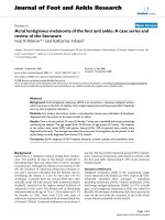
ALM (figure 1) is the only sub-type of melanoma that
occurs at the same rate in all races[11]. However, research
data have demonstrated that melanomas in acral loca-
tions account for only around 1–7% of all cutaneous
melanomas in Caucasians but has been shown to be sig-
nificantly higher in Asian[12,13], Chinese[14,15], Japa-
nese[16], Middle Eastern[17] and African
populations[18,19]. This data reflects the low incidence
of melanomas elsewhere on the body in the more pig-
mented skin types.
Aetiology
As ALM occurs equally across all races, predominantly on
an area that seldom receives much sun exposure it has
been suggested that the aetiology is different to that of
other sub-types of melanoma or that sun exposure is a
lesser risk factor than melanoma elsewhere. Green et
al[20] undertook a case control study of 275 melanomas
diagnosed on the soles and palms to investigate risk fac-
tors. Interestingly, they found that sun exposure was a sig-
nificant risk factor in the development of ALM despite
their plantar and nail bed location. Furthermore, a high
mole count on the soles and elsewhere on the body were
associated risk factors (RR = 6.3 95% CI 2.5–15.6). Rein-
forcing this belief, other studies have demonstrated that
increased sun exposure in an individual leads to the devel-
opment of higher numbers of moles, especially in chil-
dren[21].
Trauma as a cause has also been proposed as a possible
risk factor for the development of ALM[20]. Penetrative
injury of the foot showed significant association (RR = 5.0
CI 3.0–8.6) although the authors could not confirm from
the data if the ALM actually developed at the original site
of injury. In an earlier study, Briggs et al[22] reviewed a
number of cases but suggested that incidental injury to the
foot merely drew the patients attention to a pre-existing
foot problem. Kaskel et al[23] suggested that trauma in
acral areas such as the foot were to be expected more fre-
quently and could find no evidence to identify trauma as
an aetiology.
The prognosis of the disease, as with other sub-types of
melanoma, is determined by the Breslow thickness of the
lesion at diagnosis[24]. It has been suggested that ALM
itself carries a worse prognosis than other melanoma –
often as lesions are recognised later than melanoma on
other body sites[25]. Following a number of cases late
diagnosis occurring at a tertiary care centre, a study was set
up to review cases of the disease in an attempt to identify
common clinical factors.
Methods
A database search was undertaken to identify all cases of
ALM treated at the tertiary care melanoma centre located
in a central London district. From these, notes were
selected of patients presenting with a ALM (diagnosed by
histology) on the ankle or below. In the period 2000 –
2006, twenty seven patients were identified and from
their records clinical data including gender, age, ethnicity
and diagnostic information were gathered and tabulated
for review.
Acral lentiginous melanoma on the plantar surfaceFigure 1
Acral lentiginous melanoma on the plantar surface.
Journal of Foot and Ankle Research 2008, 1:11 />Page 3 of 5
(page number not for citation purposes)
Results
The cohort of patients totalled 27 (20 female and 7 male)
with a female ratio of nearly 3:1. The patients' age at diag-
nosis ranged from 35 to 96 years. The average age of the
patient at was 62.7 with no age difference between men
and women (62.5 versus 62.8 respectively). The majority
of patients reported their ethnicity as white (n = 16) in
addition there were 7 Afro-Caribbean, 1 Chinese/Oriental
and 3 unrecorded. Although not always recorded, patients
had been reviewed at a number of other clinics with their
lesions prior to reaching the dermatology department
with a definitive diagnosis. These included a range of spe-
cialities – general practice (n = 5), podiatry/chiropody (n
= 9), vascular clinics (n = 2), diabetology (n = 1) and plas-
tic surgery (n = 1).
Nineteen of the lesions were reported on the right foot
and eight on the left. All male patients exhibited ALM on
their right foot only. The majority of lesions were located
on the plantar surface (62%) with 2 on the ankle, 2 on the
dorsum of the foot, 1 on the digit and 4 located in the nail
bed (with 2 in the hallux and 2 in the fifth toe nail bed).
One lesion site was stated simply as being on the "foot"
(see table 1). Twenty-one (78%) of the lesions were
reported as melanotic, three amelanotic (11%) and three
(11%) were unknown.
Data on the time from the patient first recognising some-
thing on their foot to diagnosis was available for 19
patients. The average time for women was 12.5 months
versus 14.5 months in males. The most reported symp-
toms from patients were change in size and bleeding (see
table 2). A number of lesions were misdiagnosed as warts
(n = 4). Lesion thickness at diagnosis ranged from 0.84
mm to 13.30 mm. The mean thickness for women being
3.68 mm (n = 16) versus 4.41 mm in males (n = 6).
Discussion
This set of patients represents a small cohort (n = 27) of a
population from an urban area with a high ethnic mix.
Interestingly, despite the wide ethnic diversity of the local
area, a high proportion of this cohort were white (69%).
Despite the wide spread of ages (35 – 96), the average age
of the patient in this study was 62.6 years which concurs
with similar studies[26,27] that ALM is most frequent in
the 60–70 age group[25]. ALM appears to occur in an
older age group, other types of melanoma having a peak
incidence around 50 years of age, albeit with a wider age
spread[2]. The female preponderance to ALM was 2.8:1
slightly higher than other published data [26-28] but still
confirms that MM is a disease more common in
females[3,10].
Within this study, the prime location for ALM was the
plantar surface (65%), with 4 of these occurring under the
first metatarsal head. A smaller number were seen in nail
beds, ankle and dorsum of the foot. A similar prevalence
pattern for the plantar area has been reported by Soon et
al[27](61%) and Kuchelmeister[25] (65%) with sub-
ungual lesions making up a smaller percentage of all cases
of ALM. The four sub-ungual tumours in this study were
located exclusively on the hallux (50%) and fifth toe
(50%). The hallux has been consistently reported to be
the most common area for sub-ungual lesions in the foot.
Possible reasons for this are two-fold. Firstly, the hallux
may be the most prevalent location owing to the larger
proportion of nail tissue in this area. Secondly, one could
debate the role of trauma. The hallux is typically an area
of the forefoot more prone to abuse from footwear and
one-off injury. In one case series from Germany, 6
patients with ALM reported tight footwear as a possible
causative agent[23]. The authors went on to discuss that
patients with acral melanoma tended to report a high rate
of trauma compared to those with melanoma at other
sites but this was not found to be statistically significant.
Table 1: Summary of locations of ALM in 27 patients
Location Number
Plantar Surface 17
Plantar Forefoot 6 6
(4 located under 1st met head.)
Plantar Midfoot 5
Plantar Heel 6
Dorsum of the foot 2
Ankle 2
Nail Bed 4
(2 hallux, 2 fifth toe)
Digit (excluding nail unit) 1
Not Known 1
Total 27
Table 2: Reported symptoms/diagnoses (21 patients). Reported
symptom
Symptoms Number
Change in Size 8
Bleeding 4
Change in colour 2
Change in form 2
Pain 1
Itching 1
Previous Diagnoses
Wart 4
Fungal Infection 1
Haematoma 1
Ulcer 1
Journal of Foot and Ankle Research 2008, 1:11 />Page 4 of 5
(page number not for citation purposes)
Furthermore, one could hypothesize, if physical trauma
was associated with melanoma, one would expect the foot
show a more significant proportion of lesions on the foot
as a result of the forces of weight bearing and locomotion.
Early recognition is the key to improving survival
rates[29]. As cutaneous melanoma is a visible disease,
both the patient and practitioner play a major role in rec-
ognising suspicious lesions. Initially, the time taken to
reach a diagnosis depends on the patient's ability to recog-
nise and seek professional advice. Secondly, diagnosis
depends on the professional's capacity to recognise the
lesion. Data were available for 19 patients showing that
the time from first noticing a lesion to diagnosis ranged
from 1 – 36 months, which shows similarities to other
studies of patients with ALM[26]. Reasons for the delay
were not examined in this study but have been reviewed
by Richard et el[30]. In a series of 590 patients they exam-
ined the reasons for delay in melanoma diagnosis and dis-
covered that male gender, increasing age and a low
educational level were all risk factors for a later presenta-
tion to physicians. In a second paper[31] examining phy-
sician delays, acral locations and lack of lesion
pigmentation were factors more likely to lead to a delay in
diagnosis by a physician, particularly lesions in acral loca-
tions without pigmentation.
Within this study, symptoms or initial diagnoses were
recorded for 21 patients. The most common reported
symptom was a change in the size of the lesion (38%) fol-
lowed by bleeding (19%), change in colour (9%) and
change lesion form (becoming raised/nodular) (9%).
Bleeding is a common feature in melanoma which have
entered a vertical growth phase and have become ulcer-
ated[2] and may represent a feature of advanced disease.
The average lesion thickness in patients reporting bleed-
ing was significantly higher in those not reporting it
(mean thickness 6.13 mm v 3.8 mm) although due to the
small numbers involved it was difficult to draw firm con-
clusions.
Seven of the twenty one lesions (33%) were initially mis-
diagnosed as other conditions (warts, a fungal infection,
haematoma and an ulcer). Numerous papers have high-
lighted conditions including warts, tinea pedis, ulcera-
tion, infection, paronychia, haematoma, onychomycosis,
ischaemic necrosis, pyogenic granuloma, ganglions and
blisters which have been later discovered to be ALM
[27,28,32-36]. Misdiagnosis is a common feature of
melanoma on the foot but ALM in particular has been
shown to be more likely mis-diagnosed than other sub-
types of the disease[37]. Delays can in turn lead to a
poorer prognosis for the patient. The misdiagnosis rate in
this study was 33%, other have reported rates of between
33% – 67%[27,38].
It is appreciated that the results of this study represent a
retrospective review of patient case notes which have
some inherent bias – in particular that this data was col-
lected at a tertiary centre where possibly only more com-
plex cases are seen. However, in view of the relative rarity
of the condition, twenty-six cases represent a sizeable
cohort, which has been shown to be concurrent when
compared to literature on this topic.
This paper has highlighted an uncommon but serious
lesion which may present for the first time to Chiropo-
dists and Podiatrists. One third of the lesions, in the pre-
sented cohort, were seen prior to diagnosis by a
chiropodist or podiatrist. Unfortunately, typical features
of melanoma as exhibited by the "ABCDE" rule may not
be present in a proportion of ALM and so misdiagnosis
remains a significant risk. Therefore it is important to
remain vigilant and where there is clinical suspicion,
patients should be referred for a prompt dermatological
opinion. In suggesting ways to heighten awareness, the
typical patient profile should be borne in mind as well as
continuing the patient health education message. In addi-
tion, dermoscopy has been demonstrated as a useful, non-
invasive technique to increase sensitivity in acral
lesions[39]improving early recognition.
Conclusion
Acral lentigious melanoma is an uncommon malignant
tumour which can occur on the foot. This study provided
clinical data from 27 cases based on a mainly white, urban
population. A third of cases in this series were misdiag-
nosed before reaching the skin clinic with a proportion of
patients having been seen by a number of specialities
prior to diagnosis. Lesions were most common on the
plantar surface (62%). The average time from patients first
noticing something to diagnosis was 13.5 months. The
most common reported symptoms were a enlargement of
the lesion (38%) and bleeding (19%). Further studies are
required to better understand the aetiology and pathology
of this unusual but serious tumour.
Competing interests
The authors declare that they have no competing interests.
Authors' contributions
Please see sample text in the instructions for authors.
Acknowledgements
The authors wish to acknowledge the help of Sally King at St Thomas' in
identifying patient cases.
References
1. Lens MB, Dawes M: Global perspectives of contemporary epi-
demiological trends of cutaneous malignant melanoma. Brit-
ish Journal of Dermatology 2004, 150:179-185.
2. Bishop JN, Bataille V, Gavin A, Lens M, Marsden J, Mathews T, Wheel-
house C: The prevention, diagnosis, referral and manage-
Publish with Bio Med Central and every
scientist can read your work free of charge
"BioMed Central will be the most significant development for
disseminating the results of biomedical research in our lifetime."
Sir Paul Nurse, Cancer Research UK
Your research papers will be:
available free of charge to the entire biomedical community
peer reviewed and published immediately upon acceptance
cited in PubMed and archived on PubMed Central
yours — you keep the copyright
Submit your manuscript here:
/>BioMedcentral
Journal of Foot and Ankle Research 2008, 1:11 />Page 5 of 5
(page number not for citation purposes)
ment of melanoma of the skin: concise guidelines. Clin Med
2007, 7(3):283-290.
3. UK Skin Cancer mortality statistics [cerresear
chuk.org/cancerstats/types/skin/mortality/]
4. Australian Institute of Health and Welfare (AIHW) & Australasian
Association of Cancer (AACR): Cancer in Australia: an overview 2006
Canberra: AIHW; 2007.
5. Soong SJ, Shaw HM, Balch CM, McCarthy WH, Urist MM, Lee JY:
Predicting survival and recurrence in localized melanoma: a
multivariate approach. World J Surg 1992, 16:191-195.
6. Barnes B, Seigler H, Saxby T, Kocher M, Harrelson J: Melanoma of
the foot. J Bone Joint Surg AM 1994, 76:892-898.
7. Clark WH Jr, From L, Bernardino EA, Mihm MC: The Histogenesis
and Biologic Behavior of Primary Human Malignant Melano-
mas of the Skin. Cancer Res 1969, 29:705-727.
8. Reed R: Acral lentiginous melanoma. In New concepts in surgical
pathology of the skin Edited by: Hartmann W, Reed R. New York:
Wiley; 1976:89-90.
9. Elwood J, Gallagher R, Hill G, Spinelli J, Pearson J, Threlfall P: Pig-
mentation and skin reaction to sun as risk factors for cutane-
ous melanoma: Western Canada Melanoma Study. British
Medical Journal 1984, 288(6411):99-102.
10. Hudson DA, Krige JEJ, Stubbings H: Plantar melanoma: Results of
treatment in three population groups. Surgery 1998,
124:877-882.
11. Stalkup JR, Orengo IF, Katta R: Controversies in Acral Lentigi-
nous Melanoma. Dermatologic Surgery 2002, 28:1051-1059.
12. Chang JW, Yeh KY, Wang CH, Yang TS, Chiang HF, Wei FC, Kuo TT,
Yang CH: Malignant melanoma in Taiwan: a prognostic study
of 181 cases. Melanoma Res 2004, 14:537-541.
13. Chen YJ, Wu CY, Chen JT, Shen JL, Chen CC, Wang HC: Clinico-
pathologic analysis of malignant melanoma in Taiwan. J Am
Acad Dermatol 1999, 41:945-949.
14. Collins RJ: Melanoma in the Chinese of Hong Kong. Emphasis
on volar and subungual sites. Cancer 1984, 54:1482-1488.
15. Luk NM, Ho LC, Choi CL, Wong KH, Yu KH, Yeung WK: Clinico-
pathological features and prognostic factors of cutaneous
melanoma among Hong Kong Chinese. Clinical and Experimental
Dermatology 2004, 29:600-604.
16. Ishihara K, Saida T, Yamamoto A: Updated statistical data for
malignant melanoma in Japan. Int J Clin Oncol 2001, 6:109-116.
17. Al-Maghrabi JA, Al-Ghamdi AS, Elhakeem HA: Pattern of skin can-
cer in Southwestern Saudi Arabia. Saudi Med J 2004,
25:776-779.
18. Muchmore JH, Mizuguchi RS, Lee C: Malignant melanoma in
American black females: an unusual distribution of primary
sites. J Am Coll Surg 1996, 183:457-465.
19. Bellows CF, Belafsky P, Fortgang IS, Beech DJ: Melanoma in Afri-
can-Americans: trends in biological behavior and clinical
characteristics over two decades. J Surg Oncol 2001, 78:10-16.
20. Green A, McCredie M, MacKie R, Giles G, Young P, Morton C, Jack-
man L, Thursfield V: A case-control study of melanomas of the
soles and palms (Australia and Scotland). Cancer Causes Control
1999, 10:21-25.
21. Wiecker TS, Luther H, Buettner P, Bauer J, Garbe C: Moderate sun
exposure and nevus counts in parents are associated with
development of melanocytic nevi in childhood. Cancer 2003,
97:628-638.
22. Briggs JC: The role of trauma in the aetiology of malignant
melanoma: a review article. Br J Plast Surg 1984, 37:514-516.
23. Kaskel P, Kind P, Sander S, Peter RU, Krahn G: Trauma and
melanoma formation: a true association? British Journal of Der-
matology 2000, 143:749-753.
24. Breslow A: Prognostic Factors in the Treatment of Cutaneous
Melanoma. Journal of Cutaneous Pathology 1979, 6:208-212.
25. Kuchelmeister C, Schaumburg-Lever G, Garbe C: Acral cutaneous
melanoma in caucasians: clinical features, histopathology
and prognosis in 112 patients. 2000, 143:275-280.
26. Phan A, Touzet S, Dalle S, Ronger-Savle S, Balme B, Thomas L: Acral
lentiginous melanoma: a clinicoprognostic study of 126
cases.
British Journal of Dermatology 2006, 155:561-569.
27. Soon SL, Solomon AR Jr, Papadopoulos D, Murray DR, McAlpine B,
Washington CV: Acral lentiginous melanoma mimicking
benign disease: the Emory experience. J Am Acad Dermatol
2003, 48:183-188.
28. Fortin PT, Freiberg AA, Rees R, Sondak VK, Johnson TM: Malignant
melanoma of the foot and ankle. J Bone Joint Surg Am 1995,
77:1396-1403.
29. Roberts D, Anstey A, Barlow R, Cox N: UK guidelines on the
management of cutaneous melanoma. British Journal of Derma-
tology 2002, 146:7-17.
30. Richard MA, Grob JJ, Avril MF, Delaunay M, Gouvernet J, Wolken-
stein P, Souteyrand P, Dreno B, Bonerandi JJ, Dalac S, et al.: Delays
in diagnosis and melanoma prognosis (I): the role of patients.
Int J Cancer 2000, 89:271-279.
31. Richard MA, Grob JJ, Avril MF, Delaunay M, Gouvernet J, Wolken-
stein P, Souteyrand P, Dreno B, Bonerandi JJ, Dalac S, et al.: Delays
in diagnosis and melanoma prognosis (II): the role of doc-
tors. Int J Cancer 2000, 89:280-285.
32. Gregson CL, Allain TJ: Amelanotic malignant melanoma dis-
guised as a diabetic foot ulcer. Diabetic Medicine 2004,
21:924-927.
33. Serarslan G, Akcaly C, Atik E: Acral lentiginous melanoma mis-
diagnosed as tinea pedis: a case report. Int J Dermatol 2004,
43:37-38.
34. Rosen T: Acral lentigious melanoma misdiagnosed as verruca
plantaris: a case report. Dermatol Online J 2006, 12(4):3.
35. Dalmau J, Abellaneda C, Puig S, Zaballos P, Malvehy J: Acral
Melanoma Simulating Warts: Dermoscopic Clues to Pre-
vent Missing a Melanoma. Dermatologic Surgery 2006,
32:1072-1078.
36. Valdes A, Kulekowskis A, Curtis L: Case Report: Amelanotic
Melanoma Located on the Lower Extremity (letter).
Am Fam
Physician 2007, 76:1614.
37. Metzger S, Ellwanger U, Stroebel W, Schiebel U, Rassner G, Fierlbeck
G: Extent and consequences of physician delay in the diagno-
sis of acral melanoma. Melanoma Res 1998, 8:181-186.
38. Bennett DR, Wasson D, MacArthur JD, McMillen MA: The effect of
misdiagnosis and delay in diagnosis on clinical outcome in
melanomas of the foot. J Am Coll Surg 1994, 179:279-284.
39. Bristow I: Dermoscopy – a technique for Podiatrists to assess
pigmented lesions of the foot. World Congress in Podiatry. Den-
mark 2007.