Báo cáo y học: "Preliminary observations of muscle fibre cross sectional area of flexor digitorum brevis in cadaver feet with and without claw toes" pps
Bạn đang xem bản rút gọn của tài liệu. Xem và tải ngay bản đầy đủ của tài liệu tại đây (802.73 KB, 8 trang )
RESEARCH Open Access
Preliminary observations of muscle fibre cross
sectional area of flexor digitorum brevis in
cadaver feet with and without claw toes
Jackie Locke
*
, Stuart A Baird, Jamie Frankis
Abstract
Background: In order to facilitate normal gait, toes require to be in a rectus position during the propulsive phase.
This requires a corr ect balance and sequence of activity of the intrinsic musculature of the feet. Alteration of this
balance and sequence may lead to the development of claw toes. Atrophy of the lumbricals occurs in the
development of claw toes, but it is not known if changes occur in any other intrinsic muscles, including flexor
digitorum brevis. This study set out to investigate whether hypertrophic changes were evident in flexor digitorum
brevis in feet with claw toes.
Methods: Four cadaver feet were investigated, two with rectus toes and two with claw toes. Flexor digitorum
brevis was removed from each, and seven anatomically significant tissue sections from each muscle were routinely
processed, cut and stained. One hundred and sixty muscle fibre cross sectional areas were measured from each
section.
Results: The mean age of the donors was 81.5 years, and three of the four were female. Results showed that the
cross sectional area of fibres from feet with claw toes was 417 μg
2
significantly greater (p < 0.01) than the cross
sectional area of fibres from feet with rectus toes, which was 263 μg
2
.
Conclusions: Although this study has several limitations, preliminary observations reveal that flexor digitorum
brevis muscle fibre cross sectional area is significantly reduced in feet with claw toes. This would indicate a
relationship between muscle fibre atrophy of flexor digitorum brevis and clawing of the lesser toes.
Background
Toes have the primary functio n of increasing the total
weight bearing area of the forefoot during the stance
phase of gait, dispersing the loads under the metatarsal
heads [1]. If optimal propulsion is to occur, the toes
should be parallel with the ground at approximately 20°
of metatarsophalangeal (MTP) joint dorsiflexion in rela-
tion to the metatarsal shaft [2]. The toes must also be
stable and function in a rectus position as a rigid beam
[3]. During the propulsive phase of gait, the stable digit
becomes the point about which muscle a ctivity occurs
[3]. Correct digital f unction is a balance b etween the
intrinsic and extrinsic muscularae acting on the digits
during the gait cycle . If the rectus position is to be
maintained in order to facilitate effective propulsion, a
sequence of muscle contractions must occur. When
smooth c o-ordination of the muscle groups is compro-
mised, the dominant group gains a mechanical advan-
tage, altering the digital position during propulsion [3,4].
This, coupled with Davis ’s Law, leads to contractures
and digital deformities [3].
Claw toes are part of a group of lesser toe deformities,
which also include mallet and hammer toes. They are
best defined as a sagittal plane deformity, where there is
dorsiflexion at the MTP joint, and flexion of the inter-
mediate and distal interphalangeal (IP) joints [1,5,6].
There are numerous aetiological factors associated with
the development of claw toes including muscular spasm
resulting from an upper motor neurone lesion, rheuma-
toid arthritis, biomechanical abnormalities, inappropriate
footwear, peripheral neuropathy, neuromuscular dis-
orders and myoneural ischemia from compartment
* Correspondence:
Department of Podiatric Medicine and Surgery, Glasgow Caledonian
University, Cowcaddens Road, Glasgow, Scotland, G4 OBA, United Kingdom
Locke et al. Journal of Foot and Ankle Research 2010, 3:32
/>JOURNAL OF FOOT
AND ANKLE RESEARCH
© 2010 Locke et al; licensee BioMed Central Ltd. This is an Open Access article distributed under the terms of the Creative Commons
Attribution License ( which permits unrestricted use, distri bution, and reproduction in
any medium, provided the original work is properly cited.
syndromes of the foot and calf [1,7]. Whilst a wide spec-
trum of aetiological factors is evident, most authors are
agreed that they all result in pathomechanical changes
that cause muscular imbalance, bringing about altera-
tions in digital position.
It is accepted that clawing of the toes is associated
with atrophy of the lumbrical muscles, [3,4,8] however
no quantifiable data has been produced as evidence to
support this. Root et al. [4] suggest that the lumbricals
are stance phase muscles which act in conjunction with
flexor digitorum longus (FDL). Lumbrical function is to
extend the intermediate and distal IP joints of the lesser
toes [4] and assist in stabilizing the proximal phalanx of
the lesser toes by plantarflexing against ground reaction
force [4]. This action maintains the digits in a rectus
position during gait, but atrophy would permit overpull
of the flexors to extend the MTP joints against ground
reaction force, and flex the intermediate and distal IP
joints to create a claw toe deformity. Greene and Brekke
[3] dispute this stance phase theory, stating that lumbri-
cals are primarily swing phase muscles, which are acti-
vated to prod uce a flexion force at the MTP joints prior
to the activation of extensor digitorum longus and bre-
vis, thus creating digital stabilitypriortothetoescom-
mencing ground contact.
Atrophy or paralysis of the lumbricals [9] disrupts
both the sequence and ba lance of muscle activity during
gait, resulting in a weakened or unopposed action of the
extensors during swing. As a consequence of the
reduced flexion action of the lumbricals at the MTP
joints, there is extension of the MTP joints during the
swing phase, resulting in the digits being in an unstable
position prior to ground contact. Coupled with the
actions of FDL and flexor digitorum brevis (FDB)
through the propulsive phase, flexion of the proximal
and distal IP joints occurs, with all joints in the claw toe
position [3,8,9]. Whether stance or swing phase muscles,
or both, lumbrical atrophy or paralysis would result in
clawing of the lesser toes. However, the role of other
muscles requires inve stigation to gain a greater under-
standing of this deformity. If, as Greene and Brekke [3]
suggest, there is a mechanical overpull of FDB and FDL,
they may display evidence of hypertrophic changes.
FDB is a fusiform muscle originating from the medial
calcaneal process and the plantar aponeurosis. At its dis-
tal aspect, it divides into four, giving rise to four ten-
dons, each of which insert into the lateral four toes on
the plantar surface of the intermediate phalanx [10].
Because of the site of the tendonous insertions, any
impl ication of FDB in the development of claw toes can
only relate to changes at the MTP joints and proximal
IP joints, but not the distal IP joints. The open chain
action of FDB is to flex the MTP joints and proximal
IPJ’s, [10], but its function during gait is somewhat less
clear. A mongst the various functions described by Root
et al. [4], two relate specifically to digital stability. FDB
(i) assists flexor digitorum longus to maintain stability
of the lesser digits against the ground in propulsion, and
(ii) stabilises the intermediate phalanx of each toe pos-
terially against the proximal phalanx, and the proximal
phalanx against its respective metatarsal head. These
functions would ma intain the digits in a rectus p osition.
Root et al. [4] also describe FDB as a stance phase mus-
cle, a theory concurrent with the findings of Greene and
Brekke [3], Hughes [2] an d Perry [11]. The timing of its
activity during the stance phase of gait is disputed, with
some authors quoting as little as 33%, and others up to
75% [2,4,11]. However, all are agreed that FDB is active
from heel lift through toe off, to maintain the digits in a
rectus position, thus obtaining the necessary stability
during the propulsive phase o f gait. A ccording to
Greene and Brekke [3], FDB also works in conjunction
with FDL during normal gait to ensure the toe remains
flat against the ground during stance, to create the rigid
beam required for effective propulsion. An excessive
unbalanced action, or a pull which is unopposed by cor-
rect lumbrical function would lead to extension of the
MTP joints against ground reaction force, and flexion of
the proximal IP joints. It is reasonable to conclude that
FDB should be investigated in relation to the develop-
ment of claw toes.
The ability to produce force and movement is the most
basicpropertyofskeletalmuscle[12,13].Itdoessoby
contracting either isometrically, where tension is pro-
duced against force without the muscle shortening, or
isotonically where the muscle contracts t o produce
movement [12,13]. There are three main influences on
muscle contraction, namely fibre type, length and dia-
meter [14,15]. To investigate fibre type and length was
outwith the scope of this study. However the importance
of fibre length was recognised as individual muscle fibres
do not extend the full length of the muscle itself, but are
arranged in overlapping bundles [16]. Investigations by
Loeb et al. [17], and Ounjian et al. [18] revealed that
many muscle fibres originate and terminate within the
muscle belly, attaching to the connective tissue matrix,
which itself has an important role in fibre to tendon ten-
sion. Given this, several sites of anatomical significance
were selected for measuring. Muscle fibre diameter was
not measured directly, as f ibres are not perfectly round.
However, cross sec tional fibre area, which is directly
related to fibre diameter, was measured by computer
based image analysis. This method has been shown to
have both intra and inter tester validity and reliability
[19]. Any alterations in mechanical usage or innervation
to the muscle will result in either atrophic or hyper-
trophic changes to the muscle, which is manifest by a
decrease or increase respectively in the size of the
Locke et al. Journal of Foot and Ankle Research 2010, 3:32
/>Page 2 of 8
individu al muscle fibre [20]. The aim of the study was to
test the hypothesis that the muscle fibre cross sectional
area of FDB was greater in feet with claw toes than in
feet with rectus toes.
Methods
For the purpose of this study, fifteen feet were made
available by Glasgow University Department of Human
Anatomy. They were then sub divided into two groups,
feet with re ctus toes, and feet with claw toes. Two feet
from each sub group were then randomly selected. The
inclusion criterion for rectus toes was clinical observa-
tion of all lesser toes in a rectus position. The inclusion
criterion for claw toes was clinical observation of all les-
ser toes in the claw position as defined by Merriman
and Tollafield [6]. The exclusion criteria for rectus toes
was any single toe with another lesser toe deformity
such as hammer o r mallet toe, or an y indication of
trauma or surgical pro cedure to the f oot. The exclusi on
criteria for claw toes were any lesser toe in a rectus
position, or any indication o f trauma or surgical proce-
dure to the foot.
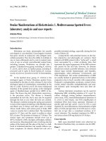
The flexor digitorum brevis of each foot was removed
by minute dissection as described by Romanes [10]. A
3 mm transverse section was then isolated from seven
sites within the muscle (Figure 1).
Each section was routinely decalcified for 7 days in
Ethylenediaminetracetic acid (EDTA), processed via the
Histokinette 2000 (Reichert-Jung, Germany) and vacuum
embedded in wax at 58°C, cooled for a minimum of
2 hours to solidify, prior to being cut to 7 μm. They
were then routinely mounted and stained using Haema-
toxylin and Eosin (H&E). Visual image analysis was con-
ducted using an Olympus BH2 RFCA (Olympus,
London) photo microscope, in conjunction with Soft
Image Analysis
®
and Viewfinder Lite
®
Version 1.0 (Pix-
era Corporation 1998 - 2000). Using this software, tissue
samples from each section were divided into 4 quad-
rants. Within each quadrant a blood vessel was located,
and the cross sectional area s of the surrounding 40
muscle fibres measured at × 40,000 magnification. This
data was recorded directly to Microsoft
®
Excel 1997
(Microsoft
®
Corporation, USA) and copied to SSPS
12.0
©
2002 SPSS Inc.for statistical analysis.
Results
FDB was dissected from four cadaver feet. Foot 2 had
no tendon attached to the 5
th
toe. Table 1 gives details
of age, gender, mean cross sectional area, standard
deviation and range for each foot. Figure 2 illustrates
that the mean cross sectional area is greater in feet with
claw toes than feet with rectus toes.
Independent sample t-tests showed;
(i) Mean cross sectional area associated with clawed
toes (417 μ
2
) was significantly greater than the cross
sectional area associated with rectus toes (263 μ
2
)(p=
< 0.01), with a difference of the means = 154 μ
2
.
(ii) A significant difference in the mean cross sectional
area (260 μ
2
and 240 μ
2
) of the two feet with rectus toes
(p < 0.01) with a difference of the means = 20 μ
2
.
(iii) A significant difference in t he mean cross sec-
tional area (342 μ
2
and 493 μ
2
) of the two feet with claw
toes (p < 0.01) with a difference of the means 151 μ
2
.
The two rectus feet displayed a similar pattern of mus-
cle fibre cross sectional area a t each anatomical site
examined. Foot 4 (claw) had a greater cross sectional
area than both rectus feet at each anatomical site,
whereas Foot 2 (claw) was only greater than both rectus
feet at 2 sites. This made a conclusion problematic and
can be viewed diagrammatically in Figure 3. In the light
of this statistical analysis, and due to alterations in ageing
muscle fibres, the cross sectional area from the 83 year
old female rectus foot was compared with the 84 year old
female claw foot at each anatomical site. A two way
ANOVA which investigated person and anatomical site
revealed that the cross sectional area of the claw foot was
significantly greater (p = < 0.01) , than the cross sectional
area of the rectus foot at all corresponding anatomical
sites (Table 2).
Discussion
The findings of this study appear to confirm the hypoth-
esis that mean cross sectional area is greater in FDB
associated with claw toes than rectus toes. This however
is a simplistic analysis, and several issues should be con-
sidered to ensure that this finding is viewed within the
appropriate context. Further investigati on revealed there
was a significant difference in the mean cross sectional
area of the two rectus feet and the two claw feet. The
difference between the means of the two claw feet was
151 μ
2
, whilst the difference of the means between claw
and rectus feet was only 3 μ
2
greater at 154 μ
2
.When
the mean cross sectional area for each of the anatomic al
sites of the four feet were contrasted, as displayed in
Figure 3 it was noted that not all anatomical sites of the
claw feet were greater than the same anatomical site in
the rectus feet.
The two rectus feet followed a similar pattern of cross
sectional area at each anatomical site within th e muscle,
whereas the two claw feet did not. Foot 4 was greater at
each anatomical site than both rectus feet, but apart
from the sites of Belly and Proximal to Division of Foot
2, the measurements from this claw foot were similar in
size and pattern to the two rectus feet. It is therefore
not entirely accurate to conclude that the cross sectional
area is greater in claw feet than rectus feet.
Locke et al. Journal of Foot and Ankle Research 2010, 3:32
/>Page 3 of 8
One of the major influences on these results, is the
age of the sample. The youngest was 71 years, with the
other t hree aged 83, 84 and 88 years. Inokuci [21] con-
cluded that muscle tissue is marked by an increase in
fat and connective tissue in the elderly, especially those
in advance of 80 years. During dissection, it was noted
in the 88 year old that visually, there appeared to be a
greater amount of fat surrounding and impregnating the
muscle. Additionally, under microscopic conditions, an
increase in the endomysial spaces was v isuall y apparent.
Hooper [22] recorded that one characteristic of a geing
muscle is an increased variation in fibre size due to
atrophy and compensatory hypertrophy. Table 1 displays
the maximum and minimum measurements, and indi-
cates an exceptionally large range of fibre sizes, giving
credence to this theory. There appear to be two theories
as to why there is fibre atrophy with compensatory
hypertrophic changes. Firstly, as age increases above
60 years, neurogenic alterations occur, resulting in
cycles of denervation and reinnervation of motor neu-
rons. With each cycle, some fibres a re permanently
denervated, resulting in atrophy and are eventually
tendons to 2nd and 3rd toes
muscle belly
tendons to 4th and 5th toe
s
proximal to divisions
origin
Figure 1 Diagramatical illustration of anatomical sites of muscle tissue sections taken from flexor digitorum brevis.
Table 1 Characteristics of feet included in the sample
Foot Foot 1 Foot 2 Foot 3 Foot 4
Type Rectus Claw Rectus Claw
Age 71 88 83 84
Gender Male Female Female Female
Mean in μ
2
259.74 341.70 239.89 430.44
(Standard Deviation) (123.08) (199.97) (106.20) (222.64)
Range in μ
2
63.86 - 1212.10 80.41- 1525.87 61.75- 912.79 89.87 1403.41
Locke et al. Journal of Foot and Ankle Research 2010, 3:32
/>Page 4 of 8
replaced by connective tissue [23]. Secondly, a reduction
in the effectiveness of the peripheral arterial supply due
to arteriosclerosis is a physiological process that occurs
with advancing age [24]. This reduces the healing prop-
erties in any muscle fibres which are injured, even dur-
ing moderate physical activity, resulting in eventual
muscle loss. As a consequence of e ither occurrence, a
reduced number of fibres would be required to maintain
the same activity, resulting in hypertrophic changes.
Grimby and Saltin [25] suggest that neurogenic changes
affecting muscle fibres, are directly related to the length
of the peripheral nerve, thereby implying there is a likely
263.1
417.1
0
50
100
150
200
250
300
350
400
45
0
r
ec
t
us c
l
a
w
mean cross-sectional area μ
2
Figure 2 Comparison of mean cross sectional areas in feet with rectus toes and feet with claw toes.
0
100
200
300
400
500
600
700
800
origin belly proximal to
division
proximal to
tendon
2n
d
toe
proximal to
tendon 3rd
toe
proximal to
tendon 4th
toe
proximal to
tendon 5th
toe
mean cross-sectional area μ2
foot 1 rectu
s
foot 2 claw
foot 3 rectu
s
foot 4 claw
Figure 3 Comparison of muscle fibre cross sectional areas within each foot.
Locke et al. Journal of Foot and Ankle Research 2010, 3:32
/>Page 5 of 8
risk of these changes affecting muscles of the foot.
Atrophic changes will also arise from muscle disuse
[13], due to reduction in ambulation as age increases. In
the light of age related changes to muscle t issue, the
83 year old rectus and 84 year old claw (both female)
feet were compared. A two-way ANOVA c onfirmed a
significant difference between claw and rectus toes, with
the cross sectional area of all corresponding anatomical
sites greater in the foot with claw toes (Table 2).
Itwouldalsobeprudenttoobservefiveotherlimita-
tions associated with this study. Firstly, the study was
carried out on muscle tissue from cadavers treated with
a Formaldehyde based embalming fluid as per Glasgow
University embalming protocols. This may result in t he
tissues becoming hard, rigid and often difficult to dissect
[26]. MacBride [27] also suggested that studies using
embalmed cadavers were often avoided because the fixa-
tion process was poor, making tissues less suitable for
histological examination. In examining the effects of var-
ious fixatives on bovine muscle, Stickland [28] observed
that Formaldehyde based solutions were amongst those
most likely to cause shrinkage to the muscle fibre. How-
ever, it should also be considered that the effects of
shrinkage on skeletal muscle of cadaveric fixation was
marked when muscle tissue was fixed in isolation from
the skeleton, but not when fixed in situ on the skeleton
[29]. Apart fro m the e ffects of the embalming solution,
the subsequent stages of dehydrating and clearing have
also been implicated in muscle fibre shrinkage. It h as
been concluded by Stickland [28] that these processes
cause shrinkage to a greater degree than fixation. Histo-
logical processin g can al so cause fragmentation of tissue
[30], and this was evident in some of the tissues under
microscopic exam ination, occasionally making measure-
ment difficult. In addition to the effects of fixatio n,
dehydrating and clearing, muscle fibres taken from cada-
vers differ ar chitecturally [31] from in vivo muscle.
Living muscle f ibres are either in an extreme of relaxa-
tion or contraction, whereas cadaveric muscle is in a
state between relaxation and contraction [31]. This is
thought to be a result of fixation occurring whilst the
muscle fibres are in the partially contracted state seen in
rigor mortis. Due to the alterations of fixation, histologi-
cal processing and rigor mortis, results of cadaveric stu-
dies cannot reflect exactly what would be found in a
living specimen. One final problem in conducting this
cadaveric study was that no medical history was avai l-
able that may have indicated an aetiological influence
on fibre atrophy, hypertrophy or development of claw
toes.
The second major limitation isthatthisinvestigation
has only looked at the muscle fibre cross sectional area,
but has not accounted for any differences associated
with the other two components of muscle contraction,
namely, fibre length and fibre type. Although fibre
length was not investigated, observation of Figure 3
demonstrates that the mean cross sectional area varies
at each of the various sections along the muscle. It is
not known whether the fibre lengths or their origins
and insertions to connective tissue varies between claw
and rectus toes. Length of fibres from FDB would be
observations in the sagittal plane, the same plane on
which claw toe deformity occurs. Man y biomechanical
abnormalities associated with the development of claw
toes are also associated with elongation of the medial
longitudinal arch of t he foot [32]. In such conditions,
Table 2 Results of two-way analysis of variance comparing muscle cross sectional area at different sites in feet with
and without claw toes
95% Confidence Interval
Anatomical site Foot number (type) Mean Std. error Lower bound Upper bound ANOVA p-value
Origin 3 (rectus) 276.57 10.99 255.02 298.12 <0.01
4 (claw) 424.30 10.99 402.75 445.86 <0.01
Belly 3 (rectus) 201.62 10.99 180.06 223.17 <0.01
4 (claw) 285.65 10.99 264.10 307.21 <0.01
Prox div 3 (rectus) 234.86 10.99 213.31 256.42 <0.01
4 (claw) 478.52 10.99 456.70 500.07 <0.01
Tendon 2
nd
3 (rectus) 325.06 10.99 303.51 346.61 <0.01
4 (claw) 676.90 10.99 655.35 698.47 <0.01
Tendon 3
rd
3 (rectus) 228.91 10.99 207.36 250.46 <0.01
4 (claw) 679.68 10.99 658.13 701.24 <0.01
Tendon 4
th
3 (rectus) 256.76 10.99 235.20 278.31 <0.01
4 (claw) 410.21 10.99 388.66 431.77 <0.01
Tendon 5th 3 (rectus) 155.43 10.99 133.87 176.98 <0.01
4 (claw) 357.70 10.99 336.15 379.25 <0.01
Locke et al. Journal of Foot and Ankle Research 2010, 3:32
/>Page 6 of 8
chronic stretching of muscle fibres could occur in FDB.
As a result the f ibre may i ncrease in length, causing the
muscle velocity, excursion and a bility to generate force
to increase [14]. This wou ld be consistent with the the-
ory that there is excessive pull of FDB associated with
claw toes.
There was no possible means of making any observa-
tions relating to fibre type during this study, as H&E
does not reveal any difference. Serrano et al. [33] illu-
strated not only the potential of muscle fibre to change
fibre type, but the existence of “hybrid fibres” which can
quickly undergo transitions from one fibre t ype to
another, in response to changes of muscle activity. Fibre
types have been seen to change in response to force,
durati on and velocity of muscle activity, all of which are
relevant within the context of gait. It would be of
extreme interest to note if there is a difference in pro-
portion of fibre type between claw and rectus toes, and
if the hypertrophic changes affect one fibre type more
than another. Any findings from such a study would
give great indication as to whether FDB is involved in
active gait or postural stability.
The third major limitat ion is that this was a morpho-
logical study, which has investigated a functional pathol-
ogy. In order to fully understand muscle function,
especially within the foot, muscle activity requires to be
studied during gait. The options for obtaining quantita-
tive data from muscle activity f rom the foot are limited
due to both t he restrictions of the instrumentation and
the anatomical positioning of foot muscles. EMG studies
have been used, but possible limitations have already
been documented. Magnetic resonance imaging has
been extremely successful in the study of cadaveric mus-
cle, but less successful when used for in vivo muscle
activity [34]. Ultrasonographic studies have been docu-
mented as the method to provide a better understanding
of the dynamic nature of skeletal muscle, and could be
used to elucidate the biomechanics of muscle contrac-
tion [31].
Fourthly, it should be recognised that this study h as
only made observations regarding FDB in isolation from
other muscles that may be implicated in the develop-
ment of claw toes. In order to gain a true understanding
of atrophic and hypertrophic changes, a comprehensive
study of all muscles attaching to the lesser toes is
required. This would facilit ate a comparison not only of
individual muscles and their differences in claw and rec-
tus toes, but how muscles of the same foot compare
between the two conditions.
A fifth point to note is that the sample used was
small, with only four feet being analysed. Although a
larger sample would give a clearer picture, conclusions
have been drawn from previous cadaver studies using a
similar sample size [31,34].
In order to gain more data, further studies in this field
are required and should endeavour to select a larger
sample with similar specimen ages to obtain a more
meaningful comparison. It is also necessary to investi-
gate all m uscles which are potentially involved in the
development of claw toes. To advance the understand-
ing of muscle differences between claw and rectus toe s
at a morphological level, the investigation of fibre type
would be advantageous. A high proportion of Type IIA
and IIB fibres would indicate an active functional role in
gait, as they produce high power output, but a high pro-
portion of Type I fibres would indicate a link to postural
stability. It is possible there may be a difference in pro-
portion of fibre types between muscles associated with
rectus toes, and those with claw toes. The study of fibre
type using sigma antibodies to fast myosin is required.
Conclusion
This research was undertaken to investigate whether
there were h ypertrophic changes associated with flexor
digitorum brevis in the development of claw toes. The
study was limited by variables such as small sample size,
alterations to tissue associated with embalming and hi s-
tological processing, only being able to investigate one
influence of muscle contraction a nd lack of cadaver
medical history. However, despite these limitations,
quantifiable data has been produced i n an area of anat-
omy that currently has received little or no investigation
Authors’ contributions
JL participated in the study design, carried out the dissection, histology, data
collection, assisted with statistics and drafted the manuscript. SB conceived
the study, and participated in its design and coordination and helped to
draft the manuscript. JF directed and supervised the statistical analysis and
interpretation of the study results, and commented on the drafts of the
manuscript. All authors read and approved the final draft.
Authors’ Information
JL is a Lecturer in Department of Podiatric Medicine and Surgery, SB Head
of Department of Podiatric Medicine and Surgery, and JF a Senior Lecturer
in Research Methods within the School of Health. JL is also an Associate
Teacher of Podiatry, Southern General Hospital, Glasgow, Scotland, United
Kingdom, G51 4TF.
Competing interests
The authors declare that they have no competing interests.
Received: 21 December 2009 Accepted: 22 December 2010
Published: 22 December 2010
References
1. Cooper PS: From Disorders and deformities of the lesser toes. In Foot and
Ankle Disorders. Volume One. Edited by: Myerson MS. Philidelphia: WB
Saunders Company; 2000:308-313.
2. Hughes J, Clark P, Klenerman L: The importance of the toes in walking.
J Bone Joint Surg 2002, 72:245-51.
3. Green DR, Brekke M: Anatomy, Biomechanics and Pathomechanics of
Lesser Digital Deformities. Clin Podiatr Med Surg 1996, 13:179-197.
4. Root ML, Orien WP, Weed JH: Normal and abnormal function of the foot.
In Clinical Biomechanics. Volume 2. Los Angeles, Clinical Biomechanics
Corporation; 1977.
Locke et al. Journal of Foot and Ankle Research 2010, 3:32
/>Page 7 of 8
5. Coughlin MJ: Mallet toes, hammer toes, claw toes and corns. Causes and
treatment of lesser-toe deformities. Postgrad Med 1984, 75:191-198.
6. Merriman LM, Tollafield DR: From Assessment of the locomotor system. In
Assessment of the Lower Limb. Edited by: Merriman LM, Tollafield DR.
Edinburgh: Churchill Livingstone; 1995:156-186.
7. Scheck M: Acquired Hammertoe Deformity. Clin Orthop Reat Res 1977,
123:63-69.
8. Price AE, Maisel R, Drennan MD: Computed Tomographic Analysis of Pes
Cavus. J Pediatr Orthop B 1993, 13:646-653.
9. Schnepp KH: Hammer Toe and Claw Foot. Am J Anat 1993, 36:351-359.
10. Romanes GJ: Cunningham’s Manual of Practical Anatomy. 15 edition. Oxford:
Oxford University Press; 1999.
11. Perry J: Gait Analysis, Normal and Pathological Function New Jersey: SLACK
Incorporated; 1992.
12. Herzog W: Biomechanics of the Musculo-skeletal System Chichester: John
Wiley & Sons; 1994.
13. Lieber RL: Skeletal Muscle, Structure, Function and Plasticity: The Physiological
Basis of Rehabilitation. 2 edition. Philadelphia: Lipcott Williams and Wilkins;
2002.
14. Goldspink G: Handbook of Physiology Bethesda: American Physiological
Society; 1983.
15. Stevens A, Lowe J: Human Histology. 2 edition. London: Mosby; 1997.
16. Loeb GE, Pratt CA, Chanaud CM, Richmond FJR: Distribution and
Innervation of short interdigitated muscles in parallel fibred muscles of
cat hind limb. J Morphol 1987, 191:1-15.
17. Ounjian M, Roy RR, Eldred E, Garfinkel A, Payne JR, Armstrong A, Toga AW,
Edgerton V: Physiological Development implications of Motor Unit
Anatomy. J Neurobiol 1996, 22:547-559.
18. Holmback AM, Askaner K, Holtas S, Downham D, Lexell J: Assessment of
Contractile and Noncontractile Components in Human Skeletal Muscle
by Magnetic Resonance Imaging. Muscle Nerve 2002, 25:251-258.
19. Keynes RD, Aidley DJ: Nerve and Muscle. 2 edition. Cambridge: Cambridge
University Press; 1991.
20. Inokuchi S, Ishikawa H, Iwamoto S, Kimura T: Age-Related Changes in the
Histological Composition of the Rectus Abdominis Muscle of the Adult
Human. Hum Biol 1975, 47:231-249.
21. Hooper ABC: Length Diameter and Number of Ageing Skeletal Muscle
Fibres. Gerontology 1981, 27:121-126.
22. Lexell MD, Henriksson-Larsen MD, Winblad B, Sjostrom M:
Distribution of
Different Fiber Types in Human Skeletal Muscles: Effects of Ageing in
Whole Muscle Cross Sections. Muscle Nerve 1983, 6:588-595.
23. Kumar P, Clark M: Clinical Medicine. 5 edition. Edinburgh: W.B. Saunders;
2002.
24. Grimby G, Saltin B: The Ageing Muscle. Clinical Physiology 1983, 3:209-218.
25. Krishnamurthy S, Powers SK: The use of Fabric Softener in Neurosurgical
Prosections. Neurosurgery 1995, 36:420-424.
26. MacBride RG: Potential Use of Embalmed Cadavers to Study Mast Cell
Presence. The Anat Rec 1998, 250:117-120.
27. Stickland NC: A Detailed Analysis of the Effects of Various Fixatives on
Animal Tissue with Particular Reference to Muscle Tissue. Stain Technol
1975, 50:255-264.
28. Cutts A: Shrinkage of muscle fibres during the fixation of cadaveric
tissue. J Anat 1988, 160:75-78.
29. Bancroft JD, Stevens A: Theory and Practice of Histological Techniques. 2
edition. Edinburgh: Churchill Livingstone; 1982.
30. Martin DC, Medri MK, Chow RS, Oxorn V, Leekam RN, Agur AM, McKee H:
Comparing human skeletal muscle architectural parameters of cadavers
with in vivo ultrasonographic measurements. J Anat 2001, 199:429-434.
31. Thomson CE, Campbell RH, Wood AR, Rendall CC: FromAdult Foot
Disorders. In Neales Disorders of the foot Diagnosis and Management. 6
edition. Edited by: Lorimer D. Edinburgh: Churchill Livingstone; 2002:142.
32. Serrano AL, Perez M, Lucia A, Chicharro JL, Quiroz-Rothe E, Rivero JJL:
Immunolabelling, histochemistry and in situ hybridisation in human
skeletal muscle fibres to detect myosin heavy chain expression at the
protein and mRNA level. J Anat 2001, 199:329-337.
33. Narici M: Human skeletal architecture studies in vivo by non-invasive
imaging techniques; functional significance and applications.
J Electromyogr Kinesiol 1999, 9:97-103.
34. Frowen P, Benjamin M: Variations in the quantity of uncalcified
fibrocartilage at the insertions of the extrinsic calf muscles in the foot.
J Anat 1995, 186:417-421.
doi:10.1186/1757-1146-3-32
Cite this article as: Locke et al.: Preliminary observations of muscle fibre
cross sectional area of flexor digitorum brevis in cadaver feet with and
without claw toes. Journal of Foot and Ankle Research 2010 3:32.
Submit your next manuscript to BioMed Central
and take full advantage of:
• Convenient online submission
• Thorough peer review
• No space constraints or color figure charges
• Immediate publication on acceptance
• Inclusion in PubMed, CAS, Scopus and Google Scholar
• Research which is freely available for redistribution
Submit your manuscript at
www.biomedcentral.com/submit
Locke et al. Journal of Foot and Ankle Research 2010, 3:32
/>Page 8 of 8