báo cáo khoa học: "Aggressive juvenile fibromatosis of the paranasal sinuses: case report and brief review" pptx
Bạn đang xem bản rút gọn của tài liệu. Xem và tải ngay bản đầy đủ của tài liệu tại đây (434.36 KB, 6 trang )
BioMed Central
Page 1 of 6
(page number not for citation purposes)
Journal of Hematology & Oncology
Open Access
Case report
Aggressive juvenile fibromatosis of the paranasal sinuses: case
report and brief review
Shaheen E Lakhan*
1
, Robert M Eager
2
and Lindsey Harle
2
Address:
1
Executive Director, Global Neuroscience Initiative Foundation, Los Angeles, CA, USA and
2
Research Consultant, Department of
Biomedical Sciences, Global Neuroscience Initiative Foundation, Los Angeles, CA, USA
Email: Shaheen E Lakhan* - ; Robert M Eager - ; Lindsey Harle -
* Corresponding author
Abstract
Desmoid fibromatoses are benign, slow growing fibroblastic neoplasms, arising from
musculoaponeurotic stromal elements. Desmoids are characterized by local invasion, with a high
rate of local recurrence and a tendency to destroy adjacent structures and organs. Desmoid
fibromatoses are rare in children, and though they may occur in the head and neck region, are
extremely rare in the paranasal sinuses. Here we report a case of extraabdominal desmoid
fibromatosis in a seven-year-old boy involving the sphenoid sinus, one of only six published reports
of desmoid fibromatosis of the paranasal sinuses. The expansile soft tissue mass eroded the walls
of the sphenoid sinus as well as the posterior ethmoid air cells extending cephalad through the base
of the skull. We discuss the clinicopathologic features of this lesion, including structural and
ultrastructural characteristics, and we review the literature regarding treatment and outcome.
Background
Desmoid tumors arise from musculoaponeurotic stromal
elements and are locally invasive, deep-seated fibrotic
tumors. They are destructive of surrounding tissue, with a
high rate of recurrence, but are not known to have the
capacity to metastasize. Desmoid tumors have two gen-
eral classifications, intraabdominal and extraabdominal.
This distinction is significant in determining proper clini-
cal management. Extraabdominal tumors are predomi-
nantly sporadic, and often can be effectively treated with
local resection; systemic treatment is generally reserved
for refractory tumors. Conversely, intraabdominal
desmoid fibromatosis, for example those seen with famil-
ial adenomatous polyposis and Gardner syndrome, are
often diffusely infiltrative and surgically unresectable; sys-
temic therapy is considered first-line treatment of intraab-
dominal desmoids. Extraabdominal tumors in the
paranasal sinus are extremely rare; to the best of our
knowledge only six cases have been reported in the litera-
ture [1-6] (Table 1).
Case presentation
An otherwise healthy seven-year-old male presented with
a six month history of chronic sinus congestion and hali-
tosis. He was initially treated for atopy and bacterial
sinusitis with no resolution of symptoms. Suspicion was
raised of a foreign body in the nose and ENT consultation
was ordered. Prior to endoscopic removal of the foreign
body, computed tomography (CT) of the head was per-
formed.
CT revealed a large expansile mass, 4 cm in greatest
dimension, expanding and eroding the walls of the sphe-
noid sinus and the posterior ethmoid air cells. Because the
mass extended cephalad into the base of the skull, mag-
netic resonance imaging (MRI) was performed. MRI
Published: 28 May 2008
Journal of Hematology & Oncology 2008, 1:3 doi:10.1186/1756-8722-1-3
Received: 23 April 2008
Accepted: 28 May 2008
This article is available from: />© 2008 Lakhan et al; licensee BioMed Central Ltd.
This is an Open Access article distributed under the terms of the Creative Commons Attribution License ( />),
which permits unrestricted use, distribution, and reproduction in any medium, provided the original work is properly cited.
Journal of Hematology & Oncology 2008, 1:3 />Page 2 of 6
(page number not for citation purposes)
found no evidence of meningeal involvement or brain
parenchymal invasion and the major intracranial arteries
appeared intact. The right optic nerve was displaced but
without evidence of impingement.
The patient underwent functional endoscopic sinus sur-
gery with biopsy of the lesion. Histological analysis
revealed a cellular myofibroblastic neoplasm suggestive of
extraabdominal desmoid fibromatosis (Figures 1, 2). Sur-
gical resection was performed and histological analysis
confirmed the diagnosis. Surgical margins were positive.
Because of the rarity of this tumor, particularly in the para-
nasal sinuses of a child, immunohistochemical examina-
tion was performed. The tumor showed focal positivity for
SMA and multifocal nuclear positivity for beta catenin;
desmin, S-100, and CD34 were negative.
Discussion
Desmoid tumors are rare, accounting for approximately
0.03% of all neoplasms, and less than 3% of all soft tissue
tumors. The estimated incidence in the general popula-
tion is 2-4/1,000,000/year, which in the US translates to
approximately 900 new tumors annually [7]. Individuals
between the ages of 15 and 60 are most often affected;
desmoid tumors are rare in the young and in the elderly.
They are slightly more common in women than in men
[8,9], and there is no significant racial or ethnic distribu-
tion. Desmoids tend to be large bulky tumors that locally
infiltrate adjacent tissue structures. Histologically, they are
characterized by small bundles of spindle cells in an abun-
dant fibrous stroma. The fibroblasts have a propensity to
concentrate at the periphery of the lesion, and the cellular-
ity is low. There are usually few mitotic figures and necro-
sis is absent. The etiology of desmoid tumors is unknown.
However, the identification of clonal chromosomal
changes in a significant fraction of cases supports the neo-
plastic nature of these tumors [10], and emerging evi-
dence implicates dysregulated wound healing in the
pathogenesis of these and other fibroblastic lesions. Tri-
somy 8 and 20 as nonrandom clonal chromosomal
changes, particularly trisomy 8, occur in at least 30% of
sporadic desmoid tumors [11-14]. Although the clinical
relevance of these genetic abnormalities is unclear, these
genetic insults appear to be associated with a higher risk
of recurrence [12].
Treatment
Because of their locally infiltrative nature, desmoid
tumors are traditionally treated by local resection with
wide surgical margins when significant morbidity can be
avoided [15,16]. Considering the potential toxicity and
morbidity associated with local and systemic therapy in
children, complete surgical excision is the treatment of
choice for aggressive juvenile fibromatosis. Because these
are benign tumors with a high rate of recurrence, surgeons
must balance the need to obtain tumor-free margins while
at the same time using function-preserving approaches to
minimize major functional and cosmetic sequelae. The
available data are conflicting with regard to the impor-
tance of complete resection. Buitendijk et al. [17] reported
that, of 187 published cases of juvenile fibromatosis, the
single greatest determinant of tumor recurrence was
incomplete resection. In another evaluation of 63 pediat-
ric patients, the only factor associated with an increased
rate of recurrence-free survival was negative surgical mar-
gins (70% versus 15% with positive margins) [18]. In con-
trast, several authors report that the risk of recurrence is
independent of margin status [19-25]. In one of the larg-
est series of 203 patients undergoing surgery for either pri-
mary or recurrent desmoid tumors, margins were
microscopically positive in 57 and negative in 146 [21].
As expected, the disease-free survival rate was significantly
better in patients with primary disease (76% versus 59%
at 10 years), but it was not significantly worse for those
with microscopically positive versus negative margins at
primary surgery (five year disease-free survival rate for
those with positive and negative margins, 79% versus
82%; at 10 years, 74% versus 77%). In patients who
Table 1: Reported cases of pediatric desmoid fibromatosis of the paranasal sinuses.
Age Location Presentation Pathology Therapy Response Reference
2 year old male Right maxillary
sinus
Nasal obstruction Aggressive
fibromatosis
Surgical resection Lost to follow up [1]
14 year old female Right parotid/
mandible
Right facial
deformity
Aggressive
fibromatosis
Surgical resection
(positive margins)
No recurrence at <
1 year
[6]
15 month old male Nasal cavity/
anterior maxilla
Facial deformity Aggressive
fibromatosis
1. Surgical resection
(positive margins) 2.
Surgical resection
(negative margins)
Recurrence in 1
month, no
recurrence
[5]
2 year old male Left maxillary sinus Nasal deformity Desmoid
fibromatosis
Surgical resection
(twice), followed by
adjuvant tamoxifen
No recurrence at 2
years
[4]
Conley et al. [2] reported a series of 40 different cases, three cases between the ages 1–10. One of these cases involved the ethmoid sinus.
Fu [3] reported two cases of juvenile fibromatosis ages 2 and 10. One of these cases involved the maxillary sinus.
Journal of Hematology & Oncology 2008, 1:3 />Page 3 of 6
(page number not for citation purposes)
undergo aggressive resection with wide margins recur-
rence rates remain at 23% to 39% [15,26-29]. When they
recur, salvage therapy with radiation therapy (RT) and/or
repeat excision is often successful. This data cast some
doubt on the current dogma of aggressive pursuit of neg-
ative surgical margins in cases that may result in excessive
morbidity [9,19]. The uncertainty as to the importance of
positive resection margins also spurs controversy with
regard to the role of postoperative RT for patients with
incompletely resected disease.
Radiation therapy
In patients for whom surgery is not an option, primary RT
is an effective alternative therapeutic course. In several
reports, RT alone (50 to 60 Gy) or combined with surgery
in patients with positive resection margins achieves long-
term control in approximately 70% to 80% of patients
with desmoids [23,25,27,29-31]. The volume of disease
does not appear to affect the probability of local control.
Local recurrence rates do not appear to correlate with the
use of higher doses. In one study of 23 patients the relapse
rate at five years was 31%, and radiation doses above 56
Gy did not improve outcome. In fact, higher dose levels
were associated with more complications: 30% (high
dose) versus 5% (low doses) at 15 years [23]. Adverse
events included the following: soft tissue necrosis, bone
fracture, radiation enteritis, peripheral neuropathy,
edema with cellulitis, limb shortening, and bone hypo-
plasia. Positive resection margins were not a prognostic
factor in this report.
Systemic Therapy
Patients with extraabdominal desmoids and multiple
locoregional recurrences despite adequate surgical and/or
radiation treatment are generally considered for systemic
therapy. Other indications for systemic therapy include
unresectable tumors and intraabdominal desmoids. In
these settings, early and aggressive systemic therapy is
important to avoid life-threatening complications. A vari-
ety of agents are active, including noncytotoxic therapy
(i.e. non-steroidal anti-inflammatory drugs (NSAIDs),
hormone manipulation, and pirfenidone) and cytotoxic
chemotherapy. The conclusions that can be drawn as to
the relative effectiveness of these agents in the treatment
of desmoid tumors are limited by the low incidence.
Unfortunately the majority of data generated on this topic
consists of case reports. Therefore, in the absence of clear
evidence, a conservative approach is appropriate. In cases
where there is no impending threat to life or function it is
reasonable to begin with less toxic approaches, such as
hormone therapy or NSAIDs. Cytotoxic chemotherapy is
a more appropriate choice for patients with rapidly grow-
ing tumors or those who are highly symptomatic.
Noncytotoxic systemic therapy
Clinical and experimental evidence suggest the hormone
dependency of desmoid growth. Clinical benefit is
reported in nearly 50% of patients with tamoxifen treat-
ment, with most of the objective responses being partial
rather than complete. Tumors are slow to manifest an
actual reduction in size, and not infrequently, shrinkage
lags behind discontinuation of therapy by months or even
years. Response durations vary to a great degree, ranging
from seven months to 12 years [32]. The mechanism is
unclear since response to treatment does not appear to
correlate with the presence of estrogen receptor alpha
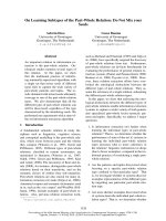
Paranasal tumor, H&E stained section, high power (40×)Figure 2
Paranasal tumor, H&E stained section, high power (40×).
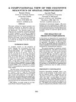
Paranasal tumor, H&E stained section, low power (10×)Figure 1
Paranasal tumor, H&E stained section, low power
(10×). The section shows a spindle cell neoplasm with taper-
ing nuclei, eosinophilic cytoplasm and minimal atypia. Focally,
myxoid features predominated.
Journal of Hematology & Oncology 2008, 1:3 />Page 4 of 6
(page number not for citation purposes)
[9,33], and the significant lag of the therapeutic response
has led some to hypothesis that the mechanism of action
is estrogen independent [46]. There are also documented
responses to NSAIDs, most often sulindac, both alone and
in combination with tamoxifen [9,34-39]. At least one
report documents the resolution of a desmoid tumor
being treated with indomethacin and ascorbic acid for 14
months [40]. Although response rates as high as 70% are
reported with combined therapy [9], regression is usually
partial and may take many months after an initial period
of tumor enlargement. In addition, response criteria for
these case reports are not standardized. Several case
reports describe objective response or prolonged periods
of disease stabilization with interferon alpha (IFN-alpha)
[41-43], in some cases following failure of sulindac and
tamoxifen [36,44,45]. However, new data suggesting that
IFN type I signaling is a positive regulator of neoplastic
growth has raised questions about the therapeutic role of
IFN-alpha in this disease [46]. An increasing number of
reports suggest clinical and radiographic benefit from the
tyrosine kinase inhibitor imatinib (Gleevec) [47,48]. This
effect is presumably due to tumor expression of activated
receptor tyrosine kinases c-kit and/or platelet-derived
growth factor receptor-alpha (PDGFRA). However the
clinical efficacy of imatinib and the mechanism underly-
ing clinical benefit in the patients who have been treated
with this agent are uncertain.
Cytotoxic systemic therapy
Although desmoid tumors as a group are generally slow
growing with low metastatic potential, there are several
highly active chemotherapy regimens that can potentially
produce durable response. The combination of low dose
methotrexate and vinblastine has shown promising
results, particularly in children [49-52]. One study of 30
patients with a median age of 27 reported 10 year progres-
sion free survival in 67% [49]. Liposomal doxorubicin has
proven to be a well tolerated and efficacious option [53].
High dose doxorubicin or ifosfamide-based regimens
have shown more activity and increased incidence of seri-
ous toxicity; thus they are usually reserved for cases that
are life threatening and refractory to other treatments [54-
57].
Conclusion
Desmoid fibromatosis are rare pediatric tumors, and the
case reported here is one of only six published accounts of
pediatric desmoid fibromatosis of the paranasal sinuses.
Aggressive juvenile fibromatoses are a group of lesions
with variable response to treatment; they are locally
aggressive but have low metastatic potential. Current
treatment ranges from traditional surgical resection to
multidisciplinary approaches involving local radiation
and/or systemic cytotoxic and cytostatic agents. However,
surgical resection with wide margins remains the primary
treatment for extraabdominal fibromatoses. Reports in
the literature are conflicting as to the importance of
obtaining tumor-free surgical margins; some retrospective
analyses have found a significant decrease in recurrence
rate with negative margins, while others have not. Based
on these reports, the optimal treatment strategy for pedi-
atric desmoids fibromatoses is patient-dependent, and
clinical decisions must be made based upon tumor loca-
tion, risk of surgical morbidity and risk of radiation-
induced damage. Radiation therapy and cytotoxic chemo-
therapy in pediatric patients should be used in cases that
are refractory to surgery and noncytotoxic systemic ther-
apy due to the potential of growth disturbance, contrac-
ture, and the development of secondary malignancy.
Abbreviations
CT: computed tomography; IFN-alpha: interferon alpha;
MRI: magnetic resonance imaging; NSAID: non-steroidal
anti-inflammatory drug; PDGFRA: platelet-derived
growth factor receptor-alpha; RT: radiation therapy; SMA:
smooth-muscle actin.
Competing interests
The authors declare that they have no competing interests.
Authors' contributions
SL, RE, and LH secured the case, conducted the literature
review, and participated in the preparation of the manu-
script. All authors read and approved the final manu-
script.
Consent
Written informed consent was obtained from the patient's
parents for publication of this case report and any accom-
panying images. A copy of the written consent is available
for review by the Editor-in-Chief of this journal.
References
1. Mannan AA, Ray R, Sharma SC, Hatimota P: Infantile fibromatosis
of the nose and paranasal sinuses: report of a rare case and
brief review of the literature. Ear Nose Throat J 2004,
83(7):481-484.
2. Conley J, Healey WV, Stout AP: Fibromatosis of the head and
neck. Am J Surg 1966, 112(4):609-614.
3. Fu YS, Perzin KH: Nonepithelial tumors of the nasal cavity,
paranasal sinuses, and nasopharynx. A clinicopathologic
study. VI. Fibrous tissue tumors (fibroma, fibromatosis, fib-
rosarcoma). Cancer 1976, 37(6):2912-2928.
4. Maillard AA, Kountakis SE: Pediatric sino-orbital desmoid
fibromatosis. Ann Otol Rhinol Laryngol 1996, 105(6):463-466.
5. Naidu RK, Aviv JE, Lawson W, Biller HF: Aggressive juvenile
fibromatosis involving the paranasal sinuses. Otolaryngol Head
Neck Surg 1991, 104(4):549-552.
6. Thompson DH, Khan A, Gonzalez C, Auclair P: Juvenile aggressive
fibromatosis: report of three cases and review of the litera-
ture. Ear Nose Throat J 1991, 70(7):462-468.
7. Reitamo JJ, Hayry P, Nykyri E, Saxen E: The desmoid tumor. I.
Incidence, sex-, age- and anatomical distribution in the Finn-
ish population. Am J Clin Pathol 1982, 77(6):665-673.
8. Enzinger FM, Weiss SW: Soft tissue tumors 3rd edition. St. Louis:
Mosby; 1995.
Journal of Hematology & Oncology 2008, 1:3 />Page 5 of 6
(page number not for citation purposes)
9. Hansmann A, Adolph C, Vogel T, Unger A, Moeslein G: High-dose
tamoxifen and sulindac as first-line treatment for desmoid
tumors. Cancer 100(3):612-620. Feb 1 2004
10. De Wever I, Dal Cin P, Fletcher CD, Mandahl N, Mertens F, Mitelman
F, Rosai J, Rydholm A, Sciot R, Tallini G, Berghe H Van Den, Vanni R,
Willén H: Cytogenetic, clinical, and morphologic correlations
in 78 cases of fibromatosis: a report from the CHAMP Study
Group. CHromosomes And Morphology. Mod Pathol 2000,
13(10):1080-1085.
11. Bridge JA, Swarts SJ, Buresh C, Nelson M, Degenhardt JM, Spanier S,
Maale G, Meloni A, Lynch JC, Neff JR: Trisomies 8 and 20 charac-
terize a subgroup of benign fibrous lesions arising in both soft
tissue and bone. Am J Pathol 1999, 154(3):729-733.
12. Fletcher JA, Naeem R, Xiao S, Corson JM: Chromosome aberra-
tions in desmoid tumors. Trisomy 8 may be a predictor of
recurrence. Cancer Genet Cytogenet 1995, 79(2):139-143.
13. Mertens F, Willén H, Rydholm A, Brosjö O, Carlén B, Mitelman F,
Mandahl N: Trisomy 20 is a primary chromosome aberration
in desmoid tumors. Int J Cancer 63(4):527-529. Nov 15 1995
14. Qi H, Dal Cin P, Hernández JM, Garcia JL, Sciot R, Fletcher C, Van
Eyken P, De Wever I, Berghe H Van den: Trisomies 8 and 20 in
desmoid tumors. Cancer Genet Cytogenet 1996, 92(2):147-149.
15. Ballo MT, Zagars GK, Pollack A, Pisters PW, Pollack RA: Desmoid
tumor: prognostic factors and outcome after surgery, radia-
tion therapy, or combined surgery and radiation therapy. J
Clin Oncol 1999, 17(1):158-167.
16. Abbas AE, Deschamps C, Cassivi SD, Nichols FC 3rd, Allen MS, Sch-
leck CD, Pairolero PC: Chest-wall desmoid tumors: results of
surgical intervention. Ann Thorac Surg 2004, 78(4):1219-1223.
discussion 1219–1223
17. Buitendijk S, Ven CP van de, Dumans TG, den Hollander JC, Nowak
PJ, Tissing WJ, Pieters R, Heuvel-Eibrink MM van den: Pediatric
aggressive fibromatosis: a retrospective analysis of 13
patients and review of literature. Cancer 104(5):1090-1099. Sep
1 2005
18. Faulkner LB, Hajdu SI, Kher U, La Quaglia M, Exelby PR, Heller G,
Wollner N: Pediatric desmoid tumor: retrospective analysis
of 63 cases. J Clin Oncol 1995, 13(11):2813-2818.
19. Merchant NB, Lewis JJ, Woodruff JM, Leung DH, Brennan MF:
Extremity and trunk desmoid tumors: a multifactorial anal-
ysis of outcome. Cancer 86(10):2045-2052. Nov 15 1999
20. Lev D, Kotilingam D, Wei C, Ballo MT, Zagars GK, Pisters PW, Lazar
AA, Patel SR, Benjamin RS, Pollock RE: Optimizing treatment of
desmoid tumors. J Clin Oncol 25(13):1785-1791. May 1 2007
21. Gronchi A, Casali PG, Mariani L, Lo Vullo S, Colecchia M, Lozza L,
Bertulli R, Fiore M, Olmi P, Santinami M, Rosai J: Quality of surgery
and outcome in extra-abdominal aggressive fibromatosis: a
series of patients surgically treated at a single institution. J
Clin Oncol 21(7):1390-1397. Apr 1 2003
22. Pignatti G, Barbanti-Bròdano G, Ferrari D, Gherlinzoni F, Bertoni F,
Bacchini P, Barbieri E, Giunti A, Campanacci M: Extraabdominal
desmoid tumor. A study of 83 cases. Clin Orthop Relat Res
2000:207-213.
23. Ballo MT, Zagars GK, Pollack A: Radiation therapy in the man-
agement of desmoid tumors. Int J Radiat Oncol Biol Phys
42(5):1007-1014. Dec 1 1998
24. Reitamo JJ: The desmoid tumor. IV. Choice of treatment,
results, and complications. Arch Surg 1983, 118(11):1318-1322.
25. Miralbell R, Suit HD, Mankin HJ, Zuckerberg LR, Stracher MA, Rosen-
berg AE: Fibromatoses: from postsurgical surveillance to
combined surgery and radiation therapy. Int J Radiat Oncol Biol
Phys 1990, 18(3):535-540.
26. Posner MC, Shiu MH, Newsome JL, Hajdu SI, Gaynor JJ, Brennan MF:
The desmoid tumor. Not a benign disease. Arch Surg 1989,
124(2):191-196.
27. Spear MA, Jennings LC, Mankin HJ, Spiro IJ, Springfield DS, Gebhardt
MC, Rosenberg AE, Efird JT, Suit HD: Individualizing manage-
ment of aggressive fibromatoses. Int J Radiat Oncol Biol Phys
40(3):637-645. Feb 1 1998
28. Karakousis CP, Mayordomo J, Zografos GC, Driscoll DL: Desmoid
tumors of the trunk and extremity. Cancer 72(5):1637-1641.
Sep 1 1993
29. Nuyttens JJ, Rust PF, Thomas CR Jr, Turrisi AT 3rd:
Surgery versus
radiation therapy for patients with aggressive fibromatosis
or desmoid tumors: A comparative review of 22 articles.
Cancer 88(7):1517-1523. Apr 1 2000
30. Goy BW, Lee SP, Eilber F, Dorey F, Eckardt J, Fu YS, Juillard GJ, Selch
MT: The role of adjuvant radiotherapy in the treatment of
resectable desmoid tumors. Int J Radiat Oncol Biol Phys
9(3):659-665. Oct 1 1997
31. Zlotecki RA, Scarborough MT, Morris CG, Berrey BH, Lind DS,
Enneking WF, Marcus RB Jr: External beam radiotherapy for pri-
mary and adjuvant management of aggressive fibromatosis.
Int J Radiat Oncol Biol Phys 54(1):177-181. Sep 1 2002
32. Janinis J, Patriki M, Vini L, Aravantinos G, Whelan JS: The pharma-
cological treatment of aggressive fibromatosis: a systematic
review. Ann Oncol 2003, 14(2):181-190.
33. Sorensen A, Keller J, Nielsen OS, Jensen OM: Treatment of
aggressive fibromatosis: a retrospective study of 72 patients
followed for 1–27 years. Acta Orthop Scand 2002, 73(2):213-219.
34. Tsukada K, Church JM, Jagelman DG, Fazio VW, McGannon E,
George CR, Schroeder T, Lavery I, Oakley J: Noncytotoxic drug
therapy for intra-abdominal desmoid tumor in patients with
familial adenomatous polyposis. Dis Colon Rectum 1992,
35(1):29-33.
35. Izes JK, Zinman LN, Larsen CR: Regression of large pelvic
desmoid tumor by tamoxifen and sulindac. Urology 1996,
47(5):756-759.
36. Bauernhofer T, Stöger H, Schmid M, Smola M, Gürtl-Lackner B,
Höfler G, Ranner G, Reisinger E, Samonigg H: Sequential treat-
ment of recurrent mesenteric desmoid tumor. Cancer
77(6):1061-1065. Mar 15 1996
37. Lackner H, Urban C, Kerbl R, Schwinger W, Beham A: Noncyto-
toxic drug therapy in children with unresectable desmoid
tumors. Cancer 80(2):334-340. Jul 15 1997
38. Waddell WR, Kirsch WM: Testolactone, sulindac, warfarin, and
vitamin K1 for unresectable desmoid tumors. Am J Surg 1991,
161(4):416-421.
39. Dominguez-Malagon HR, Alfeiran-Ruiz A, Chavarria-Xicotencatl P,
Duran-Hernandez MS: Clinical and cellular effects of colchicine
in fibromatosis.
Cancer 69(10):2478-2483. May 15 1992
40. Brooks MD, Ebbs SR, Colletta AA, Baum M: Desmoid tumours
treated with triphenylethylenes. Eur J Cancer 1992, 28A(6–
7):1014-1018.
41. Raguse JD, Gath HJ, Oettle H, Bier J: Interferon-induced remis-
sion of rapidly growing aggressive fibromatosis in the tempo-
ral fossa. Int J Oral Maxillofac Surg 2004, 33(6):606-609.
42. Fernberg JO, Brosjo O, Larsson O, Soderlund V, Strander H: Inter-
feron-induced remission in aggressive fibromatosis of the
lower extremity. Acta Oncol 1999, 38(7):971-972.
43. Leithner A, Schnack B, Katterschafka T, Wiltschke C, Amann G,
Windhager R, Kotz R, Zielinski CC: Treatment of extra-abdom-
inal desmoid tumors with interferon-alpha with or without
tretinoin. J Surg Oncol 2000, 73(1):21-25.
44. Plukker JT, van Oort I, Vermey A, Molenaar I, Hoekstra HJ, Panders
AK, Dolsma WV, Koops HS: Aggressive fibromatosis (non-
familial desmoid tumour): therapeutic problems and the
role of adjuvant radiotherapy. Br J Surg 1995, 82(4):510-514.
45. Geurs F, Kok TC: Regression of a great abdominal desmoid
tumor by interferon alpha. J Clin Gastroenterol 1993,
16(3):264-265.
46. Tjandra SS, Hsu C, Goh YI, Gurung A, Poon R, Nadesan P, Alman BA:
IFN-{beta} signaling positively regulates tumorigenesis in
aggressive fibromatosis, potentially by modulating mesen-
chymal progenitors. Cancer Res 67(15):7124-7131. Aug 1 2007
47. Heinrich MC, McArthur GA, Demetri GD, Joensuu H, Bono P, Her-
rmann R, Hirte H, Cresta S, Koslin DB, Corless CL, Dirnhofer S, van
Oosterom AT, Nikolova Z, Dimitrijevic S, Fletcher JA: Clinical and
molecular studies of the effect of imatinib on advanced
aggressive fibromatosis (desmoid tumor). J Clin Oncol
24(7):1195-1203. Mar 1 2006
48. Mace J, Sybil Biermann J, Sondak V, McGinn C, Hayes C, Thomas D,
Baker L: Response of extraabdominal desmoid tumors to
therapy with imatinib mesylate. Cancer 95(11):2373-2379. Dec
1 2002
49. Azzarelli A, Gronchi A, Bertulli R, Tesoro JD, Baratti D, Pennacchioli
E, Dileo P, Rasponi A, Ferrari A, Pilotti S, Casali PG: Low-dose
chemotherapy with methotrexate and vinblastine for
patients with advanced aggressive fibromatosis. Cancer
92(5):1259-1264. Sep 1 2001
50. Weiss AJ, Lackman RD: Low-dose chemotherapy of desmoid
tumors. Cancer 64(6):1192-1194. Sep 15 1989
Publish with BioMed Central and every
scientist can read your work free of charge
"BioMed Central will be the most significant development for
disseminating the results of biomedical research in our lifetime."
Sir Paul Nurse, Cancer Research UK
Your research papers will be:
available free of charge to the entire biomedical community
peer reviewed and published immediately upon acceptance
cited in PubMed and archived on PubMed Central
yours — you keep the copyright
Submit your manuscript here:
/>BioMedcentral
Journal of Hematology & Oncology 2008, 1:3 />Page 6 of 6
(page number not for citation purposes)
51. Skapek SX, Ferguson WS, Granowetter L, Devidas M, Perez-Atayde
AR, Dehner LP, Hoffer FA, Speights R, Gebhardt MC, Dahl GV, Grier
HE, Pediatric Oncology Group: Vinblastine and methotrexate
for desmoid fibromatosis in children: results of a Pediatric
Oncology Group Phase II Trial. J Clin Oncol 25(5):501-506. Feb
10 2007
52. Reich S, Overberg-Schmidt US, Buhrer C, Henze G: Low-dose
chemotherapy with vinblastine and methotrexate in child-
hood desmoid tumors. J Clin Oncol 1999, 17(3):1086.
53. Wehl G, Rossler J, Otten JE, Boehm N, Uhl M, Kontny U, Niemeyer
C: Response of progressive fibromatosis to therapy with lipo-
somal doxorubicin. Onkologie 2004, 27(6):552-556.
54. Lynch HT, Fitzgibbons R Jr, Chong S, Cavalieri J, Lynch J, Wallace F,
Patel S: Use of doxorubicin and dacarbazine for the manage-
ment of unresectable intra-abdominal desmoid tumors in
Gardner's syndrome. Dis Colon Rectum 1994, 37(3):260-267.
55. Patel SR, Evans HL, Benjamin RS: Combination chemotherapy in
adult desmoid tumors. Cancer 72(11):3244-3247. Dec 1 1993
56. Pilz T, Pilgrim TB, Bisogno G, Knietig R, Koscielniak E, Carli M, Tre-
uner J: [Chemotherapy in fibromatoses of childhood and ado-
lescence: results from the Cooperative soft tissue sarcoma
study (CWS) and the Italian Cooperative study group (ICG-
AIEOP)]. Klin Padiatr 1999, 211(4):291-295.
57. Okuno SH, Edmonson JH: Combination chemotherapy for
desmoid tumors. Cancer 97(4):1134-1135. Feb 15 2003