báo cáo khoa học: "Rosai dorfman disease of the orbit" pot
Bạn đang xem bản rút gọn của tài liệu. Xem và tải ngay bản đầy đủ của tài liệu tại đây (1.14 MB, 7 trang )
BioMed Central
Page 1 of 7
(page number not for citation purposes)
Journal of Hematology & Oncology
Open Access
Research
Rosai dorfman disease of the orbit
Geeta K Vemuganti*
1
, Milind N Naik
2
and Santosh G Honavar
2
Address:
1
Ophthalmic Pathology Service, Hyderbad Eye Research Centre, L V Prasad Eye Institute, Hyderbad, India and
2
Division of Ophthalmic
Plastic Surgery, Orbit, and Ocular Oncology, LV Prasad Eye Institute, Hyderabad, India
Email: Geeta K Vemuganti* - ; Milind N Naik - ; Santosh G Honavar -
* Corresponding author
Abstract
Objective: To report the clinico-histopathologic features, management and outcome of Rosai-
Dorfman disease of the orbit.
Design: Non-comparative case series.
Results: Rosai-Dorfman disease of the orbit constituted 0.09% of all ocular specimens received at
our Institute, presenting with a firm rubbery mass causing proptosis; bilateral in 4 (57%) cases. The
median age at presentation was 13 years (range 5–65); median duration of symptoms was 6 (range
3–15) years. Lymphadenopathy was noted in 4 (57%); extranodal involvement in 3 (43%). After
biopsy, 3 cases were treated with systemic corticosteroids, 2 cases developed local recurrence that
responded to systemic corticosteroid therapy. Polymorphous population of lymphocytes, plasma
cells, and characteristic S-100-positive histiocytes showing emperipolesis were pathognomonic
histologic features.
Conclusion: Rosai-Dorfman disease of the orbit, although rare, should be considered in young
individuals with chronic proptosis with rubbery masses. Excision and corticosteroid therapy
provide a favorable outcome.
Background
Rosai-Dorfman disease, also known as sinus histiocytosis
with massive lymphadenopathy is a rare disorder charac-
terized by nonmalignant proliferation of distinctive histi-
ocytes within lymph node sinuses and other extranodal
sites. It is a self-limiting disorder of unknown etiology that
occurs worldwide in children and young adults [1-5].
Classically, Rosai-Dorfman disease manifests as chronic
painless cervical lymphadenopathy with pyrexia, leucocy-
tosis, increased erythrocyte sedimentation rate and hyper-
gammaglobulinemia. About 43% of patients have
extranodal manifestation in the eye, upper respiratory
tract, salivary gland, skin, bone, meninges and central
nervous system and testis [3-5]. The reported ophthalmic
manifestations include eyelid and orbital mass, and rarely
uveitis [6-9]. Orbital involvement could be an isolated
extranodal manifestation or associated with concurrent
systemic disease. Orbital mass in Rosai-Dorman disease
could mimic lymphoma, lacrimal gland tumors, and
other histiocytic tumors. We herein report the clinical
manifestations, management, and outcome of a series of
seven histopathologically proven cases of Rosai-Dorfman
disease of the orbit.
Published: 28 June 2008
Journal of Hematology & Oncology 2008, 1:7 doi:10.1186/1756-8722-1-7
Received: 17 May 2008
Accepted: 28 June 2008
This article is available from: />© 2008 Vemuganti et al; licensee BioMed Central Ltd.
This is an Open Access article distributed under the terms of the Creative Commons Attribution License ( />),
which permits unrestricted use, distribution, and reproduction in any medium, provided the original work is properly cited.
Journal of Hematology & Oncology 2008, 1:7 />Page 2 of 7
(page number not for citation purposes)
Results
Clinical Profile
Of the 7537 ocular specimens received during the study
period, seven patients were diagnosed as Rosai-Dorfman
disease, constituting 0.09 % of all ocular specimens and
2.3% of orbital lesions (7 of 300). The median age was 13
years (range 5–65), with male: female ratio of 3:4. Median
duration of symptoms at presentation was 6 years (range,
3–15 years). The clinical profile of these patients is shown
in [Additional File 1]. The presenting symptoms were
painless progressive proptosis in all except two patients
who presented with an eyelid mass. The referral diagnosis
included orbital lymphoma, xanthogranuloma, and lac-
rimal gland tumor. Three patients had earlier undergone
biopsy earlier elsewhere with an inconclusive diagnosis
(slides not available for review).
At baseline evaluation, the visual acuity was 20/20 in all
except one patient (patient 4) [Additional File 1] who had
senile cataract. Diffuse conjunctival congestion was noted
in 3. There was restricted ocular motility in 5 patients and
mechanical ptosis in one. All patients had proptosis,
which was unilateral in 3 (43%) and bilateral in 4 (57%).
All patients had a palpable well-defined, nontender, rub-
bery, firm orbital mass with variable intrinsic mobility.
The mass was preseptal in 2 patients (figure 1a), anterior
orbital in 1, diffuse orbital in 3 and involved the orbital
lobe of the lacrimal gland in 1. The preauricular, sub-
mandibular and anterior cervical lymph nodes were mas-
sive, confluent, nontender and rubbery in 4 patients.
Computed tomography scans revealed a homogeneous
soft tissue mass in a preseptal location in 2, in the anterior
orbit in 2, diffuse in 2, and involving the lacrimal gland in
1.
Concurrent extranodal locations included the parotid
gland in 1, paranasal sinus in 2, and nasopharynx in 1.
Two patients had normocytic hypochromic anemia. The
erythrocyte sedimentation rate was within the normal
range in all patients. None had evidence of hepat-
osplenomegaly on clinical examination or imaging.
The primary management consisted of surgery in all cases.
In three patients the preseptal and anterior orbital mass
was well defined so as to allow easy dissection and sepa-
ration from the surrounding structures; they underwent
complete excision (figure 1b). Three patients with diffuse
orbital mass and one with lacrimal gland involvement
underwent an incisional biopsy (near total excision).
Three patients with diffuse orbital mass received systemic
corticosteroids at 1 mg/kg body weight initially and
tapered over 6 weeks to 3 months. The mean duration of
follow-up was 18.3 ± 17.7 months (range 0–80 months).
Of six patients who had follow-up evaluation, two devel-
oped local recurrence. The first patient, who had under-
gone macroscopically apparent complete excision of a
bilateral anterior orbital mass, developed unilateral dif-
fuse orbital recurrence 2 months later. The second patient
developed diffuse bilateral orbital recurrence and concur-
rent involvement of the maxillary sinus 10 months fol-
lowing complete excision of bilateral eyelid mass. Both
these patients had not received systemic corticosteroid
therapy as part of the primary treatment [Additional File
1]. The recurrence was treated with systemic corticoster-
oids. At the final follow-up, all patients had improvement
in proptosis and cosmetic appearance. None had residual
ocular motility restriction or ptosis.
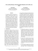
Bilateral upper eyelid and preseptal orbital mass in a 16-year-old female with complete mechanical ptosis (1a)Figure 1
Bilateral upper eyelid and preseptal orbital mass in a
16-year-old female with complete mechanical ptosis
(1a). Published with permission from Elsevier. This fig-
ure was published in the Clinical Ophthalmic Oncology,
Vemuganti GK, Honavar SH, Eyelid Stroma Tumors, Page
105, Copyright Elsevier 2007. Three-month post-operative
appearance following complete excision of the mass (1b).
Journal of Hematology & Oncology 2008, 1:7 />Page 3 of 7
(page number not for citation purposes)
Histopathology
All lesions had similar histopathologic characteristics. On
gross examination, the specimens measured 20 to 50 mm
in dimension (figure 2a), was firm to rubbery in consist-
ency with a lobulated surface. Cut sections revealed focal
yellowish areas within the lesion (figure 2b). Microscopi-
cally, there was a polymorphous population of histio-
cytes, plasma cells and mature lymphocytes separated by
fibrous septa (figure 3). A few lymphoid follicles were
seen with germinal centers. In addition, there were prom-
inent histiocytes which contained abundant pale to vacu-
olated cytoplasm and a large nucleus with prominent
nucleoli. Many of these cells showed lymphophagocytosis
or emperipolesis (figure 4). These histiocytes were immu-
noreactive to the S-100 antibody (figure 5). The plasma
cells were seen clustered with extracellular and intracellu-
lar immunoglobulin deposits. Immunophenotyping
revealed polyclonal population of plasma and lym-
phocytes. Imprint smears prepared from the 4 unfixed
fresh specimens available, revealed characteristic histio-
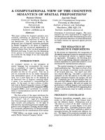
Histopathologic section showing a polymorphous population of cells consisting of mature lymphocytes and plasma cells interspersed with histiocytesFigure 4
Histopathologic section showing a polymorphous population
of cells consisting of mature lymphocytes and plasma cells
interspersed with histiocytes. Histiocytes are large with a
vesicular nuclei and abundant cytoplasm with engulfed lym-
phocytes and plasma cells, a phenomenon called lym-
phophagocytosis or emperipolesis, a hallmark of Rosai-
Dorfman disease (arrow) (× 500, hematoxylin, eosin). Pub-
lished with permission from Elsevier. This figure was pub-
lished in the Clinical Ophthalmic Oncology, Vemuganti GK,
Honavar SH, Eyelid Stroma Tumors, Page 105, Copyright
Elsevier 2007.
The gross specimen of the excised mass with lobulated and smooth surface (2a)Figure 2
The gross specimen of the excised mass with lobu-
lated and smooth surface (2a). The cut section of the
specimen shows a solid appearance with a few yellowish
areas (2b).
Histopathologic section showing a lymphoid follicle (arrow) surrounded by a cuff of lymphocytes and plasma cells with a few pale areas consisting of sheets of histiocytes (asterisk) (× 50, hematoxylin, eosin)Figure 3
Histopathologic section showing a lymphoid follicle (arrow)
surrounded by a cuff of lymphocytes and plasma cells with a
few pale areas consisting of sheets of histiocytes (asterisk) (×
50, hematoxylin, eosin).
Journal of Hematology & Oncology 2008, 1:7 />Page 4 of 7
(page number not for citation purposes)
cytes with emperipolesis in a background of plasma cells
and lymphocytes (figure 6).
Discussion
In 1969, two pathologists Juan Rosai and Ronald Dorf-
man reported a distinct histiocytic disorder in young black
males presenting with bilateral, painless, massive cervical
lymphadenopathy with a protracted clinical course, and
in most instances associated with fever, anemia, neu-
trophilia, elevated erythrocyte sedimentation rate, and
polyclonal gammopathy[1]. The clinicopathological con-
glomerate that they named sinus histiocytosis with mas-
sive lyphadenopathy has now come to be known as Rosai-
Dorfman disease[1]. Subsequently it became clear that the
disease had no specific predilection for geographic loca-
tion or race [5].
Painless lymphadenopathy is the most frequent systemic
presenting symptom and involves the cervical region in
up to 90% of patients [5]. Other locations such as
inguinal (26%), axillary (24%) and mediastinal lymph
nodes (15%) are also reported to be involved [5].
Extranodal disease is documented in 43% of patients, in
some without associated lymphadenopathy, which may
or may not develop later in the disease course [5]. The
most common extranodal sites, in the decreasing order of
frequency, are skin, nasal cavity and paranasal sinus, eye-
lid, orbit, bone, salivary gland and central nervous system
[Additional File 2] [5]. The simultaneous involvement of
multiple extranodal sites is not unusual [5]. Hepat-
osplenomegaly, unlike in other histiocytic disorders, is
uncommon [5].
The high prevalence of this disease in this series (2.3% of
orbital lesions and 0.09% of ocular specimens) is possibly
because this center is a tertiary eye referral center with a
dedicated Ocular Oncology Service. With increasing
number of ocular specimens received at our centre to
nearly 4,000 samples per year, the prevalence may now
match the same as seen in any general hospital, i.e 0.03%
[5/15,000 cases based on the personal communication
received from a surgical pathologist ]. The reported oph-
thalmic manifestations of Rosai-Dorfman disease include
orbital and eyelid involvement, lacrimal gland involve-
ment, optic nerve compressive neuropathy and uveitis
[3,6-12]. Orbital involvement is the most common of
ophthalmic manifestations [3,6-12]. While a majority of
patients with orbital involvement have concurrent lym-
phadenopathy, some may present with orbit as the sole
extranodal site of involvement without synchronous
nodal disease, and a minority may have concurrent
involvement of other extranodal sites such as the parana-
sal sinus [3]. In our series of seven patients, six were under
20 years of age and had chronic symptoms ranging from
3–15 years. All had painless progressive proptosis and two
had an eyelid mass. Four patients had bilateral manifesta-
tions, 4 had synchronous nodal disease and 3 had concur-
rent extranodal involvement.
Clinical laboratory findings in Rosai-Dorfman disease
include hematological abnormalities such as normocytic
or microcytic anemia, hemolytic anemia, elevated erthro-
cyte sedimentation rate and polyclonal hypergammaglob-
ulinemia [5]. Two patients in our series had normocytic
hypochromic anemia. However, erythrocyte sedimenta-
tion rate was within the normal range in all patients and
serum electrophoresis did not reveal hypergammaglob-
ulinemia.
According to the Writing Group of the Histiocyte Society
[13], the histiocytic syndromes can be subdivided based
on whether the proliferating histiocytes are the Langer-
hans cells or not and whether the process is benign or
malignant. Rosai-Dorfman disease is one of the non-
Langerhans cell benign histiocytosis where predomi-
nantly the sinuses of the lymph nodes, and less com-
monly the interfollicular area of the lymph nodes are
infiltrated with distinctive histiocytes with round or oval
vesicular nuclei with well-defined, delicate nuclear mem-
branes and a single prominent nucleolus [1,2,5]. Nuclear
atypia and mitoses are infrequent. The hallmark of Rosai-
Dorman disease is lymphophagocytosis or emperipolesis,
wherein the viable lymphocytes are located in well-
defined cytoplasmic vacuoles of intact histiocytes. Plasma
cells, neutrophils and red blood cells may also occupy this
unique intracytoplasmic niche. The involved histiocytes
are activated macrophages with features of phagocytic
cells as well as immune accessory cells and thus express S-
100 protein, HAM 56, α1 antitrypisn, α1 chymotrypsin,
lysozyme, Mac 387, Ki-1 (CD 30, Ber-H2), but are nega-
tive for CD 1a (leu 6) [14]. Rosai-Dorfman disease involv-
ing extranodal sites shows similar morphologic features to
its nodal counterpart with more fibrosis and fewer histio-
cytes with emperipolesis. The histological differential
diagnosis includes hemophagocytic syndromes, storage
disorder, inflammatory lesions, necrobiotic xanthogranu-
loma and lymphoreticular malignancies [15]. The pres-
ence of benign histiocytes with emperipolesis, absence of
cellular atypia, immunohistochemical profile, and associ-
ated clinical features distinguish Rosai-Dorfman disease
from other simulating disorders.
Cytologic features of Rosai-Dorfman syndrome are well
recognized and the role of fine-needle aspiration cytology
in its diagnosis has been demonstrated [16,17]. We used
the impression cytology technique for rapid intraopera-
tive diagnosis of Rosai-Dorfman disease based on known
cytological characteristics in 4 patients in this series. The
Journal of Hematology & Oncology 2008, 1:7 />Page 5 of 7
(page number not for citation purposes)
cytologic diagnosis correlated with the final histopathol-
ogy in all four patients.
Despite its well-recognized clinical presentation, the precise
etiology of Rosai-Dorfman disease remains unknown. The
etiologic factors implied in the pathogenesis of this disease
are bacterial (Klebsiella), virus (Epstein barr virus, parvovi-
rus B 19), immune dysfunction, or an aberrant response to
an unspecified antigen, HHV-6 or EBV [18-21]. Current
thinking is that the defective Fas/FasL signaling leading to
altered apoptosis may be an important mechanism
whereby uncontrolled histiocytic proliferation is triggered
[20]. The presence of characteristic histiocyte, derived from
circulating mononuclear cells, long history and an
increased incidence of serum autoreactive antibodies dur-
ing active disease suggest a possible pathogenic correlations
with a dysregulatory process. In a recent report, the evi-
dence points towards a viral etiology as suggested by the
immunolocalization of parvovirus B19 (B19) virus using
antibodies against B19 capsid proteins VP1/VP2[21]. The
relative increase of cases from this part of the world, also
prompts us to believe that there could be a possible envi-
ronmental factor, thereby warranting further studies in this
direction. Though not done in this series, immunological
studies, specifically for viral etiology, liver function along
with follow-up to identify known risk factors like airway
compression, would be beneficial in understanding more
about this rare disease [21,22].
The clinical course of Rosai-Dorfman disease is chronic
and variable with episodes of exacerbation alternating
with periods of remission, where the timing and duration
of each phase is entirely unpredictable. Foucar et al
reported stable disease in 54%, spontaneous regression in
21%, and progressive disease in only 1% [5].
The ideal treatment for Rosai-Dorfman disease is yet
unestablished. Only about 50% of patients with Rosai-
Dorfman disease need some form of treatment [20]. Man-
agement options include observation for mild manifesta-
tions with no cosmetic or functional abnormality, surgical
excision or debulking for lesions in surgically accessible
locations, and systemic corticosteroids, chemotherapy or
radiotherapy in patients with severe symptoms where vital
organ function is compromised [23-27]. Radiotherapy
and antimetabolite treatment has been considered in a
few cases but the literature review by Pulsoni et al does not
suggest any conclusive role of these treatment modalities
[27]. The treatment of orbital manifestations of Rosai-
Dorfman disease aims to control the functional and cos-
metic abnormalities. Orbital involvement, being cosmeti-
cally disturbing and surgically accessible, may be more
often considered for surgical treatment. Massive or recur-
rent orbital disease or significant residual lesion following
surgical debulking may be treated with systemic corticos-
teroids, chemotherapy or radiotherapy. Chemotherapy
has also been used to relieve the sight threatening optic
nerve compression [10].
All patients in our series underwent surgery – 3 with well-
defined localized mass underwent surgical excision and 3
with diffuse orbital involvement and 1 with lacrimal
gland involvement underwent an incisional biopsy. Three
patients with diffuse orbital involvement received sys-
temic corticosteroid therapy. Local recurrence was noted
in 2 of 7 (29%) cases, both within one year of primary
treatment, and these patients responded to systemic corti-
costeroids.
Imprint cytology shows large histiocytes (arrow) with vesicu-lar nucleus and phagocytosed lymphocytes and plasma cells (asterisk) within the cytoplasm (× 500, Giemsa)Figure 6
Imprint cytology shows large histiocytes (arrow) with vesicu-
lar nucleus and phagocytosed lymphocytes and plasma cells
(asterisk) within the cytoplasm (× 500, Giemsa).
Histopathologic section showing large histiocytes with emperipolesis, immunoreactive for S-100 antigenFigure 5
Histopathologic section showing large histiocytes with
emperipolesis, immunoreactive for S-100 antigen. (× 200,
DAB).
Journal of Hematology & Oncology 2008, 1:7 />Page 6 of 7
(page number not for citation purposes)
Conclusion
To conclude, Rosai-Dorfman disease may be suspected in
young individuals with unilateral or bilateral slowly pro-
gressive proptosis manifesting with a rubbery firm mass,
with or without massive cervical lymphadenopathy. Sys-
temic evaluation is necessary to document other extran-
odal sites of involvement. A biopsy will help confirm the
diagnosis. Patients with cosmetic and/or functional
abnormality secondary to the orbital mass could be con-
sidered for debulking or complete excision. Diffuse, resid-
ual, or recurrent lesions may be treated with systemic
corticosteroids.
Methods
We reviewed consecutive cases of orbital tumors on the
registry of the Ophthalmic Pathology Service at a tertiary
care centre between January 1996 and December 2002
and included patients with a histopathologic diagnosis of
Rosai-Dorfman disease in this series. The medical records
of these patients were reviewed for the demographic data,
clinical manifestations and radiologic features, manage-
ment and outcome.
An experienced ophthalmic pathologist reviewed forma-
lin-fixed, paraffin-embedded hematoxylin-eosin stained
sections. Immunohistochemistry was performed on the
sections using S-100 protein and monoclonal antibodies
against T-and B-cell markers, and lambda and kappa
chains of immunoglobulins.
Competing interests
The authors declare that they have no competing interests.
Authors' contributions
GKV conceived the idea, carried out the histopathologic
studies, drafted the manuscript and reviewed the review of
literature, MN participated in the study design, and
reviewed the cases, SGH participated in the study design
and provided critical inputs into the study. All authors
read and approved the final manuscript
Consent
The patients have given their consent for the medical
records to be reviewed for research and publications
through the informed consent.
Additional material
Acknowledgements
We acknowledge the financial support from Hyderabad Eye Research and
C-TRACER, Prof Brien Holden Eye Research Center, Hyderabad Eye
Research Foundation, LV Prasad Eye Institute, Hyderabad, India. Part of this
work was presented at the American Academy of Ophthalmic Pathologist
Meeting 2002 at Orlando, Florida.
References
1. Rosai J, Dorfman RF: Sinus histiocytosis with massive lymphad-
enopathy. A newly recognized benign clinicopathological
entity. Arch Pathol 1969, 87:63-70.
2. Rosai J, Dorfman RF: Sinus histiocytosis with massive lymphad-
enopathy: a pseudolymphomatous benign disorder. Analysis
of 34 cases. Cancer 1972, 30:1174.
3. Foucar E, Rosai J, Dorfman RF: The ophthalmologic manifesta-
tions of sinus histiocytosis with massive lymphadenopathy.
Am J Ophthalmol 1979, 87:354-367.
4. Sanchez R, Rosai J, Dorfman RF: Sinus histiocytosis with massive
lymphadenopathy. An analysis of 113 cases with special
emphasis on its extranodal manifestations. Lab Invest 1977,
36:21-22.
5. Foucar E, Rosai J, Dorfman RF: Sinus histiocytosis with massive
lymphadenopathy (Rosai-Dorfman disease): Review of the
entity. Semin Diagn Pathol 1990, 7(1):19-73.
6. Friendly DS, Font RL, Rao NA: Orbital involvement in sinus his-
tiocytosis – a report of four cases. Arch Ophthalmol 1977,
95:2006-2011.
7. Child HA, Kim RY: Radiation response of Rosai-Dorfman dis-
ease presenting with involvement of the orbits. Am J Clin Oncol
1999, 22:526-528.
8. Marion JR, Geisinger KR: Sinus histiocytosis with massive lym-
phadenopathy, bilateral orbital involvement spanning 17
years. Ann Ophthalmol 1989, 21:55-58.
9. Vemuganti GK, Sekhar GC, Indira K: Multifocal Rosai-Dorfman
disease of periorbital tissues spanning 15 Years – a case
report. Orbit 2001, 20:297-300.
10. Lee-Wing M, Oryschak A, Attariwala G, Ashenhurst M: Rosai-Dor-
fman disease presenting as bilateral lacrimal gland enlarge-
ment. Am J Ophthalmol 2001, 131:677-678.
11. Goldberg S, Mahadevia P, Lipton M, Rosenbaum PS: Sinus histiocy-
tosis with massive lymphadenopathy involving the orbit:
reversal of compressive optic neuropathy after chemother-
apy.
J Neuroophthalmol 1998, 18:270-275.
12. Meyer CH, Sel S, Horle S, et al.: Rosai-Dorfman disease with
bilateral serous retinal detachment. Arch Ophthalmol 2003,
12(5):733-735.
13. Writing Group of the Histiocytic Society: Histiocytosis syndrome
in children. Lancet 1987, 1:208.
14. Eisen RN: Immunophenotypic characteristic of sinus histiocy-
tosis with massive lymphadenopathy. Semin Diagn Pathol 1990,
7:74.
15. Wieczorek R: Familial erythrophagocytic lymphohistiocytio-
sis: Immunophenotypic, immunohistochemical, and
ultrastructural demonstration of the relation to sinus histio-
cytosis. Human Pathol 1986, 17:55.
16. Layfield LJ: Fine needle aspiration cytologic findings in a case
of sinus histiocytosis with massive lymphadenopathy (Rosai-
Dorfman syndrome). Acta Cytol 1990, 34:767-770.
Additional file 1
Table 1 Clinical features of seven patients with Rosai-Dorfman disease of
the orbit. This table describes the clinical features of all cases of Rosai Dor-
fman Disease of the orbit included in this study.
Click here for file
[ />8722-1-7-S1.doc]
Additional file 2
Table 2: Clinical manifestations of Rosai-Dorfman disease. This table
describes the site, frequency and the clinical manifestation of Rosai Dorf-
man Disease. This research was originally published in Blood. McClain
KL, Natkunam Y, Swerdlow SH. Atypical cellular disorders. Blood.
2004;:283–96.
©
American Society of Hematology.
Click here for file
[ />8722-1-7-S2.doc]
Publish with BioMed Central and every
scientist can read your work free of charge
"BioMed Central will be the most significant development for
disseminating the results of biomedical research in our lifetime."
Sir Paul Nurse, Cancer Research UK
Your research papers will be:
available free of charge to the entire biomedical community
peer reviewed and published immediately upon acceptance
cited in PubMed and archived on PubMed Central
yours — you keep the copyright
Submit your manuscript here:
/>BioMedcentral
Journal of Hematology & Oncology 2008, 1:7 />Page 7 of 7
(page number not for citation purposes)
17. Deshpande AH, Nayak S, Munshi MM: Cytology of sinus histiocy-
tosis with massive lymphadenopathy (Rosai-Dorfman dis-
ease). Diagn Cytopathol 2000, 22:101-105.
18. Lampert F, Lennert K: Sinus histiocytosis with massive lym-
phadenopathy: fifteen new cases. Cancer 1976, 37:783-789.
19. Harley EH: Sinus histiocytosis with massive lymphadenopathy
(Rosai-Dorfman disease) in a patient with elevated Epstein-
Barr virus titers. J Natl Med Assoc 1991, 83(10):922-924.
20. McClain KL, Natkunam Y, Swerdlow H: Atypical cellular disor-
ders. Hematology Am Soc Hematol Educ Program 2004, 1:283-96.
21. Mehraein Y, Wagner M, Remberger K, Füzesi L, Middel P, Kaptur S,
Schmitt K, Meese E: Parvovirus B19 detected in Rosai-Dorfman
disease in nodal and extranodal manifestations. J Clin Pathol
2006, 59:1320-6.
22. Henter JI, Tondini C, Pritchard J: Histiocytic Disorders. Critical
Reviews in Oncology/Hematology 2004, 50:157-174.
23. Horneff G, Jurgens H, Hort W, Karitzky D, Gobel U: Sinus histio-
cytosis with massive lymphadenopathy (Rosai-Dorfman dis-
ease): response to methotrexate and mercaptopurine. Med
Pediatr Oncol 1996, 27:187-192.
24. Komp DM: The treatment of sinus histiocytosis with massive
lymphadenopathy (Rosai-Dorfman disease). Semin Diagn Pathol
1990, 7:83.
25. Remadi S, Anagnostopoulou ID, Jlidi R, Cox JN, Seemayer TA:
Extranodal Rosai-Dorfman disease in childhood. Pathol Res
Pract 1996, 192(10):1007-1015.
26. Carbone A, Passannante A, Gloghini A, Devaney KO, Rinaldo A, Fer-
lito A: Review of sinus histiocytosis with massive lymphaden-
opathy (Rosai-Dorfman disease) of head and neck. Ann Otol
Rhinol Laryngol 1999, 108(11 Pt 1):1095-1104.
27. Pulsoni A, Anghel G, Falcucci P, Matera R, Pescarmona E, Ribersani M,
Villivà N, Mandelli F: Treatment of sinus histiocytosis with mas-
sive lymphadenopathy (Rosai-Dorfman disease): report of a
case and literature review.
Am J Hematol 2002, 69:67-71.